Báo cáo khoa học: " An analysis of the subtypes of dengue fever infections in Barbados 2003–2007 by reverse transcriptase polymerase chain reaction" pptx
Bạn đang xem bản rút gọn của tài liệu. Xem và tải ngay bản đầy đủ của tài liệu tại đây (341.26 KB, 6 trang )
BioMed Central
Page 1 of 6
(page number not for citation purposes)
Virology Journal
Open Access
Research
An analysis of the subtypes of dengue fever infections in Barbados
2003–2007 by reverse transcriptase polymerase chain reaction
M Gittens-St Hilaire*
1,2
and Nicole Clarke-Greenidge
2
Address:
1
University of the West Indies, Faculty of Medical Sciences, Queen Elizabeth Hospital, Martindales Road, St. Michael, Barbados and
2
Leptospira Laboratory, Ministry of Health, Enmore #2, Lower Collymore Rock, St. Michael, Barbados
Email: M Gittens-St Hilaire* - ; Nicole Clarke-Greenidge -
* Corresponding author
Abstract
Background: To perform a retrospective analysis of patients with IgM antibodies to dengue fever
infection to determine the serotypes present by molecular techniques. A representative sample
(~20%/per year) of patients diagnosed with dengue fever infection were selected based on the
detection of IgM antibodies in the acute phase serum sample. RNA was extracted from each sample
and reverse transcribed. Following this, the amplicons were electrophoresed and serotyped based
on band sizes.
Results: This study consisted of 71 males and 101 females ranging in age from 0 – 50+ yrs giving
a total of 172 persons with an average of 34.4 patients per year. Onset averaged 6.9 days ranging
from 0–90 days. Common symptoms were as follows: fever (69%), headache (52%), arthralgia
(36%), ocular pain (32%), emesis (15%) and lumbar pain (15%). All patients investigated with the
exception of one, were infected with DENV-3.
Conclusion: DENV-3 is currently circulating on the island and not DENV-1 or DENV-2 as in
previous years. This has implications for the enhancement of clinical, laboratory and environmental
surveillance systems.
Background
Dengue is a homonym for the African ki denga pepo, which
appeared in English literature during an 1827–28 Carib-
bean outbreak. The first definite clinical report of dengue
is attributed to Benjamin Rush in 1789, but the viral aeti-
ology and its mode of transmission via mosquitoes were
not established until the early 20th century [1].
Dengue has been called the most important mosquito-
transmitted viral disease in terms of morbidity and mor-
tality occurring in most tropical and subtropical regions.
Dengue fever is currently endemic in over 100 tropical
and non-tropical countries, and imported cases have been
reported in several non-endemic countries. The major dis-
ease burden occurs in South East Asia, the Americas and
the western Pacific. Four serotypes of dengue virus are
transmitted corresponding to a geographical area of
between 35°N and 35°S latitude where there is the distri-
bution of A. aegypti, the principal mosquito vector. Aedes
albopictus, Aedes polynesiensis, and other species can trans-
mit the virus in specific circumstances [2]. The annual
incidence of dengue fever and dengue hemorrhagic fever
(DHF) has increased dramatically around the world in
recent decades [3,4]; the World Health Organization
(WHO) estimates that over 2.5 billion people are cur-
rently at risk from dengue viruses globally [5].
Published: 17 December 2008
Virology Journal 2008, 5:152 doi:10.1186/1743-422X-5-152
Received: 7 August 2008
Accepted: 17 December 2008
This article is available from: />© 2008 Gittens-St Hilaire and Greenidge; licensee BioMed Central Ltd.
This is an Open Access article distributed under the terms of the Creative Commons Attribution License ( />),
which permits unrestricted use, distribution, and reproduction in any medium, provided the original work is properly cited.
Virology Journal 2008, 5:152 />Page 2 of 6
(page number not for citation purposes)
Classic dengue fever is an acute febrile disease with head-
aches, musculoskeletal pain and rash, but the severity of
illness and clinical manifestations vary with age. Infection
is often asymptomatic or non-specific consisting of fever,
malaise, pharyngeal infection, upper respiratory symp-
toms, and rash – particularly in children. Classic dengue
primarily occurs in nonimmune, nonindigenous adults
and children. After an incubation period of 4 to 7 days,
fever, often with chills, severe frontal headache, and retro-
orbital pain-develops abruptly with a rapid progression to
prostration, severe musculoskeletal and lumbar back
pain, and abdominal tenderness. DHF is a more serious
clinical entity. DHF/DSS usually occurs during a second
dengue infection in persons with pre-existing actively or
passively (maternally) acquired immunity to a heterolo-
gous dengue virus serotype. Illness begins abruptly with a
minor stage of 2–4 days' duration followed by rapid dete-
rioration. Increased vascular permeability, bleeding, and
possible DIC may be mediated by circulating dengue anti-
gen-antibody complexes, activation of complement, and
release of vasoactive amines. In the process of immune
elimination of infected cells, proteases and lymphokines
may be released and activate complement coagulation
cascades and vascular permeability factors. In 20–30% of
DHF cases, the patient develops shock, known as the den-
gue shock syndrome (DSS). Worldwide, children younger
than 15 years comprise 90% of DHF subjects; however, in
the Americas, DHF occurs in both adults and children.
There is no real 'safe' season, although there seems to be a
cyclical pattern and a rise in infections during rainy sea-
sons [5]. Rising rainfall in some regions has contributed to
an extension of the season in recent years.
With the increasing frequency of dengue outbreaks and
concurrent circulation in the Caribbean region of all sero-
types, places the Caribbean populations at risk for DHF/
DSS. Dengue haemorrhagic fever was recorded for the first
time in Trinidad in 1992–1993, while in 1995; Jamaica
recorded 108 cases of DHF and 3 cases of DSS, with a total
of 4 deaths.
Large outbreaks of dengue occurred in Barbados in 1995
and 1997 and were associated with circulation of serotype
1 (1995) and serotype 2 (1997), placing the population at
increased risk of DHF. Dengue haemorrhagic fever was
first detected in Barbados in 1995 and five fatalities due to
DHF occurred in 1997 [6].
This study sought to determine the subtypes of dengue
virus circulating in the island over the last five years by
molecular techniques and so assess the efficacy of this
method in the adaptation of the current investigation pro-
tocol to facilitate rapid turnaround times in patient care.
Results
The total number of requests fluctuated over the last five
years ranging from 775 (2003) to 434 (2006) which cor-
related with the number of cases (presence of IgM anti-
bodies). It should be noted that the number of persons
with IgG antibodies outnumbered those with IgM anti-
bodies and the number of new cases was proportional to
the number of dengue fever requests. (Table 1)
In 2003, a diagnosis of dengue fever was confirmed in 454
(58.6%) of 775 patients by having dengue IgM antibod-
ies. There is an average positivity of 38.5% in patients sus-
pected of having dengue fever over the past five years.
However, this figure dipped in 2005, where an average
detection rate of 15.6% was obtained over the 12 months
of that year (Fig 1).
On average, 34.4 persons were selected per year retrospec-
tively for the study with six seronegative control speci-
mens included (Table 2). It should be noted that these
patients were serologically (IgM positive) identified as
being dengue positive and selected for confirmation and
serotyping by reverse transcriptase polymerase chain reac-
tion. In 2003, 45 specimens were analysed of which 14
were males and 31 were females. There were 22, 22, 22
and 61 patients selected from 2004–2007 respectively
(Table 3). This gave a total of 71 males and 101 females
included in the study. An average of 20% of the total
number of cases selected per year for analysis. Twenty-two
patients had unknown dates of onset, however the aver-
age duration of onset was 6.88 days with a range of 0 – 90
days. Symptoms included fever (68.5%), headache
(52.4%), vomiting (10%), arthralgia (36.2%), retroor-
bital (ocular) pain (31.5%), jaundice (8.9%), cough
(8.9%), chills (12.1%), emesis (15.3%), myalgia
(10.5%), diarrhoea (9.7%), lumbar pain (14.5%), rash
(12.9%), malaise (12.1%) and thrombocytopenia (3.2%)
(Table 4).
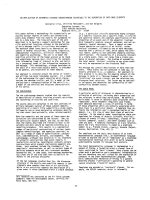
Based on the molecular analysis of all patients, dengue
type 3 was recorded for all patients (99.4%) with the
exception of 1(0.6%) male who had an infection with
dengue type 2 (Fig. 2).
Table 1: Dengue cases per year in Barbados tested for IgM and
IgG antibodies, 2003–2007
2003 2004 2005 2006 2007
Total requests 775 450 501 434 550
IgM + 454 195 78 153 219
IgG + 613 342 376 320 375
IgM-, IgG- 78 72 66 66 94
IgM + only 84 37 3 41 48
Serotype 33333>2
Virology Journal 2008, 5:152 />Page 3 of 6
(page number not for citation purposes)
Discussion
The lack of a vaccine or a cure for dengue fever makes the
development of laboratory-based surveillance systems all
the more important and to provide essential information
for effective vector control programmes. It is of considera-
ble importance to determine the serotypes of circulating
dengue virus, when and where since previous infection
with one of the four serotypes can be an important risk
factor for the development of dengue haemorrhagic fever
and dengue shock syndrome (DHF/DSS) upon infection
with a heterotypic serotype. The current "gold standard"
for typing dengue virus involves isolation of the virus in
cultured cells or mosquitoes followed by indirect immun-
ofluorescence. However, this requires cell culture facilities
or mosquito colonies, which are difficult to maintain in
laboratories in many developing countries. Serological
techniques such as immunoglobulin M or G enzyme
linked immunosorbent assays (ELISA) with a single serum
sample does not provide information on the serotype of
the virus. However, single-step reverse transcriptase
polymerase chain reaction (RT-PCR) detection and typing
of dengue virus offers a sensitive, specific, reproducible
and rapid alternative that requires only one acute-phase
serum sample [7,8].
The diagnosis of dengue fever by this one-step RT-PCR
procedure has reduced the amount of time in getting the
results to the patient. In comparison with the IgM ELISA
or IgG ELISA methods, antibodies are detectable in the
range of 5–10 days, whereas viraemia occurs after a few
days of onset (1–8 days) and can be readily measured by
RT-PCR which is more sensitive and also provides the
serotype of the virus.
A previous study on Barbadian patients with dengue who
were initially investigated for leptospirosis, indicated that
serotypes during that period (1995–1997) where 1, 2 or 4
[9]. The current study investigating patients during the
five year span (2003–2007), revealed that the majority
(>99%) of cases were caused by serotype 3. This serotype
first presented in Belize in 1963 and has subsequently
spread to the other Caribbean countries. Over the last
three decades, dengue serotypes 1, 2 and 4 have become
Percentage of dengue cases 2003–2007Figure 1
Percentage of dengue cases 2003–2007.
0
10
20
30
40
50
60
70
2003 2004 2005 2006 2007
Year
%
0
10
20
30
40
50
60
70
80
90
New Cases IgM + IgG + IgM -, IgG -
Table 2: Percentage of patients selected per year for the study
based on the presence of IgM antibodies
Year Cases No. of cases selected % of cases selected
2003 454 45 11
2004 195 22 11
2005 79 22 28
2006 153 22 14
2007 219 61 35
Average 220 34.4 20
Virology Journal 2008, 5:152 />Page 4 of 6
(page number not for citation purposes)
endemic in the Caribbean sub-region, producing numer-
ous epidemics at irregular intervals. Co-circulation of
these virus types is occurring in many islands such that
dengue fever is considered hyperendemic in this region
[10].
With the current dengue epidemic in Barbados, where the
population's majority are immunologic virgins to this
specific serotype, the heightened risk of DHF/DSS acquisi-
tion is imminent. Accordingly, over 74.4% of patients
were previously infected or had evidence of current expo-
sure in the recent past. Although the actual number of per-
sons with DHF/DSS over the five year period is unknown
based on the observation of clinical and laboratory fea-
tures, at least 4% of patients had suggestive symptoms.
This figure gives an average of at least 16 cases of DHF/
DSS per year based on the number of patients with IgG
antibodies. The ten patients included in the study with
known symptoms of DHF/DSS (thrombocytopenia,
bleeding gums, haematuria etc), ranged in age from 8
months to 57 years and did not conform to the suggested
age range of less than 15 years.
Although the different DENV serotypes can lead to vary-
ing clinical and epidemiologic profiles, defining precisely
which clinical characteristics are associated with the dis-
Table 3: Specimens tests prospectively and retrospectively by RT-PCR
Year Sex 0–16 17–20 21–30 31–40 41–50 50+ Age Unknown Total Total (male & female)
2003 Male212421
Female5386233 31
2004 Male1023121 1022
Female4020015 12
2005 Male1033111 1022
Female0232302 12
2006 Male1100111 522
Female3232115 17
2007 Male16365331 3261
Female14273041 29
Total Male 21 5 13 15 8 8 6 71 172
Female 26 9 23 13 6 10 16 101
Total 47143628141822 172
Table 4: List of clinical signs and symptoms in patients tested
Clinical/Laboratory feature n = 124 Number of cases Percentage (%)
Pyrexia 85 69
Arthralgia 45 63
Headache 65 52
Ocular pain 39 32
Emesis 19 15
Lumbar pain 18 15
Rash 16 13
Chills 15 12
Malaise 15 12
Myalgia 13 11
Diarrhoea 12 10
Jaundice 11 9
Cough 11 9
Abdominal pain 8 7
Thrombocytopenia 5 4
Neck pains 5 4
Haematuria 4 3
Hepatitis 4 3
Sore throat 4 3
Bleeding gums 3 2
Blood-shot eyes 1 0.8
Petechiae 1 0.8
Shock 1 0.8
Virology Journal 2008, 5:152 />Page 5 of 6
(page number not for citation purposes)
tinct serotypes has been elusive. Several reports have indi-
cated that DENV-2 and DENV-3 may cause more severe
disease than other serotypes and that DENV-4 is responsi-
ble for milder illness [11,12]. Certain genotypes within
particular serotypes have been associated with epidemics
of DHF [13] versus classic dengue, but no correlation with
specific clinical features has been reported.
In a Nicaraguan study [14], DENV-2 was associated with
greater disease severity, followed by DENV-3, which led to
greater hospitalizations in primary illness, while individ-
uals experiencing a primary DENV-2 infection tended to
exhibit less notable clinical disease. They also observed
that secondary infections were a risk factor for the pres-
ence of severe manifestations of dengue in Nicaragua
when DENV-2 was the dominant serotype, but not when
DENV-1 or DENV-3 predominated. This gives credence to
our study, as the number of DHF/DSS occurring yearly
was inappreciable, and this can be reflected by the pre-
dominant serotype. Also, although, more that 70% of
patients were previously exposed due to the detection of
IgG antibodies, severe manifestation of the infection were
minimal. Previous studies have shown that DENV-3 and
DENV-2 were particularly virulent to young children. This
observation was corroborated in our study, where numer-
ous infections occurred in those less than 16 years partic-
ularly in 2007. These children where immunological
virgins and could not mount an appreciable immune
response to this infection since most of these children
were hospitalized and never have detectable IgG antibod-
ies.
When DENV-1 predominated in the Nicaraguan study,
they observed a higher percentage of laboratory confirma-
tions of dengue but the clinical manifestations were
milder. Our retrospective analysis has indeed shown that
the number of laboratory confirmed cases has decreased
particularly in 2005 and this may be attributed to the sero-
type of dengue present within our population and not
reflective of total adherence to preventative methods or a
reduction in rainfall. However, there has been a steady
increase in infections caused by DENV-3.
Conclusion
In conclusion, it would therefore be expected that with the
predominance of DENV-3 circulating in the population,
although this infection may not be severe as previous
when DENV-2 predominated, there will be a higher pro-
portion of children being continuously infected with the
greater producing severe manifestations. This may overall
cause an increase in the apparent number of cases, but
with the present system, although more cases are detected
with DENV-1 is present, the RT-PCR will enhance the
detection particularly where the IgM and IgG antibodies
are undetectable by ELISA especially for those samples
that under 5 days after symptom onset. This study has
essentially characterized the serotypes circulating on the
island over the last five years and presents a plausible
explanation for the apparent reduction in cases. This study
also enhances the surveillance mechanisms for dengue
serotype detection and rapid turnaround time particularly
for those who may have DHF/DSS. Further characteriza-
tion of the genotypes is necessary to enhance the epidemi-
ologic profile. Recently, DEN-3/4 composite types have
been seen in our population and this could be similar in
other Caribbean islands.
Methods
Specimen collection
One hundred and seventy-two whole blood specimens
collected during the period January 2003 to August 2007
were analyzed for dengue fever infection with onset rang-
ing from 1 to 5 days. These patients were either hospital-
ized at the Queen Elizabeth Hospital (QEH), Ministry of
Health, Barbados, sent through private physicians or
through one of the eight (8) outpatient polyclinics on the
RT-PCR detection and typing of dengue virus in serum from patients infected during the period of 2003–2007 in BarbadosFigure 2
RT-PCR detection and typing of dengue virus in serum from
patients infected during the period of 2003–2007 in Barba-
dos. RNA was extracted from serum samples and was ampli-
fied by the two-enzyme single tube RT-PCR assay as
described in the Materials and Methods section. Lane 1: Den-
gue 1 RNA (positive control); Lane 2: Dengue 2 RNA (posi-
tive control); Lane 3: Dengue 3 RNA (positive control); Lane
4: Dengue 4 RNA (positive control), Lane 5 – 21: patients
(DENV- 3 positive); Lane 22: negative control (water): Lane
A: 100 bp DNA ladder.
A 12 13 14 15 16 17 18 19 20 21 22
Lanes
A 1 2 3 4 5 6 7 8 9 10 11
Lanes
Virology Journal 2008, 5:152 />Page 6 of 6
(page number not for citation purposes)
island. Peripheral blood specimens were separated for
serum and stored at -20°C until processing.
Serology
IgM and IgG antibody-capture ELISAs were performed
according to manufacturer's instructions (Focus Diagnos-
tics, Cypress, CA, 90630 USA). Briefly 100 μl of 1:101
diluted serum sample or control sample is added to the
washed 96-microwell polystyrene plate previously coated
with anti-human antibody specific for IgM or IgG and 100
μl of antigen solution (inactivated lyophilized dengue
fever virus antigen with equal amounts of DEN 1–4) was
then added. After a 2-hour incubation, followed by wash-
ing, 100 μl of affinity-purified and peroxidase mouse anti-
flavivirus IgM conjugate was added. Following incubation
and washing, 100 μl of substrate (tetramethylbenzidine
and hydrogen peroxide) was added. Colour development
was stopped with IM sulphuric acid and the OD was read
at 450 nm using a microplate autoreader. The levels of
specific antibodies were calculated from OD values.
RT-PCR and amplification
RT-PCR was performed according to manufacturer's
instructions. Briefly, RNA was extracted from 280 μl of
serum using the QIAamp Viral RNA minikit. Reverse tran-
scription and polymerase chain reaction was performed
using the One-Step Superscript III/RT/Platinum Taq Mix
(Invitrogen), 0.1 M dithiothreitol (DTT), 5' primer D1
and 3' primer TS1 at a concentration of 0.5 μM each and
3' primers TS2, TS3 and DEN4 at a concentration of 0.25
μM each in a total volume of 50 μl containing 5 μl of RNA.
A negative control was included in each run to identify
contamination. One cycle of 60°C for 30 min for the
reverse transcription was followed by 94°C for 2 minutes,
52°C for 1 min, 60°C for 1 minute and with a final exten-
sion of 60°C for 7 minutes [7].
The expected sizes of the amplification products were as
follows: 482 bp (DENV-1), 119 bp (DENV-2), 290 bp
(DENV-3) and 389 (DENV-4). Ten microlitres of the fifty
microlitre mixture was electrophoresed on a 1.5% TAE
agarose gel with a 100 bp DNA ladder (Invitrogen).
Competing interests
The authors declare that they have no competing interests.
Authors' contributions
MGS conceived the study and participated in its designed
and coordination and drafted the manuscript. NCG par-
ticipated in the design and coordination of the study and
performed the molecular studies.
Authors' information
Marquita Gittens-St. Hilaire is a lecturer in microbiology
at the University of the West Indies, Cave Hill Campus
adjunct Director of the Leptospira Laboratory (a govern-
mental institution). Her research interests include infec-
tious diseases, with primary focus on dengue,
leptospirosis and other zoonotic infections. Nicole Clarke
Greenidge is a medical laboratory technology at the Lept-
ospira Laboratory. Her research interests include microbi-
ology, surveillance and epidemiology.
Acknowledgements
We would like to thank the Ministry of Health for providing the funds to
support this project and the laboratory staff of the Leptospira Laboratory
and Dr. Rosa Salabas (Virology Division, CAREC) for the provision of the
control serotype specific RNA.
References
1. Emedicine Web site [ />topic124.htm]
2. Mandell GL, Douglas RG Jr, Bennett JE, Monath TP, eds: Principles
and practice of infectious disease. 6th edition. Churchill Living-
stone, Inc; 2005:1927-1950.
3. Gubler DJ: The global emergence/resurgence of arboviral dis-
eases as public health problems. Arch Med Res 2002, 33:330-342.
4. CDC traveler's health page: Dengue fever. Centers for Dis-
ease Control and Prevention Web site [http://
www2.ncid.cdc.gov/travel/yb/utils/ybGet.asp?section=dis&obj]
5. Pan American Health Organization: Number of reported cases of
dengue & dengue hemorrhagic fever (DHF), Region of the
Americas (by country and subregion). 2005 [http://
www.paho.org/English/AD/DPC/CD/dengue-cases-2005.htm].
6. Dengue Virus Types Identified in CAREC Member 1997–
2000 Web Site [ />dengue_virus_types_1997-2000.htm]
7. Halstead SB: Pathogenesis of dengue challenges to molecular
biology. Science 1998, 239:476-481.
8. Morens DM: Antibody-dependent enhancement of infection
and the pathogenesis of viral disease. Clin Infect Dis 1994,
19:500-512.
9. Levett PN, Branch SL, Edwards CN: Detection of dengue infec-
tion in patients investigated for leptospirosis in Barbados.
Am J Trop Med Hyg 2000, 62(1):112-114.
10. Harris E, Roberts TG, Smith L, Selle J, Kramer LD, Valle S, Sandoval
E, Balmaseda A: Typing of dengue viruses in clinical specimens
and mosquitoes by single-tube multiplex reverse tran-
scriptase PCR. J Clin Microbiol 1998, 36(9):2634-2639.
11. Nisalak A, Endy TP, Nimmanniya S, Kalayanarooj S, Thisayakorn U,
Scott RM, Burke DS, Hoke CH, Innis BL, Vaugh DN: Serotype-spe-
cific dengue virus circulation and dengue disease in Bangkok,
Thailand from 1973–1979. Am J Trop Med Hyg 2003, 68:191-202.
12. Vaugh DW, Green S, Kalayanarooj S, Innis BL, Nimmannitya S, Sun-
tayakorn S, Endy T, Raengsakulrach B, Rothman Al, Ennis FA, Nisalak
A: Dengue viremia titre antibody response pattern and virus
serotype correlate with disease severity. J Infect Dis 2000,
181:2-9.
13. Messer WB, Gubler DJ, Hams E, Sivananthank de Silva AM: Emer-
gence and global spread of a dengue serotype 3, subtype III
virus. Emerg Infect Dis 2003, 9:800-809.
14. Balmaseda A, Hammond SA, Perex Y, Tellez Y, Saborio SI, Mercado
JC, Cuadra R, Rocha J, Perez MA, Silva S, Rocha C, Harris E: Sero-
type-specific differences of clinical manifestations of dengue.
Am J Trop Med Hyg 2006, 74(3):449-456.