Báo cáo khoa học: "Epstein-Barr Nuclear Antigen 1 modulates replication of oriP-plasmids by impeding replication and transcription fork migration through the family of repeat" ppt
Bạn đang xem bản rút gọn của tài liệu. Xem và tải ngay bản đầy đủ của tài liệu tại đây (522.25 KB, 15 trang )
BioMed Central
Page 1 of 15
(page number not for citation purposes)
Virology Journal
Open Access
Research
Epstein-Barr Nuclear Antigen 1 modulates replication of
oriP-plasmids by impeding replication and transcription fork
migration through the family of repeats
Ashok Aiyar*
1,2
, Siddhesh Aras
2
, Amber Washington
†2
, Gyanendra Singh
†1
and Ronald B Luftig
2
Address:
1
Stanley S. Scott Cancer Center, LSU Health Sciences Center, 533 Bolivar Street, New Orleans, LA 70112, USA and
2
Department of
Microbiology, LSU Health Sciences Center, 1901 Perdido Street, New Orleans, LA 70112, USA
Email: Ashok Aiyar* - ; Siddhesh Aras - ; Amber Washington - ;
Gyanendra Singh - ; Ronald B Luftig -
* Corresponding author †Equal contributors
Abstract
Background: Epstein-Barr virus is replicated once per cell-cycle, and partitioned equally in latently
infected cells. Both these processes require a single viral cis-element, termed oriP, and a single viral
protein, EBNA1. EBNA1 binds two clusters of binding sites in oriP, termed the dyad symmetry
element (DS) and the family of repeats (FR), which function as a replication element and partitioning
element respectively. Wild-type FR contains 20 binding sites for EBNA1.
Results: We, and others, have determined previously that decreasing the number of EBNA1-
binding sites in FR increases the efficiency with which oriP-plasmids are replicated. Here we
demonstrate that the wild-type number of binding sites in FR impedes the migration of replication
and transcription forks. Further, splitting FR into two widely separated sets of ten binding sites
causes a ten-fold increase in the efficiency with which oriP-plasmids are established in cells
expressing EBNA1. We have also determined that EBNA1 bound to FR impairs the migration of
transcription forks in a manner dependent on the number of EBNA1-binding sites in FR.
Conclusion: We conclude that EBNA1 bound to FR regulates the replication of oriP-plasmids by
impeding the migration of replication forks. Upon binding FR, EBNA1 also blocks the migration of
transcription forks. Thus, in addition to regulating oriP replication, EBNA1 bound to FR also
decreases the probability of detrimental collisions between two opposing replication forks, or
between a transcription fork and a replication fork.
Background
Epstein-Barr virus (EBV) is replicated once per cell-cycle as
an episome in proliferating latently infected cells [1,2].
Episomal replication requires a viral sequence in cis,
termed oriP, and a single viral protein EBNA1 [3,4]. OriP
contains two sets of binding sites for EBNA1, the region of
dyad symmetry (DS), that contains four sites of low affin-
ity for EBNA1, and the family of repeats (FR) that contains
twenty high-affinity sites for EBNA1 [5,6]. DNA synthesis
initiates at DS, in a manner dependent upon the associa-
tion of the cellular origin recognition complex (ORC)
proteins and minichromosome maintenance (MCM) pro-
Published: 5 March 2009
Virology Journal 2009, 6:29 doi:10.1186/1743-422X-6-29
Received: 7 February 2009
Accepted: 5 March 2009
This article is available from: />© 2009 Aiyar et al; licensee BioMed Central Ltd.
This is an Open Access article distributed under the terms of the Creative Commons Attribution License ( />),
which permits unrestricted use, distribution, and reproduction in any medium, provided the original work is properly cited.
Virology Journal 2009, 6:29 />Page 2 of 15
(page number not for citation purposes)
teins with DS [7-9]. Recent evidence indicates that EBNA1
recruits the ORC proteins to DS through an RNA-medi-
ated interaction with ORC1 [10].
FR functions as a plasmid maintenance and partitioning
element [11,12]. FR from the prototypic B95-8 strain of
EBV contains 20 high-affinity sites for EBNA1, which
binds each of these sites as a dimer [13,14]. EBNA1 bound
to FR tethers viral episomes or oriP plasmids to cellular
chromosomes [15-19]; an association that facilitates the
plasmids to piggy-back into daughter cells at each met-
aphase [20,21]. In addition to its role in genome parti-
tioning, two-dimensional gel analysis by Schildkraut and
co-workers has indicated that the migration of replication
forks through FR is attenuated, so that for the circular EBV
genome or an oriP-plasmid, the bidirectional replication
fork that initiates at DS is terminated at FR [22]. This abil-
ity of EBNA1 bound to FR to attenuate replication forks
has been recapitulated in biochemical assays performed in
vitro; such assays reveal that DNA binding domain of
EBNA1 bound to FR impede the migration of replication
forks from an SV40 origin on the same template [23].
Using assays for transcription activation and plasmid
maintenance, we have examined the binding site require-
ments for EBNA1 in the EBV FR in detail [24]. Our analy-
ses indicated that although the wild-type FR contains 20
binding sites, plasmids with 10 binding sites are main-
tained far more efficiently in colony formation assays
than the former (ibid). A similar finding has been reported
for deletion mutants constructed within the natural FR, in
that a plasmid with nine binding sites replicated more
efficiently than a plasmid with twenty binding sites [25].
Thus these results concur in that the wild-type number of
EBNA1-binding sites in FR limits the replication of oriP-
plasmids by acting in cis.
In this study, we have examined the mechanism by which
the wild-type number of binding sites limits the replica-
tion of oriP-plasmids. Our results indicate that EBNA1
bound to FR limits replication by impeding the migration
of replication forks from DS. In addition, we have deter-
mined that EBNA1 bound to FR severely impairs the
migration of transcription forks through FR. We discuss
both these findings in the context of the stable replication
of EBV episomes.
Methods
Bacterial strains and plasmid purification
All plasmids were propagated in the E. coli strains DH5α,
MC1061/P3, or STBL2 (Invitrogen, Carlsbad, CA). Plas-
mids used for transfection were purified on isopycnic
CsCl gradients [26].
Plasmids
Plasmids AGP73, and AGP74 have been described previ-
ously [24], and contain 10 and 20 EBNA1-binding sites in
the FR respectively. These plasmids are constructed in the
backbone of pPUR, and also contain EBV's DS and the
EBV sequences between FR and DS. AGP81 contains 40
EBNA1-binding sites in FR and was constructed by dimer-
izing the FR in AGP74. AGP82 contains 80 EBNA1-bind-
ing sites in FR and was constructed by dimerizing the FR
in AGP81. AGP83 has been described previously and is a
control plasmid that only contains DS and completely
lacks FR. AGP212, and AGP213 contain 20 EBNA1-bind-
ing sites split into two FRs each containing ten binding
sites as described in the Results section. They were con-
structed as derivatives of AGP73. AGP212 was constructed
by recovering an MfeI-EcoRV fragment containing FR from
AGP73 and inserting it into the EcoRI-BamHI sites of that
plasmid. AGP213 was constructed by inserting an EcoRV-
Acc65I fragment from AGP73 into the Acc65I site of the
same plasmid. Plasmid 2380 contains wild-type oriP
cloned in pPUR, and was a gift from Bill Sugden. Plasmids
AGP39, AGP40, and AGP41 were constructed as deriva-
tives of pRSVL, by inserting 10, 20, or 40 EBNA1 binding
sites between the end of the luciferase open reading frame
and the SV40 polyadenylation signal in that plasmid.
Plasmid 1606 has been described previously and
expresses the large T antigen of SV40 under the control of
the CMV immediate early promoter [27]. Plasmid 1160
has been described previously and expresses the DNA
binding domain of EBNA1 under the control of the CMV
immediate early promoter [28]. The empty expression
vector, pcDNA3, was used as a control plasmid. Plasmid
2145 has been described previously and expresses EGFP
under the control of the CMV immediate early promoter
[17].
Cell culture and transfections
The human cell line 293 [29], and its EBNA1-expressing
derivative, 293/EBNA1, were used in this study. Both cell-
types were grown in DMEM supplemented with 10% fetal
bovine serum. G418 was added at a concentration of 200
mg/L to the media for 293/EBNA1 cells. Cells were grown
at 37°C in a humidified 5% CO
2
atmosphere. Plasmids
were introduced into cells by the calcium phosphate
method as described previously [17,18,24]. Transfections
were normalized by the inclusion of a CMV-EGFP expres-
sion plasmid, 2145, in each transfection. Upon harvest, a
fraction of the cells were profiled using a Becton-Dickin-
son FACSCalibur. Transfection efficiency was measured as
the fraction of GFP-expressing, live cells quantified using
CellQuest software from Becton-Dickinson (Franklin
Lakes, NJ).
Virology Journal 2009, 6:29 />Page 3 of 15
(page number not for citation purposes)
Colony formation assays to assess plasmid maintenance
and partitioning
Ten μg of AGP74 or an equivalent number of moles of
plasmids AGP73, AGP81, 2380, AGP82, AGP83, AGP212,
and AGP213, were co-transfected with 1 μg of 2145 into 1
× 10
7
293/EBNA1 cells on a 10 cm dish. Cells were split
eight hours post-transfection so that they would not be
confluent at 48 hours post-transfection, at which time
cells were harvested, FACS profiled to measure GFP
expression, and re-plated in duplicate at 2 × 10
5
, 2 × 10
4
,
and 2 × 10
3
GFP-positive, live cells per culture dish. Cells
were placed under selection with 0.5 μg/ml puromycin
four days post-transfection. After two weeks of selection,
the resulting puromycin-resistant colonies were fixed with
formamide and subsequently stained with methylene
blue. Colonies that were at least 2 mm in size were scored
as positive. Colonies were counted using a colony count-
ing macro written for NIH Image as described previously
[17,18].
Southern hybridization analysis to assess plasmid
replication
Ten μg of AGP74 or an equivalent number of moles of
plasmids AGP73, 2380, AGP212, and AGP213 were co-
transfected with 1 μg of 2145 into 1 × 10
7
293/EBNA1
cells on a 10 cm dish. Cells were placed under puromycin
selection 48 hours post-transfection. After three weeks of
selection, episomal DNAs were extracted from cells in
puromycin resistant colonies that were pooled. Episomal
DNAs were extracted from 2 × 10
7
– 10
8
puromycin-resist-
ant cells as described previously [11,30]. Extracted DNAs
were digested with 200 units of DpnI, 20 units of BamHI,
and 20 units of XbaI in a final volume of 100 μl overnight
at 37°C. Restriction endonucleases were purchased from
New England Biolabs (Beverly, MA), and used as per the
manufacturer's instructions. Digestions were extracted
with phenol:chloroform (1:1), precipitated and electro-
phoresed on a 0.8% agarose gel. DNAs were transferred
from the gel to Hybond membrane (Amersham, Bucking-
hamshire, UK) using an Appligene vacuum transfer appa-
ratus (Boekel Scientific, Feasterville, PA). Radioactive
probes were prepared by the incorporation of α-
32
P-dCTP
(6000 Ci/mmol) (Amersham) during Klenow synthesis
using random primers and PstI-digested AGP83 as tem-
plate. Probe specific activities ranged from 1 × 10
9
cpm/μg
to 3 × 10
9
cpm/μg. Southern hybridization was performed
as described by Hubert and Laimins [31,32]. Southern
blots were visualized and quantified by phosphorimage
analysis using a Molecular Dynamics Storm phosphorim-
ager (Molecular Dynamics, Sunnyvale, CA).
Transfection of linear plasmid DNAs to assess replication
fork migration in vivo
Ten μg of PvuII-linearized AGP73 or AGP74 was trans-
fected into 293/EBNA1 cells as described above along
with 1 μg of 1606. Hirt extracts were prepared from 2 ×
10
7
transfected cells 14 – 16 hours post-transfection and
digested exhaustively with DpnI (200 units). The digested
extracts were then digested with HindIII (10 units)
&Acc65I (10 units) to release a 1063 bp fragment between
the SV40 origin and FR, and with BsrGI (10 units) &SpeI
(20 units) to release a 637 bp fragment that lies immedi-
ately after FR. The digested products were separated on a
1.5% agarose gel electrophoresed in 0.5× TBE, and trans-
ferred to Hybond membrane and probed as described
above. Probe was synthesized using random primers and
the HindIII-Acc65I fragment, as well as the BsrGI-SpeI frag-
ment as template. In control experiments, probes were
hybridized against purified fragments to confirm that the
BsrGI-SpeI fragment bound approximately two-thirds as
much probe as the HindIII-Acc65I fragment.
Transcription reporter assays
100 ng of pRSVL [33], or an equivalent number of moles
of AGP39, AGP40, or AGP41 was co-transfected with 1 μg
of 2145 and 10 μg of pcDNA3 or 10 μg of 1160 into 293
cells. Cells were split eight hours post-transfection so that
they would not have reached confluence when harvested
72 hours post-transfection. A fraction of the harvested
cells were then counted twice using a Coulter counter, and
FACS profiled to normalize for the fraction of live trans-
fected cells. The remainder of the cells were pelleted, and
lysed in reporter lysis buffer (provided along with a luci-
ferase assay kit from Promega, Madison, WI) at a concen-
tration of 1 × 10
5
cells/μl. Lysates were spun for 5 minutes
at 1000 g to remove nuclei, and then frozen at -80°C until
assay. Luminescence assays were performed as per manu-
facturer's instructions, using a Zylux FB 15 luminometer.
RT-PCR analysis to measure migration of transcription
forks through FR
Total RNA was extracted from transfected 293 cells using
the SV Total RNA Isolation System from Promega (Madi-
son, WI). PolyA+ RNA was extracted from transfected 293
cells using the PolyATract mRNA Isolation System from
Promega (Madison, WI). Either 5 μg of total RNA or 1 μg
of polyA RNA was used in RT-PCR reactions using the fol-
lowing primers to detect firefly luciferase:
AGO83: 5' GGAATACTTCGAAATGTCCG
AGO84: 5' TCATTAAAACCGGGAGGTAG
Control RT-PCR reactions amplifying the glyceraldehyde
phosphate dehydrogenase (GAPDH) transcript were per-
formed using the following two primers:
AGO81: 5' CTCAGACACCATGGGGAAGGTGA
AGO82: 5' ACTTGATTTTGGAGGGATCTCG
Virology Journal 2009, 6:29 />Page 4 of 15
(page number not for citation purposes)
RT-PCR reactions were performed using the AccessQuick
one-tube RT-PCR System purchased from Promega (Mad-
ison, WI).
Results
The number of viable colonies decreases with an increasing
number of EBNA1 binding sites in FR
During our studies to determine the optimal number of
binding sites in FR, as well as the spacing between adja-
cent sites, we determined that plasmids with ten high-
affinity EBNA1 binding sites in a synthetic FR formed
puromycin resistant colonies in 293/EBNA1 that were 2
mm in size and larger more efficiently than colonies with
20 binding sites in FR [24]. The EBNA1-binding sites in
the synthetic FRs are identical, and were chosen using the
sequence of the EBNA1-binding site found most fre-
quently in the natural FR (seven times out of 20) (ibid).
There is a small amount of sequence variation between
binding sites in the natural FR. The most frequent site is
repeated seven times, and an additional 11 sites are single
nucleotide variations of this site [6,34]. To eliminate the
possibility that an FR with 20 identical EBNA1-binding
sites behaves differently than the natural FR, we compared
the colony formation efficiency of plasmids containing a
synthetic FR with 20 binding sites versus the natural FR,
and found their efficiencies to be indistinguishable (Table
1). Therefore our observation that plasmids with ten iden-
tical EBNA1-binding sites in FR form colonies more effi-
ciently than plasmids 20 identical EBNA1-binding sites in
FR recapitulates the observations made with natural FR, or
deletion derivatives thereof [25]. These authors have
determined that a plasmid containing a deletion muta-
tion of the natural FR with only nine EBNA1-binding sites
replicates more efficiently than a plasmid with the intact
natural FR containing 20 binding sites (ibid). Next, it was
determined whether additional increases in the number
of EBNA1-binding sites would continue to decrease the
efficiency of replication and therefore decrease the
number of colonies formed. For this, the colony forma-
tion efficiency of reporter plasmids with FRs containing
40 and 80 binding EBNA1-binding sites was measured in
293/EBNA1 cells. The results of this assay are summarized
in Table 1. The summarized results indicate several obser-
vations: 1) In concordance with our previous results, plas-
mids with ten binding sites in FR form colonies
approximately four times more efficiently than colonies
with 20 binding sites in FR. This difference is statistically
significant with a p-value of 0.02 by the Wilcoxon rank-
sum test; 2) The FR with 20 identical EBNA1-binding sites
cannot be distinguished statistically from the natural FR
in colony formation assays and replication assays (see
below); 3) Most surprisingly, the efficiency of colony for-
mation decreases sharply for replication reporters con-
taining FRs with 40 or 80 EBNA1-binding sites, such that
a plasmid with 40 binding sites in FR formed puromycin-
resistant colonies approximately one log less efficiently
than a plasmid with 20 binding sites in FR, and a plasmid
with 80 binding sites in FR formed colonies two logs less
efficiently than a plasmid with 20 binding sites in FR.
Both these decreases were found to be highly significant (p
< 0.01 by the Wilcoxon rank-sum test). Indeed a plasmid
with 80 EBNA1-binding sites formed 2 mm and larger col-
onies with the same efficiency as replication reporter that
only contained the DS element (Table 1).
The colony formation assay we employ only counts colo-
nies that are 2 mm or larger in size by 18 days post-trans-
fection. We observed that 293/EBNA1 cells transfected
with replication reporter plasmids containing 40 or 80
binding sites in FR formed a large number of colonies that
were substantially smaller than 2 mm in size, and never
increased in size despite two additional weeks of growth
in selective media (Figure 1, and data not shown). Figure
1 contains examples of colony formation assays per-
formed with plasmids that contain only DS, or DS with
increasing numbers of EBNA1-binding sites in FR. As seen
in the figure, while a plasmid containing DS alone forms
very few colonies, plasmids with 40 or 80 EBNA1-binding
sites in FR form a large number of colonies that are much
smaller than 2 mm in size. In contrast, the majority of col-
onies formed by cells transfected with plasmids contain-
Table 1: A greater than wild-type number of EBNA1 binding sites in the family of repeats causes a decrease in the number of
puromycin resistant colonies obtained in colony formation assays.
Replication reporter transfected Number of EBNA1 binding sites in FR
a
Colonies per 10
5
live, transfected cells plated
b
AGP83 0 2 ± 1.8
AGP73 10 4390 ± 311.1
AGP74 20 1296 ± 106.1
2380 20
c
1230 ± 28.3
AGP81 40 78 ± 14.2
AGP82 80 3 ± 1.7
a
plasmids contain DS, EBV sequences between FR and DS, and a synthetic FR containing the indicated number of EBNA1 binding sites.
b
puromycin resistant colonies present 18 days post-transfection that are 2 mm and larger in size.
c
plasmid 2380 contains wild-type FR from the B95-8 strain of EBV.
Virology Journal 2009, 6:29 />Page 5 of 15
(page number not for citation purposes)
ing ten or 20 EBNA1-binding sites in FR are larger than 2
mm in size.
The large number of tiny colonies formed upon transfec-
tion of plasmids containing 40 or 80 binding sites in FR is
consistent with the behavior of plasmids that confers
puromycin resistance to transfected cells but are not dis-
tributed to daughter cells at mitoses, thus preventing the
formation of a large puromycin-resistant colonies. This
could happen either due to a defect in plasmid partition-
ing or due to a failure in plasmid replication. We favor a
defect in plasmid replication, because the colony forma-
tion phenotype of these two plasmids is strikingly differ-
ent from that of a plasmid containing only DS (Figure 1).
DS-only plasmids are replicated transiently but not parti-
tioned, and thus give rise to a few puromycin-resistant col-
onies that contain integrated copies of the plasmid [24].
For the reporter plasmids containing 40 and 80 EBNA1-
binding sites in FR, the presence of a large number of col-
onies that do not expand in size suggests that the initially
transfected plasmids are partitioned, but are poorly repli-
cated, if at all. Therefore, the cells that nucleate a colony
cannot give rise to drug-resistant daughters upon cell pro-
liferation, as the latter lack plasmids to confer drug resist-
ance. In this study we have examined why increasing the
number of EBNA1-binding sites in FR decreases the effi-
ciency of plasmid replication.
There are two possible reasons for this defect, illustrated
by the models in Figure 2. In Figure 2A we have schemat-
ically depicted the "replication factor titration" model
proposed earlier [25]. In this model, EBNA1 bound to FR
is proposed to non-functionally titrate cellular replication
factors, such as the ORC proteins, away from EBNA1
bound to DS. This non-functional recruitment of proteins
such as ORC decreases the replication potential of plas-
mids, by reducing the frequency of replication initiation
as DS, as the number of EBNA1-binding sites in FR is
increased. An alternative model is suggested by the results
of Gahn and Schildkraut [22], who have demonstrated
that FR forms a barrier that attenuates the migration of
replication forks initiated at DS. If the efficiency of atten-
uation is dependent upon the number of EBNA1-binding
sites in FR, then increases in binding site number are pre-
dicted to decrease replication efficiency. Thus in this
model, termed the "replication fork barrier" model, and
depicted in Figure 2B, EBNA1 bound to FR suppresses rep-
lication from DS by attenuating the migration of the rep-
lication fork after initiation of DNA synthesis. If this latter
model underlies the relative inefficiency in the replication
of plasmid with 20 binding sites compared to a plasmid
with ten binding sites, then it is predicted that presenting
the 20 binding sites as two widely-separated sets of ten
binding sites on a plasmid should revert the observed
decrease. On the other hand, the replication factor titra-
tion model predicts that a plasmid with two FRs, each
with ten binding sites, should replicate with the same effi-
ciency as a plasmid with a single FR containing 20 EBNA1-
binding sites.
Replication of plasmids with split FRs containing ten
binding sites each
To test the models presented in Figure 2, two additional
replication reporter plasmids illustrated in Figure 3A were
constructed. In the first, AGP212, two FRs with ten bind-
ing sites each were placed on either side of DS, and sepa-
rated from DS by the EBV sequences normally present
between FR and DS. In the second, AGP 213, two FRs with
ten binding sites each were placed in tandem, but sepa-
rated from each other by the EBV sequences normally
present between FR and DS. AGP212 and AGP213 were
transfected into 293/EBNA1 cells and their ability to form
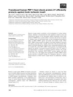
Plasmids with an FR containing more than 20 EBNA1 binding sites form minute colonies under selectionFigure 1
Plasmids with an FR containing more than 20 EBNA1 binding sites form minute colonies under selection. The
indicated plasmids were transfected into 293/EBNA1 cells, which were subjected to puromycin selection for 18 days in colony
formation assays as described in the Materials and Methods section. Representative images of methylene blue stained colonies
are shown. The identity of the transfected plasmid is indicated above each image, and the number of EBNA1 binding sites
present in the FR of each plasmid is indicated below each image. As a negative control, an assay was also performed with
AGP83, a DS-only plasmid that replicates transiently, but is not partitioned, and forms colonies with very low efficiency. The
colonies formed from cells transfected with AGP81 and AGP82 did not increase in size even after several weeks of growth in
selective media. The number of colonies formed in such assays that were 2 mm in size and larger is indicated in Table 1.
Virology Journal 2009, 6:29 />Page 6 of 15
(page number not for citation purposes)
puromycin resistant colonies was evaluated (Table 2). As
indicated in the table, both plasmids containing 20
EBNA1-binding sites split into two sets of ten binding
sites form puromycin resistant colonies far more effi-
ciently than a plasmid containing 20 contiguous EBNA1
binding sites in FR, or a plasmid that contains wild-type
FR (p-value < 0.05 by the Wilcoxon rank-sum test). Not
only do AGP212 and AGP213 form colonies more effi-
ciently than AGP74, they also give rise to puromycin-
resistant colonies more efficiently than AGP73 that con-
tains a single block of ten EBNA1-binding sites. This result
favors the "replication fork barrier" model over the "repli-
cation factor titration" model.
To verify that AGP212 and AGP213 are replicated episo-
mally, episomal DNA from 293/EBNA1 cells transfected
independently with AGP73, AGP74, 2380, AGP212 and
AGP213, was extracted 18 days post-transfection, digested
exhaustively with DpnI, linearized with XbaI, and exam-
ined by Southern blot. These results are shown in Figure
3B, and tabulated in Table 3. These results indicate that all
the plasmids are replicated episomally and maintained at
Two models to explain decreases in copy number when oriP plasmids contain an FR with 20 or more EBNA1 binding sitesFigure 2
Two models to explain decreases in copy number when oriP plasmids contain an FR with 20 or more EBNA1
binding sites. DS is represented as a striped oval, and EBNA1-binding sites in FR are represented as black filled circles. For
simplicity, only ten binding sites are shown. EBNA1 dimers bound to DS or FR are represented as gray ovals. (A) Replication
factor titration model. EBNA1 bound to FR is proposed to non-functionally titrate cellular replication factors, such as ORC
proteins, away from EBNA1 bound to DS, thus decreasing replication initiation events at DS. The titration efficiency is propor-
tional to the amount of EBNA1 at FR, which in turn is dependent on the number of EBNA1 binding sites in FR. (B) The replica-
tion fork barrier model in which EBNA1 bound to FR is proposed to act post-initiation to impede the progression of
replication forks initiated at DS. A decreased efficiency of progression is indicated by the gradation in line color from black to
light gray. The strength of this barrier is proportional to the amount of EBNA1 present at FR, which is also dependent on the
number of EBNA1 binding sites in FR.
Virology Journal 2009, 6:29 />Page 7 of 15
(page number not for citation purposes)
Split FRs distinguish between the replication factor titration and replication fork barrier modelsFigure 3
Split FRs distinguish between the replication factor titration and replication fork barrier models. (A) Schematic
representation of the oriP region from plasmids designed to distinguish between the replication factor titration and the replica-
tion fork barrier models. The identity of the plasmid is indicated to the left of each schematic. DS is represented as a striped
oval, and the EBNA1-binding sites in FR as filled black circles. The number of EBNA1 binding sites within each FR is indicated
above each FR. FRs are separated from each other (plasmid AGP213) or from DS (plasmid AGP212) by the EBV sequences
normally present between FR and DS. (B) Stable replication of oriP replication reporters under selection in 293/EBNA1 cells.
293/EBNA1 cells were transfected with the indicated plasmid, placed under puromycin selection for 18 days, at which time
replicated DpnI-resistant episomal DNAs were recovered and quantified as described in the Methods. "M" indicates the migra-
tion position of standards used for quantitation, and the amounts of standards loaded are indicated above each lane. The iden-
tity of the transfected plasmid is indicated above each lane. "A" indicates the migration position of DpnI-resistant, linearized
plasmid DNAs.
Virology Journal 2009, 6:29 />Page 8 of 15
(page number not for citation purposes)
copy numbers varying between ~25 and ~80 molecules
per transfected cell under selection.
EBNA1 bound to 20 contiguous binding sites in FR impedes
the migration of replication forks within cells
The results described above are interpreted to indicate that
EBNA1 bound to 20 contiguous binding sites limits repli-
cation from oriP in a manner consistent with it impeding
the migration of replication forks. To determine whether
the efficiency with which EBNA1 bound to FR impedes
replication fork migration is dependent upon the number
of binding sites in FR, the experiment schematically
depicted in Figure 4A was performed. 293/EBNA1 cells
were co-transfected with linear DNAs containing the SV40
origin, and FRs with either ten or 20 EBNA1-binding sites,
along with a large T-antigen expression plasmid. Fourteen
to 16 hours post-transfection, low molecular DNAs were
recovered by the method of Hirt and digested exhaustively
with DpnI to remove any unreplicated linear DNAs
present from the transfection. The DpnI-treated DNA was
then digested with HindIII and Acc65I to release a 1 kb
fragment (labeled fragment ONE) that lies immediately
between the SV40 origin and FR, and with BsrGI and SpeI
to release a 0.6 kb fragment (labeled fragment TWO) that
lies immediately after FR. The digested DNAs were electro-
phoresed on a 1.5% agarose gel, transferred to nylon and
probed for each of the fragments. If EBNA1 bound to FR
does not function as block to the migration of replication
forks from the SV40 origin in vivo, we expect that equiva-
lent amounts of fragment ONE and TWO will be synthe-
sized. In contrast, if EBNA1 bound to FR efficiently blocks
replication forks from the SV40 origin in vivo, we expect a
smaller amount of DpnI-resistant, replicated fragment
TWO relative to fragment ONE. It is pertinent to note that
because fragment TWO is smaller than fragment ONE, a
TWO/ONE ratio of approximately 0.6 is indicative of
equivalent amounts of both fragments. The results of two
independent experiments are shown in Figure 4B. As can
be seen from the Figure, when FR in the transfected plas-
mid contained ten EBNA1 binding sites, the TWO/ONE
ratio averaged 0.67, indicating that pieces of DNA on
either side of FR were synthesized equivalently. In con-
trast, when FR contained 20 binding sites, the TWO/ONE
ratio averaged 0.12, indicating that the fragment before FR
was synthesized five-times as much as the fragment after
FR. This experiment provides strong in vivo molecular evi-
dence that EBNA1 bound to 20 contiguous binding sites
attenuates the migration of replication forks. Further, the
strength of attenuation is dependent upon the number of
binding sites for EBNA1, and is non-existent when only
ten contiguous binding sites are on the template.
Thus, we conclude that plasmids containing the wild-type
number of binding sites in FR are replicated less well than
plasmids with fewer binding sites in FR (Table 1, Figure 1,
Figure 4). The apparent conundrum posed by this data is
to explain why the EBV genome has evolved to contain a
plasmid-partitioning element that reduces the efficiency
with which the genome is replicated. One possible reason
for this is that it provides a mechanism for EBV to limit the
replication of its latent replicon and maintain copy
number control in latently infected cells. An increase in
genome copy number may result in the unfettered expres-
sion of viral genes, and thereby compromise the ability of
Table 2: Splitting twenty contiguous EBNA1 binding sites into two sets of ten binding sites increases the efficiency of replication as
estimated by colony formation.
Replication reporter transfected Arrangement of EBNA1 binding sites
a
Colonies per 10
5
live, transfected cells plated
b
AGP73 FR(10), DS 4390 ± 311.1
c
AGP74 FR(20), DS 1296 ± 106.1
c
AGP212 FR(10), DS, FR(10) 10048 ± 371.7
AGP213 FR(10), FR(10), DS) 6132 ± 180.2
a
The arrangement of EBNA1 binding sites in FR is schematically depicted in Figure 3.
b
puromycin resistant colonies present 18 days post-transfection that are 2 mm and larger in size.
c
values are taken from Table 1, and shown here for convenience,
Table 3: Copy number of replicated, DpnI-resistant, plasmids detected 18 days after transfection into 293/EBNA1 cells.
Replication reporter transfected Arrangement of EBNA1 binding sites Plasmid copy number
AGP73 FR(10), DS 38 ± 11
AGP74 FR(20), DS 47 ± 8
AGP212 FR(10), DS, FR(10) 60 ± 11
AGP213 FR(10), FR(10), DS 51 ± 11
2380 wild-type oriP 44 ± 9
a
Numbers represent the average number of DpnI-resistant episomal plasmid molecules per transfected cell detected in three experiments along
with the standard deviation.
Virology Journal 2009, 6:29 />Page 9 of 15
(page number not for citation purposes)
Figure 4 (see legend on next page)
Virology Journal 2009, 6:29 />Page 10 of 15
(page number not for citation purposes)
latently infected cells to evade immune surveillance. We
believe it likely that there are additional reasons that
EBNA1 bound to FR attenuates fork migration. It has been
demonstrated that plasmids with active transcription
units suppress the use of replication origins on the same
plasmid [35,36]. This could possibly arise from the colli-
sion of transcription and replication forks on the same
plasmid, resulting in the faster transcription forks stalling
the slower migrating replication forks [37-40], possibly
generating of double-strand breaks (DSBs) [41]. In its nat-
ural context, oriP is immediately adjacent to the EBER
genes that are heavily transcribed during latency, which is
also when oriP is active as a replication origin. The EBERs
are transcribed toward DS, the replication origin within
oriP, and separated from DS by FR. Therefore, we wished
to test whether EBNA1 bound to FR could terminate the
migration of transcription forks, and thereby protect rep-
lication forks initiated at DS.
EBNA1 bound to FR impedes the progression of
transcription forks
The transcription reporter plasmid pRSVL [33] was modi-
fied to introduce ten, 20, or 40 contiguous EBNA1 bind-
ing sites between the end of the luciferase open reading
frame and the SV40 polyadenylation sequence in that
plasmid. The structure of these reporter plasmids is shown
in Figure 5A. These reporter plasmids were then co-trans-
fected into 293 cells with a control expression plasmid
(pcDNA3), or plasmid 1160 that expresses the DNA bind-
ing domain of EBNA1 (DBD). Cells were harvested two
days post-transfection, FACS profiled to normalize for live
transfected cells, following which luciferase levels were
measured. This analysis is shown in Figure 5B. In the
absence of EBNA1 binding sites on the reporter plasmid,
the co-transfected DBD expression plasmid had no effect
on luciferase expression. Similarly, pcDNA3 had no effect
on luciferase expression from reporter plasmids that con-
tained ten, 20 or 40 EBNA1 binding sites. However when
the luciferase reporter plasmids had 20 or 40 EBNA1
binding sites, and were co-transfected with the DBD
expression plasmid, there was a sharp decrease in the
expression of luciferase dependent upon the number of
number binding sites placed 5' to the polyadenylation sig-
nal.
It was speculated that this decrease in luciferase expres-
sion resulted from prematurely terminated luciferase tran-
scripts formed as a consequence of DBD bound to EBNA1
binding sites functioning as a transcription fork-block. To
test this hypothesis, the distribution of luciferase RNA in
total and polyA+ RNA pools was examined by reverse-
transcriptase PCR (RT-PCR), with the following rationale.
Prematurely terminated transcripts should be transiently
detected in the total RNA pool but not the polyA+ pool,
while the mature luciferase mRNA should be present in
both pools of RNA. The rationale is depicted schemati-
cally in Figure 5A, and the experimental outcome is
shown in Figure 5C. As seen in the figure, the DBD did not
effect amplification of the target sequence by RT-PCR
from both the total RNA and mRNA pools when cells were
transfected with pRSVL, or a derivative of pRSVL contain-
ing ten EBNA1-binding sites before the polyadenylation
signal. In contrast, for derivatives of pRSVL containing 20
or 40 EBNA1-binding sites before the polyadenylation sig-
nal, there was a clear decrease in amplification of the luci-
ferase target sequence by RT-PCR from polyA+ RNA pool,
mirroring the decrease in luciferase expression observed
in Figure 5B. However, the target was amplified from the
total RNA pool recovered from cells transfected with these
plasmids (ibid). We interpret this analysis to indicate that
the decrease in luciferase signal observed in Figure 5B
results from EBNA1 bound to FR acting to terminate the
migration of transcription forks, and that this termination
can be observed when FR contains the wild-type number
of 20 binding sites, but not ten binding sites.
Discussion
In this study we have demonstrated that the wild-type
number of EBNA1-binding sites in EBV's FR region is sub-
optimal for the efficient replication of oriP-plasmids. A
plasmid with ten binding sites in FR formed colonies
more efficiently than a plasmid with the wild-type
EBNA1 bound to FR blocks the progression of replication forks in transfected cellsFigure 4 (see previous page)
EBNA1 bound to FR blocks the progression of replication forks in transfected cells. A) Plasmids containing the
SV40 replication origin, and FR regions with ten or 20 EBNA1-binding sites were linearized, and co-transfected into 293/
EBNA1 cells with large T-antigen expression plasmid. A schematic representation of bidirectional replication fork movement
from the SV40 origin is indicated above and below the linear transfected DNA, with the position of FR and the SV40 origin
indicated. The leading strands from the SV40 origin are indicated as long arrows, and Okazaki fragments as the short arrows.
Dark lines indicate unimpeded fork progression, while light gray lines indicated segments where diminished DNA synthesis is
predicted. The positions and identities of restriction enzyme recognition sites to liberate fragments "ONE" and "TWO" from
replicated DNA are shown. (B) Hirt extraction was sued to recover DNAs from transfected 293/EBNA1 cells that were sub-
sequently digested with DpnI and the specified restriction endonucleases to release fragments ONE and TWO, which were
separated by electrophoresis, and quantified by Southern blot. Two independent experiments are shown with the migration of
fragments ONE and TWO, and the number of EBNA1-binding sites in FR indicated. The TWO:ONE ratio is also shown.
Virology Journal 2009, 6:29 />Page 11 of 15
(page number not for citation purposes)
Figure 5 (see legend on next page)
Virology Journal 2009, 6:29 />Page 12 of 15
(page number not for citation purposes)
number of 20 binding sites. Increasing the number of
binding sites in FR beyond 20 further decreased the effi-
ciency of replication (Table 1). These results corroborate
those of Leight and Sugden who have demonstrated that
an oriP-plasmid with a deletion that removes approxi-
mately one-half of FR is replicated more efficiently than a
plasmid with wild-type FR [25].
We have tested two models to explain why plasmids with
fewer binding sites in FR are replicated more efficiently
than plasmids with the wild-type number of binding sites.
Our results support a model wherein EBNA1 bound to FR
impedes the progression of replication forks that originate
from DS. It was determined that this effect correlates with
the number of contiguous EBNA1 binding sites in FR.
Attenuation of fork migration is not readily detected with
ten contiguous sites, but is easily observed with 20 contig-
uous binding sites. As has been observed previously in
vitro [23], we found that EBNA1 bound to FR also
impedes replication forks from the SV40 origin within
transfected cells. The SV40 origin was used for this analy-
sis because it fires multiple times in a single cell-cycle, per-
mitting facile evaluation of the reduction in fork
migration. The major difference between the SV40 replica-
tion fork and replication forks that initiate from DS lies in
the nature of the leading strand helicase. The hexameric
large T-antigen helicase in the SV40 replication fork has
approximately the same mass as the hexameric MCM hel-
icase present at replication forks that initiate from DS [7].
Both forks progress at similar rates, with elongation being
estimated at approximately 100 bp/min for the SV40 rep-
lication fork [42,43], and at between 10 – 50 bp/min for
EBV replication [44]. Given the similar biophysical char-
acteristics of both forks, we believe that EBNA1 bound to
FR will impede the progression of replication forks that
fire from DS in a manner dependent on the number of
binding sites.
Our data indicates that split-FR plasmids containing two
FRs with ten binding sites each are replicated more effi-
ciently than plasmids containing a single FR with twenty
contiguous EBNA1 binding sites (Table 2). EBNA1 bound
to FR tethers oriP-plasmids to chromosomes to facilitate
their maintenance and partitioning in proliferating cells
[17-19]. The efficiency of this process is dependent upon
the number of binding sites in FR, such that an EBNA1
mutant which is partially defective in chromosomal asso-
ciation can be rescued by increasing the number of bind-
ing sites in FR [18]. However, with 20 contiguous sites,
this increase in partitioning efficiency is offset by a
decrease in replication efficiency. Splitting the 20-binding
site FR into separated FRs with ten binding sites each, no
longer impedes replication, but retains the advantage of
having 20 binding sites for efficient oriP-plasmid parti-
tioning. The data obtained with AGP212 and AGP213
(Table 2) also indicates that the replication factor titration
model proposed previously is unlikely. Both these plas-
mids contain 20 EBNA1 binding sites and replicate more
efficiently than a plasmid that contains ten binding sites.
Were the titration model to be correct, replication of these
plasmids would be less efficient than replication of a plas-
mid with ten EBNA1 binding sites in FR.
The ability of EBNA1 to impede replication fork migration
likely impacts replication of EBV genomes. Besides DS,
there are other replication origins on the EBV genome also
used during latency [45], such as an origin that lies in the
BamHI-A fragment [46,47]. It is known that collision of
replication forks can lead to fork collapse, and the conse-
quential generation of double-stranded breaks (DSBs)
[48]. Such events can lead to irregular recombination
events, and a large number of DSBs causes apoptosis [49-
51]. We propose that EBNA1 bound to FR acts as a buffer
to prevent two replication forks from running into each
other and thereby protects cells latently infected by EBV
from undergoing apoptosis as a consequence of DSB gen-
eration.
There is a striking parallel between the function of EBNA1
at FR and TTFI at Sal repeats that terminate ribosomal
DNA replication. Both proteins impede the progression of
replication forks dependent on the number of binding
EBNA1 bound to FR impedes transcription fork progressionFigure 5 (see previous page)
EBNA1 bound to FR impedes transcription fork progression. (A) Representation of transcription reporter plasmids
used here. In pRSVL, the RSV LTR drives transcription of the luciferase gene, and the SV40 late polyadenylation signal is used
for polyadenylation. Derivatives of pRSVL with ten, 20 or 40 EBNA1-binding sites (filled black circles) between the luciferase
gene and the polyadenylation signal were constructed. Primary transcripts, and prematurely terminated transcripts present in
total RNA preparations are indicated, as are mature luciferase mRNAs present in total and polyA+ RNA preparations. Primers
used for RT-PCR are indicated as arrows. (B) Luciferase expression from the reporter plasmids described above. Plasmids
were co-transfected with either pcDNA3 (stippled bars), or a EBNA1 DNA binding domain expression plasmid (black bars)
into 293 cells. The number of EBNA1 binding sites in the reporter plasmid is indicated below each pair of bars. Luciferase activ-
ity is reported relative to the activity observed when pRSVL was co-transfected with pcDNA3. (C) RT-PCRs to detect luci-
ferase and GAPDH transcripts in total or polyadenylated RNAs recovered from the transfected cells described in B. PCR
products were visualized with ethidium bromide and the identity of the transfected plasmid is indicated above each lane.
Virology Journal 2009, 6:29 />Page 13 of 15
(page number not for citation purposes)
sites for the protein on the template DNA [52]. Addition-
ally, just as TTFI bound to the Sal repeats blocks the pro-
gression of transcription forks and terminates them [53],
we have found that EBNA1 bound to FR blocks the pro-
gression of transcription forks in a manner dependent
upon the number of binding sites (Figure 5). In its natural
chromosomal context the ribosomal DNA replication
fork block is required for the proper termination of rRNA
transcripts. Within the EBV genome, the EBER RNA genes
are immediately 5' of FR and transcribed toward it [34].
The EBERs are pol III transcripts [54,55], and it is now
known that some cellular pol III transcripts are terminated
by pol II transactivators acting as transcription fork blocks
[56]. On this basis, we speculate that EBNA1 bound to FR
participates in the proper termination of EBER RNAs. It is
also possible that FR prevents transcription forks emanat-
ing from the EBER genes colliding with replication forks
emanating from DS. Similar to collisions between replica-
tion forks, such collisions also cause replication-fork col-
lapse, with the consequent pro-apoptotic generation of
DSBs.
In conclusion, there are several reasons for EBV to have an
FR that is sub-optimal for plasmid replication. It is clear
that EBNA1 bound to FR activates transcription from mul-
tiple viral promoters [57-59], a property of EBNA1 neces-
sary for naïve B-cells to be immortalized by EBV [60]. We
and others have demonstrated that ability of EBNA1 to
activate transcription is proportional to the number of
binding sites in FR [24,61]; EBNA1 bound to 20 binding
sites activates transcription approximately two to three
times as well as EBNA1 bound to ten binding sites [24].
Thus, while the number of EBNA1-binding sites in FR is
sub-optimal for replication of oriP-plasmids, this number
of binding sites is likely necessary for EBNA1 to transacti-
vate effectively. It is also intriguing that when bound to 20
binding sites, EBNA1 functions effectively as a transcrip-
tion and replication fork-block, leading us to conjecture
that the latter activity protects latently infected cells by
preventing DNA damage resulting from collisions
between a replication fork originating at DS, and tran-
scription or replication fork-blocks emanating from else-
where in the EBV genome.
Conclusion
We conclude from this data that upon binding FR, EBNA1
limits the replication of oriP-plasmids by impeding the
progression of replication forks through FR. The imped-
ance is dependent on the number of EBNA1-binding sites
within FR, and is observed with the wild-type number of
binding sites. Splitting the wild-type number of binding
sites in FR into two sets of ten binding sites creates oriP-
plasmids that maintained up to ten-fold more efficiently
than wild-type oriP-plasmids. EBNA1 bound to FR also
impedes the progression of transcription forks through
FR. This data permits us to propose that in addition to
limiting the replication of EBV genomes during latency,
EBNA1 bound to FR may prevent the formation of dou-
ble-stranded breaks as a consequence of fork collision.
Competing interests
The authors declare that they have no competing interests.
Authors' contributions
AA was responsible for experimental design, conducting
experiments, and writing the manuscript. SA was respon-
sible for experimental design, and conducting experi-
ments. AW was responsible for conducting experiments.
GS was responsible for conducting experiments. RBL was
responsible for experimental design, and writing the man-
uscript.
Acknowledgements
Some constructions used in this study were made by C. Ott. We thank Tim
Foster for critiquing the manuscript. AA and GS were supported by funds
from the Stanley S. Scott Cancer Center at LSUHSC. SA and AW are grad-
uate students in the Department of Microbiology, Immunology, and Parasi-
tology at LSUHSC. Support from the South Louisiana Institute for
Infectious Diseases Research (SLIIDR), sponsored by the Louisiana Board
of Regents is acknowledged. An award from the National Cancer Institute
(R01CA112564) to AA supported this work.
References
1. Adams A: Replication of latent Epstein-Barr virus genomes in
Raji cells. J Virol 1987, 61:1743-1746.
2. Yates JL, Guan N: Epstein-Barr virus-derived plasmids repli-
cate only once per cell cycle and are not amplified after entry
into cells. J Virol 1991, 65:483-488.
3. Lupton S, Levine AJ: Mapping genetic elements of Epstein-Barr
virus that facilitate extrachromosomal persistence of
Epstein-Barr virus-derived plasmids in human cells. Mol Cell
Biol 1985, 5:2533-2542.
4. Yates JL, Warren N, Sugden B: Stable replication of plasmids
derived from Epstein-Barr virus in various mammalian cells.
Nature 1985, 313:812-815.
5. Ambinder RF, Shah WA, Rawlins DR, Hayward GS, Hayward SD:
Definition of the sequence requirements for binding of the
EBNA-1 protein to its palindromic target sites in Epstein-
Barr virus DNA. J Virol 1990, 64:2369-2379.
6. Rawlins DR, Milman G, Hayward SD, Hayward GS: Sequence-spe-
cific DNA binding of the Epstein-Barr virus nuclear antigen
(EBNA-1) to clustered sites in the plasmid maintenance
region. Cell 1985, 42:859-868.
7. Chaudhuri B, Xu H, Todorov I, Dutta A, Yates JL: Human DNA
replication initiation factors, ORC and MCM, associate with
oriP of Epstein-Barr virus. Proc Natl Acad Sci USA 2001,
98:10085-10089.
8. Dhar SK, Yoshida K, Machida Y, Khaira P, Chaudhuri B, Wohlschlegel
JA, Leffak M, Yates J, Dutta A: Replication from oriP of Epstein-
Barr virus requires human ORC and is inhibited by geminin.
Cell 2001, 106:287-296.
9. Schepers A, Ritzi M, Bousset K, Kremmer E, Yates JL, Harwood J, Dif-
fley JF, Hammerschmidt W: Human origin recognition complex
binds to the region of the latent origin of DNA replication of
Epstein-Barr virus. EMBO J 2001, 20:4588-4602.
10. Norseen J, Thomae A, Sridharan V, Aiyar A, Schepers A, Lieberman
PM: RNA-dependent recruitment of the origin recognition
complex. EMBO J 2008,
27:3024-3035.
11. Aiyar A, Tyree C, Sugden B: The plasmid replicon of EBV con-
sists of multiple cis-acting elements that facilitate DNA syn-
thesis by the cell and a viral maintenance element. EMBO J
1998, 17:6394-6403.
Virology Journal 2009, 6:29 />Page 14 of 15
(page number not for citation purposes)
12. Krysan PJ, Haase SB, Calos MP: Isolation of human sequences
that replicate autonomously in human cells. Mol Cell Biol 1989,
9:1026-1033.
13. Ambinder RF, Mullen MA, Chang YN, Hayward GS, Hayward SD:
Functional domains of Epstein-Barr virus nuclear antigen
EBNA-1. J Virol 1991, 65:1466-1478.
14. Bochkarev A, Barwell JA, Pfuetzner RA, Bochkareva E, Frappier L,
Edwards AM: Crystal structure of the DNA-binding domain of
the Epstein-Barr virus origin-binding protein, EBNA1, bound
to DNA. Cell 1996, 84:791-800.
15. Grogan EA, Summers WP, Dowling S, Shedd D, Gradoville L, Miller
G: Two Epstein-Barr viral nuclear neoantigens distinguished
by gene transfer, serology, and chromosome binding. Proc
Natl Acad Sci USA 1983, 80:7650-7653.
16. Marechal V, Dehee A, Chikhi-Brachet R, Piolot T, Coppey-Moisan M,
Nicolas JC: Mapping EBNA-1 domains involved in binding to
metaphase chromosomes. J Virol 1999, 73:4385-4392.
17. Sears J, Kolman J, Wahl GM, Aiyar A: Metaphase chromosome
tethering is necessary for the DNA synthesis and mainte-
nance of oriP plasmids but is insufficient for transcription
activation by Epstein-Barr nuclear antigen 1. J Virol 2003,
77:11767-11780.
18. Sears J, Ujihara M, Wong S, Ott C, Middeldorp J, Aiyar A: The amino
terminus of Epstein-Barr Virus (EBV) nuclear antigen 1 con-
tains AT hooks that facilitate the replication and partitioning
of latent EBV genomes by tethering them to cellular chro-
mosomes. J Virol 2004, 78:11487-11505.
19. Hung SC, Kang MS, Kieff E: Maintenance of Epstein-Barr virus
(EBV) oriP-based episomes requires EBV-encoded nuclear
antigen-1 chromosome-binding domains, which can be
replaced by high-mobility group-I or histone H1. Proc Natl
Acad Sci USA 2001, 98:1865-1870.
20. Calos MP: Stability without a centromere. Proc Natl Acad Sci USA
1998, 95:4084-4085.
21. Nanbo A, Sugden A, Sugden B: The coupling of synthesis and par-
titioning of EBV's plasmid replicon is revealed in live cells.
EMBO J 2007, 26:4252-4262.
22. Gahn TA, Schildkraut CL: The Epstein-Barr virus origin of plas-
mid replication, oriP, contains both the initiation and termi-
nation sites of DNA replication. Cell 1989, 58:527-535.
23. Ermakova OV, Frappier L, Schildkraut CL: Role of the EBNA-1
protein in pausing of replication forks in the Epstein-Barr
virus genome. J Biol Chem 1996, 271:33009-33017.
24. Hebner C, Lasanen J, Battle S, Aiyar A: The spacing between adja-
cent binding sites in the family of repeats affects the func-
tions of Epstein-Barr nuclear antigen 1 in transcription
activation and stable plasmid maintenance. Virology 2003,
311:263-274.
25. Leight ER, Sugden B: The cis-acting family of repeats can inhibit
as well as stimulate establishment of an oriP replicon. J Virol
2001, 75:10709-10720.
26. Sambrook J, Fritsch EF, Maniatis T: Molecular Cloning: A Laboratory
Manual 2nd edition. Cold Spring Harbor, NY: Cold Spring Harbor
Laboratory Press; 1989.
27. Aiyar A, Sugden B: Fusions between Epstein-Barr viral nuclear
antigen-1 of Epstein-Barr virus and the large T-antigen of
simian virus 40 replicate their cognate origins. J Biol Chem
1998, 273:33073-33081.
28. Kirchmaier AL, Sugden B: Dominant-negative inhibitors of
EBNA-1 of Epstein-Barr virus. J Virol 1997, 71:1766-1775.
29. Graham FL, Smiley J, Russell WC, Nairn R: Characteristics of a
human cell line transformed by DNA from human adenovi-
rus type 5. J Gen Virol 1977, 36:59-74.
30. Hirt B: Selective extraction of polyoma DNA from infected
mouse cell cultures. J Mol Biol 1967, 26:365-369.
31. Hubert WG, Kanaya T, Laimins LA: DNA replication of human
papillomavirus type 31 is modulated by elements of the
upstream regulatory region that lie 5' of the minimal origin.
J Virol 1999, 73:1835-1845.
32. Southern EM: Detection of specific sequences among DNA
fragments separated by gel electrophoresis. J Mol Biol 1975,
98:503-517.
33. de Wet JR, Wood KV, DeLuca M, Helinski DR, Subramani S: Firefly
luciferase gene: structure and expression in mammalian
cells. Mol Cell Biol 1987, 7:725-737.
34. Baer R, Bankier AT, Biggin MD, Deininger PL, Farrell PJ, Gibson TJ,
Hatfull G, Hudson GS, Satchwell SC, Seguin C, et al.: DNA sequence
and expression of the B95-8 Epstein-Barr virus genome.
Nature 1984, 310:207-211.
35. Haase SB, Heinzel SS, Calos MP: Transcription inhibits the repli-
cation of autonomously replicating plasmids in human cells.
Mol Cell Biol 1994, 14:2516-2524.
36. Hashizume T, Shimizu N: Dissection of mammalian replicators
by a novel plasmid stability assay. J Cell Biochem 2007,
101:552-565.
37. Liu B, Alberts BM: Head-on collision between a DNA replica-
tion apparatus and RNA polymerase transcription complex.
Science 1995, 267:1131-1137.
38. Krings G, Bastia D: Sap1p binds to Ter1 at the ribosomal DNA
of Schizosaccharomyces pombe and causes polar replication
fork arrest. J Biol Chem 2005, 280:39135-39142.
39. Krings G, Bastia D: swi1- and swi3-dependent and independent
replication fork arrest at the ribosomal DNA of Schizosac-
charomyces pombe. Proc Natl Acad Sci USA 2004,
101:14085-14090.
40. Deshpande AM, Newlon CS: DNA replication fork pause sites
dependent on transcription. Science 1996, 272:1030-1033.
41. Kohzaki M, Hatanaka A, Sonoda E, Yamazoe M, Kikuchi K, Vu Trung
N, Szuts D, Sale JE, Shinagawa H, Watanabe M, Takeda S: Coopera-
tive roles of vertebrate Fbh1 and Blm DNA helicases in
avoidance of crossovers during recombination initiated by
replication fork collapse. Mol Cell Biol 2007, 27:2812-2820.
42. Tack LC, Proctor GN: Two major replicating simian virus 40
chromosome classes. Synchronous replication fork move-
ment is associated with bound large T antigen during elon-
gation. J Biol Chem 1987, 262:6339-6349.
43. Wiekowski M, Schwarz MW, Stahl H: Simian virus 40 large T
antigen DNA helicase. Characterization of the ATPase-
dependent DNA unwinding activity and its substrate
requirements. J Biol Chem
1988, 263:436-442.
44. Norio P, Schildkraut CL: Visualization of DNA replication on
individual Epstein-Barr virus episomes. Science 2001,
294:2361-2364.
45. Norio P, Schildkraut CL: Plasticity of DNA replication initiation
in Epstein-Barr virus episomes. PLoS Biol 2004, 2:e152.
46. Little RD, Schildkraut CL: Initiation of latent DNA replication in
the Epstein-Barr virus genome can occur at sites other than
the genetically defined origin. Mol Cell Biol 1995, 15:2893-2903.
47. Wang CY, Sugden B: Identifying a property of origins of DNA
synthesis required to support plasmids stably in human cells.
Proc Natl Acad Sci USA 2008, 105:9639-9644.
48. Davidson IF, Li A, Blow JJ: Deregulated replication licensing
causes DNA fragmentation consistent with head-to-tail fork
collision. Mol Cell 2006, 24:433-443.
49. Huang LC, Clarkin KC, Wahl GM: Sensitivity and selectivity of
the DNA damage sensor responsible for activating p53-
dependent G1 arrest. Proc Natl Acad Sci USA 1996, 93:4827-4832.
50. Manthey KC, Opiyo S, Glanzer JG, Dimitrova D, Elliott J, Oakley GG:
NBS1 mediates ATR-dependent RPA hyperphosphorylation
following replication-fork stall and collapse. J Cell Sci 2007,
120:4221-4229.
51. Sogo JM, Lopes M, Foiani M: Fork reversal and ssDNA accumu-
lation at stalled replication forks owing to checkpoint
defects. Science 2002, 297:599-602.
52. Evers R, Grummt I: Molecular coevolution of mammalian
ribosomal gene terminator sequences and the transcription
termination factor TTF-I. Proc Natl Acad Sci USA 1995,
92:5827-5831.
53. Smid A, Finsterer M, Grummt I: Limited proteolysis unmasks
specific DNA-binding of the murine RNA polymerase I-spe-
cific transcription termination factor TTFI. J Mol Biol 1992,
227:635-647.
54. Howe JG, Shu MD: Epstein-Barr virus small RNA (EBER)
genes: unique transcription units that combine RNA
polymerase II and III promoter elements. Cell
1989,
57:825-834.
55. Howe JG, Shu MD: Upstream basal promoter element impor-
tant for exclusive RNA polymerase III transcription of the
EBER 2 gene. Mol Cell Biol 1993, 13:2655-2665.
56. Wang Z, Bai L, Hsieh YJ, Roeder RG: Nuclear factor 1 (NF1)
affects accurate termination and multiple-round transcrip-
Publish with BioMed Central and every
scientist can read your work free of charge
"BioMed Central will be the most significant development for
disseminating the results of biomedical research in our lifetime."
Sir Paul Nurse, Cancer Research UK
Your research papers will be:
available free of charge to the entire biomedical community
peer reviewed and published immediately upon acceptance
cited in PubMed and archived on PubMed Central
yours — you keep the copyright
Submit your manuscript here:
/>BioMedcentral
Virology Journal 2009, 6:29 />Page 15 of 15
(page number not for citation purposes)
tion by human RNA polymerase III. EMBO J 2000,
19:6823-6832.
57. Gahn TA, Sugden B: An EBNA-1-dependent enhancer acts
from a distance of 10 kilobase pairs to increase expression of
the Epstein-Barr virus LMP gene. J Virol 1995, 69:2633-2636.
58. Puglielli MT, Woisetschlaeger M, Speck SH: oriP is essential for
EBNA gene promoter activity in Epstein-Barr virus-immor-
talized lymphoblastoid cell lines. J Virol 1996, 70:5758-5768.
59. Reisman D, Sugden B: trans activation of an Epstein-Barr viral
transcriptional enhancer by the Epstein-Barr viral nuclear
antigen 1. Mol Cell Biol 1986, 6:3838-3846.
60. Altmann M, Pich D, Ruiss R, Wang J, Sugden B, Hammerschmidt W:
Transcriptional activation by EBV nuclear antigen 1 is essen-
tial for the expression of EBV's transforming genes. Proc Natl
Acad Sci USA 2006, 103:14188-14193.
61. Wysokenski DA, Yates JL: Multiple EBNA1-binding sites are
required to form an EBNA1-dependent enhancer and to
activate a minimal replicative origin within oriP of Epstein-
Barr virus. J Virol 1989, 63:2657-2666.