Báo cáo khoa học: " Pathogenesis of swine influenza virus (Thai isolates) in weanling pigs: an experimental t" doc
Bạn đang xem bản rút gọn của tài liệu. Xem và tải ngay bản đầy đủ của tài liệu tại đây (826.59 KB, 11 trang )
BioMed Central
Page 1 of 11
(page number not for citation purposes)
Virology Journal
Open Access
Research
Pathogenesis of swine influenza virus (Thai isolates) in weanling
pigs: an experimental trial
Donruethai Sreta
†1
, Roongtham Kedkovid
†1
, Sophon Tuamsang
†2
,
Pravina Kitikoon
†1
and Roongroje Thanawongnuwech*
1
Address:
1
Chulalongkorn University, Bangkok, Thailand and
2
National Institute of Animal Health, Bangkok, Thailand
Email: Donruethai Sreta - ; Roongtham Kedkovid - ; Sophon Tuamsang - ;
Pravina Kitikoon - ; Roongroje Thanawongnuwech* -
* Corresponding author †Equal contributors
Abstract
Background: The objective of this study is to investigate the pathogenesis of swine influenza virus
(SIV) subtype H1N1 and H3N2 (Thai isolates) in 22-day-old SPF pigs.
Results: The study found that all pigs in the infected groups developed typical signs of flu-like
symptoms on 1–4 days post- infection (dpi). The H1N1-infected pigs had greater lung lesion scores
than those of the H3N2-infected pigs. Histopathological lesions related to swine influenza-induced
lesions consisting of epithelial cells damage, airway plugging and peribronchial and perivascular
mononuclear cell infiltration were present in both infected groups. Immunofluorescence and
immunohistochemistry using nucleoprotein specific monoclonal antibodies revealed positive
staining cells in lung sections of both infected groups at 2 and 4 dpi. Virus shedding was detected
at 2 dpi from both infected groups as demonstrated by RT-PCR and virus isolation.
Conclusion: The results demonstrated that both SIV subtypes were able to induce flu-like
symptoms and lung lesions in weanling pigs. However the severity of the diseases with regards to
lung lesions both gross and microscopic lesions was greater in the H1N1-infected pigs. Based on
phylogenetic analysis, haemagglutinin gene of subtype H1N1 from Thailand clustered with the
classical H1 SIV sequences and neuraminidase gene clustered with virus of avian origin, whereas,
both genes of H3N2 subtype clustered with H3N2 human-like SIV from the 1970s.
Background
Swine influenza is an acute, highly contagious, respiratory
disease caused by type A influenza virus infection. Cur-
rently, 16 haemagglutinin (HA) subtypes and 9 neurami-
nidase (NA) subtypes are identified. Three main subtypes
currently circulating in the pig population are classical
swine influenza virus (SIV) and reassortant viruses of
H1N1, H3N2 and H1N2 [1]. However, pigs can also be
infected with other subtypes of influenza A viruses. Pig
plays a substantially important role in the ecology of
influenza A virus [2] since they can act as a 'mixing vessel'.
When co-infections among human, avian or swine influ-
enza viruses occur within a specific host, any new subtype
can be produced by antigenic reassortment [3].
Normally, SIV infects the epithelial lining of the respira-
tory tract producing clinical signs consisting of cough,
fever, lethargy and anorexia. SIV-associated gross lung
Published: 25 March 2009
Virology Journal 2009, 6:34 doi:10.1186/1743-422X-6-34
Received: 3 March 2009
Accepted: 25 March 2009
This article is available from: />© 2009 Sreta et al; licensee BioMed Central Ltd.
This is an Open Access article distributed under the terms of the Creative Commons Attribution License ( />),
which permits unrestricted use, distribution, and reproduction in any medium, provided the original work is properly cited.
Virology Journal 2009, 6:34 />Page 2 of 11
(page number not for citation purposes)
lesions observed in pigs are characterized by multifocal
well-demarcated purplish-red lesions in the cranioventral
areas of lung lobes known as a checker-board lung. SIV-
induced microscopic lesions consist of epithelial disrup-
tion and attenuation in the bronchioles with later found
hyperplastic proliferation and bronchiolitis obliterans.
Mild to moderate peribronchiolar and perivascular lym-
phocytic infiltration occurs at nearly all levels of the air-
ways. Viral antigen can be detected in epithelial cells of
airways by immunohistochemistry (IHC) staining [4].
In Thailand, H1N1 SIV was the first subtype isolated from
pigs with an influenza-like symptom in 1990 [5]. Cur-
rently, both H1N1 and H3N2 subtypes are commonly
found among the pig population in the country according
to serological studies and virus isolation [6]. Subse-
quently, in 2005 a new subtype H1N2 was isolated from
pigs in Saraburi province [6]. Wang et al. [7] reported that
the H1 HA antigen was more resistant to natural cleavage
into its two subunits (HA1 and HA2 subunits) than H3
HA antigen. It is possible that H3 virus could easily bind
to the specific receptors resulting in better ability to infect
cells than H1 virus. Moreover, human H3N2 virus could
induce higher antibody response than that of H1N1 virus
as revealed by hemagglutination-inhibition (HI) titers [8].
In addition, Van Reeth et al. [9] demonstrated that pigs
infected with a European H3N2 virus induced higher HI
titers compared to a European H1N1 virus.
In Thailand, pathogenesis of SIV subtype H1N1 and
H3N2 infection in swine has never been studied. Since
different subtypes of the influenza type A viruses isolated
from pigs are found to cause different pathogenic levels in
pigs, the objective of this study is to investigate the patho-
genesis of SIV (Thai isolates) subtype H1N1 (A/swine/
Thailand/HF6/05) and H3N2 (A/swine/Thailand/S1/05)
in weanling SPF pigs. Genetic characterization of the HA
gene of both studied viruses were also performed in this
report.
Results
Clinical evaluation
All pigs in the SIV infected groups showed clinical respira-
tory signs such as nasal discharge, coughing, sneezing and
conjunctivitis by 1–4 dpi with mean clinical scores from
1.5 to 2.0. However, there were no significant differences
between the infected groups. The negative control group
showed no clinical respiratory signs. All studied pigs had
no fever (≤ 40°C).
Pathological evaluation
At necropsy, lung macroscopic lesions characterized by
dark plum-colored, consolidated areas on lung lobes were
observed in both-infected pigs. Grossly, the lung lesions
were seen mostly in the cranioventral areas. Percentages of
gross lung lesions are shown in Table 1. The lung lesions
were severe the most at 2 dpi, especially in the H1N1-
infected pigs. The lungs of all control pigs had no macro-
scopic lesions at all necropsied dates.
Microscopic lung lesions in both infected groups showed
a thin layer bronchiolar lining of attenuated epithelium.
Subsequently, necrosis and sloughing off of the epithelial
cells with loose lymphocytes infiltrating around the bron-
chiole was evident. The microscopic lung lesions were
scored 2, 1 and 0 in both infected groups at 2, 4 and 12
dpi, respectively. The lungs of all control pigs had no
microscopic lesion scores at any necropsied date.
SIV antigen was observed in the lung tissues by both IFA
and IHC (Table 1). IFA using nucleoprotein specific mon-
oclonal antibodies was done at the necropsied dates
revealing positive apple-green color in the nuclei of the
alveolar, bronchial and bronchiolar epithelial cells in all
infected pigs as early as 2 dpi. Later, IHC also demon-
strated strong positive dark brown staining in the nuclei of
those lung cells similar to the IFA which were correlated
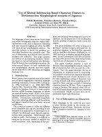
well with the lung lesions. However, IHC positive staining
cells were also found in macrophages located mainly in
the lung lesion at the cranioventral areas (Figure 1).
Virus isolation
There were no differences in the virus titers in both exper-
imental SIV-infected groups. The virus titers at 2 dpi were
between 10
2
– 10
3
TCID
50
/ml either from lung tissues or
from bronchoalveolar lavage fluid (BALF) in both
infected groups. There was no virus titre at 4 or 12 dpi nei-
ther from lung nor from BALF in both infected groups or
in the control pigs at any necropsied date (data not
shown).
RT-PCR
RT-PCR using specific primers for the matrix protein gene
to confirm the presence of SIV in the nasal swabs revealed
positive results only between 2–4 dpi in the H1N1-
Table 1: Percentages of gross lung lesions and the presence of
SIV-specific antigen based on immunohistochemistry (IHC)
staining
mock* H1N1** H3N2**
dpi Pig 1 (%) Pig 1 (%) Pig 2 (%) Pig 1 (%) Pig 2 (%)
2 0.0(-) 36.0(+) 33.0(+) 20.0(+) 2.0(+)
4 0.0(-) 5.0(+) 3.0(+) 2.0(+) 1.0(-)
12 0.0(-) 6.0(+) 0.5(-) 0.0(-) 0.0(-)
*n = 1, **n = 2.
dpi = days post infection
(-) negative antigen detection by IHC, (+) positive antigen detection
by IHC
Virology Journal 2009, 6:34 />Page 3 of 11
(page number not for citation purposes)
infected group and only at 2 dpi in the H3N2-infected
group. SIV genetic material was not found in the nasal
swabs or the sera of the negative control pigs.
Haemagglutination-inhibition (HI) assay
Sera from pigs in the control group had no antibody titres (≤
1:10 HI titre) to either H1N1 or H3N2 subtypes at all col-
lected dates. Sera from pigs in the H3N2-infected group had
no antibody titres (≤ 1:10 HI titre) to the H1N1 subtype.
Similarly, sera from pigs in the H1N1-infected group had no
antibody titres (≤ 1:10 HI titre) to the H3N2 subtype. H3N2-
infected pigs had 1:40 HI titre at 4 and 12 dpi to the H3N2
subtype. Interestingly, H1N1-infected group had the HI titre
(1:160 HI titre) to the H1N1 subtype at 12 dpi.
Pathogenic bacterial culture and identification
Pathogenic bacterial culture yielded negative results from
the tracheal swabs and lung tissues in all groups at 2, 4
and 12 dpi. In addition, Mycoplasma hyopneumoniae were
not found by PCR tested from lung tissues in all pigs.
DNA Sequencing and Phylogenetic analysis
The nucleotide sequences in this study contained 1700 bp
(36–1736) HA gene of H1N1, 1686 bp (37–1723) HA
gene of H3N2 and 1409 bp NA gene of both subtypes
from the selected Thai isolates. The HA and NA gene
nucleotide sequences of the Thai isolates of both subtypes
from Chonburi province in this study were analyzed by
BLAST (Basic Local Alignment Search Tool) program
available at /> demon-
strating that both HA and NA sequences of the H1N1
virus had the highest homology to A/swine/Chonburi/
05CB1/2005 (H1N1) and the H3N2 virus had the highest
homology to A/swine/Chonburi/05CB2/2005 (H3N2)
(Table 2). Geographic influence definitely played a major
role in those studied viruses.
SIV antigen staining by IHC, (A) negative control, (B) dark brown staining cells (arrow) of the SIV-positive control, (C) SIV-pos-itive staining on alveolar epithelial cells and (D) bronchiolar epithelial cells.Figure 1
SIV antigen staining by IHC, (A) negative control, (B) dark brown staining cells (arrow) of the SIV-positive
control, (C) SIV-positive staining on alveolar epithelial cells and (D) bronchiolar epithelial cells.
A
B
C
D
Virology Journal 2009, 6:34 />Page 4 of 11
(page number not for citation purposes)
Phylogenetic analysis of the HA gene from the selected
isolates revealed that nucleotide sequences of H1 viruses
had three major clusters, classical swine, avian and
human influenza virus (Figure 2) and nucleotide
sequences of H3 viruses had four major clusters, Euro-
pean, American and Asian swine, avian and human influ-
enza viruses (Figure 3). Phylogenetic analysis of the NA
gene from the selected isolates revealed that nucleotide
sequences of N1 viruses contained three clusters, Ameri-
can swine (classical swine), Human, and European swine
and avian influenza virus as shown in Figure 4. The nucle-
otide sequences of N2 viruses from the selected isolates
had three clusters, American and Asian swine, European
swine and human and avian influenza virus as shown in
Figure 5. The results showed that the nucleotide sequences
of the HA [GenBank:FJ688266
] gene of A/Thailand/HF6/
05 (H1N1) belonged to the classical swine H1 lineage and
the NA [GenBank:FJ688267
] gene belonged to avian N1
lineage, and both HA [GenBank:FJ688268
] and NA [Gen-
Bank:FJ688269
] genes of A/Thailand/S1/05 (H3N2)
belonged to the human H3N2 lineage.
Discussion
The results demonstrated that both SIV subtypes (Thai
isolates) were able to induce the flu-like symptoms and
lesions compatible with viral pneumonia in the craniov-
entral areas and were able to cause broncho-interstitial
pneumonia similar to previous reports [4,10-12]. The pig
lung is certainly the major site of swine influenza virus
replication [1] since we did not find any viraemic pig or
SIV antigen detection outside the lung tissue at any day of
infection. IHC and RT-PCR seemed to be more sensitive
than the virus isolation. The course of infection was lim-
ited to less than a week in both SIV-infected groups as SIV
antigen detection was found positive only at 2–4 dpi. The
SIV antigen was found in the nuclei of the bronchial and
bronchiolar epithelial cells, pneumocytes and pulmonary
macrophages with similar levels in both SIV-infected
groups indicating no differences between the two sub-
types in the viral protein production or replication. It
should be noted that both studied Thai isolates of both
subtypes replicated only in the respiratory tract of pigs and
shed the virus in the nasal secretions similar to other SIV.
The clinical respiratory signs and lung pathology in swine
influenza-infected pigs are commonly induced by the pro-
inflammatory cytokines such as IFN-α, TNF-α, IL-1 α and
β, and IL-6 in bronchoalveolar lavage fluids and the
amount of viral load in the lung tissue [13]. Our results
showed that pigs in both SIV-infected groups showed
more severe clinical respiratory signs and had higher body
temperature compared to the control pigs. Although,
none of the studied pigs had fever, pigs in both SIV-
infected groups had slight elevated body temperature.
However, cytokines responsible for those clinical signs
and lesions were not done in this study.
It is generally believed that there is no cross-protection
between H1N1 and H3N2 viruses [9]. The HI test between
H1N1 and H3N2-infected groups had no cross HI anti-
body reaction in this study. Commonly, infection with
H3N2 viruses induces higher HI titers than those of H1N1
infection [9,14]. Wang et al. [7] found that the cleavage
site of HA antigen of H3 virus (A/Panama/2007/99) was
cleaved easier than that of H1 virus (A/New Caledonia/
20/99) in transiently transfected 293T cells possibly mak-
ing the H3 viruses more immunogenic. Similarly, a
recombinant vaccine study in elderly humans using A/
Panama/2007/99 (H3N2), A/New Caledonia/20/99
(H1N1) or influenza virus type B as virus sources showed
that H3N2 virus induced significant higher HI titres than
that of H1N1 virus [15]. In contrast, the H1N1-infected
pigs in this study induced higher HI titres than that of the
H3N2-infected pigs at 12 dpi. Similarly, Kitikoon et al.
[14] showed that HI titre of the H1N1-infected pigs at 7
dpi was higher than that of the H3N2-infected pigs. How-
ever, the HI titres at 14 and 21 dpi in the H3N2 virus-
Table 2: Subtype and homology analysis of HA and NA genes of the H1N1 and H3N2 challenge viruses
Virus Subtype Virus with homology of HA gene %* Virus with homology of NA gene %*
A/swine/Thailand/HF6/05 H1N1 A/swine/Chonburi/05CB1/05 (H1N1) 100 A/swine/Chonburi/05CB1/05 (H1N1) 100
A/swine/Chonburi/06CB2/06 (H1N1) 99 A/swine/Chonburi/06CB2/06 (H1N1) 99
A/Thailand/271/05 (H1N1) 97 A/Thailand/271/05 (H1N1) 97
A/swine/Tennessee/4/78 (H1N1) 91 A/Swine/England/195852/92 (H1N1) 93
A/swine/Tennessee/2/78 (H1N1) 91 A/swine/Cotes d'Armor/1488/99 (H1N1) 93
A/swine/Thailand/S1/05 H3N2 A/swine/Chonburi/05CB2/05 (H3N2) 99 A/swine/Chonburi/05CB2/05 (H3N2) 99
A/swine/Nakhon pathom/NIAH586-1/05
(H3N2)
97 A/swine/Nakhon pathom/NIAH586-1/05
(H3N2)
97
A/swine/Nakhon pathom/NIAH586-2/05
(H3N2)
96 A/swine/Nakhon pathom/NIAH586-2/05
(H3N2)
96
A/swine/Chachoengsao/NIAH586/05 (H3N2) 98 A/swine/Chachoengsao/NIAH586/05 (H3N2) 95
A/Albany/20/74(H3N2) 88 A/Albany/20/74 (H3N2) 90
* Percent homology of nucleotide sequence from BLAST analysis
Virology Journal 2009, 6:34 />Page 5 of 11
(page number not for citation purposes)
Phylogenetic tree of the HA [GenBank:FJ688566] gene of 1700 bp (36–1736) H1 influenza A virusesFigure 2
Phylogenetic tree of the HA [GenBank:FJ688566
] gene of 1700 bp (36–1736) H1 influenza A viruses.
Virology Journal 2009, 6:34 />Page 6 of 11
(page number not for citation purposes)
Phylogenetic tree of the HA [GenBank:FJ688268] gene of 1686 bp (37–1723) H3 influenza A virusesFigure 3
Phylogenetic tree of the HA [GenBank:FJ688268
] gene of 1686 bp (37–1723) H3 influenza A viruses.
Virology Journal 2009, 6:34 />Page 7 of 11
(page number not for citation purposes)
infected pigs were higher in that study. Different viruses
used in the studies may yield different results due to the
origin of those isolates. Interestingly, the antigenic site of
the H1N1 used in this study contained the changed in the
amino acid sequence of haemagglutinin which may result
in increased immunogenicity and induced more severe
clinical diseases. However, the antigenic site of the H3N2
virus used in this study had no amino acid changes. In
addition, we did not have the HI results after 12 dpi in our
study.
Recently, genetic analysis of the HA gene of Thai SIV [16]
showed that the recent Thai H1N1 virus belonged to the
classical swine H1N1 lineage and had the highest homol-
ogy to the human isolate A/Thailand/271/05(H1N1). The
H3N2 subtype belonged to the human H3N2 lineage and
Phylogenetic tree of the NA [GenBank:FJ688267
] gene of 1409 bp N1 influenza A virusesFigure 4
Phylogenetic tree of the NA [GenBank:FJ688267
] gene of 1409 bp N1 influenza A viruses.
Virology Journal 2009, 6:34 />Page 8 of 11
(page number not for citation purposes)
Phylogenetic tree of the NA [GenBank:FJ688269] gene of 1409 bp N2 influenza A virusesFigure 5
Phylogenetic tree of the NA [GenBank:FJ688269
] gene of 1409 bp N2 influenza A viruses.
Virology Journal 2009, 6:34 />Page 9 of 11
(page number not for citation purposes)
showed highest homology to the H3N2 human influenza
virus from the 1970s A/Bilthoven/2600/75(H3N2) [16].
Similarly, in our study nucleotide sequences of both HA
and NA genes of A/Thailand/HF6/05(H1N1) also
belonged to the classical swine H1N1 lineage and A/Thai-
land/S1/05(H3N2) belonged to the human H3N2 line-
age. Since those viruses were isolated from the same
province and in the same year, geographic distribution
could be an explanation for the similarity of those viruses.
Interestingly, both viruses in this study were isolated from
the same farm in August 2005. We did not find any
genetic shift in those viruses or any reassortant virus such
as H1N2 subtype occurring in the farm (unpublished
data). Whole genome sequencing of all 8 genes is needed
to be evaluated in both viruses. It is possible that the new
reassortant virus might occur in Thailand since both stud-
ied viruses were isolated from the same farm. However,
our results showed that the Thai SIV subtypes, H1N1 and
H3N2, are still circulating in the pig population in Thai-
land since the first report in 1990. Our studied viruses
were able to induce flu-like lesions in weanling pigs. Sur-
veillance and molecular investigation of SIV in the coun-
try should be done continuously to prevent the future
emerging or re-emerging influenza A virus in pigs popula-
tion.
Conclusion
The results of this study may assist in the prevention and
control of SIV infection in Thailand, especially for H1N1
and H3N2 subtypes. Based on the percentages of craniov-
entral pneumonic lesion and times of virus shedding, the
H1N1 virus might play a major role in respiratory diseases
in weanling pigs in that farm. More works are needed in
the co-infection model with other respiratory pathogens
and in the prevention and control of the SIV-related dis-
eases in Thailand. In this study, investigations on virus
transmissibility between sentinel animals housing
together with infected animals were not performed.
Therefore, whether these Thai H1N1 and H3N2 subtypes
will be transmitted efficiently in the field situation
requires further experimental and epidemiologic studies.
Methods
Viruses
Swine influenza virus subtype H1N1 (A/swine/Thailand/
HF6/05) and H3N2 (A/swine/Thailand/S1/05) were iso-
lated from weanling pigs with respiratory signs in the
same farm in Chonburi province in 2005. The viruses
were propagated in embryonated chicken eggs for 3 pas-
sages and the virus concentration (10
7
TCID
50
/ml) was
evaluated in Madin-Darby canine kidney (MDCK) cells
and stored at -80°C until used.
Experimental DesignAnimal usage and handling protocols
were approved by Chulalongkorn University-Faculty of
Veterinary Science Animal Care and Use Committee (pro-
tocol No. 55/2549). The animals were housed at the BSL2
animal facility of the National Institute of Animal Health,
Bangkok, Thailand throughout the experiment. Fifteen 22-
day-old crossbred SPF pigs serologically negative for por-
cine reproductive and respiratory syndrome virus (PRRSV),
Pseudorabies virus (PRV), Mycoplasma hyopneumoniae and
SIV were obtained from a commercial farm and assigned to
3 different groups. The infected groups contained six pigs
each and were intratracheally inoculated with H1N1 and
H3N2 viruses (5 ml of 10
7
TCID
50
/ml) in group 1 and 2,
respectively. Group 3 served as a negative control group
containing three pigs which were mockedly infected with 5
ml of media (minimal essential medium (MEM)). Two pigs
from the infected groups and one pig from the control
group were necropsied at 2, 4 and 12 days post-infection
(dpi). Nasal swabs were collected at 0, 1, 2, 3, 4, 5, 7, 10 and
12 dpi. Following collection, nasal swabs were immedi-
ately placed in the infected MEM with 5% BSA, 300 U/ml
penicillin, 300 μg/ml streptomycin and 1–2 μg/ml trypsin)
and stored at -80°C for evaluation of virus shedding. At
each necropsy, sera were collected for assessing antibody
response, bronchoalveolar lavage fluid (BALF) were col-
lected for virus isolation and tissue samples from all organs
were collected for microscopic lesion detection. In addi-
tion, tracheal swabs were tested for bacterial culture and
identification.
Clinical evaluation
Pigs were evaluated daily for respiratory disease symp-
toms including coughing, sneezing, tachypnea, dyspnea,
nasal discharge and conjunctivitis. Pigs were observed and
scored for the respiratory signs (ranged from 0 to 5) as
previously described [12]. Rectal temperature was also
measured daily. Fever was recorded when the rectal tem-
perature ≥ 40°C.
Pathological evaluation
At necropsy, significant macroscopic lesions of all organs
and percentage of lung lesions were recorded. Tissue sam-
ples from all lung lobes were collected and immediately
identified for the presence of SIV-antigen by immunoflu-
orescence assay (IFA) using anti-influenza A nucleopro-
tein monoclonal antibody (HB654404 B.V. EUROPEAN
VETERINARY LABORATORY, the Netherlands).
Tissue samples from all organs were fixed in 10% neutral
buffered-formalin, processed and embedded in paraffin
as described previously [4]. Sections were cut approxi-
mately 4–6 μm thick for histopathological and immuno-
histochemistry (IHC) tests using anti-influenza A
nucleoprotein monoclonal antibody as previously
described [17]. Microscopic lesions were scored as previ-
ously described [18] and the scores were ranged from 0 to
3; 0 = normal bronchioles, 1 = mild bronchiolitis, 2 =
moderate bronchiolitis, 3 = severe bronchiolitis.
Virology Journal 2009, 6:34 />Page 10 of 11
(page number not for citation purposes)
Virus isolation
SIV was isolated from nasal swabs, sera, BALF and lung tis-
sues according to Kitikoon et al. [12] and the titres
(TCID
50
/ml) were calculated according to Reed and
Muench [19]. Briefly, ten-fold serial dilutions of the nasal
swab solution were inoculated onto MDCK cells and incu-
bated until the cytopathic effect (CPE) was observed for at
least 3 cell culture passages. Virus was identified by influ-
enza A virus-specific staining. Prior to staining, cells were
fixed with 4% phosphate-buffered formalin and washed
with 0.5% Tween-20 in PBS (washing solution). Subse-
quently, the cells were incubated for 1 h with anti-influ-
enza A nucleoprotein monoclonal antibody diluted
1:1,000 in the washing solution containing 1% BSA
(diluting solution). After washing, the cells were incu-
bated 1 h with the rabbit anti-mouse IgG conjugated
horseradish peroxidase (Dako Cytomation, Carpinteria,
California) diluted 1:400 with the diluting solution. The
color was developed using a chromogen aminoethyl car-
bazole substrate (Sigma, St. Louis, Missouri). Each proce-
dure contained mock-infected negative control cells and
positive control cells infected with a known-titreed virus.
RT-PCR
Sera and nasal swabs were performed to evaluate viraemia
and virus shedding using M-gene specific RT-PCR as pre-
viously described [10]. Viral RNAs were extracted using
QIAamp Viral RNA Mini Kit (Qiagen, Valencia, CA) from
200 μl volume of each nasal swabs and sera. The RT-PCR
was performed using Promega One step RT-PCR
(Promega, USA) containing a specially formulated
enzyme blend for both reverse transcription and PCR. The
forward and reward primers were 5' TGA TCT TCT TGA
AAA TTT GCA G 3'and 5' TGT TGA CAA AAT GAC CAT CG
3', respectively [20]. The expected amplicon size is 276 bp.
The RT-PCR was run at 94°C for 3 min for reverse tran-
scription followed by 30 cycles of denaturing at 94°C for
30 s, annealing at 55°C for 30 s and extension at 72°C for
45 s and ended with a final extension step at 72°C for 7
min. The amplified PCR products were analyzed on a
1.5% agarose gel electrophoresis.
Haemagglutination-inhibition (HI) assay
Sera were evaluated for the HI antibody titres to both of
the SIV subtypes, H1N1 and H3N2 using 0.5% chicken
erythrocytes for haemagglutination. All sera were
absorbed with Trypsin-Heat-Periodate to reduce nonspe-
cific inhibitors before HI testing [3]. Virus antigens uti-
lized in the HI assays were the challenge viruses, H1N1
(A/swine/Thailand/HF6/05) and H3N2 (A/swine/Thai-
land/S1/05).
DNA Sequencing
Full-length HA and NA genes of the studied subtype
H1N1 and H3N2 were amplified using universal and spe-
cific primers [21,22] with some modifications. The RT-
PCR products were analyzed by 1% agarose gel electro-
phoresis, purified by NucleoSpin Extract II (Macherey-
Nagel, Düren, Germany) and cloned into pGEMT Easy
(Promega, Madison, WI, USA). Plasmids containing the
viral genes were purified by NucleoSpin Plasmid (Mach-
erey-Nagel, Düren, Germany) and sequenced by using
synthetic oligonucleotides. Sequence data were edited and
analyzed using Bioedit software. The phylogenetic trees
were conducted in MEGA4 [23] using neighbor-joining
method with 1000 times bootstrapping replicates [24].
Competing interests
The authors declare that they have no competing interests.
Authors' contributions
DS carried out the virology, pathology, molecular genetic
studies and animal experiment, analyzing data and draft-
ing the manuscript, RK helped in animal work and molec-
ular work, ST helped animal work and pathology, PK
virology and revising the manuscript, RT experimental
design, pathology and drafting the manuscript and final
approval. All authors have read and approved the final
manuscript.
Acknowledgements
The authors would like to thank Drs. S. Kesdangsakonwut, T. Paphavasithi
and R. Tantilertcharoen and Mr. S. Wangnaitham for their technical assist-
ance and Dr. Sudarat Damrongwatanapokin, United States Agency for
International Development (USAID)/Regional Development Mission-Asia
for her kindness as an external examiner. This research was supported by
The Rachadapiseksompoch Endowment Fund #R011_2550 and the
National Research Council of Thailand #2551-231.
References
1. Dee SA: Respiratory Disease of Pigs. In The Merck Veterinary Man-
ual 9th edition. Pensylvania: National Publishing Inc; 2005:1228.
2. Easterday BC, Van Reeth K: Swine Influenza. Desease of Swine; Iowa
8th edition. 1999:277-287.
3. Webster R, Cox N, Stohi K: WHO Manual on Animal Influenza Diagno-
sis and Surveillance World Health Organization; 2002:15-67.
4. Thacker EL, Thacker BJ, Janke BH: Interaction between Myco-
plasma hyopneumoniae and Swine Influenza Virus. J Clin
Microbiol 2001, 39:2525-2530.
5. Kupradinun S, Peanpijit P, Bhodhikosoom C, Yoshioka Y, Endo A,
Nerome K: The first isolation of swine H1N1 influenza viruses
from pigs in Thailand. Arch Virol 1991, 118:289-297.
6. Damrongwatanapokin S, Pinychon W, Parchariyanon S, Damrongwa-
tanapokin T: Serological study and isolation of influenza A
virus infection of pigs in Thailand. Proceedings of the 19th IPVS
Congress; 16–19 July 2006; Denmark 2006:136.
7. Wang S, Taaffe J, Parker C, Solorzano A, Cao H, Garcia-Sastre A, Lu
S: Hemagglutinin (HA) Proteins from H1 and H3 Serotypes
of Influenza A Viruses Require Different Antigen Designs for
the Induction of Optimal Protective Antibody Responses as
Studied by Codon-Optimized HA DNA Vaccines. J Virol 2006,
80:11628-11637.
8. Saurwein-Teissl M, Steger MM, Gluck R, Cryz S, Grubeck-Loeben-
stein B: Influenza vaccination in a healthy geriatric popula-
tion: preferential induction of antibodies specific for the
H3N2 influenza strain despite equal T cell responsiveness to
all vaccine strains. Vaccine 1998, 16:196-200.
9. Van Reeth K, Gregory V, Hay A, Pensaert M: Protection against a
European H1N2 swine influenza virus in pigs previously
Publish with BioMed Central and every
scientist can read your work free of charge
"BioMed Central will be the most significant development for
disseminating the results of biomedical research in our lifetime."
Sir Paul Nurse, Cancer Research UK
Your research papers will be:
available free of charge to the entire biomedical community
peer reviewed and published immediately upon acceptance
cited in PubMed and archived on PubMed Central
yours — you keep the copyright
Submit your manuscript here:
/>BioMedcentral
Virology Journal 2009, 6:34 />Page 11 of 11
(page number not for citation purposes)
infected with H1N1 and/or H3N2 subtypes. Vaccine 2003,
21:1375-1381.
10. Landolt GA, Karasin AI, Phillips L, Olsen CW: Comparison of the
pathogenesis of two genetically different H3N2 influenza A
viruses in pigs. J Clin Microbiol 2003, 41:1936-1941.
11. Jung K, Ha Y, Chae C: Pathogenesis of swine influenza virus sub-
type H1N2 infection in pigs. J Comp Pathol 2005, 132:179-184.
12. Kitikoon P, Nilubol D, Erickson BJ, Janke BH, Hoover TC, Sornsen
SA, Thacker EL: The immune response and maternal antibody
interference to a heterologous H1N1 swine influenza virus
infection following vaccination. Vet Immunol Immunopathol 2006,
112:117-128.
13. Van Reeth K: Cytokines in the pathogenesis of influenza. Vet
Microbiol 2000, 74:109-116.
14. Kitikoon P, Strait EL, Thacker EL: The antibody responses to
swine influenza virus (SIV) recombinant matrix 1 (rM1),
matrix 2 (M2), and hemagglutinin (HA) proteins in pigs with
different SIV exposure. Vet Microbiol 2008, 126:51-62.
15. Treanor JJ, Schiff GiM, Couch RB, Cate TR, Brady RC, Hay CM, Wolff
M, She D, Cox MM: Dose-Related Safety and Immunogenicity
of a Trivalent Baculovirus-Expressed Influenza-Virus
Hemagglutinin Vaccine in Elderly Adults. J Infect Dis 2006,
193:1223-1228.
16. Chutinimitkul S, Thippamom N, Damrongwatanapokin S, Payungporn
S, Thanawongnuwech R, Amonsin A, Boonsuk P, Sreta D, Bunpong N,
Tantilertcharoen R, et al.: Genetic characterization of H1N1,
H1N2 and H3N2 swine influenza virus in Thailand. Arch Virol
2008, 153:1049-1056.
17. Haines DM, Waters EH, Clark EG: Immunohistochemical detec-
tion of swine influenza A virus in formalin-fixed and paraffin-
embedded tissues. Can J Vet Res 1993, 57:33-36.
18. Richt JA, Lager KM, Janke BH, Woods RD, Webster RG, Webby RJ:
Pathogenic and Antigenic Properties of Phylogenetically
Distinct Reassortant H3N2 Swine Influenza Viruses Cocircu-
lating in the United States. J Clin Microbiol
2003, 41:3198-3205.
19. Reed LJ, Muench H: A simple method of estimating fifty per-
cent endpoints. Am J Hyg 1938, 27:493-497.
20. Payungporn S, Phakdeewirot P, Chutinimitkul S, Theamboonlers A,
Keawcharoen J, Oraveerakul K, Amonsin A, Poovorawan Y: Single-
step multiplex reverse transcription-polymerase chain reac-
tion (RT-PCR) for influenza A virus subtype H5N1 detection.
Viral Immunol 2004, 17:588-593.
21. Hoffmann E, Stech J, Guan Y, Webster RG, Perez DR: Universal
primer set for the full-length amplification of all influenza A
viruses. Arch Virol 2001, 146:2275-2289.
22. Zou S: A practical approach to genetic screening for influenza
virus variants. J Clin Microbiol 1997, 35:2623-2627.
23. Tamura K, Dudley J, Nei M, Kumar S: MEGA4: Molecular Evolu-
tionary Genetics Analysis (MEGA) Software Version 4.0. Mol
Biol Evol 2007, 24:1596-1599.
24. Saitou N, Nei M: The neighbor-joining method: a new method
for reconstructing phylogenetic trees. Mol Biol Evol 1987,
4:406-425.