Báo cáo khoa học: "A novel virus that infecting hypovirulent strain XG36-1 of plant fungal pathogen Sclerotinia sclerotiorum" potx
Bạn đang xem bản rút gọn của tài liệu. Xem và tải ngay bản đầy đủ của tài liệu tại đây (4.18 MB, 9 trang )
BioMed Central
Page 1 of 9
(page number not for citation purposes)
Virology Journal
Open Access
Research
A novel virus that infecting hypovirulent strain XG36-1 of plant
fungal pathogen Sclerotinia sclerotiorum
Liyan Zhang
1,2
, Yanping Fu
2
, Jiatao Xie
2
, Daohong Jiang*
1,2
, Guoqing Li
1,2
and Xianhong Yi
2
Address:
1
State Key Laboratory of Agricultural Microbiology, Huazhong Agricultural University, Wuhan 430070, Hubei Province, PR China and
2
The Provincial Key Lab of Plant Pathology of Hubei Province, College of Plant Science and Technology, Huazhong Agricultural University,
Wuhan, 430070, Hubei Province, PR China
Email: Liyan Zhang - ; Yanping Fu - ; Jiatao Xie - ;
Daohong Jiang* - ; Guoqing Li - ; Xianhong Yi -
* Corresponding author
Abstract
Background: Sclerotinia sclerotiorum is a notorious plant fungal pathogen which spreads across the
world. Hypovirulence is a phenomenon where the virulence of fungal pathogens is decreased, even
lost, due to mycovirus infection. The potential of hypoviruses for biological control of the chestnut
blight fungus (Cryphonectria parasitica) has attracted much interest, and has led to discovery of new
hypovirulent strains in other fungi.
Results: A hypovirulent strain, strain XG36-1, was isolated from a typical lesion on the stem of
rapeseed (Brassica napus) caused by Sclerotinia sclerotiorum. Strain XG36-1 grew on PDA very slowly
(average 2.5 ± 0.1 mm/d) with sectoring, and developed abnormal colony morphology with few
sclerotia. Unlike health strains (such as wildtype strain XG-13), it was unable to induce lesions on
detached leaves of rapeseed. Sclerotia of strain XG36-1 produced apothecia rarely. A sexual
progeny test showed that the phenotypes of all 104 sexual progeny were not different from
wildtype strain XG-13 which shows normal phenotype of S. sclerotiorum, and protoplast
regeneration tests showed that 25.5% of the regenerants of strain XG36-1 were recovered fully.
Furthermore, the hypovirulence and its associated traits could be transmitted to XG36-1A34
R
, a
hygromycin-resistance gene labelled sexual progeny of strain XG36-1, by hyphal anastomosis.
Transmission electron microscope (TEM) observation showed that the cytoplasm of strain XG36-
1 was destroyed and granulated; the membranes of nuclei and mitochondria were disintegrated;
and mitochondrial cristae were cavitated. Viral particles (about 40 nm) in hyphae of strain XG36-
1, but not in its sexual progeny and wildtype strain XG-13, could be observed with TEM, and
several virus-like particles were uniquely enveloped by single layer membrane in the cells of strain
XG36-1. Furthermore, the viral particles could be co-transmitted with the hypovirulence traits
through hyphal anastomosis.
Conclusion: Hypovirulence and its associated traits of strain XG36-1 could be mediated by a
fungal virus. Currently, we could not know the characteristic of this virus, but it likely represent a
new type of mycovirus in S. sclerotiorum, and possibly in fungi.
Published: 7 July 2009
Virology Journal 2009, 6:96 doi:10.1186/1743-422X-6-96
Received: 26 May 2009
Accepted: 7 July 2009
This article is available from: />© 2009 Zhang et al; licensee BioMed Central Ltd.
This is an Open Access article distributed under the terms of the Creative Commons Attribution License ( />),
which permits unrestricted use, distribution, and reproduction in any medium, provided the original work is properly cited.
Virology Journal 2009, 6:96 />Page 2 of 9
(page number not for citation purposes)
Background
Rapeseed (Brassica napus) is one of the most important
oilseed crops, and offers the potential for biodiesel pro-
duction to relieve the pressure of the current energy short-
age. The area planted with rapeseed in China is currently
7.4 million hectares, and the Chinese government encour-
ages farmers to plant more winter rapeseed during late fall
to early summer in central China />gzdt/2008-01/11/content_855731.htm. Sclerotinia sclero-
tiorum (Lib.) de Bary is an important fungal plant patho-
gen which damages a wide variety of crops throughout the
world [1]. In China, this fungus causes stem rot of rape-
seed and is responsible for serious losses every year; in
2008, more than 15–70% of rapeseed plants were killed
by this pathogen in Hubei Province. Due to the shortage
of disease-resistant cultivars, chemical control is currently
the only choice to control stem rot. However, there are
problems associated with chemical control of stem rot.
Firstly, fungicide control requires treatment during the
bloom stage of rapeseed, but this is not practical because
the chemical does not arrive at the stems efficiently
through heavy canopy. Secondly, fungicide-resistant
strains of S. sclerotiorum have been frequently isolated in
the field [2]. Non-fungicidal alternatives for the control of
stem rot of rapeseed are necessary.
Hypovirulence or hypovirulence is a phenomenon where
the virulence of fungal pathogens is decreased, even lost,
due to mycovirus infection. Hypovirulence was first
reported in the chestnut blight, a destructive disease
caused by Cryphonectria parasitica by Grente [3]. The suc-
cessful control of chestnut blight with hypovirulent
strains of C. parasitica represented an alternative approach
to biological control fungal diseases other than with myc-
oparasites and antagonists [4,5]. The potential of hypovir-
ulence for biological control of plant fungal diseases has
Hypovirulence and its associated traits of Sclerotinia sclerotiorum strain XG36-1Figure 1
Hypovirulence and its associated traits of Sclerotinia sclerotiorum strain XG36-1. A, abnormal colony morphology
developed on 20 ml PDA plate at 20–22 C for 15 days, a typical colony morphology of S. sclerotiorum (strain XG-13) developed
at the same condition was showed as control. B and E, strain XG36-1 did not induce any typical lesion on detached leaf of
rapeseed (Brassica napus) as strain XG-13 did; lesions were photographed and measured at 60 hpi. C and D, comparing to
strain XG-13, strain XG36-1 grew on PDA plate slowly and produced less biomass (grown out on cellophane membranes on
top of 20 ml PDA plate at 20°C for 72 h).
Virology Journal 2009, 6:96 />Page 3 of 9
(page number not for citation purposes)
attracted much interest, and has lead to discovery of new
hypovirulent strains in other fungi. Other mycoviruses
causing hypovirulence or hypovirulence of plant fungal
pathogens include: mitovirus in C. parasitica [6], Ophios-
toma novo-ulmi [7], Sclerotinia homoeocarpa [8,9], Chalara
elegans [10] and Botrytis cinerea [11]; mycoreoviruses in C.
parasitica [12], and Rosellinia necatrix [13]; and some
unclassified mycoviruses, such as SsDRV in the family
Flexiviridae in S. sclerotiorum [14], DaRV in Diarporthe
ambigua [15], FgV-DK21 in Fusarium graminearum [16,17]
and a 33-nm isometric mycovirus in B. cinerea [18].
Hypovirulent strains have been reported in S. sclerotiorum,
such as isolate 91, strain Ep-1PN and isolate S10 [19-21];
and mycoviruses that associated with hypovirulence of S.
sclerotiorum were isolated from strain Ep-1PN [16,22].
Hypovirulence in S. sclerotiorum is likely common since
we often isolate some mild strains, even non-virulence
strains, from fields. In this paper, we report on a hypovir-
ulent strain isolated from a typical lesion on stem of rape-
seed and called XG36-1 which possibly differs from
previously reported hypovirulent strains of S. sclerotiorum.
Results
Strain XG36-1 showed hypovirulence phenotype
The colony of strain XG36-1 on PDA was thick with many
sectors at the colony margin; only a few sclerotia were pro-
duced and distributed in the colony irregularly. The col-
ony morphology of strain XG36-1 was abnormal and
obviously different from strain XG-13, a healthy wildtype
strain isolated from the same filed as strain XG36-1 (Fig
1A). Hypha extended slowly on PDA plate, and the
Diverse phenotypes of protoplast regenarants of S. sclerotiorum hypovirulent strain XG36-1Figure 2
Diverse phenotypes of protoplast regenarants of S. sclerotiorum hypovirulent strain XG36-1. A, three types of col-
ony morphologies, namely Type I, Type IIand Type III, were developed on PDA plate at 20–22 C for 15 days. Regenarants in
Type Iwere not significantly different from wildtype strain XG-13, while regenarants in Type III were similar to strain XG36-1
(see Figure 1), Type II were intermediate type between Type Iand Type III. B and C, comparison of the hyphal growth rate on
PDA plate and virulence on detached rapeseed leaves among three types of regenarants. The growth and virulence of rege-
narants in TypeI were fully recovered, and that in Type II were partially recovered; the growth and virulence of regenarants in
Type III was not significantly different from strain XG36-1.
Virology Journal 2009, 6:96 />Page 4 of 9
(page number not for citation purposes)
hyphal tips often branch excessively. The growth rate of
XG36-1 was 4.1 ± 0.1 mm/d, which was significantly
lower than 19.0 ± 1.1 mm/d found for a wildtype strain
XG-13 (Fig 1C). The biomass produced by strain XG36-1
on a 9-cm-diam. PDA plate was 8.6 ± 0.1 mg after 72 h
incubation, while that produced by strain XG-13 was 26.3
± 2.1 mg (Fig 1D). Unlike strain XG-13, strain XG36-1
produced fewer sclerotia in the mature colony, with the
average number of sclerotia at 5 sclerotia/plate, while
strain XG-13 had 12 sclerotia/plate. Strain XG36-1 was
almost incapable of inducing any lesions on detached
leaves of rapeseed at 60 h post inoculation (hpi), while
wildtype strain XG-13 could induce typical lesions on
detached leaves, averaging 2.75 ± 0.14 cm at 60 hpi (Fig
1B, E). Thus, the strain XG36-1 was judged to be a hypo-
virulent strain of S. sclerotiorum.
Multi-phenotype of protoplast regenerants of strain
XG36-1
Fifty-five protoplast regenerants of strain XG36-1 were
obtained. Their growth rates, colony morphology and
pathogenicity were tested, and the results showed that the
phenotypes of these regenerants were significantly
diverse. Based on growth rate and colony morphology,
these regenerants could divide into three groups, namely
TypeI, TypeII and Type III. Type I regenerants grew on
PDA just like wildtype strain XG-13, developing normal
colony morphology of S. sclerotiorum, and capable of
inducing typical lesions on detached leaves of rapeseed
(Fig 2). Approximately 25.5% of the regenerants (14/55)
belonged to Type I. Type II regenerants grew on PDA
much faster than hypovirulent parental strain XG36-1,
but slower than wildtype strain XG-13. These regenerants
could cover an entire 9-cm-diam. PDA plate by 14 days
with an average growth rate of 9.7 mm/d. These regener-
ants could induce small lesions on detached leaves with
an average size of 1.0 cm across (Fig 2). Approximately
23.8% of the regenerants (13/55) belonged to type II.
Type III regenerants were not significantly different from
hypovirulent parental strain XG36-1 (Fig 2), comprising
51.7% of the regenerants (28/55).
Normal phenotype of sexual progeny of strain XG36-1
Only a few of sclerotia of strain XG36-1 could be success-
fully induced to form apothecia. 104 single-ascospore-iso-
lation sexual progeny were obtained. The cultural
Sexual progeny of hypovirulent strain XG36-1 showed normal phenotypes of S. sclerotiorumFigure 3
Sexual progeny of hypovirulent strain XG36-1 showed normal phenotypes of S. sclerotiorum. A and B, the colony
morphology and virulence on rapeseed detached leaves of a randomly selected sexual progeny XG36-1A34; C, the growth rate
of 104 tested sexual progeny.
Virology Journal 2009, 6:96 />Page 5 of 9
(page number not for citation purposes)
characteristics and pathogenicity of these 104 sexual prog-
eny were tested, and the results showed that all sexual
progeny had a typical wildtype phenotype of S. sclerotio-
rum. Compared to strain XG-13, the grow rate, colony
morphology of sexual progeny were not significantly dif-
ferent (Fig 3).
Transmission of hypovirulence phenotype of strain XG36-1
After contacting with strain XG36-1 on PDA, the hyphae
around the colony margin of hygromycin-resistance gene
labelled sexual progeny XG36-1A34
R
branched excessively
(Fig. 4), subcultures from this region showed hypoviru-
lence traits (Fig 4). The growth rate, the sectoring, the col-
ony morphology, and the pathogenicity of infected XG36-
1A34
R
were not significantly different from strain XG36-1.
Furthermore, the hypovirulence phenotype obtained by
XG36-1A34
R
could be transmitted to XG36-1A34 and
other sexual progeny.
Virus particles observed in strain XG36-1
Under TEM, the cytoplasm of strain XG36-1 was seen to
be destroyed and granulated; the membranes of nuclei
and mitochondria were disintegrated. Only a few mito-
chondria were seen in cell, while, the mitochondrial cris-
tae were cavitated (Fig 5A). However, the nuclei and
mitochondria in wildtype strain XG-13 were not
destroyed, and the cytoplasm were well-distributed and
filled with plentiful mitochondria, nuclei and mitochon-
dria was not destroyed (Fig 5B). Viral particles could be
observed in the cells of strain XG36-1, but not in the cells
of wildtype strain XG-13 or sexual progeny XG36-1A34.
The viral particles were almost isometric, with a diameter
of ~40 nm. Several viral particles were enveloped by a sin-
gle layer membrane (Fig 5C). However, viral particles
could be observed in the cells of subcultures of strain
XG36-1A34
R
after contacting the colony of strain XG36-1.
Thus, the viral particles are transmissible and associated
Transmission of hypovirulence and its associated traits of strain XG36-1Figure 4
Transmission of hypovirulence and its associated traits of strain XG36-1. A, the colony of hygromycin-resistant gene
labelled strain XG36-1A34
R
was converted when dual culturing with hypovirulent strain XG36-1(red triangle). B, the hyphae at
the colony margin of strain XG36-1A34
R
branched excessively as strain XG36-1 did. C, converted strain XG36-1A34
R
also lost
virulence on detached rapeseed leaves.
Virology Journal 2009, 6:96 />Page 6 of 9
(page number not for citation purposes)
with hypovirulence of strain XG36-1. Viral particles could
also be extracted from the hyphae of strain XG36-1 after
ultra-centrifugation in a Cesium chloride (CeCl) gradient
medium, but only very few viral particles could be
observed through TEM.
Viral nucleic tides not extracted from strain XG36-1
All attempts to extract dsRNA were not successful. Dou-
ble-stranded RNA could not be extracted directly from
hyphae of strain XG36-1, but could be extracted from pre-
viously reported hypovirulent strain Ep-1PN (positive
control). No viral RNA sample could be extracted from the
pellets precipitated with ultracentrifugation.
Discussion
Our experiments showed that strain XG36-1 was a hypo-
virulent strain of S. sclerotiorum. The protoplast regener-
ants test suggested that the hypovirulence-associated
element (HAE) in cells of strain XG36-1 did not distribute
equally, and hypovirulence of regenerants derived from
protoplasts that without DAE or with a low concentration
of HAE were cured or partially cured. All tested sexual
progeny showed the wildtype phenotype of S. sclerotiorum
suggesting that chromosomal or DNA changes (nuclear
genomic mutations) were not responsible for the hypovir-
ulence of strain XG36-1, and that there is likely some
mechanism to eliminate HAE during sexual reproduction.
Transmission tests showed that the hypovirulence of
strain XG36-1 could be transmitted to wildtype strains
efficiently. The hypovirulence and its associated traits of
strain XG36-1 is similar to previously reported hypoviru-
lent strain Ep-1PN [14,20,23]. Thus, the HAE in strain
XG36-1 could be transmissible genetic elements.
In fungi, both fungal plasmids and mycoviruses are trans-
missible genetic elements, and both mycoviruses, and
some fungal plasmids may cause hypovirulence to their
hosts [24]. Fungal plasmids are not likely to be HAE of
strain XG36-1. Extra-chromosomal DNA segments were
not observed with agarose gel electrophoresis analysis of
whole DNA samples (data not shown). Meanwhile, viral
particles were observed in hyphae of strain XG36-1, but
not in its sexual progeny nor in wildtype strain XG-13,
and viral particles were always associated the transmission
of hypovirulence traits of strain GX36-1. Furthermore, the
observed elimination of viruses and hypovirulence during
sexual reproduction of strain XG36-1 is in accord with the
sexual reproductive behaviour of virus-infected ascomyce-
tous fungal hosts, where the viruses are not transmitted to
progeny [25]. Thus, the transmissible DAE in strain XG36-
1 is likely to be a mycovirus.
The viral particles in strain XG36-1 is possibly a new type
of mycovirus that infecting S. sclerotiorum. The distinct
characteristic of the viral particles in strain XG36-1 is that
the isometric viral particles are enveloped with a single
layer membrane which is possibly derived from its host.
Viral particles (double membranes bodies, DMB) have
been observed in hypovirulent isolate 275 of S. sclerotio-
rum [26], but not in hypovirulent strain Ep-1PN and iso-
late S10 [14,27]. Furthermore, the average size of DMBs in
isolate 275 was about 70 nm in diameter which was much
larger that that of viral particles in strain XG36-1.
This unique envelopment of particles has not been
observed in other mycoviruses which may or may not
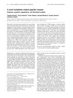
Ultrastructure and virus-like particles (viral particles) in the cell of S. sclerotiorum hypovirulent strain XG36-1 observed under transmission electron microscopes (TEM)Figure 5
Ultrastructure and virus-like particles (viral parti-
cles) in the cell of S. sclerotiorum hypovirulent strain
XG36-1 observed under transmission electron
microscopes (TEM). A, hyphal ultrastructure of hypoviru-
lent strain XG36-1, the cytoplasm was granulated, the mem-
branes of nuclei (N) and mitochondria (M) was disintegrated;
only a few mitochondria existed, but the mitochondrial cris-
tae was cavitated. B, hyphal ultrastructure of wildtype strain
XG-13, the cytoplasm was well-distributed, plentiful mito-
chondria, and the membranes of nuclei (N) and mitochondria
(M) was not destroyed. C, viral particles (white arrow) in cell
of hypovirulent strain XG36-1, the size of individual particle
is about 40 nm, several particles were enveloped by single-
layer membrane. D, A few viral particles (red triangles) could
be observed after negatively stained with 1% uranyl acetate
on carbon-coated 400 mesh copper grids. Ultrastructure
observation was carried out under FEI Tecnai G
2
20 TWIN
transmission electron microscope).
Virology Journal 2009, 6:96 />Page 7 of 9
(page number not for citation purposes)
encode coat protein. Viruses infect in all the major groups
of fungi kingdom, and RNA viruses in the family Chryso-
viridae, Hypoviridae, Narnaviridae (Mitovirus), Partitiviridae
and Totiviridae are typical fungal viruses [28]. However,
more and more fungal viruses were characterized on
molecular level, the plenty diversity of fungal viruses in
nature is becoming more and more clear. Viral particles in
strain XG36-1 are enveloped uniquely, and could cause
severe debilitation of host with low titre, suggest that virus
infecting strain XG36-1 is most likely to be a novel myco-
virus associated with hypovirulence of plant fungal path-
ogen.
Conclusion
Our work, here, proved that the hypovirulence and it asso-
ciated traits of S. sclerotiorum strain XG36-1 could not be
transfer to sexual progeny vertically, and the hypoviru-
lence associated element (HAE) is not distributed equally
in cells of strain XG36-1. Thus, the hypovirulence of strain
XG36-1 is not due to the genome mutation. Hypoviru-
lence and its associated traits could be transferred effi-
ciently to vegetative compatible strain XG36-1A34
R
, a
hygromycin resistance gene labelled sexual progeny of
strain XG36-1, through hyphal anastomosis. Thus, the
HAE in strain XG36-1 is a mobile element.
The cytoplasm of strain XG36-1 was granulated and not
well-distributed, the membranes of nuclei and mitochon-
dria were disintegrated; and mitochondrial cristae were
cavitated. Viral particles could be observed in cells of
strain XG36-1, but not in wildtype strain XG-13 and sex-
ual progeny XG36-1A34. Viral particles could also be
extracted with ultracentrifugation from the hyphae of
strain XG36-1. Although the viral nucleic acids were not
extracted and identified currently, however, comparing to
previously reported hypovirulence or debilitation associ-
ated mycoviruses, the virus in strain XG36-1 is unique; it
is most likely to be a novel mycovirus associated with
hypovirulence of plant fungal pathogen.
Methods
Fungal strains, media and culture
S. sclerotiorum strain XG36-1 was isolated from a typical
lesion on stem of rapeseed at Xiaogan County, Hubei
Province, P R China. Strain XG-13, a healthy wildtype
strain, was also isolated from another typical lesion in the
same rapeseed field as strain XG36-1. Hypovirulent strain
Ep-1PN was originally isolated from diseased eggplant
[21]. All strains and their derivatives were grown on PDA
(potato dextrose agar, PDA) at 20°C, and stored on PDA
slants at 4–6°C.
Comparison of cultural characteristics
Strains XG36-1 and XG-13 were maintained on Petri
dishes containing 20 ml PDA, and incubated at 20°C for
3 days. To assess growth rates, 5-mm-diameter agar disks
from actively growing colony margins of XG36-1 and XG-
13 were transferred onto 9-cm-diam Petri dishes contain-
ing 20 ml PDA, and then incubated at 20°C. The diameter
of colonies of XG-13 and XG36-1 was measured at 24
hour post inoculation (hpi) and 48 hpi, respectively; the
hyphal growth rate of the two strains was calculated as fol-
lows: growth rate (cm/d) = (48 hpi diam. - 24 hpi diam.)/
2. To compare the biomass between XG36-1 and XG-13
produced on PDA, these two strains were grown out on
cellophane membranes on top of PDA (20 ml) at 20°C
for 48 h, and then the mycelial mass was rolled from the
membrane, placed in an 80°C oven for 10 h, and the dry
weights were recorded. To compare the colony morphol-
ogy, the colonies were grown on 20 ml PDA plates at
20°C for up to 15 days.
Pathogenicity test of XG36-1
Agar disks (5-mm-diam.) from actively-growing colony
margins of strain XG36-1 and its derivatives and strain
XG-13 were placed on the leaves of rapeseed with the myc-
elial side facing the leaf surface, and then the inoculated
leaves were placed in an incubator at 20°C and 100% rel-
ative humility for 60 h. Lesion diameter on each inocu-
lated leaf was measured. There were five replicates for each
treatment.
Protoplast preparation and regeneration
To obtain protoplasts of strain XG36-1, mycelial-agar
discs (5-mm-diam.) cut from actively growing colony
margins of strain XG36-1 were transferred onto cello-
phane membranes overlaying PDA. After 2 days, the myc-
elia were collected from cellophane membranes, and then
ground with sterilized mortar and pestle to make hyphal
fragments. Approximately 1 ml of hyphal fragment mush
was transferred into a 250 ml flask containing 80 ml PDB
(Potato Dextrose Broth, PDB), and shaken at 150 rpm for
up to 20 h at 20°C. The filtrate was collected by passing
through two layers of sterilized cheesecloth, and the myc-
elium was washed twice with 100 ml of potassium chlo-
ride (KCl) buffer (0.6 mol/L). The mycelial mass was
squeezed to remove liquids, and then re-suspended with
digestion buffer which contained 1.5 mg/ml Lysing
enzymes from Trichoderma harzianum (Sigma-Aldrich,
Inc), and then incubated at 32°C for 3 h. The liquid was
filtered through four layers for sterilized cheesecloth and
than passed through two layers of sterilized filter paper to
remove the debris and undigested hyphal fragments. Pro-
toplasts were collected by centrifugation for 10 min at
4000 rpm. The precipitate was washed twice with 0.6 mol/
L KCl solution by re-suspension and centrifugation at
4000 rpm for 5 min. The final precipitate was re-sus-
pended with in 0.6 mol/L KCl solution to give 1–2 × 10
3
protoplasts/ml for regeneration. One hundred microliter
of protoplast suspension was gently mixed with 20 ml
Virology Journal 2009, 6:96 />Page 8 of 9
(page number not for citation purposes)
regeneration medium (RM: sucrose 0.7 M; yeast extract
0.5 g/L, agar 1.5 g/L, adding KCl to a final concentration
0.6 mol/L before use), and poured into a Petri dish (diam.
90 mm). The plates were incubated at 20–22°C for 3–4
days, and then small colonies were observed on the RM
plates and transferred onto fresh PDA plates. These sub-
cultures were considered as protoplast regenerants of
strain XG36-1. The cultural characteristics and patho-
genicity of regenerants were tested with measures
described above.
Sexual reproduction and progeny isolation
To collect sclerotia, strain XG36-1 was allowed to grow on
sterilized carrot at 20°C for up to one month. After that,
sclerotia were harvested and washed with tap water to
remove mycelia and debris, and dried at room tempera-
ture for up to two weeks. To induce carpogenic germina-
tion of sclerotia, the dry sclerotia were placed at -20°C for
up to one month, and then the low-temperature treated
sclerotia were surface sterilized with 70% ethanol and
sowed onto sterilized wet sand and incubated at 15–17°C
for up to two months. A few sclerotia then produced
apothecia. To obtain ascospores, mature apothecia were
placed into a 50 ml syringe with 10 mL sterile water, and
then the syringe was capped with silica gel, and then the
piston was pushed and pulled several times to allow
apothecia to release ascospores. The ascospores suspen-
sion was collected and adjusted to a concentration of ~10
3
spores/ml. To create mono-ascospore cultures, 200 ml of
the spore suspension was spreaded over a thin layer of
water agar (10 ml water agar in a 90-mm-diam. plate),
and then placed at 20°C for 24 h Under light microscopy,
the mycelium formed from a single ascospore was excised,
and transferred to fresh PDA plate. The cultural character-
istics and pathogenicity of mono-ascospore cultures were
assessed with the measures described above.
Transmission of hypovirulence
To test the possible transmission of hypovirulence from
strain XG36-1, a non-hypovirulent progeny of strain
XG36-1, strain XG36-1A34, was randomly selected for
labelling with hygromycin B resistance gene (hph) medi-
ated by Agrobacterium transformation [29]; and one hph-
labelled insert which was similar to strain XG36-1A34,
named as strain XG36-1A34
R
, was chosen for the trans-
mission test. Then strain XG36-1A34
R
was dual cultured
with strain XG36-1 in a PDA plate allowing the two colo-
nies to intermingle according to Jiang et al [23]. Mycelial
agar plugs at the colony margin of strain XG36-1A34
R
were placed onto fresh PDA containing 50 mg/mL hygro-
mycin (where unlabelled strains could not grow), and
placed at 20°C for 3 to 4 days. Mycelial plugs were taken
from the new colonies and transferred into fresh PDA
plate without any hygromycin. The cultural characteristics
and pathogenicity of subcultures of XG36-1A34
R
after
contacting strain XG36-1 were tested with measures
described above.
Transmission electron microscopy (TEM) observation
Strain XG36-1, strain XG-13, strain 36-1A34
R
and its sub-
cultures after contacting strain XG36-1 were grown on
PDA plates for 2–3 days at 20°C, and the mycelia of each
strain were collected for TEM observation (FEI Tecnai G
2
20 TWIN transmission electron microscope). The
approach for TEM observation followed Boland et al [26].
To extract viral particles, strain XG36-1 was grown out on
cellophane membranes on top of PDA for 3 days, and
mycelia were harvested for extracting virus-like particles
(viral particles) according to Ghabrial and Havens [30].
The viral particles were observed under TEM after negative
staining with uranyl-acetate. TEM observation was carried
out at Institute of Virology, Chinese Academy of Sciences,
Wuhan, P R China.
Extraction and confirmation of dsRNA
Mycelia for dsRNA isolation were grown out on cello-
phane membranes on top of PDA for 2 to 10 days, respec-
tively. Following harvesting, the mycelium was stored at
80°C before use. The procedure for dsRNA extraction
described by Xie et al [14] was used with minor modifica-
tions. The RNA sample was first digested with RNase-free
DNaseI, treated with S1 nuclease, and then subjected to
electrophoretic analysis on 1% agrose gel.
Data analysis
Each test had three to five replicates, and data from the
experiments were analyzed using an analysis of variance
(ANOVA) in SAS (SAS Software, NJ). Treatment means
were compared with the test of least significant difference
(LSD) at the p = 0.05.
Competing interests
The authors declare that they have no competing interests.
Authors' contributions
DJ, YF, LZ, JX and GL designed the experimental strategy;
LZ and JX conducted the experiments; DJ, LZ, YF, JX, GL
and XY were involved in the data analysis and their
processing; and DJ and YF wrote the manuscript. All
authors approved the final manuscript.
Acknowledgements
This work was financial supported by the Commonweal Specialized
Research Fund of China Agriculture (3–21) and Program for New Century
Excellent Talents in University (NCET-06-0665). We thank Dr Tom Hsiang
of the University of Guelph, Canada for his editorial assistance.
References
1. Boland GF, Hall R: An index of plant hosts susceptible to Scle-
rotinia sclerotiorum. Can J Plant Pathol 1994, 16:93-108.
Publish with Bio Med Central and every
scientist can read your work free of charge
"BioMed Central will be the most significant development for
disseminating the results of biomedical research in our lifetime."
Sir Paul Nurse, Cancer Research UK
Your research papers will be:
available free of charge to the entire biomedical community
peer reviewed and published immediately upon acceptance
cited in PubMed and archived on PubMed Central
yours — you keep the copyright
Submit your manuscript here:
/>BioMedcentral
Virology Journal 2009, 6:96 />Page 9 of 9
(page number not for citation purposes)
2. Shi ZQ, Zhou MG, Ye ZY, Shi JR, Chen HG, Wang YZ: Resistance
monitoring of Sclerotinia sclerotiorum to carbendazim. Jiangsu
Journal of Agricultural Science 2000, 16:226-229.
3. Grente J: Les formes hypovirulences d' Endothia parasitica et
les espoirs de lutte contrele chancre du chataignier. C R Acad
Agric France 1965, 51:1033-1037.
4. Anagnostakis SL: Biological control of chestnut blight. Science
1982, 215:466-471.
5. Anagnostakis SL, Chen B, Geletka LM, Nuss DL: Hypovirus trans-
mission to ascospore progeny by field-released transgenic
hypovirulent strains of Cryphonectria parasitica. Phytopathology
1998, 88:598-604.
6. Polashock JJ, Hillman BI: A small mitochondrial double-stranded
(ds)RNA element associated with a hypovirulent strain of
the chestnut blight fungus and ancestrally related to yeast
cytoplasmic T and W dsRNAs. Proc Natl Acad Sci U S A. 1994,
91(18):8680-8684.
7. Hong Y, Dover SL, Cole TE, Brasier CM, Buck KW: Multiple mito-
chondrial viruses in an isolate of the Dutch Elm disease fun-
gus Ophiostoma novo-ulmi. Virology 1999, 258:118-127.
8. Deng F, Xu R, Boland GJ: Hypovirulence-associated double-
stranded RNA from Sclerotinia homoeocarpa is conspecific
with Ophiostoma novo-ulmi mitovirus 3a-Ld. Phytopathology
2003, 93:1407-1414.
9. Deng F, Boland GJ: A satellite RNA of Ophiostoma novo-ulmi
mitovirus 3a in hypovirulent isolates of Sclerotinia homoeo-
carpa. Phytopathology 2004, 94:917-923.
10. Park Y, Chen XB, Punja ZK: Molecular and biological character-
ization of a mitovirus in Chalara elegans (Thielaviopsis basi-
cola). Phytopathology 2006, 96:468-479.
11. Wu MD, Zhang L, Li GQ, Jiang D, Hou M, Huang HC: Hypoviru-
lence and double-stranded RNA in Botrytis cinerea. Phytopa-
thology 2007, 97:1590-1599.
12. Hillman BI, Supyani S, Kondo H, Suzuki N: A reovirus of the fungus
Cryphonectria parasitica that is infectious as particles and
related to the Coltivirus genus of animal pathogens. Journal of
Virology 2004, 78:892-898.
13. Sasaki A, Kanematsu S, Onoue M, Oikawa Y, Nakamura H, Yoshida
K: Artificial infection of Rosellinia necatrix with purified viral
particles of a member of the genus Mycoreovirus reveals its
uneven distribution in single colonies. Phytopathology 2007,
97:278-286.
14. Xie J, Wei D, Jiang D, Fu Y, Li G, Ghabrial SA, Peng Y: Characteri-
zation of hypovirulence-associated mycovirus infecting the
plant-pathogenic fungus Sclerotinia sclerotiorum. J Gen Virol
2006, 87:241-249.
15. Preisig O, Moleleki N, Smit W, Wingfield BD, Wingfield MJ: A novel
RNA mycovirus in a hypovirulent isolate of the plant patho-
gen Diaporthe ambigua. J Gen Virol 2000, 81:3107-3114.
16. Chu YM, Jeon JJ, Yea SJ, Kim YH, Yun SH, Lee YW, Kim KH: Double-
stranded RNA mycovirus from Fusarium graminearum. Appl
Environ Microbiol 2002, 68:2529-2534.
17. Kwon SJ, Lim WS, Park SH, Park MR, Kim KH: Molecular charac-
terization of a dsRNA mycovirus, Fusarium graminearum
Virus-DK21, which is phylogenetically related to hypoviruses
but has a genome organization and gene expression strategy
resembling those of plant potex-like viruses. Mol Cells. 2007,
23(3):304-315.
18. Castro M, Kramer K, Valdivia L, Ortiz S, Castillo A: A double-
stranded RNA mycovirus confers hypovirulence-associated
traits to Botrytis cinerea. FEMS Microbiol Lett 2003, 228:87-91.
19. Boland GJ: Hypovirulence and double-stranded RNA in Scle-
rotinia sclerotiorum. Can J Plant Pathol 1992, 14:10-17.
20. Li G, Jiang D, Wang D, Zhu B, Rimmer R: Double-stranded RNAs
associated with the hypovirulence of Sclerotinia sclerotiorum
strain Ep-1PN. Progress in Natural Science 1999, 9:836-841.
21. Li G, Wang D, Huang HC, Zhou Q: Polymorphisms of Sclerotinia
sclerotiorum isolated from eggplant in Jiamusi, Heilongjiang
Province. ACTA Phytopathologica Sinica 1996, 26:237-242.
22. Liu H, Fu Y, Jiang D, Li G, Xie J, Peng Y, Yi X, Ghabrial SA: A novel
mycovirus that is related to the human pathogen Hepatitis E
virus and rubi-like viruses. Journal of Virology 2009, 83:1981-1991.
23. Jiang D, Li G, Fu Y, Yi X, Wang D: Transmissible hypovirulent
element in isolate Ep-1PN of Sclerotinia sclerotiorum. Chinese
Science Bulletin 1998, 43:779-781.
24. Ramesh M, Arunasalam N: Senescence in fungi: the view from
Neurospora. FEMS Microbiol Lett 2008, 280:135-143.
25. Buck KW: Fungal Virology-An overview. Fungal Virology
1986:85-108.
26. Boland GJ, Mould MJR, Robb J: Ultrastructure of a hypovirulent
isolate of Sclerotinia selerotiorum containing double-stranded
dsRNA. Physiol and Mol Plant Pathol 1993, 43:21-32.
27. Li G, Huang HC, Laroche A, Acharya SN: Occurrence and charac-
terization of hypovirulence in the tan sclerotial isolate S10 of
Sclerotinia sclerotiorum. Mycological Research 2003,
107:1350-1360.
28. Ghabrial SA, Suzuki N: Fungal viruses. In Encyclopedia of Virology Vol-
ume 2. 3rd edition. Edited by: Mahy BWJ, Van Regenmortel MHV.
Oxford, Elsevier; 2008:284-291.
29. Li M, Gong X, Zheng J, Jiang D, Fu Y, Hou M: Transformation of
Coniothyrium minitans, a parasite of Sclerotinia sclerotiorum,
with Agrobacterium tumefaciens. FEMS Microbiol Lett 2005,
243:323-329.
30. Ghabrial SA, Havens WM: The Helminthosporium victoriae 190S
mycovirus has two forms distinguishable by capsid protein
composition and phosphorylation state. Virology 1992,
188:657-665.