báo cáo khoa học: " Lutein is needed for efficient chlorophyll triplet quenching in the major LHCII antenna complex of higher plants and effective photoprotection in vivo under strong light" pps
Bạn đang xem bản rút gọn của tài liệu. Xem và tải ngay bản đầy đủ của tài liệu tại đây (1.92 MB, 20 trang )
BioMed Central
Page 1 of 20
(page number not for citation purposes)
BMC Plant Biology
Open Access
Research article
Lutein is needed for efficient chlorophyll triplet quenching in the
major LHCII antenna complex of higher plants and effective
photoprotection in vivo under strong light
Luca Dall'Osto
1
, Chiara Lico
2
, Jean Alric
4,5
, Giovanni Giuliano
2
,
Michel Havaux
3
and Roberto Bassi*
1,4
Address:
1
Dipartimento Scientifico e Tecnologico, Università di Verona, Strada Le Grazie 15, I-37134 Verona, Italy,
2
Ente per le Nuove tecnologie,
l'Energia e l'Ambiente (ENEA), Unità Biotecnologie, Centro Ricerche Casaccia, C.P. 2400, Roma 00100, Italy,
3
CEA/Cadarache, DSV, DEVM,
Laboratoire d'Ecophysiologie de la Photosynthèse, UMR 6191 CEA-CNRS-Aix Marseille II, F-13108 Saint-Paul-lez-Durance, France,
4
Laboratoire
de Génétique et Biophysique des Plantes (LGBP), Département d'Ecophysiologie Végétale et Microbiologie – UMR 163 CEA-CNRS Université de
la Méditerranée Aix-Marseille II, 163 Avenue de Luminy, Marseille, France and
5
Institut de Biologie Physico-Chimique (IBPC), rue Pierre et Marie
Curie 13, Paris, France
Email: Luca Dall'Osto - ; Chiara Lico - ; Jean Alric - ;
Giovanni Giuliano - ; Michel Havaux - ; Roberto Bassi* -
* Corresponding author
Abstract
Background: Lutein is the most abundant xanthophyll in the photosynthetic apparatus of higher plants.
It binds to site L1 of all Lhc proteins, whose occupancy is indispensable for protein folding and quenching
chlorophyll triplets. Thus, the lack of a visible phenotype in mutants lacking lutein has been surprising.
Results: We have re-assessed the lut2.1 phenotypes through biochemical and spectroscopic methods. Lhc
proteins from the lut2.1 mutant compensate the lack of lutein by binding violaxanthin in sites L1 and L2.
This substitution reduces the capacity for regulatory mechanisms such as NPQ, reduces antenna size,
induces the compensatory synthesis of Antheraxanthin + Zeaxanthin, and prevents the trimerization of
LHCII complexes. In vitro reconstitution shows that the lack of lutein per se is sufficient to prevent
trimerization. lut2.1 showed a reduced capacity for state I – state II transitions, a selective degradation of
Lhcb1 and 2, and a higher level of photodamage in high light and/or low temperature, suggesting that
violaxanthin cannot fully restore chlorophyll triplet quenching. In vitro photobleaching experiments and
time-resolved spectroscopy of carotenoid triplet formation confirmed this hypothesis. The npq1lut2.1
double mutant, lacking both zeaxanthin and lutein, is highly susceptible to light stress.
Conclusion: Lutein has the specific property of quenching harmful
3
Chl* by binding at site L1 of the major
LHCII complex and of other Lhc proteins of plants, thus preventing ROS formation. Substitution of lutein
by violaxanthin decreases the efficiency of
3
Chl* quenching and causes higher ROS yield. The phenotype
of lut2.1 mutant in low light is weak only because rescuing mechanisms of photoprotection, namely
zeaxanthin synthesis, compensate for the ROS production. We conclude that zeaxanthin is effective in
photoprotection of plants lacking lutein due to the multiple effects of zeaxanthin in photoprotection,
including ROS scavenging and direct quenching of Chl fluorescence by binding to the L2 allosteric site of
Lhc proteins.
Published: 27 December 2006
BMC Plant Biology 2006, 6:32 doi:10.1186/1471-2229-6-32
Received: 01 August 2006
Accepted: 27 December 2006
This article is available from: />© 2006 Dall'Osto et al; licensee BioMed Central Ltd.
This is an Open Access article distributed under the terms of the Creative Commons Attribution License ( />),
which permits unrestricted use, distribution, and reproduction in any medium, provided the original work is properly cited.
BMC Plant Biology 2006, 6:32 />Page 2 of 20
(page number not for citation purposes)
Background
The pigment composition of the photosynthetic appara-
tus of higher plants is extremely well conserved: chloro-
plast-encoded photosynthetic reaction center complexes
bind β-carotene and chlorophyll a, while nuclear-encoded
light harvesting proteins bind Chl a, chlorophyll b and the
three xanthophylls lutein, violaxanthin and neoxanthin.
In addition, plants exposed to excess light conditions syn-
thesize antheraxanthin and zeaxanthin by a two step de-
epoxidation of the existing violaxanthin [1]. β-carotene is
also bound to the light harvesting complex of Photosys-
tem I [2]. The conservation of carotenoid composition
and distribution across a range of plant taxa suggests that
each xanthophyll species serves a specific role. However,
the reason for the co-existence of different xanthophyll
species is not completely clear. In fact, all of the above-
mentioned xanthophylls possess similar absorption char-
acteristics in the visible region of the spectrum and are
capable of quenching harmful chlorophyll triplets and
reactive oxygen species produced during oxygenic photo-
synthesis [3]. Also, the energy level of the S1 state of dif-
ferent xanthophylls, which is critical for energy transfer
from chlorophyll, is very similar both in solution and
when bound to Lhc proteins [4,5]. Although a small frac-
tion of xanthophylls is likely to be free into the thylakoid
lipids, where they catalyze ROS scavenging and reduce
lipid peroxidation [6,7], xanthophylls are mainly bound
to the Lhc proteins of both PSI and PSII [8]. Recent work,
using both recombinant proteins and carotenoid biosyn-
thesis mutants, has suggested that the function of individ-
ual xanthophyll species can be understood within the
framework of their binding to proteins of the Lhc family
[9]. It was shown that the competitive binding of violax-
anthin and zeaxanthin to the allosteric site L2 of Lhc pro-
teins controlled the transitions between two
conformations with respectively long and short fluores-
cence lifetime. This change is assumed to contribute to the
regulation of light harvesting efficiency and of dissipation
of excess light energy (reviewed in [10]).
Lutein is the most abundant carotenoid in the higher
plant photosynthetic apparatus and the only ligand for
site L1 in Lhc proteins, whose occupancy is essential for
protein folding and the quenching of
3
Chl* [9]. Early
studies reported isolation of viable lutein-deficient
mutants, showing no visible phenotype in laboratory con-
ditions [11]). Later studies have shown that the lut2
mutant has alterations in NPQ kinetics, antenna size, and
reduced LHCII trimer stability [12]. However, none of
these studies reported an "in vivo" phenotype correspond-
ing to the observed biochemical lesions and could suggest
a specific functional role for lutein wth respect to other
xanthophyll species but for a recent report of decreased
growth and Fv/Fm upon stress in lut2 [13]. In this manu-
script we report on the function of lutein in photosynthe-
sis, through the isolation of a knock-out ε-cyclase mutant
of Arabidopsis thaliana, lut2.1, and its characterization
through biochemical and physiological methods.
Detailed analysis in vivo and purified xanthophyll bind-
ing proteins allows individuate specific functional pheno-
types, which are consistent with lutein being more
efficient in chlorophyll triplet quenching than violaxan-
thin and suggesting that each xanthophyll species has a
specific effect in chloroplast photoprotection.
Results
Pigment composition and photosynthetic functions
In agreement with previous results on lut2 mutant [14],
lut2.1 plants showed similar organ size compared to WT
plants, but a slightly lower Chl content per fresh weight
and leaf surface. When analyzed for their pigment compo-
sition [see Additional file 1] it appeared that the Chl a/b
ratio was higher in lut2.1 with respect to WT as was the
Chl/Car ratio. Lutein was completely absent from the
mutant; a strong compensatory increase of violaxanthin
was observed. WT dark-adapted plants did not contain
any antheraxanthin or zeaxanthin which were, instead,
found in lut2.1 leaves to low, but detectable amounts
[14]. When exposed to strong light for 20 min, lut2.1
plants accumulated A+Z to levels approx 3 times higher
than WT. In agreement with previous results [14], the
quantum yield of PSII photochemistry (F
v
/F
m
chlorophyll
fluorescence ratio) was not significantly different in lut2.1
with respect to WT. However, we found that the fluores-
cence quantum yield of Chl in dark-adapted plants was
always lower in lut2.1 with respect to WT [see Additional
file 2]. This observation suggests that some kind of consti-
tutive thermal dissipation mechanism, resulting in the
quenching of chlorophyll fluorescence, is activated in
lut2.1 chloroplasts. According to [11], NPQ was higher in
WT with respect to lut2.1 leaves [see Additional file 6]. The
two genotypes differ for the initial rate of qE, which is
much slower in lut2.1. The PSII antenna size was deter-
mined by measuring the half time in the rise of chloro-
phyll fluorescence in the presence of the photosynthetic
electron transport inhibitor DCMU [15]. The half time
was 65 ms in WT vs. 81 ms in lut2.1, suggesting that the
functional antenna size was 20% smaller in the mutant
[see Additional file 2]. These results support suggestions
by Lokstein et al. [12] based on different methods.
State I- State II transitions are impaired in lut2.1
The antenna sizes of PSI and PSII adapt to light quality by
phosphorylating LHCII. Upon phosphorylation, this
complex is disconnected from the PSII reaction center and
diffuses to PSI complexes, where it increases light harvest-
ing and electron transport capacity. This mechanism has
been called state transition (see [16] for a review). We
assayed the capacity for performing State I – State II tran-
sitions by measuring the increase in oxygen evolution
BMC Plant Biology 2006, 6:32 />Page 3 of 20
(page number not for citation purposes)
when a far red light was superimposed to a background of
blue-green light (Emerson effect). The state transition
phenomenon was clearly visible in WT, with the Emerson
effect being low in state II (ca. 5.5%, indicating an almost
even distribution of the blue-green light energy between
PSI and PSII) and high in state I (ca. 30%, indicating a
strong imbalance in light energy in favor of PSII). In
lut2.1, the change in the Emerson effect was very small,
indicating that the capacity for change in antenna size of
PSI through state I – state II transitions was severely
impaired (Table 1). To our knowledge, this is the first evi-
dence for a specific need of lutein in the mechanism of
state transitions.
Supramolecular organization of pigment binding
complexes
Reduced stability of LHCII trimers has been previously
reported in the lut2 mutant [12]. Such phenotype could
be, in principle, due to the altered pigment composition,
or to altered protein composition of the complexes, or
both. Thus, we decided to further these observations using
sucrose density gradient fractionation of solubilized thyl-
akoids, followed by SDS-PAGE of the fractions, and HPLC
analysis of the pigment content of the fractions. The
results of the fractionation are shown in Figure 1A. Five
bands are visualized in the WT: Band 1 is yellow and con-
tains free carotenoid pigments; band 2 contains the minor
antenna complexes CP24, CP29 and CP26, and LHCII
monomers; band 3 contains LHCII trimers; band 4 con-
tains the LHCII-CP29-CP24 complex; band 5 contains the
PSII core complex; and band 6 the PSI-LHCI complex.
Mutant thylakoid membranes show the complete absence
of band 3 (trimeric LHCII), while band 2 (monomeric
LHCII) is much more represented than in WT. Upon nor-
malization to the Chl content of the PSII core complex
band, the Chl content associated to Lhc proteins in band
2+3 is lower in lut2.1 by approx. 10%, in agreement with
the smaller functional PSII antenna size indicated by our
fluorescence measurements and a previous report [12],
while that associated to the PSI-LHCI complex is
unchanged. SDS-PAGE analyses show that band 2 from
lut2.1 contain the same polypeptides as the corresponding
band from WT, although the relative amount of the
Lhcb1-3 polypeptides, components of LHCII, is increased
(Figure 1B). Overall, the data confirm that LHCII is
present in the lut2.1 mutant but its aggregation state is
monomeric rather than trimeric [12].
HPLC analyses of bands 2 and 3 (Table 2) indicate that V,
A and Z are associated to the Lhcb proteins in band 2 of
lut2.1, while in WT only V, N and L are found in bands 2
and 3.
We then asked if the lack of lutein and its substitution by
violaxanthin in Lhc proteins, per se, was the actual reason
for LHCII monomerization in lut2.1. In order to verify this
point, we used recombinant Lhcb1 protein, overexpressed
in bacteria, for reconstitution with different xanthophyll
species plus Chl a and Chl b. Refolded proteins were then
separated from free pigment by Ni
2+
column chromatog-
raphy and fractionated by sucrose gradient ultracentrifu-
gation in order to resolve different aggregation states. The
results (Figure 1C) indicate that Lhcb1 reconstituted with
a mix containing all pigments, as well as the complex with
lutein only, did produce trimers. Conversely, if violaxan-
thin was supplied in the absence of lutein, a violaxanthin-
binding complex was obtained which did not produce
trimers. For the first time, our measurements show that
the binding of lutein per se is sufficient for LHCII trimeri-
zation, and that violaxanthin cannot substitute for lutein
in this function.
Lutein binds to specific sites within LHCII complexes
[17], termed sites L1 and L2, while neoxanthin binds to
site N1 and V+A+Z to the external site V1 [18]. Different
binding sites provide slightly different protein environ-
ments, which are reflected in different shifts of the absorp-
tion maxima of the bound xanthophylls [19] (see legend
to Table 3). Thus, it is possible, by applying a spectral
deconvolution analysis, using spectral forms of Chl and
carotenoids in protein environment [20] to deduce the
protein environment in which a carotenoid is bound. The
complete data set for spectral deconvolution is given [see
Additional file 8], while relevant results are summarized
in Tab. 3.
Since, in LHCII monomers from lut2.1, lutein is com-
pletely substituted by violaxanthin, we asked if this xan-
thophyll occupies the same sites L1 and L2 occupied by
lutein in the WT. We used for this analysis IEF-purified
LHCII proteins, in which the external V1 site is empty
[19]. The results are summarized in Table 3. The low
amplitude Viola spectral form at site L2 (492 nm) [18] is
maintained in lut2.1 with a 4-fold higher amplitude,
meaning that this site is now completely occupied by vio-
laxanthin. A new violaxanthin spectral form, with a simi-
lar amplitude and an unusually high red-shift (505 nm)
appears at site L1. Neoxanthin spectral forms and energy
transfer are instead unaltered in lut2.1 with respect to WT.
Both violaxanthin spectral forms in lut2.1 show high effi-
ciency of energy transfer (80–90%) to Chl a. Since energy
transfer is strongly influenced by the pigments' mutual
distance and orientation, these data strongly suggest that
the two violaxanthins occupy, in lut2.1, the L1 and L2
sites. The unusually high red-shift and energy transfer effi-
ciency of Viola at site L1 is probably due by the "unnatu-
ral" binding of this pigment at this site, normally
occupied by lutein.
BMC Plant Biology 2006, 6:32 />Page 4 of 20
(page number not for citation purposes)
Unaltered thermal stability of purified Lhcb proteins
binding violaxanthin
In order to identify a possible effect of the altered pigment
composition on the stability of Lhc proteins, we measured
the heat denaturation dependence of the major CD signal
at 492 nm [21,22] [see Additional file 7]. In band 2 from
lut2.1 and WT, two inflection points showing essentially
the same values were found, suggesting that both LHCII
and minor Lhcb complexes had, on the average, the same
stability to heat denaturation, irrespective of whether they
bound violaxanthin or lutein. In order to distinguish
between the contributions of individual Lhc gene prod-
ucts to the above determination, the band 2 from WT and
lut2.1 was fractionated by preparative IEF and the frac-
tions analyzed for polypeptide composition [see Addi-
tional file 9]. Fractions containing the same Lhcb
apoproteins, as determined by SDS-PAGE, were analyzed
for their stability to heat denaturation and their pigment
composition [see Additional file 3]. It clearly appeared
that not only LHCII, but also other Lhcb proteins folded
correctly and showed unaltered stability when violaxan-
thin was substituted for lutein. The Chl a/b ratio was sig-
nificantly lower in LHCII isoforms from lut2.1 with
respect to WT, while IEF bands with less acidic pI,
enriched in minor Lhc proteins, were less affected in their
Chl a/b ratio.
Photoprotection and carotenoid triplet formation in lutein
vs violaxanthin-binding Lhc proteins
Strong illumination of chlorophyll-proteins in the pres-
ence of oxygen leads to
3
Chl* formation, which reacts
with molecular oxygen forming
1
O
2
*. Singlet oxygen
causes bleaching of Chl with kinetics inversely dependent
on the efficiency of chlorophyll triplet quenching by
bound xanthophylls. The photobleaching behavior of
pigment-proteins from sucrose gradient bands (Figure
1A) was determined as previously described [9]. The
results are shown in Figure 2A. The highest resistance was
found in WT band 3, containing trimeric LHCII, while
band 2, containing mostly minor Lhcbs, was more prone
to photobleaching in agreement with previous findings
[23]. In the case of band 2 from lut2.1, the resistance to
photobleaching was, surprisingly, only slightly higher
than in the case of WT, although the LHCII content was
much higher (the LHCII/minor antennae ratio was 2.5 in
band 2 from WT and 3.7 in lut2.1, see Figure 1B). This sug-
gests that either the presence of violaxanthin, rather than
lutein, within these proteins, or the monomerization of
LHCII, caused a decreased resistance to photobleaching.
To clarify this point, we analyzed the photobleaching
behavior of monomeric LHCII from WT and lut2.1 puri-
fied by IEF (Figure 2B). The lut2.1 complex was clearly
more sensitive to photobleaching than that from WT. An
increase in resistance to photobleaching was detected in
trimeric LHCII from WT with respect to the monomeric
form, thus indicating that trimerization per se contributes
to photoprotection. Into LHCII, site L1 was shown to be
essential for
3
Chl* quenching and consequently for pro-
tection from photobleaching in the presence of oxygen,
while site L2 had little relevance in this respect [9]; there-
fore, we conclude that the reduced resistance to photob-
leaching is due not only to the monomerization of LHCII
subunits, but also to the substitution of lutein in site L1 by
violaxanthin.
In order to further substantiate this conclusion, we per-
formed direct measurements of the kinetics of carotenoid
triplet formation and triplet chlorophyll quenching by
time-resolved spectroscopy of lutein- vs. violaxanthin-
containing monomeric Lhcb1 proteins. Time-resolved
absorbance changes were recorded, subsequently to chlo-
rophyll excitation at 650 nm. Consistent with previous
results [9] recombinant proteins binding violaxanthin
showed faster photobleaching than those binding lutein
(not shown). The data shown in Figure 3 refer to in vitro
reconstituted, recombinant proteins.
3
Car* formation
and decay can be followed as the changes in absorbance
at 505 nm, while
1
Chl* gives a negative signal at 440–460
nm (panels B and D, see Experimental Procedures for a
detailed discussion of the spectral deconvolution proce-
dure). Spectra measured on lutein- and violaxanthin-con-
taining Lhcb1 gave similar half-times for
3
Car* decay (2–
2.5 μs) but evidenced a rise-time for violaxanthin triplet
(~50 ns) slower than for lutein (~20 ns). Analysis of puri-
fied monomeric LHCII proteins purified from WT and
lut2.1 membranes by IEF yielded similar results (data not
shown).
Table 1: Emerson enhancement of oxygen evolution measured on WT and lut2.1 leaves.
genotypes State II State I
WT 5.7 ± 0.9 28.8 ± 2.8
lut2.1 15.3 ± 3.3 19.1 ± 4.8
O
2
evolution was measured with the photoacoustic method (see Experimental Procedures for details). The Emerson enhancement was determined
by comparing state I (obtained after 10 min. illumination with far-red light) to state II (obtained after 10 min. illumination with blue-green light).
BMC Plant Biology 2006, 6:32 />Page 5 of 20
(page number not for citation purposes)
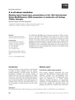
A. Sucrose density gradient profiles of WT and lut2.1 solubilized thylakoidsFigure 1
A. Sucrose density gradient profiles of WT and lut2.1 solubilized thylakoids. Thylakoid membranes from WT and
lut2.1 plants were solubilized with α-DM and loaded on sucrose gradient; for each gradient, fractions harvested (left) and chlo-
rophyll distribution (% of total Chl loaded) in the gradient along gradients (right) are indicated. Chlorophyll levels of each band
were normalized to the Chl content of WT band 5. Data are expressed as mean ± SD, n = 3. B. Gel electrophoresis of
sucrose gradient fractions. Tris-Tricine SDS-PAGE analyses of gradient bands from Figure 1A. Main protein components of
each fraction are indicated. Figure abbreviations: B, band; Thy, thylakoids; MW, molecular weight marker. C. Trimerization
behavior of recombinant LHCII proteins. LHCII were reconstituted in vitro with different xanthophyll species and trimer-
ization of monomeric subunits was allowed by adding PG, a lipid factor essential for trimerization [67]. LHCII containing a mix
of xanthophylls (L,V,N) or only lutein (L) produced trimers, while violaxanthin-binding complexes (V) did not produce trimers.
See Experimental Procedures for details. FP, free pigments; MON, monomeric subunits; TRIM, trimeric complexes.
BMC Plant Biology 2006, 6:32 />Page 6 of 20
(page number not for citation purposes)
The effect of high light growth conditions
The biochemical data suggest a deficit in the efficiency of
photoprotection at the level of Lhcb proteins, particularly
LHCII, in the lut2.1 mutant, caused by the substitution of
lutein with violaxanthin in site L1. It can thus be expected
that growth at high light intensity may reveal additional
features of the lut2.1 phenotype. WT and lut2.1 plants
were grown for 3 weeks in control conditions (120 μmol
m
-2
s
-1
) at 21°C and then either exposed to high light
(1400 μmol m
-2
s
-1
) or grown at the same light intensity
for three additional weeks (Figure 4A). After treatment,
leaves were analyzed for pigment composition [see Addi-
tional file 4] and thylakoid protein composition (Figure
4B–C). Growth in high light produced damages consist-
ing into reddening and bleaching of older leaves. The
damages were more pronounced in mutant plants.
Thylakoid membranes were isolated from low- and high-
light grown plants and analyzed by SDS-PAGE (Figure
4B–C). The relative abundance of thylakoid proteins was
evaluated by densitometry of Coomassie-stained gels
upon identification of individual selected bands by
immunoblotting with specific antibodies (not shown).
Both WT and lut2.1 thylakoids showed a decrease in the
LHCII/PSII ratio in high light, as evaluated by the level of
the 33 kDa oxygen evolving complex 1 polypeptide (Fig-
ure 4B). WT plants decreased their content in Lhcb1+2
polypeptides upon growth in high light by 15% with
respect to control plants while other Lhcb proteins were
marginally affected. lut2.1 plants showed a similar effect,
but the amplitude of the decrease in LHCII was much
higher, suggesting that mutant plants over-react to
increasing light by degrading their major antenna com-
plex and thus avoiding photoinhibition (Figure 4C).
In agreement with previous results [12] the Chl a/b ratio
increased in WT and lut2.1 with respect to control condi-
tions, the amplitude of the change being higher in the
mutant. lut2.1 had increased Chl a/b ratios even in control
conditions. Growth in high light decreased the Chl/Car
ratio in WT and lut2.1. WT plants did not contain any A+Z
in low light, and low levels in high light conditions. lut2.1
plants contained low, but detectable levels of A+Z in low
light conditions [14], and their increase in high light was
8 times higher than in WT plants. Although the increase in
A+Z was the highest, all carotenoid species increased their
relative amount with respect to Chls. This effect was
stronger in lut2.1 with respect to WT plants [see Addi-
tional file 4].
Photooxidation at low temperature
Our results strongly suggest that lut2.1 plants are affected
in their capacity to prevent photooxidation of their
antenna system, due to the lower efficiency of violaxan-
thin, with respect to lutein, in quenching
3
Chl*. Growth
in low temperature conditions should enhance the ampli-
Table 2: Pigment composition of monomeric Lhcb (from WT and lut2.1) and trimeric LHCII (from WT).
Chl a/b Chl/Car Neo Viola Anthera Lute Zea beta-Car
WT – band 2 1.8 ± 0.1 3.6 ± 0.1 5.9 ± 0.3 2.3 ± 0.1 nd 10.5 ± 0.4 nd nd
WT – band 3 1.5 ± 0.1 3.8 ± 0.1 5.9 ± 0.2 1.6 ± 0.1 nd 10.7 ± 0.4 nd nd
lut2.1 – band
2
1.5 ± 0.1 4.1 ± 0.1 9.8 ± 0.1 10.5 ± 0.1 3.2 ± 0.1 nd 1.1 ± 0.1 nd
Bands 2 and 3 were isolated from solubilized thylakoid membranes by sucrose gradient ultracentrifugation. Data are normalized to 100 Chl a+b,
and they are expressed as mean ± SD, n = 3. nd, not detected.
Table 3: Xanthophyll spectral forms and efficiency of energy transfer to Chl a in LHCII monomeric preparations purified by non-
denaturing IEF from WT and lut2.1 thylakoids.
Site
L1 L2 N1
LHCII WT Spectral form Lutein1 (489 nm) Lutein2 (495 nm) Viola1 (492 nm) Neoxanthin (486.5
nm)
Efficiency 81.0% 79.0% 60.0%
LHCII lut2.1 Spectral form Viola2 (505 nm) Viola1 (493.5 nm) Neoxanthin (486.5
nm)
Efficiency 93.8% 79.0% 60.0%
Spectral deconvolution analysis and calculation of energy transfer efficiency were as in Croce et al.,, 1999 [18]. The data, normalized to the WT, are
relative to a 100% Chl a-to-Chl a ET efficiency. The error in the ET efficiency was <4%, with the exception of Viola1 in WT (>10%). Xanthophyll
absorption maxima in ethanol are 477.2, 472.8 and 468.4 nm, respectively, for violaxanthin, lutein and neoxanthin. Binding to sites L2 and L1 shifts
violaxanthin absorption from 477.2 to 492 and 505 nm respectively; lutein is shifted from 472.8 to 489 and 495 nm, respectively. Binding to site N1
shifts neoxanthin from 468.4 to 486.5 nm.
BMC Plant Biology 2006, 6:32 />Page 7 of 20
(page number not for citation purposes)
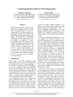
Photobleaching behaviour of isolated LhcbFigure 2
Photobleaching behaviour of isolated Lhcb. (A) Monomeric Lhcb isolated from solubilized thylakoids of WT and lut2.1,
and trimeric LHCII from WT were analyzed by following the Q
y
-transition absorbance decay during strong illumination. (B)
Sucrose bands 2 and 3 from WT and lut2.1 were fractionated by flat bed IEF in order to purify LHCII subunits in their mono-
meric and trimeric form. Kinetics of Q
y
-transition absorbance decay were measured on isolated complexes as described in
Experimental Procedures. Chlorophyll concentrations of Lhcb were set to 8 μg/ml. Samples were cooled to 10°C during meas-
urements.
BMC Plant Biology 2006, 6:32 />Page 8 of 20
(page number not for citation purposes)
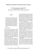
Flash-induced absorbance changes due to carotenoid triplet formation in LHCII recombinant proteins reconstituted with lutein (panels A and B) or violaxanthin (panels C and D)Figure 3
Flash-induced absorbance changes due to carotenoid triplet formation in LHCII recombinant proteins reconsti-
tuted with lutein (panels A and B) or violaxanthin (panels C and D). Panels A and C show the complete difference spectra
recorded at different time points (2.5 ns, 52.5 ns and 5 μs). Panels B and D show absorbance changes at 505 nm (
3
Car*) and
440–460 nm (*Chl). Data have been normalized on the amount of excited chlorophyll measured at 440 – 460 nm, and fitted to
a biphasic model (solid symbols in panels B and D).
BMC Plant Biology 2006, 6:32 />Page 9 of 20
(page number not for citation purposes)
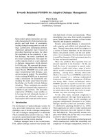
Phenotypes of WT and lut2.1 grown in normal and high light conditionsFigure 4
Phenotypes of WT and lut2.1 grown in normal and high light conditions. (A) Three-weeks-old WT (Fig. 1,3) and
lut2.1 (Fig. 2,4) plants were grown for 3 additional weeks in normal light conditions (21°C, 120 μmol m
-2
s
-1
- LL) (Fig. 1,2) or in
high light conditions (21°C, 1400 μmol m
-2
s
-1
- HL) (Fig. 3,4). (B) Tris-Tricine SDS-PAGE analyses of thylakoid from LL or HL
plants. Main protein components are indicated. (C) Relative level of thylakoid antenna proteins evaluated by densitometry of
bands identified by immunoblotting.
BMC Plant Biology 2006, 6:32 />Page 10 of 20
(page number not for citation purposes)
tude of photodamage [24]. We have thus evaluated the
effect of growing plants at 4°C at either low (20 μmol m
-
2
s
-1
) or high light conditions (800 μmol m
-2
s
-1
). The
experiment was performed on WT, lut2.1, npq1 (previ-
ously shown to have a decreased resistance to oxidative
stress under light stress conditions [6]) and the double
mutant npq1lut2.1. While WT and lut2.1 are able to
increase A+Z content at the expense of Viola upon light
treatment, npq1 and npq1lut2.1 plants cannot [see Addi-
tional file 1].
Plants were grown at 120 μmol m
-2
s
-1
, 21°C for three
weeks (t
o
) and then transferred at 4°C at either low light
or high light for three additional weeks. In low light, none
of the genotypes showed an evident stress effect, while in
high light, plants were affected to different extents (Figure
5): in WT, older leaves showed photobleaching accompa-
nied by accumulation of anthocyanin, an indicator of
stress in Arabidopsis [25,26]. These symptoms were much
stronger in npq1 and lut2.1 mutants, extending to the
younger leaves, while many of the older leaves were
almost completely bleached. Consistently with previous
reports [27], the npq1lut2.1 genotype was more light-sen-
sitive than either npq1 or lut2.1, suggesting that the lack of
zeaxanthin exacerbates the photodamage induced by the
lack of lutein. More quantitative analyses were performed
on detached leaves, choosing leaves that remained green
over the entire period of the experiment [see Additional
file 5].
Plants of WT and mutants, grown in standard conditions
(120 μmol m
-2
s
-1
) were treated for 30 hours at high light
and low temperature (1100 μmol m
-2
s
-1
, 8 hours light
photoperiod, 8°C). Following stress, the level of photoin-
hibition was assayed by chlorophyll fluorometry (F
v
/F
m
)
(Figure 6A), while lipid peroxidation was quantified by
measuring leaf chemiluminescence [28,29] (Figure 6B).
Our results clearly show that the highest levels of lipid
peroxidation and photoinhibition were obtained in the
npq1lut2.1 genotype, in accordance with evidences
obtained on C. reinhardtii lor1npq1 double mutant [30];
npq1 had intermediate levels and lut2.1 did not show a
significant difference from WT. Similar results were
obtained in a shorter experiment in which detached
leaves, floating in water at 10°C, were treated at high light
(1100 μmol m
-2
s
-1
) for 20 h (data not shown).
Discussion
The conservation of plant xanthophyll composition
strongly suggests that each xanthophyll species has a spe-
cific function. Lutein is the major xanthophyll species in
plants, accounting for approx. 60% of total xanthophylls
and 40% of total carotenoids in leaves. In LHCII com-
plexes, it binds to site L1, whose occupancy is essential for
protein folding and chlorophyll triplet quenching, and,
promisquously with other xanthophylls, site L2, essential
for photoprotection by violaxanthin/zeaxanthin exchange
[9] (Figure 7). Still, it has been reported that lutein is not
essential for photosynthesis [14]. Additional studies have
shown alterations, in the lut2 mutant, in NPQ, LHCII
antenna size and trimerization, and an increased accumu-
lation of A+Z [31] while and recent publication showed
decreased growth rate in a large range of light conditions.
We have confirmed and extended some of these observa-
tions (see Additional files). It is worth noting that our
lut2.1 mutant was isolated in Wassilewskija genetic back-
ground, while previous described lutein-less mutant
[12,14] are in the Columbia ec. It seems proper to ask if
differences between our and previous results are related to
the different genetic background. We have addressed this
question by confirming in Wassilewskija ec. results pre-
viouly obtained in Columbia ec. We concluded that the
level of sensitivity to stress and other photosynthetic
parameters were the same in boh ecotypes. Furthermore,
we obtained several confirmatory results using lut2.1
mutant, which closely match those previouly obtained in
the Columbia ec. [12]. We conclude that the two mutants
are, in every respect, comparable. Finally, in a later stage
of the study, we succeeded in isolating an equivalent
mutant from the Columbia background [32] which had
the same properties as those described here for lut2.1.
A complete disruption of the LHCII trimeric organization
was observed in the lut2.1 mutant even upon solubiliza-
tion of thylakoids with the mild detergent α-DM, which is
very effective in retaining trimers in WT. Protein gel anal-
yses of purified LHCI and LHCII monomers show that
they have unaltered protein composition, and HPLC anal-
yses show that only violaxanthin and neoxanthin are
bound to LHCII complexes. Previous work with recom-
binant proteins has shown that lutein, violaxanthin and
zeaxanthin can bind to sites L1 and L2 of Lhc proteins
[18,33] while the site for neoxanthin binding is site N1.
This was recently confirmed by X-ray crystallography [17].
We found a novel, red-shifted form of violaxanthin in
LHCII from lut2.1, consistent with the red-shift observed
for lutein in site L1 of WT LHCII [18]. This strongly sug-
gests that, in lut2.1, violaxanthin replaces lutein in site L1.
LHCII from lut2.1 contains more than one neoxanthin
molecule per polypeptide suggesting that this xanthophyll
can compete with violaxanthin in either sites L1 or L2.
Since reconstitution with neoxanthin only was unable to
yield a pigment-protein complex in all Lhc proteins, and
occupancy of site L1 was shown to be needed for refolding
[9,34,35], we conclude that, in LHCII, neoxanthin can
compete with violaxanthin for site L2 in the absence of
lutein. This is consistent with previous results [36]
obtained in vitro using low stringency reconstitution of
BMC Plant Biology 2006, 6:32 />Page 11 of 20
(page number not for citation purposes)
Phenotypes of WT and different mutants grown in different light conditions at 4°CFigure 5
Phenotypes of WT and different mutants grown in different light conditions at 4°C. Plants were growth for 3
weeks in control conditions (21°C, 120 μmol m
-2
s
-1
, (CTRL) and then transferred for 3 additional weeks at 4°C at either 20
μmol m
-2
s
-1
(LL) or 800 μmol m
-2
s
-1
(HL).
BMC Plant Biology 2006, 6:32 />Page 12 of 20
(page number not for citation purposes)
Analyses of the sensitivity to photoxidative stress on WT and mutant genotypesFigure 6
Analyses of the sensitivity to photoxidative stress on WT and mutant genotypes. Whole plants (WT and different
mutants) were exposed for 30 hours to a high photon flux density (1100 μmol m
-2
s
-1
) at low temperature (10°C). (A) Meas-
urements of the chlorophyll fluorescence ratio F
v
/F
m
, an index of photoinhibition of PSII. (B) Analyses of heat-induced lumines-
cence emission of WT and mutant leaves; the 135°C emission band was used as an index of lipid peroxidation and oxidative
stress.
BMC Plant Biology 2006, 6:32 />Page 13 of 20
(page number not for citation purposes)
recombinant proteins and with the binding of neoxanthin
to L2 site in the homologous proteins CP29 and CP26.
LHCII monomerization appears to be due to the lack of
lutein in sites L1 and/or L2 per se, as indicated by in vitro
reconstitution and trimerization of the Lhcb1 apoprotein.
We demonstrated, for the first time, that trimers can only
be formed if lutein is present in the reconstitution mix-
ture. Any substitution of lutein with other xanthophyll
species leads to monomerization, as shown also for the
npq2lut2.1 mutant [37]. Violaxanthin-binding LHCII
monomers have the same stability to heat denaturation as
lutein-binding monomers, implying that binding of vio-
laxanthin impairs trimerization but not protein stability.
Violaxanthin-containing LHCII from lut2.1 is equally effi-
cient in light harvesting, since the small decrease in func-
tional antenna size in lut2.1 is quantitatively consistent
with the lower LHCII content determined by biochemical
methods.
lut2.1 plants were unable to perform state I – state II tran-
sitions. We first considered the possibility that the prefer-
ential absorption of far-red light by the PSI-LHCI
complex, which is at the basis of state I – state II transi-
tions, could be somehow affected by the absence of lutein.
This possibility was ruled out by the observation that,
consistent with previous results in Chlamydomonas PSI
[38], the PSI-LHCI complex in sucrose gradient ultracen-
trifugation was very stable and conserved the red-shifted
absorption tail typical of WT complex (data not shown).
The loss of state transitions could either be due to the spe-
cific loss of a particular LHCII subpopulation able to
migrate from grana to stroma membranes [39] or an
impaired capacity for LHCII phosphorylation in the
mutant. The first hypothesis is ruled out by the results of
Proposed role of xanthophylls in higher plant antennaeFigure 7
Proposed role of xanthophylls in higher plant antennae. LHCII structure is taken from [17].
BMC Plant Biology 2006, 6:32 />Page 14 of 20
(page number not for citation purposes)
non-denaturing IEF analyses showing the presence of the
same isoforms in WT and lut2.1. We suggest that LHCII
phosphorylation is somehow affected in lut2.1. Phospho-
rylation experiments are in progress in order to verify this
hypothesis. Alternatively, the modified conformation of
violaxanthin-binding LHCII might affect the conforma-
tional changes [40], which are involved in detachment
from PSII and docking to PSI [41].
Recent reports have suggested the hypothesis that trimer
to monomer transition is the basis of NPQ [42]. Since
lut2.1 LHCII is completely monomeric, but still shows
residual NPQ, the data contradict this hypothesis. Recent
work has shown that NPQ is first elicited in the PSII core
complex and is then propagated to the antenna system
[43]. Quenching in isolated LHCII has been proposed to
be catalyzed by interactions between chlorophyll mole-
cules bound to binding sites A1 and/or A2 with the lutein
in site L1, elicited by a conformational change [44]. Thus,
the substitution of lutein by violaxanthin in site L1 may
limit the efficiency of the process.
WT and lut2.1 plants grow similarly in moderate light
conditions according to an early report [14]. In high light,
stress effects such as anthocyanin accumulation and
bleaching of older leaves were more pronounced in lut2.1
plants. Pigment composition clearly showed that the
increase in carotenoids on a Chl basis was stronger in
lut2.1 vs. WT. These changes are commonly observed
upon exposure to light stress [45] and correlate with an
increased resistance to excess light [46]. Further reactions
to excess light consist into a decrease in PSII antenna size
[47] by specific proteolysis of the LHCII complex [48].
Determination of Lhc protein content in thylakoids of WT
and lut2.1 plants showed that the amount of LHCII was
lower in lut2.1 grown in low light and that further
decrease upon exposure to high light was higher in the
mutant with respect to WT plants. This is possibly due to
the accumulation of zeaxanthin in Lhc complexes, favor-
ing degradation of the major LHCII complex [37]. Thus,
the lut2.1 mutant is more sensitive to light than WT and
overreacts to an increase in light intensity through the
enhanced operation of known mechanisms of photopro-
tection [49]. The over-operation of these mechanisms
likely compensates for the primary lesion brought about
by the lack of lutein.
What is the primary effect of the lut2.1 mutation? Previous
work with recombinant proteins obtained by reconstitut-
ing in vitro Lhc apoproteins with different xanthophylls,
has shown that LHCII binding violaxanthin in sites L1
and L2 undergoes a more rapid photobleaching when
illuminated in the presence of oxygen with respect to the
LHCII binding lutein [9,18]. Photobleaching is the effect
of the
1
O
2
* produced by the reaction of
3
Chl* with molec-
ular oxygen, which is a triplet in its ground state. In vivo,
3
Chl* is produced by intersystem crossing from
1
Chl* and
is efficiently quenched by carotenoids, leading to heat dis-
sipation of triplet energy [5]. LHCII purified from lut2.1 is
more prone to photobleaching than the complex from
WT. Since the fluorescence quantum yield, and thus the
1
Chl* concentration on LHCII binding Viola + Zea is
essentially the same, we conclude that violaxanthin is less
efficient than lutein in quenching
3
Chl*, thus resulting in
increased
1
O
2
* formation and photobleaching.
The first excited triplet state of carotenoids lies below the
energy level of chlorophyll triplet and singlet oxygen.
Therefore, the generation of
3
Car* can quench
3
Chl* and
scaveng
1
O
2
*.
3
Car* decay into the ground state without
emission of radiation, and thus act as safe repositories of
excess energy. The increased photobleaching of lut2.1 sug-
gests that, for the same amount of
3
Chl*, the relative con-
centration of
1
O
2
* is increased. This could be due to a
decreased formation of
3
Car*. As
3
Car* decay is very fast
(~2 μs) we addressed this question by flash-induced,
time-resolved spectroscopy. Violaxanthin triplets exhibit
an extinction coefficient (from 3·10
5
to 6·10
5
at 490 nm)
larger than lutein (about 2·10
5
at 500 nm) [50]. Despite
this difference, after normalization for the *Chl signal, we
observed that absorbance changes due to lutein triplet for-
mation were 1.5-fold larger than for violaxanthin, and the
kinetics for triplet formation were faster for lutein. This
proves that lutein bound to LHCII proteins is more effi-
cient as a
3
Chl* quencher than violaxanthin.
The binding site responsible for the increased
1
O
2
* pro-
duction is, likely, site L1, since the non-occupancy of site
L2 did not significantly affect photobleaching in recom-
binant LHCII [9,18]. It is possible that the different con-
formation of LHCII protein binding violaxanthin modify
the Chl-to-xanthophyll distance, which is crucial for tri-
plet quenching [51]. This effect is independent from the
stability of LHCII protein folding as assessed by thermal
denaturation, but is dependent on the aggregation size of
the complex, implying that trimerization increases the
"special" Chl to carotenoid interaction responsible for
optimal photoprotection. This hypothesis is supported by
the report that monomerization of LHCII yields into the
loss of a specific red-shifted (510 nm) spectral form of
lutein [19].
Thus, the primary lesion in lut2.1 mutation is the
enhanced production of singlet oxygen in the major PSII
antenna complex, partially compensated by increased
zeaxanthin production. In order to verify this hypothesis,
we have studied the behavior, under enhanced stress con-
ditions (high light + low temperature), of the lut2.1
mutant compared to the double mutant npq1lut2.1, which
cannot synthesize zeaxanthin upon exposure to excess
BMC Plant Biology 2006, 6:32 />Page 15 of 20
(page number not for citation purposes)
light. Zeaxanthin free in the membrane, upon release of
violaxanthin from LHCII and de-epoxidation by VDE, has
been suggested to protect from photooxidation, by a ROS
scavenging effect [6,30] that supplements the action of
vitamin E [52]. A previous paper [27] reported phenotipic
evidences of a higher sensitivity of double mutant
npq1lut2 to photoxidative stress. We performed a detailed
description of this genotype, showing both its behaviou
during short high-light treatment, and acclimation to
long-term stress. The npq1lut2.1 plants underwent
stronger photoinhibition, anthocyanin biosynthesis and
lipid oxidation not only with respect to WT but also with
respect to both npq1 and lut2.1 single mutants. Our results
Thus point to a strong photosensitive phenotype in higher
plant mutant due to to lack of both lutein and zeaxanthin;
such data are in agreement with those obtained in C. rein-
hardtii npq1lor1 mutant [30]. In contrast, a recent publica-
tion [13] failed to evidence a synergistic effect of lutein
and zeaxanthin in promoting growth and preventing
stress; these results could be ascribed to the different set-
ting of stress conditions used with respect to our work.
Our data, in agreement with [27] and [30], let to conclude
that zeaxanthin is effective in photoprotection of plants
lacking lutein. This is due to the multiple effects of zeax-
anthin in photoprotection, including ROS scavenging
[6,30,53] and direct quenching of Chl fluorescence by
binding to the L2 allosteric site of Lhc proteins [54].
Conclusion
The conservation of carotenoid composition across the
plant kingdom implies a specific function for each xan-
thophyll species. Lutein has the specific property of
quenching harmful
3
Chl* by binding at site L1 of the
major LHCII complex and of other Lhc proteins of plants,
thus preventing ROS formation. Substitution of lutein by
violaxanthin decreases the efficiency of
3
Chl* quenching
and causes higher ROS yield. The phenotype of lut2.1
mutant in low light is weak only because rescuing mecha-
nisms of photoprotection, namely zeaxanthin synthesis,
compensate for the ROS production, as also supported
from the lower antenna size observed in low light. Thus,
the light sensitive phenotype becomes evident in condi-
tions enhancing photooxidative stress or when the addi-
tional mechanisms are eliminated by the npq1 mutation.
Excess light and low temperature are commonly experi-
enced by plants during their lifecycle. Therefore, perform-
ing photosynthesis without lutein is like driving without
a seat belt. At low speed, one can get away with it as addi-
tional protective mechanisms (airbags = zeaxanthin) par-
tially compensate for the damage. But, in stress
conditions, the additional mechanisms fail and the dam-
age becames evident.
The specificity of site L1 for lutein in all Lhc proteins [10]
probably derives from the co-evolution of the carotenoid
biosynthesis pathway and Lhc proteins with multiple
binding sites dedicated to different functions. Lutein is the
only xanthophyll containing one beta and one epsilon
ring. Beta and epsilon carotenoid cyclases have diverged
early during plant evolution, and are both found in all
higher plant taxa, as well as in green algae like Scenedes-
mus or Chlamydomonas [55]. In most plants, lutein is
not undergoing epoxidation-deepoxidation reactions in
response to environmental conditions, while the beta-
beta xanthophylls do. Since site L1 is dedicated to the
essential function of Chl triplet quenching, it may be
desirable, for the plant, that the chromophore catalyzing
this function is not involved in rapid concentration
changes in response to environmental conditions as hap-
pens for the beta-beta xanthophylls involved in the xan-
thophyll cycle [56].
Methods
Screening procedure
We analyzed 20000 independent T-DNA insertion lines of
Arabidopsis thaliana (accession Wassilewskija-2) available
from the Institut National de la Recherche Agronomique
(INRA, Versailles, France). Four hundred seed pools, each
representing one row or one column of the grid (100
lines), were prepared and then combined into 80 super-
pools, each representing 500 lines.
The presence of T-DNA insertion in the lycopene ε-cyclase
gene (lyec) was assessed by PCR amplification on DNA
from each of the superpools, followed by a nested PCR.
The first (10 cycles: 94° 2'; 65°-1°/cycle 30"; 72° 2'. 35
cycles: 94° 15"; 55° 30"; 72° 1') was performed by using
the lyec specific primers 5'-AGTTAGTCGACGTTTGCTC-
CATG-3' and 5'-CAATGGTAATAGGCTTGTCATC-3' and
the T-DNA specific primers 5'-CTACAAATTGCCTTTTCT-
TATCGA-3' and 5'-CTGATACCAGACGTTGCCCGCATAA-
3' The nested PCR (35 cycles: 94° 45"; 56° 45"; 72° 1')
was performed by using the lyec specific primers 5'-GAG-
GAGGTAAAGTATGGTTCCAC-3' and 5'-CTCTCTCCAAA-
CATGCTCAATAC-3' and the T-DNA specific primers 5'-
CATGTACATCAAGCTTATCGATAC-3' and 5'-TAC-
GAATATCTGCATCGGCGAAC-3'. One mutant line, con-
taining a T-DNA insertion in the sixth exon of the gene,
was identified in the superpools and then in the pools,
and kindly provided by INRA. To identify homozygous
lines, PCR analyses was performed using primers 5'-
AAGCTTCTTCCGTACTTTC-3' and 5'-CAATCG-
TAAACAATATAAGCG-3', flanking the site of insertion,
and the T-DNA specific primer 5'-CATGTACATCAAGCT-
TATCGATAC-3'. This mutant will be indicated as lut2.1 in
order to avoid confusion with the original lut2 mutant
[14] solely to indicate that it is made in Wassilewskija eco-
type. After completion of this work we have obtained the
same mutation in Columbia ecotype and verified that it
BMC Plant Biology 2006, 6:32 />Page 16 of 20
(page number not for citation purposes)
showed the same behaviour as the lut2.1 with respect to
sensitivity to light stress.
Plant material
WT plants of Arabidopsis thaliana ecotype Wassilewskija
and mutants npq1 [57], were obtained from the Arabidop-
sis Stock center. Genotype npq1 lut2.1 was obtained by
crossing single mutant plants. Plants were grown for three
weeks in controlled conditions (~120 μmol m
-2
s
-1
, 21°C,
8 h light/16 h dark). For long term treatment, 3 weeks old
seedlings were exposed (a) to light conditions of 120 or
1400 μmol m
-2
s
-1
for 3 weeks at 21°C, and (b) to light
conditions of 20 or 800 μmol m
-2
s
-1
for 3 weeks at 4°C.
Short-term high light treatment was performed for 20
minutes at 1200 μmol m
-2
s
-1
.
Chlorophyll fluorescence and photosynthetic oxygen
evolution in vivo
Chlorophyll fluorescence from intact leaves or from leaf
discs was measured with a PAM-2000 fluorimeter (Walz),
as previously described [37]. The maximal quantum yield
of PSII photochemistry was measured in dark-adapted
leaves from the maximal fluorescence level (F
m
) and the
initial level (F
o
): (F
m
-F
o
)/F
m
= F
v
/F
m
. Variable fluorescence
was induced in leaf discs, infiltrated with DCMU 2.5 10
-5
M, with a red light of 8 μmol m
-2
s
-1
produced by a light
emitting diode. The half-time of the fluorescence rise was
taken as a measure of the functional antenna size of PSII
[15]. Photosynthetic O
2
evolution was measured with the
photoacoustic method, as described by Havaux et al., [37]
(for a review, see [58]). The Emerson enhancement (E) of
O
2
evolution was determined in state I or in state II by
adding a continuous far-red light (>715 nm, 34 W m
-2
) to
the modulated blue-green light (obtained with a BG38
Schott filter; photon flux density 24 μmol m
-2
s
-1
). E (%)
= [(Φ (+FR) - Φ (-FR))/Φ (-FR)] × 100, where Φ (+FR) is
the amplitude of oxygen evolution signal in the presence
of the far-red light and Φ (-FR) is the signal measured with
the modulated exciting light only. State II was reached by
illuminating leaves with blue-green light for 10 min, and
state I was obtained after 10-min illumination with far-
red light.
Non-photochemical quenching of chlorophyll fluores-
cence was measured with a PAM 101–103 fluorimeter
(Walz). NPQ was calculated according to the following
equation [59]: NPQ = (F
m
-F'
m
)/F'
m
, where F
m
is the maxi-
mum Chl fluorescence from dark-adapted leaves and F'
m
the maximum Chl fluorescence under actinic light exposi-
tion.
Thylakoid isolation and sample preparation
Unstacked thylakoid membranes were isolated from dark-
adapted leaves as previously described [39].
Membranes corresponding to 500 μg of chlorophylls were
washed with 5 mM EDTA and then solubilized in 1 ml
with 0.6% α-DM, 10 mM HEPES pH 7.5. Solubilized sam-
ples were then fractionated by ultracentrifugation in a
0.1–1 M sucrose gradient containing 0.06% α-DM, 10
mM HEPES pH 7.5 (22 h at 280,000 × g, 4°C).
Monomeric Lhcb proteins were further fractionated by
flat-bed isoelectric focusing at 4°C as previously described
[60].
Pigment analyses
The pigments were extracted either from whole leaves,
thylakoid membranes and isolated antenna complexes
with 80% acetone, then separated and quantified by
HPLC [61] and by fitting of the spectrum of the acetone
extract with the spectra of individual pigments [21].
Gel electrophoresis
SDS-PAGE analyses was performed with the Tris-Tricine
buffer system as previously described [62]. Gel images
were acquired using a Bio-Rad GS710 scanner. The picture
was then analysed with GEL-PRO ANALYZER software
(Media Cybernetics Inc., MD, USA) that quantifies the
staining of the bands as IOD (optical density integrated
on the area of the band).
In vitro reconstitution of LHCII pigments complexes and
trimerization assay
The construct over-expressing Lhcb1 was obtained as
described [63], except for a sequence coding for a His
6
tail
inserted at the 3' end before stop codon. In vitro reconsti-
tution of LHCII with altered xanthophyll composition
was performed as previously described [18]. Purification
of reconstituted holocomplexes was performed by a Ni
2+
chelating column (Pharmacia Source 15S) as described
[9]. Reconstitutions were accomplished with a mix of xan-
thophylls as follow: LHCII Viola, 100% violaxanthin;
LHCII Lute, 100% lutein; the Chl a/b ration in the mixture
was 2.3. In order to perform in vitro trimerization of
reconstituted complexes, we followed a method previ-
ously described [64] with some modifications. Ni
2+
col-
umn and bound reconstituted LHCII were washed with
trimerization buffer: 0.1 mg/ml PG, 0.06% β-DM, 0.2 M
NaCl, 20 mM phosphate buffer pH 7.5, 10 mM imida-
zole. Pigment-protein complexes were collected by wash-
ing column with eluting buffer: 0.5 M imidazole, 20 mM
phosphate buffer pH 7.5, 0.2 M NaCl, 0.06% β-DM;
LHCII trimers were separated from monomers by sucrose
gradient ultracentrifugation [19].
Spectroscopy
Steady state spectra were obtained using samples in 10
mM HEPES pH 7.5, 0.06% α-DM, 0.2 M sucrose. Absorp-
BMC Plant Biology 2006, 6:32 />Page 17 of 20
(page number not for citation purposes)
tion measurements were performed using a SLM-Aminco
DW-2000 spectrophotometer at RT.
Time-resolved spectroscopy:
absorbance changes were
monitored with a home-built pump and probe laser spec-
trophotometer, basically described in [65], and modified
as follows. The wavelength of monochromatic 10-ns light
pulses used for the detection of absorbance changes is
tuned from 410 nm to 590 nm by an optical parametric
oscillator pumped by the third harmonic of a Nd:YAG
laser (Surelite, Continuum). The 10-ns excitation flash is
provided by a home-built broadband dye laser cell filled
with DCM in methanol (650 nm) and pumped by the sec-
ond harmonic of a Nd:YAG laser (Minilite, Continuum).
The delay between the pump and probe pulses is adjusted
with a 100 MHz National Instruments PCI-6552 digital
waveform generator. Detection beam is split in two ahead
of measure and reference 10 × 10 mm cuvettes. Transmit-
ted light is filtered through a combination of 6 mm-thick
BG39 colored Schott filter and CVI low-pass dielectric fil-
ter (600 nm). Light intensity is measured with large area
silicon photodiodes fitted to AC-coupled preamplifiers.
The difference between measure and reference analog sig-
nals is achieved by a Tektronix differential amplifier (gain
10). Acquisition of difference and reference signals is per-
formed via a 16-bit National Instrument digitizer PCI-
6052E. Optical density of the sample was adjusted to 1 in
the Qy band of chlorophylls. The flash-induced signal
exhibits negative peaks around 430 nm, characteristic of
the bleaching of the carotenoid S
2
←S
0
transitions, and a
strong positive peak around 510 nm attributed to the car-
otenoid T
2
←T
1
transition. The spectra are thus denoted
triplet-minus-singlet difference absorption spectra. The
extent of the carotenoid absorbance change at 5 μs is
approximately the same at 440 nm and 460 nm. This led
to the choice of these two wavelengths as isosbestic points
of the carotenoid TmS spectrum. At 2.5 ns, a contribution
of excited chlorophyll (Chl*) could be detected within the
time-resolution of the instrument, contributing negatively
at 440 nm and positively at 460 nm. Consequently, the
kinetics of pure Chl* decay have been symbolized by the
difference at these two wavelengths (Figure 3, panels B
and D). This deconvolution procedure is validated by the
fact that the Chl* signal decays to zero within 200 ns and
stays stable until the end of the kinetics, i.e. 5 μs. This pro-
cedure also allowed us to normalize the data on the 440–
460 signal at 2.5 ns, so as to compare the relative forma-
tions of
3
Car* in either lutein- or violaxanthin reconsti-
tuted proteins. Kinetic data were fitted to a biexponential
model (solid symbols in Figure 3B,D).
Protein thermal stability
This was analyzed as described [21] by following the
decrease of the 492 nm CD signal detected by a Jasco 600
spectropolarimeter at increasing temperatures, from 20 to
80°C (scan rate 1°C/minute and step 0.2°C). Protein
thermal stability was measured as denaturation T°
1/2
,
determined by inflection point of CD denaturation
curves.
Photobleaching assay
The kinetics of antennae photobleaching was measured as
described [18] but with a higher light intensity of ca 6000
μmol m
-2
s
-1
and sample cooling at 10°C. Initial and max-
imal absorbance was 0.6.
Determination of the sensitivity to photoxidative stress
Photoxidative stress was induced in whole plants by a
strong light treatment at low temperature. Whole Arabi-
dopsis plants were exposed to high light (1100 μmol pho-
ton m
-2
s
-1
with a photoperiod of 8 h) at low temperature
(7°C/8°C, day/night air temperature), as described previ-
ously [66].
Photoinhibition of PSII was measured by chlorophyll
fluorometry (F
v
/F
m
ratio) with a PAM-2000 fluorimeter
(Walz). Photoxidative stress was measured by thermolu-
minometry with a custom-made apparatus that has been
described [7]. The amplitude of the TL peak at 135°C was
used as an index of lipid peroxidation.
List of abbreviations
α(β)-DM, α(β)-dodecyl maltoside; A, antheraxanthin;
Chl, chlorophyll;
1
Chl*, chlorophyll excited singlet state;
3
Chl*, chlorophyll excited triplet state; Car, caroten-
oid;
3
Car*, carotenoid excited triplet state; CD, circular
dichroism; DCMU, 3-(3,4-dichlorophenyl)-1,1-dimethyl-
urea; IEF, isoelectric focusing; lhc, light-harvesting com-
plex; LHCII, major light harvetsing complex of
photosystem II; L, lutein; N, neoxanthin; NPQ, non-pho-
tochemical quenching;
1
O
2
*, singlet oxygen; ; PSII (I),
photosystem II (I); qE, ΔpH-dependent portion of non-
photochemical quenching; ROS, reactive oxygen species;
V, violaxanthin; VDE, violaxanthin de-epoxidse; Z, zeax-
anthin.
Authors' contributions
LD carried out the crossing to obtain double mutant npq1
lut2.1, the characterization of either whole pants and iso-
lated Lhc complexes, the photoxidative treatments and
stress measurements. CL carried out the isolation of lut2.1
mutant. JA was involved in measurements of time-
resolved spectroscopy for carotenoid triplet formation.
MH carried out thermoluminescence and photosynthetic
O
2
evolution measurements. RB e GG conceived of the
study, and participated in its design and coordination and
helped to draft the manuscript. All authors read and
approved the final manuscript.
BMC Plant Biology 2006, 6:32 />Page 18 of 20
(page number not for citation purposes)
Additional material
Acknowledgements
We like to thank R. Croce (Trento, Italy) for help in deconvolution analyses
of isolated LHCII and S. Cazzaniga (Verona, Italy) for technical support. This
work was supported by FIRB RBLA0345SF_002 and GENEFUN (functional
genetic) program.
References
1. Yamamoto HY, NAKAYAMA TO, Chichester CO: Studies on the
light and dark interconversions of leaf xanthophylls. Arch Bio-
chem Biophys 1962, 97:168-173.
2. Lam E, Ortiz W, Malkin R: Chlorophyll a/b proteins of photosys-
tem I. FEBS Lett 1984, 168:10-14.
3. Britton G, Liaaen-Jensen S, Pfander H: Carotenoids. Basel, Switzer-
land, Birkhauser Verlag; 1998.
4. Polivka T, Herek JL, Zigmantas D, Akerlund HE, Sundström V: Direct
observation of the (Forbidden) S-1 state in carotenoids. Proc
Natl Acad Sci USA 1999, 96:4914-4917.
5. Polivka T, Zigmantas D, Sundström V, Formaggio E, Cinque G, Bassi
R: Carotenoid S-1 state in a recombinant light-harvesting
complex of photosystem II. Biochemistry 2002, 41:439-450.
6. Havaux M, Niyogi KK: The violaxanthin cycle protects plants
from photooxidative damage by more than one mechanism.
Proc Natl Acad Sci USA 1999, 96:8762-8767.
7. Havaux M: Spontaneous and thermoinduced photon emis-
sion: new methods to detect and quantify oxidative stress in
plants. Trends in Plant Science 2003, 8:409-413.
8. Bassi R, Pineau B, Dainese P, Marquardt J: Carotenoid-Binding
Proteins of Photosystem-II. Eur J Biochem 1993, 212:297-303.
9. Formaggio E, Cinque G, Bassi R: Functional architecture of the
major Light-harvesting Complex from Higher Plants. J Mol
Biol 2001, 314:1157-1166.
10. Morosinotto T, Caffarri S, Dall'Osto L, Bassi R: Mechanistic
aspects of the xanthophyll dynamics in higher plant thyla-
koids. Physiologia Plantarum 2003, 119:347-354.
11. Pogson BJ, Niyogi KK, Bjorkman O, DellaPenna D: Altered xantho-
phyll compositions adversely affect chlorophyll accumula-
tion and nonphotochemical quenching in Arabidopsis
mutants. Proc Natl Acad Sci U S A 1998, 95:13324-13329.
12. Lokstein H, Tian L, Polle JE, DellaPenna D: Xanthophyll biosyn-
thetic mutants of Arabidopsis thaliana: altered nonphoto-
chemical quenching of chlorophyll fluorescence is due to
changes in Photosystem II antenna size and stability. Biochim
Biophys Acta 2002, 1553:309-319.
13. Kalituho L, Rech J, Jahns P: The roles of specific xanthophylls in
light utilization. Planta 2007, 225:423-439.
14. Pogson B, McDonald KA, Truong M, Britton G, DellaPenna D: Ara-
bidopsis carotenoid mutants demonstrate that lutein is not
essential for photosynthesis in higher plants. Plant Cell 1996,
8:1627-1639.
15. Malkin S, Armond PA, Mooney HA, Fork DC: Photosystem II pho-
tosynthetic unit sizes from fluorescence induction in leaves.
Correlation to photosynthetic capacity. Plant Physiol 1981,
67:570-579.
16. Allen JF, Forsberg J: Molecular recognition in thylakoid struc-
ture and function. Trends Plant Sci 2001, 6:317-326.
Additional file 1
Pigment composition of leaf tissue from WT and mutant genotypes, both
dark adapted, and after light stress (1200
μ
mol m-2 s-1, 20' at RT).
Click here for file
[ />2229-6-32-S1.pdf]
Additional file 2
Photosynthetic parameters Fo, Fm, Fv/Fm and T1/2 measured on WT and
lut2.1 leaves.
Click here for file
[ />2229-6-32-S2.pdf]
Additional file 3
Pigment composition and stability to heat denaturation of fractions
obtained by preparative IEF of monomeric Lhcb isolated from WT and
lut2.1.
Click here for file
[ />2229-6-32-S3.pdf]
Additional file 4
Leaf pigment composition of 3-weeks-old WT and lut2.1 plants grown for
three additional weeks LL (120
μ
mol m
-2
s
-1
) or HL (1400
μ
mol m
-2
s
-1
)
conditions.
Click here for file
[ />2229-6-32-S4.pdf]
Additional file 5
Measurements of Fv/Fm ratio and chlorophyll content of WT and mutant
leaves during growth at low temperature and different light conditions.
Click here for file
[ />2229-6-32-S5.pdf]
Additional file 6
Kinetics of NPQ.
Click here for file
[ />2229-6-32-S6.pdf]
Additional file 7
Thermal stability of Lhcb proteins isolated from WT and lut2.1 thyla-
koids.
Click here for file
[ />2229-6-32-S7.pdf]
Additional file 8
Fitting of the Soret region of LHCII 1-T spectra from WT and lut2.1.
Click here for file
[ />2229-6-32-S8.pdf]
Additional file 9
Flat bed isoelectric focusing fractionation.
Click here for file
[ />2229-6-32-S9.pdf]
BMC Plant Biology 2006, 6:32 />Page 19 of 20
(page number not for citation purposes)
17. Liu Z, Yan H, Wang K, Kuang T, Zhang J, Gui L, An X, Chang W:
Crystal structure of spinach major light-harvesting complex
at 2.72 A resolution. Nature 2004, 428:287-292.
18. Croce R, Weiss S, Bassi R: Carotenoid-binding sites of the major
light-harvesting complex II of higher plants. J Biol Chem 1999,
274:29613-29623.
19. Caffarri S, Croce R, Breton J, Bassi R: The major antenna com-
plex of photosystem II has a xanthophyll binding site not
involved in light harvesting. J Biol Chem 2001, 276:35924-35933.
20. Cinque G, Croce R, Bassi R: Absorption spectra of chlorophyll a
and b in Lhcb protein environment. Photosynth Res 2000,
64:233-242.
21. Croce R, Canino , Ros F, Bassi R: Chromophore organization in
the higher-plant photosystem II antenna protein CP26. Bio-
chemistry 2002, 41:7334-7343.
22. Gastaldelli M, Canino , Croce R, Bassi R: Xanthophyll binding sites
of the CP29 (Lhcb4) subunit of higher plant photosystem II
investigated by domain swapping and mutation analysis. Jour-
nal of Biological Chemistry 2003, 278:19190-19198.
23. Bassi R, Croce R, Cugini D, Sandona D: Mutational analysis of a
higher plant antenna protein provides identification of
chromophores bound into multiple sites. Proc Natl Acad Sci USA
1999, 96:10056-10061.
24. Zhang S, Scheller HV: Photoinhibition of Photosystem I at chill-
ing temperature and subsequernt recovery in Arabidopsis
thaliana. Plant Cell Physiol 2004, 45:1595-1602.
25. Dixon.R.A., Paiva NL: Stress-induced phenylpropanoid metab-
olism. Plant Cell 1995, 7:1085-1097.
26. Xiang C, Werner B, Christensen E, Oliver D: the biological func-
tions of glutathione revisited in Arabidopsis transgenic
plants with altered glutathione levels. Plant Physiol 2001,
126:564-574.
27. Niyogi KK, Shih C, Chow WS, Pogson BJ, DellaPenna D, Bjorkman O:
Photoprotection in a zeaxanthin- and lutein-deficient double
mutant of Arabidopsis.
Photosynth Res 2001, 67:139-145.
28. Hideg E, Vass I: The 75-degrees-C thermoluminescence band
of green tissues - Chemiluminescence from membrane-chlo-
rophyll interaction. Photochem Photobiol 1993, 58:280-283.
29. Vavilin DV, Ducruet JM: The origin of 115-130 degrees C ther-
moluminescence bands in chlorophyll-containing material.
Photochem Photobiol 1998, 68:191-198.
30. Baroli I, Gutman BL, Ledford HK, Shin JW, Chin BL, Havaux M, Niyogi
KK: Photo-oxidative stress in a xanthophyll-deficient mutant
of Chlamydomonas. J Biol Chem 2004, 279:6337-6344.
31. Gilmore AM: Xanthophyll cycle-dependent nonphotochemi-
cal quenching in Photosystem II: mechanistic insights gained
from Arabidopsis thaliana L. mutants that lack violaxanthin
deepoxidase activity and/or lutein. Photosynthesis Research 2001,
67:89-101.
32. Fiore A, Dall'Osto L, Fraser PD, Bassi R, Giuliano G: Elucidation of
the beta-carotene hydroxylation pathway in Arabidopsis
thaliana. FEBS Lett 2006, 580:4718-4722.
33. Croce R, Remelli R, Varotto C, Breton J, Bassi R: The neoxanthin
binding site of the major light harvesting complex ( LHC II )
from higher plants. FEBS Lett 1999, 456:1-6.
34. Hobe S, Niemeier H, Bender A, Paulsen H: Carotenoid binding
sites in LHCIIb - Relative affinities towards major xantho-
phylls of higher plants. Eur J Biochem 2000, 267:616-624.
35. Giuffra E, Cugini D, Croce R, Bassi R: Reconstitution and pig-
ment-binding properties of recombinant CP29. Eur J Biochem
1996, 238:112-120.
36. Kleima FJ, Hobe S, Calkoen F, Urbanus ML, Peterman EJG, van Gron-
delle R, Paulsen H, Van Amerongen H: Decreasing the chlorophyll
a/b ratio in reconstituted LHCII: Structural and functional
consequences. Biochemistry 1999, 38:6587-6596.
37. Havaux M, Dall'Osto L, Cuine S, Giuliano G, Bassi R: The effect of
zeaxanthin as the only xanthophyll on the structure and
function of the photosynthetic apparatus in Arabidopsis thal-
iana.
J Biol Chem 2004, 279:13878-13888.
38. Polle JE, Niyogi KK, Melis A: Absence of lutein, violaxanthin and
neoxanthin affects the functional chlorophyll antenna size of
photosystem-II but not that of photosystem- I in the green
alga Chlamydomonas reinhardtii. Plant Cell Physiol 2001,
42:482-491.
39. Bassi R, Rigoni F, Barbato R, Giacometti GM: Light-harvesting
chlorophyll a/b proteins (LHCII) populations in phosphor-
ylated membranes. Biochim Biophys Acta 1988, 936:29-38.
40. Allen JF, Nilsson A: Redox signalling and the structural basis of
regulation of photosynthesis by protein phosphorylation.
Physiol Plant 1997, 100:863-868.
41. Allen JF: How does protein phosphorylation regulate photo-
synthesis? Trends Biochem Sci 1992, 17:12-17.
42. Garab G, Cseh Z, Kovacs L, Rajagopal S, Varkonyi Z, Wentworth M,
Mustardy L, Der A, Ruban AV, Papp E, Holzenburg A, Horton P:
Light-induced trimer to monomer transition in the main
light-harvesting antenna complex of plants: thermo-optic
mechanism. Biochemistry 2002, 41:15121-15129.
43. Finazzi G, Johnson GN, Dallosto L, Joliot P, Wollman FA, Bassi R: A
zeaxanthin-independent nonphotochemical quenching
mechanism localized in the photosystem II core complex.
Proc Natl Acad Sci U S A 2004, 101:12375-12380.
44. Wentworth M, Ruban AV, Horton P: Thermodynamic investiga-
tion into the mechanism of the chlorophyll fluorescence
quenching in isolated photosystem II light-harvesting com-
plexes. Journal of Biological Chemistry 2003, 278:21845-21850.
45. Verhoeven AS, Adams WW, Demmig-Adams B, Croce R, Bassi R:
Xanthophyll cycle pigment localization and dynamics during
exposure to low temperatures and light stress in Vinca
major. Plant Physiology 1999, 120:727-737.
46. Davison PA, Hunter CN, Horton P: Overexpression of beta-car-
otene hydroxylase enhances stress tolerance in Arabidopsis.
Nature 2002, 418:203-206.
47. Horton P, Ruban AV, Walters RG: Regulation of Light harvesting
in Green Plants. Annu Rev Plant Physiol Plant Mol Biol 1996,
47:
655-684.
48. Lindahl M, Spetea C, Hundal T, Oppenheim AB, Adam Z, Andersson
B: The thylakoid FtsH protease plays a role in the light-
induced turnover of the photosystem II D1 protein. Plant Cell
2000, 12:419-431.
49. Demmig-Adams B, Winter K, Kruger A, Czygan FC: Light stress
and photoprotection related to the carotenoid zeaxanthin in
higher plants. In Photosynthesis. Plant Biology Vol.8 Edited by: Briggs
WR. New York, Alan R. Liss; 1989:375-391.
50. Jhutti CS, Jàvorfi T, Merzlyak MN, Naqvi KR: Triplet-triplet
absorption spectra and extinction coefficients of lutein,
neoxanthin and violaxanthin. In Photosynthesis: mechanisms and
effects Garab, G. edition. Netherlands, Kluwer Academic Publisher;
1998:491-494.
51. Peterman EJG, Dukker FM, van Grondelle R, Van Amerongen H:
Chlorophyll a and carotenoid triplet states in light-harvest-
ing complex II of higher plants. Biophys J 1995, 69:2670-2678.
52. Havaux M, Eymery F, Porfirova S, Rey P, Dormann P: Vitamin E
Protects against Photoinhibition and Photooxidative Stress
in Arabidopsis thaliana. Plant Cell 2005, 17:3451-3469.
53. Muller-Moule P, Havaux M, Niyogi KK: Zeaxanthin deficiency
enhances the high light sensitivity of an ascorbate-deficient
mutant of Arabidopsis. Plant Physiol 2003, 133:748-760.
54. Moya I, Silvestri M, Vallon O, Cinque G, Bassi R: Time-Resolved
Fluorescence Analysis of the Photosystem II Antenna Pro-
teins in Detergent Micelles and Liposomes. Biochemistry 2001,
40:12552-12561.
55. Cunningham FX, Gantt E: Genes and enzymes of carotenoid bio-
synthesis in plants. Ann Rev Plant Physiol Plant Mol Biol 1998,
49:557-583.
56. Demmig-Adams B, Adams WW: Harvesting sunlight safely.
Nature 2000, 403:371-374.
57. Niyogi KK, Grossman AR, Björkman O: Arabidopsis mutants
define a central role for the xanthophyll cycle in the regula-
tion of photosynthetic energy conversion. Plant Cell 1998,
10:1121-1134.
58. Malkin S, Canaani O: The use and characteristics of the photoa-
coustic method in the study of photosynthesis.
Annu Rev Plant
Physiol Plant Mol Biol 1994:493-526.
59. Van Kooten O, Snel JFH: The use of chlorophyll fluorescence
nomenclature in plant stress physiology. Photosynt Res 1990,
25:147-150.
60. Dainese P, Hoyer-hansen G, Bassi R: The resolution of chloro-
phyll a/b binding proteins by a preparative method based on
flat bed isoelectric focusing. Photochem Photobiol 1990,
51:693-703.
Publish with BioMed Central and every
scientist can read your work free of charge
"BioMed Central will be the most significant development for
disseminating the results of biomedical research in our lifetime."
Sir Paul Nurse, Cancer Research UK
Your research papers will be:
available free of charge to the entire biomedical community
peer reviewed and published immediately upon acceptance
cited in PubMed and archived on PubMed Central
yours — you keep the copyright
Submit your manuscript here:
/>BioMedcentral
BMC Plant Biology 2006, 6:32 />Page 20 of 20
(page number not for citation purposes)
61. Gilmore AM, Yamamoto HY: Zeaxanthin Formation and
Energy-Dependent Fluorescence Quenching in Pea Chloro-
plasts Under Artificially Mediated Linear and Cyclic Electron
Transport. Plant Physiol 1991, 96:635-643.
62. Schägger H, von Jagow G: Tricine-sodium dodecyl sulfate-poly-
acrylamide gel electrophoresis for the separation of proteins
in the range from 1 to 100 kDa. Anal Biochem 1987, 166:368-379.
63. Giuffra E, Cugini D, Pagano A, Croce R, Sandona D, Bassi R: In vitro
reconstitution and pigment binding properties of recom-
binant CP29 an CP24. In Photosynthesis: From Light to Biosphere Vol.I
Edited by: Mathis P. Dordrecht, Kluwer Academic Publishers;
1995:271-274.
64. Hobe S, Förster R, Klingler J, Paulsen H: N-proximal sequence
motif in light-harvesting chlorophyll a/b- binding protein is
essential for the trimerization of light- harvesting chloro-
phyll a/b complex. Biochemistry 1995, 34:10224-10228.
65. Beal D, Rappaport F, Joliot P: A new high-sensitive 10-ns time-
resolution spectrophotometric technique adapted to in vivo
analysis of the photosynthetic apparatus. Rev Sci Instrum 1999,
70:202-207.
66. Hutin C, Nussaume L, Moise N, Moya I, Kloppstech K, Havaux M:
Early light-induced proteins protect arabidopsis from pho-
tooxidative stress. Proc Natl Acad Sci USA 2003, 100:4921-4926.
67. Hobe S, Prytulla S, Kuhlbrandt W, Paulsen H: Trimerization and
crystallization of reconstituted light-harvesting chlorophyll
a/b complex. EMBO J 1994, 13:3423-3429.