báo cáo khoa học: " Bioinformatic analysis of the CLE signaling peptide family" ppt
Bạn đang xem bản rút gọn của tài liệu. Xem và tải ngay bản đầy đủ của tài liệu tại đây (887.32 KB, 15 trang )
BioMed Central
Page 1 of 15
(page number not for citation purposes)
BMC Plant Biology
Open Access
Research article
Bioinformatic analysis of the CLE signaling peptide family
Karsten Oelkers
1,4
, Nicolas Goffard
2,4
, Georg F Weiller
2,4
,
Peter M Gresshoff
3,4
, Ulrike Mathesius*
1,4
and Tancred Frickey
2,4
Address:
1
School of Biochemistry and Molecular Biology, The Australian National University, Canberra, ACT, Australia,
2
Research School of
Biological Sciences, The Australian National University, Canberra, ACT, Australia,
3
The University of Queensland, Brisbane, QLD, Australia and
4
The Australian Research Council Centre of Excellence for Integrative Legume Research
Email: Karsten Oelkers - ; Nicolas Goffard - ;
Georg F Weiller - ; Peter M Gresshoff - ; Ulrike Mathesius* - ;
Tancred Frickey -
* Corresponding author
Abstract
Background: Plants encode a large number of leucine-rich repeat receptor-like kinases. Legumes
encode several LRR-RLK linked to the process of root nodule formation, the ligands of which are
unknown. To identify ligands for these receptors, we used a combination of profile hidden Markov
models and position-specific iterative BLAST, allowing us to detect new members of the CLV3/ESR
(CLE) protein family from publicly available sequence databases.
Results: We identified 114 new members of the CLE protein family from various plant species, as
well as five protein sequences containing multiple CLE domains. We were able to cluster the CLE
domain proteins into 13 distinct groups based on their pairwise similarities in the primary CLE
motif. In addition, we identified secondary motifs that coincide with our sequence clusters. The
groupings based on the CLE motifs correlate with known biological functions of CLE signaling
peptides and are analogous to groupings based on phylogenetic analysis and ectopic overexpression
studies. We tested the biological function of two of the predicted CLE signaling peptides in the
legume Medicago truncatula. These peptides inhibit the activity of the root apical and lateral root
meristems in a manner consistent with our functional predictions based on other CLE signaling
peptides clustering in the same groups.
Conclusion: Our analysis provides an identification and classification of a large number of novel
potential CLE signaling peptides. The additional motifs we found could lead to future discovery of
recognition sites for processing peptidases as well as predictions for receptor binding specificity.
Background
Genomes of higher plants contain a large number of
receptor-like kinases (RLK) [1,2]. Leucine-rich repeat RLK
(LRR-RLK) form the largest subfamily within plant RLK
and mediate protein-protein interactions [3,4]. A group of
potential receptor ligands for LRR-RLK are CLV3/ESR
(CLE) signaling peptides, first described by Cock and
McCormick [5], and recently reviewed [6-8]. Altogether,
65 CLE members are known from a variety of monocoty-
ledonous and dicotyledonous plants. The single CLE sig-
naling peptide known to be present in a non-plant species
is encoded by the plant parasitic nematode Heterodera gly-
Published: 3 January 2008
BMC Plant Biology 2008, 8:1 doi:10.1186/1471-2229-8-1
Received: 24 August 2007
Accepted: 3 January 2008
This article is available from: />© 2008 Oelkers et al; licensee BioMed Central Ltd.
This is an Open Access article distributed under the terms of the Creative Commons Attribution License ( />),
which permits unrestricted use, distribution, and reproduction in any medium, provided the original work is properly cited.
BMC Plant Biology 2008, 8:1 />Page 2 of 15
(page number not for citation purposes)
cines [9], and it has been proposed that the parasite
acquired the plant signal to alter its host's behavior
[10,11]. Apart from this single exception, it has been sug-
gested that CLE signaling peptides are plant-specific
[5,12].
Cock and McCormick [5] reported a CLV3-like gene fam-
ily, that they identified using iterative searches with posi-
tion-specific iterative BLAST (PSI-BLAST). The authors
were able to detect 42 sequences from genomic and
expressed sequence tag (EST) databases, yielding 39
related protein sequences. The protein family was termed
CLV3/ESR-related (CLE) and is characterized by a con-
served domain at the C-terminus spanning 12 residues
and a hydrophobic signal peptide at the N-terminus. The
variable region (N-terminal relative to the CLE motif) of
the protein is thought to have no specific function, as it
can be substituted with nucleotides from other genes [13].
The first identified CLE members were termed ESR genes
as they were shown to be specifically expressed in the
embryo surrounding region (ESR) of Zea mays endosperm
[14] and their mRNA constitutes the major proportion of
the mRNA in the ESR region [15]. The best described
member of the CLE family is CLAVATA 3 (CLV3) which is
presumed to be the ligand of a CLV1/CLV2 receptor com-
plex. The receptor complex is required for limiting the
number of stem cells at the shoot apical meristem (SAM)
and forms the paradigm of plant LRR-RLK signaling. A
variety of analyses suggest that CLV3 is the ligand per-
ceived by a CLV1/CLV2 receptor heterodimer [16-19].
However, direct binding of the ligand to the receptor has
not yet been shown. Overexpression of CLV3 in Arabidop-
sis thaliana hampers the initiation of organs at the SAM
after emergence of the first leaves. In clv3 loss-of-function
mutants, stem cells accumulate at the centre of shoot and
floral meristems, additional organs or undifferentiated
tissue are formed [17].
Functional characterization of CLE members showed
them to be involved in a variety of developmental mech-
anisms in plants, such as the SAM, the root apical meris-
tem (RAM) or vascular cell differentiation [10,13,20-26].
The exact function of individual CLE signaling peptides
remains, however, largely unknown. Analyses in A. thal-
iana showed similar phenotypes after ectopic expression
of 18 different CLE signaling peptides and resulted in the
classification of CLE members into four groups according
to their overexpression phenotypes. This classification
correlates with sequence characteristics of the conserved
domain [12]. However, the in vivo function of the peptides
might lead to more specific phenotypes, as their expres-
sion pattern in the plant might be local, and not correlate
with the ectopic application of active peptides as per-
formed in the assays.
In legumes, the formation of root nodules is triggered by
nitrogen fixing bacteria generically called rhizobia [27].
Rhizobia induce new meristems inside the legume root.
This process involves at least two known LRR-RLKs. At the
early stages of infection, an LRR-RLK, named NORK
(NOdulation Receptor Kinase, Medicago sativa) [28],
DMI2 (Doesn't Make Infections 2, M. truncatula) [28],
SYMRK (SYMbiosis Receptor Kinase, Lotus japonicus) [29],
or SYM19 (SYMbiosis 19, Pisum sativum) [30] perceives a
so far unknown ligand which then activates a signaling
cascade leading to nodulation. The proliferation of nod-
ule meristems is limited by the plant. This process, so-
called autoregulation of nodulation, is under control of
the CLV1-like LRR-RLK NARK (Nodulation Autoregula-
tion Receptor Kinase, Glycine max) [31], HAR1 (Hyper-
nodulation Aberrant Root 1, L. japonicus) [32], SUNN
(SUperNumerary Nodules, M. truncatula) [33], and
SYM29 (SYMbiosis 29, P. sativum) [34]. In all four of these
legume species, loss-of-function mutations in this protein
result in an uncontrolled proliferation of nodule meris-
tems. The regulation of nodulation is also linked to the
nitrogen supply of the plant. If enough nitrogen is availa-
ble in the soil, nodulation is suppressed [35]. Interest-
ingly, CLE signaling peptides could be involved in the
response of plants to nitrogen as an altered expression of
CLE2 in A. thaliana was observed under nitrogen depriva-
tion [36].
Several authors suggest that a CLE signaling peptide could
act as ligand for the autoregulation of nodulation receptor
kinase in legumes [21,37]. It is therefore conceivable that
CLE domain proteins may play a crucial role in nodule
meristem initiation and/or maintenance. So far, only
seven CLE members from legumes have been identified.
Their role remains unknown. To characterize CLE domain
proteins functionally and to test for an involvement in the
repression of root nodule meristem formation it is neces-
sary to identify more members from this family. Because
of the limited number of known CLE domain proteins
from legumes, we systematically surveyed CLE sequences
in a large number of plant sequence databases. We ana-
lyzed sequences of legumes against a background of
known and new non-legume CLE sequences to find out
whether any legume-specific CLE domain proteins could
be identified.
Due to their size, many small proteins, including poten-
tial signaling peptides, are frequently not detected by
automated annotation programs. More refined bioinfor-
matics approaches are therefore necessary to identify
potential plant signaling peptides, either at the protein or
nucleotide level [5,38-42]. In regards to the CLE family,
the majority of members were identified using PSI-BLAST
and relying on sequence similarity to known CLE mem-
bers [5,43]. MEME/MAST, a motif detection and search
BMC Plant Biology 2008, 8:1 />Page 3 of 15
(page number not for citation purposes)
tool, was used to search for CLE sequences in H. glycines
[9,44]. Several studies also used BLAST for the identifica-
tion of a limited number of CLE signaling peptides
[12,26,45].
Results
The approach we used for the identification of CLE
domain proteins is analogous to the one used in the first
report of the CLE family [5]. However, our approach
relied on identification of potential CLE family members
using a novel combination of PSI-BLAST and HMMer
[43,46,47]. PSI-BLAST was used instead of BLAST to detect
potential sequence homologues, as PSI-BLAST combines
the speed of BLAST with a higher sensitivity, by taking the
results of former searches into account and adapting the
scoring matrix for subsequent searches. This allows the
scoring matrix to better reflect the protein family being
analyzed and allows detection of remote members of the
sequence family that simple pairwise comparison would
fail to detect. HMMer, on the other hand, generates a pro-
file hidden Markov model (HMM) of a sequence family
based on a multiple sequence alignment. Given that a
high-quality sequence alignment is used, this can provide
an even better representation of the sequence family and
allow more distant family members to be identified. The
downside is that HMMer searches against large sequence
databases are quite time consuming. To utilize the best of
both approaches we used HMMaccel [48], a program
combining PSI-BLAST with HMMer. PSI-BLAST is used in
a first step to reduce a large sequence database to a smaller
set of sequences showing a minimal amount of sequence
similarity to the protein family of interest. In this case, the
reduced database consisted of those sequences generating
high scoring sequence pairs up to E-values of 10,000. This
smaller set of sequences can then be searched using the
slower but more exact HMM approach. Thanks to an
increased knowledge of CLE domain proteins we could
use the previously identified additional sequence charac-
teristics, N-terminal signal sequence and C-terminal con-
served domain, as further criteria for assigning motif
containing protein sequences to the family.
Identification of CLE signaling peptides
A custom database using sequence resources from a vari-
ety of plant species was generated. We combined sequence
data from genome projects for M. truncatula, Oryza sativa,
Populus trichocarpa and A. thaliana, as well as ESTs from the
TIGR Gene Indices [49], and TIGR Plant Transcript Assem-
blies [50] from legume species and various plants. This
yielded a database containing data from a variety of
sequencing projects and incorporating a maximum of
sequence information, albeit in a redundant form. We
included the moss Physcomitrella patens and the green alga
Chlamydomonas reinhardtii, to infer the evolutionary origin
of the CLE protein family. The primary input for the iter-
ative search using HMMaccel consisted of a multiple
sequence alignment of 45 of the CLE sequences known at
the start of the project. A sequence alignment was gener-
ated using ClustalW [51] and manually refined. This
alignment served as input for HMMaccel, which was used
to iteratively search the above mentioned plant databases
with a combination of PSI-BLAST and HMMer to detect
further homologs. Iteration one produced 169 candidates,
iteration two 227 and iteration three 811. Examination of
iteration three showed that many sequences were being
detected that, while showing some sequence similarity to
the known CLE sequences, did not adequately represent
the conserved 12 amino acids at the C-terminus. This indi-
cated our HMM having reached the limits of what could
be reliably detected based solely on the sequence conser-
vation in this family. To reduce the number of false-posi-
tives in the dataset, we analyzed the 811 candidate CLE
sequences in CLANS [52,53]. All sequences that did not
connect to the central cluster containing the known CLE
sequences at a P-value threshold of 1E-04 were removed
from the dataset. This threshold was chosen, as none of
the excluded sequences contained the 12 amino acids of
the CLE motif, whereas increasing the threshold to 1E-05
excluded valid representatives from the dataset. Having
refocused the set of sequences to what we believed to be
true-positive hits, the remaining 499 sequences were used
to seed a fourth iteration of the HMMaccel search. The
aim of this search was to detect all true CLE representa-
tives rather than generating a set of sequences containing
only true hits and no false-positives. This final iteration
also served to recover any true positive sequences we may
have inadvertently discarded in the CLANS filtering proce-
dure or that were missed in the third iteration due to a
degeneration of the HMM. Iteration four returned 659
sequences. The fact that less sequences were found in iter-
ation four than in iteration three, although more
sequences were used to seed the search in iteration four,
points to iteration three having returned many true-posi-
tive as well as some false-positive sequences and the sub-
sequent CLANS filtering having succeeded in excluding
most of the false-positive hits and refocusing the search
on true CLE sequences. Iteration four concluded our
search for putative CLE signaling peptide sequences.
As a control, we determined whether 20 recently identi-
fied members of the family, that had not been included in
the initial set of 45 sequences, but had been present in the
database, were correctly identified in iteration four. All 20
sequences could be found in the final dataset. Starting
from the initial 45 sequences, we also tested whether any
of the sequences from previous iterations were lost in sub-
sequent iterations, which would indicate a drift of the
dataset. This was performed for the first three iterations
but was not applicable for the fourth, as sequences had
been manually removed from the dataset. We could not
BMC Plant Biology 2008, 8:1 />Page 4 of 15
(page number not for citation purposes)
detect a noticeable drift of the dataset as, at most, three
sequences were lost between successive iterations. The 45
CLE members, serving as initial seeds for the search per-
formed in iteration one, were consistently recovered
throughout the following iterations. The only known CLE
sequences we were unable to detect were CLE8 (A. thal-
iana) [5,53] and CLE15 (O. sativa) [5], as these were not
present in our database. The closest homologues we could
identify for CLE8 were other known CLE members with
high sequence identity in the conserved CLE domain. We
were unable to detect any sequence showing a high degree
of similarity to CLE8 over the entire length of the protein.
For CLE15, we were able to identify two close homologues
(O. sativa TIGR EST entries TC281944_+1 and
NP936837_+1). A multiple sequence alignment revealed
that both EST entries do not contain a CLE motif, but are
identical with CLE15 in the remaining sequence. This
indicates that the assembly of the EST changed. Therefore,
we concluded that the sequences originally identified as
CLE8 and CLE15 had been removed from the database
version that was used for this study. All other known CLE
sequences were identified in the course of this iterative
search.
Next, we eliminated false positive candidates from the
659 sequences obtained in the final HMMaccel search.
There is no stereotype CLE member in regards to the pri-
mary protein sequence and slight variations in the
sequence of the CLE motif occur throughout the known
family members. Consequently, the tandem repeats
described by Strabala et al. [12] and stringent criteria
based on the primary sequence were set up to reliably
assign candidates to the CLE family. The primary charac-
teristic of the CLE family is the amino acid sequence of the
conserved C-terminal region. As second criteria, protein
length (60–120 amino acids) and relative position of the
motif in the sequence were considered. Commonly, the
motif is localized at the C-terminus, well within the last
third of the full-length sequence. As a third criterion the
isoelectric point was considered, as the vast majority of
known CLE sequences have a basic pI. Of the 659
sequences, we eliminated 303 sequences that did not con-
form to the above criteria leaving 356 potential CLE
domain proteins.
Many sequences were represented multiple times with
varying identifiers as our custom database was generated
by pooling multiple sequence databases together. To
reduce the redundancy of our final set, we grouped the
356 sequences by sequence similarity using CD-Hit [54].
CD-Hit clusters were calculated with different thresholds
ranging from 70–100% identity. To make the dataset non-
redundant, sequences were sorted according to their 70%
identity-threshold and all sequences assigned to the same
cluster were grouped. Groups containing sequences with
less than 99% identity were manually validated using
MultAlin [55]. This process resulted in a final set of 179
non-redundant sequences, which included the 65 known
and 114 novel CLE domain proteins (Table 1, Additional
File 1).
There is confusion in the nomenclature of the family. We
attempted to keep naming of the CLE family members
objective and consistent. Similar the approach by Cock
Table 1: Known and identified CLE signaling peptides
Species Overall redundant New non-redundant Known non-redundant Overall non-redundant
Arabidopsis thaliana 83 1 31 32
Brassica napus 5213
Chlamydomonas reinhardtii 2101
Glycine max 43 13 2 15
Gossypium hirsutum ND ND 1 1
Heterodera glycines 1011
Lotus japonicus 1101
Lycopersicum esculentum 7314
Medicago truncatula 31 11 5 16
Nicotiana tabacum 2101
Oryza sativa 89 31 13 44
Phaseolus coccineus 1101
Phaseolus vulgaris 2202
Physcomitrella patens 2101
Populus trichocarpa 35 26 0 26
Solanum tuberosum 9505
Triticum aestivum ND ND 3 3
Zea mays 41 15 6 21
Zinnia elegans ND ND 1 1
Overview of the identification of potential CLE signaling peptides from plant species with newly identified and known CLE members. ND – not
determined in this study.
BMC Plant Biology 2008, 8:1 />Page 5 of 15
(page number not for citation purposes)
and McCormick [5] every member was consecutively
numbered and prefixed with "CLE", independent of spe-
cies origin. We also assigned CLE numbers to those mem-
bers which had not yet been included in a systematic
nomenclature (e.g., CLV3, TDIF, HgCLE, BnCLE19).
Independently, we searched a custom database containing
sequences from symbiotic bacteria (Bradyrhizobium japon-
icum, Sinorhizobium meliloti, Mesorhizobium loti), patho-
genic bacteria (Agrobacterium tumefaciens, Agrobacterium
rhizogenes), symbiotic fungi (Glomus interadices, Laccaria
bicolor) and a range of pathogenic fungi (e.g., Ustilago may-
dis, Botrytis cinerea, Phytophthora sojae) to see whether any
non-plant CLE sequence could be detected. No CLE can-
didate sequences could be detected in any of these species.
Finally, we searched the non-redundant protein database
from NCBI (nr) using the HMM derived from the filtered
results of iteration three. CLE sequences returned by this
search were solely from plants, with the single exception
of the previously identified CLE member from H. glycines
[10]. In addition, searching the nr database did not reveal
any sequences we had not previously identified using our
custom plant database.
CLE members with multiple and regularly arranged CLE
domains
A general characteristic of the CLE family is that members
contain a single conserved domain. Surprisingly, we
found five sequences (CLE75, CLE76, CLE68, CLE30,
CLE31) from three plant species which contained multi-
ple CLE motifs (Table 2). The sequences encoding CLE75
and CLE76 had one entry each in the O. sativa genome,
originating from two different genomic loci on chromo-
some 5. CLE68 had one entry in the M. truncatula genome.
CLE30 and CLE31 from T. aestivum were identified by
Cock and McCormick and originate from the T. aestivum
EST database [5]. In all five cases, the conserved CLE
motifs within one protein sequence are very similar to one
another and carry the same variations within the CLE
motif. CLE68 from M. truncatula is an exception, as the
third domain is different from the first two domains in the
protein sequence. In all cases, the CLE domains are regu-
larly arranged, with the first domain occurring after 50–75
amino acids, which is typical for standard CLE members,
and further domains occurring at intervals of approxi-
mately 30 amino acids (Figure 1). Again, CLE68 from M.
truncatula forms an exception with a larger gap between
the first and the second domain. The sequences posi-
tioned in between consecutive CLE motifs are similar to
one another, indicating a fusion of tandem duplications
of the gene or a mis-annotation of the genome or EST
entry.
Sequence analysis
The majority of the overall protein sequence of CLE mem-
bers appears unrelated; sequence similarity within the
family is essentially confined to a conserved domain of
12–18 amino acids at the C-terminus. We carried out
detailed sequence analyses, firstly to search for similarity
within the CLE motif (12–18 amino acids), and secondly
to test whether there is any sequence similarity outside the
CLE motif. We performed cluster analyses of the con-
served domains of the family using CLANS [52,53].
CLANS is a Java tool to visualize and analyze protein
sequence similarity based on pairwise similarity (BLAST)
and well suited for the analysis of large sets of sequences.
CLANS does not allow drawing phylogenetic conclusions,
it solely allows analyzing protein sequence similarity. The
clustering of the sequences led to the classification of 136
sequences into 13 groups (Figure 2). We excluded the five
CLE members carrying multiple CLE domains from the
graph, as these complicated the visualization. 38
sequences, which comprise known as well as newly iden-
tified CLE members, could not be reliably assigned to any
of the 13 groups.
After clustering, we analyzed the sequence similarity of
the entire protein sequence to see whether the sequences
grouped by their CLE motif had similar sequence regions
outside the motif. We built sequence logos to visualize
conserved residues within and outside the 12 amino acid
CLE motif. Within the CLE motif, the sequence consensus
over the whole family reveals that there are six residues
which are almost invariant (Figure 3). These include R, P,
G, P, P and H, of which the first two P residues were found
to be hydroxylated [24]. Because of the central conserved
position of G, we assigned G to the position zero and
numbered the positions of the other amino acids relative
to this G. There are two positions which have an equal
probability of occurrence for N and D as well as for N and
H. These conserved residues might provide a framework
for the receptor interaction of the presumed ligands. Some
rare variations in these conserved residues occur in posi-
tion 0 (C instead of G in group 8 only) and position +1 (S
instead of the predominant hydroxylated P in groups 6
and 12). Other positions in the domain are rather varia-
ble, such as positions -4 and -1. We were able to identify
group-specific residues, i.e. residues that are responsible
for the separation into distinct groups based on CLANS,
which are highlighted in Figure 3.
An analysis of the protein sequence regions adjacent to
the CLE motif showed that, rather than being random,
certain regions outside the CLE motif were conserved (Fig-
ure 3). Interestingly, these conserved motifs followed the
groupings based on CLANS. This shows that the sequence
of the primary CLE motif correlates with further regions of
BMC Plant Biology 2008, 8:1 />Page 6 of 15
(page number not for citation purposes)
sequence similarity, possible secondary sequence motifs,
in other parts of the coding region of CLE proteins.
Biological function of identified CLE signaling peptides in
Medicago truncatula
To confirm the biological activity of the in silico identified
CLE members we tested synthetic peptides corresponding
to the conserved CLE domain in a peptide assay. Since the
majority of CLE sequences are predicted to have an effect
on the growth of the RAM, we used peptides that we
expected to have an effect on the RAM based our grouping
(Figure 2). We synthesized two peptides, peptide 1
(SKRKVPSCPDPLHN) and peptide 2 (SKRRVPNGPD-
PIHN). The length of 14 amino acids was chosen, as such
peptides were shown to be active in previous reports [22].
Peptide 1 was only found in one CLE member, CLE67 of
M. truncatula, which clustered in group 9 (Figure 2, Addi-
tional File 2). Peptide 2 was present in a total of eight CLE
sequences from various plant species CLE34, CLE36,
CLE64, CLE78, CLE80, CLE117, CLE118 and CLE163,
due to the redundancy in the conserved domain. Because
the CLE domain that was used for clustering included up
to 18 amino acids, some of the latter CLE sequences were
grouped into different groups, including group 7 (CLE34,
CLE78, CLE80, CLE117, CLE118, CLE163), group 8
(CLE64) and one was ungrouped but located close to
groups 7 and 8 (CLE36). As a control, we used two pep-
tides with individually randomized sequence (peptide 3
and peptide 4) having the same amino acid composition,
molecular weight and isoelectric point as peptide 1 and 2,
respectively.
M. truncatula seedlings were grown with the peptide as
growth media additive [22]. A termination of root growth
was clearly observable six days after treatment in all of the
seedlings treated with peptides 1 and 2 compared to con-
trol plants in the absence of either peptide and compared
to the randomized peptides (Figure 4, Figure 5). After six
days of treatment, root growth of the plants treated with
peptide 1 and peptide 2 was significantly (p < 0.0001,
one-way analysis of variance) reduced compared to the
no-peptide and the random peptide controls. After 20
days, almost no further root growth was observed in seed-
lings treated with peptide 1 or 2. We noted an increased
formation of lateral roots in both peptide treatments.
Similar to the RAM, the newly formed meristems of the
lateral roots terminated their growth shortly after lateral
root emergence. We tested the reversibility of the peptide
treatment by transferring half of the plants to a fresh plate
not containing peptides. The RAM recovered within two
weeks. In some cases the main root terminated its growth,
and a lateral root elongated instead. We also observed that
the main root could recover its growth after release from
the peptide-containing medium. In this experiment,
shoot growth was not noticeably affected by the presence
of peptide in the agar, although shoots were not in direct
contact with the agar.
Discussion
Identification of CLE members
The aim of this study was to identify new members of the
CLE signaling peptide family in plants, in particular from
legumes. The overall criteria for assignment of candidates
to the family were stringent and limiting, allowing us to
eliminate many false positive hits. The number of redun-
dant sequences retrieved from our custom database was
much larger than the number of sequences in the final
non-redundant set. This indicates that, in many cases, sev-
eral redundant sequence entries from EST and genome
databases were combined under one CLE number. That
the same CLE sequences were reproducibly recovered
from both EST and genomic data makes it highly likely
that these proteins are actually expressed in the plant.
However, the number of CLE signaling peptides identified
from plant species with a sequenced genome so far cannot
be considered complete. This is because our analysis was
based on the proteins predicted from the genome, which
are annotated by automated open reading frame detec-
tion. This automatic detection frequently fails to detect
small proteins like members of the CLE family [38-42]. As
such we would expect improvements in prediction of
expressed proteins to, possibly, identify further CLE sign-
aling peptides. The set of sequences that we were able to
identify consisted of 65 known and 114 new CLE
sequences bringing the number of identified potential
CLE signaling peptides to 179. The dataset included 28
new legume CLE sequences. Sequence similarity of the
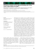
Multidomain CLE sequencesFigure 1
Multidomain CLE sequences. The potential multidomain
CLE signaling peptides CLE75, CLE76, CLE68, CLE31 and
CLE30 are represented. The figure is a scaled representation
of the domain organization. The relative positions of the first
amino acid of the motifs are specified.
BMC Plant Biology 2008, 8:1 />Page 7 of 15
(page number not for citation purposes)
CLE family was analyzed not based on phylogenetic trees
but on pairwise sequence comparisons. As pointed out by
Floyd and Bowman, the restricted sequence conservation
of 14 amino acids hampers phylogenetic analysis in case
of the CLE family [56].
So far, we were able to identify one representative of the
CLE family from Physcomitrella patens using the EST data-
base, although more might be found once the genome of
this organism is made publicly available. From the green
alga Chlamydomonas reinhardtii, of which we used the
genome as well as the EST database and TIGR transcript
assemblies, we could only identify one CLE sequence,
which did not cluster with any of the groups (Figure 2).
The biological function of this putative CLE signaling pep-
tide in Chlamydomonas will need to be established in
future studies. It will be interesting to find out if the CLE
sequence of Chlamydomonas has a different role to the
function of CLE signaling peptides in higher plants, which
show cell differentiation and meristem activity, and
whether CLE signaling peptides are part of an essential
genetic equipment required for plant development [56].
A new finding was the identification of CLE protein
sequences carrying multiple CLE motifs. We were able to
detect multidomain CLE proteins carrying two to six
motifs from O. sativa, T. aestivum and M. truncatula, but
not in any other plant species. The sequences originated
from different databases and sequencing projects. To
reduce the probability that mis-assembly of the genome
or TC-entries is responsible for the occurance of proteins
containing multiple CLE-domains, we examined the
genomic positions and EST coverage of the proteins.
Using the TIGR O. sativa genome browser, we determined
that the motifs in CLE75 and CLE76 originated from a sin-
gle exon. Examining the TC-entries for CLE30 and CLE32
from T. aestivum we were able to find 25 individual
sequence reads (EST's) for CLE30 and five sequence reads
for CLE31 covering at least two CLE motifs. This provides
evidence that both of the multi-CLE proteins from T. aes-
tivum are transcribed in the predicted manner and are
unlikely to be an artifact of TC-assembly. We hypothesize
that the full protein sequence releases several active sign-
aling peptides after processing, which could provide an
amplification effect.
Clustering of CLE motifs and identification of new
secondary motifs
Cluster analysis of the CLE sequences using CLANS
showed that these sequences could be assigned to 13
groups. The grouping we observed based on sequence
similarity corresponds to the classification of ectopic CLE
overexpression phenotypes in A. thaliana made by Stra-
bala et al.[12]. Furthermore, it is equivalent to the phylo-
genetic grouping and consistent with observations of
effects on the root apical meristem and tissue differentia-
Table 2: Detailed characteristics of multi-CLE domain proteins
CLE Database Length Motif Start Stop Motif Sequence Distance
CLE75 O. sativa
genome
250 1 51 63 IGVGKRLTPTGPNPVHNEFQP 51
287 99IGNGKRLTPTGPDPIHNEFQP 36
3123135IGDGKRLTPTGPDPVHNKFQP 36
4155167IGDGKRLTPTGPDPIHNEFQP 32
5191203IGDGKRLTPIGPDPIHNEFPP 36
6223235IGDGKRLTPTGPDPVHNEFQP 36
CLE76 O. sativa
genome
195 1 65 77 DFSVLRKVPTGPDPITSDPPP 65
294106QFSVLRKVPTGPDPITSDPPP 29
3119131EFPVLREVPSGPDPITSDPPP 25
4146158EFPVLREVPSGPDPITSDPPP 27
CLE68 M. truncatula
genome
181 1 70 82 EIGELRKVPSSPDPIHNSDID 70
2128140QIRGLTKVPTSPDPIHNSDSV 58
3157169QIGRARMVSSGPNPLHNRLIN 29
CLE31 T. aestivum
ESTs
175 1 75 97 IMMAPRPVPSGPDPIHHCPPA 75
2 112 124 AMVAPRPVPSGPNPIHHRPPH 37
3145157VMVAPMPIPSGPDPIHHCPPA 33
CLE30 T. aestivum
ESTs
175 1 61 73 VMVAPRPVPSGPDPIHHRPHA 61
2 98 110 VMVAPRPVPSGPNPIHHFPAP 37
Detailed characteristics of identified CLE members that carry multiple CLE motifs. The table contains information about database origin, protein
length in amino acids, and motif position as well as motif sequences and distance in amino acids between the motifs in the protein sequence.
BMC Plant Biology 2008, 8:1 />Page 8 of 15
(page number not for citation purposes)
tion [8,24]. We observed a close spatial arrangement of
known functional orthologs in the graph (e.g., FON4 and
CLV3, see group 3, Figure 2) [26]. The established group-
ing allows the interspecies identification of further
orthologs. We hypothesize that CLE125, located in the
same group as CLV3 and FON4, is the functional ortholog
of CLV3 in P. trichocarpa, and CLE143 and/or CLE147 in
Z. mays, respectively. The grouping also allows narrowing
down the number of candidate CLE genes from which the
nematode H. glycines may have acquired its CLE signaling
peptide. The H. glycines CLE sequence clustered tightly
with group 2. Provided a lateral gene transfer occurred,
this points to the nematode having acquired a CLE mem-
ber from group 2 and may allow insights as to the func-
tions of group-2 CLE signaling peptides as well as to the
function of the H. glycines CLE signaling peptides. Overall,
the results indicate that there is a connection between the
sequence similarities leading to distinct groups of CLE
members and the observed effect in case of excess peptide
supply (ectopic expression or peptide addition) [8,12,21-
24]. However, as ectopic expression might lead to pheno-
types that do not reflect the in vivo role of CLE signaling
peptides, future studies could focus on characterizing the
exact biological function of each signaling peptide.
In a peptide assay we confirmed that two in silico identi-
fied signaling peptides had biological function in M. trun-
catula. Both peptides arrested the activity of the root apical
meristem and lateral root meristems, resulting in reduced
root growth. The sequences of these peptides were found
in CLE members grouping either in group 7, 8 or 9 (Figure
1). Other CLE peptides that clustered in these groups were
also found to have a negative effect on the root apical mer-
istem, for example CLE25 and CLE26 in studies in A. thal-
iana and Zinnia elegans [8,24]. In addition, members of
CLE sequences in group 9, including CLE9–CLE13 also
showed an effect on the RAM [8,24].
One of the main questions remaining is why plants
encode such a large number of LRR-RLKs, and what their
function and ligands are. CLE signaling peptides could
bind to LRR-RLKs related to the CLV1/CLV2 receptor, but
so far little is known about specificity between CLE pep-
tide ligands and their receptors. Group specific and invar-
iant residues as well as variations of conserved residues
identified through sequence analysis could determine a
selective specificity for receptor subgroups targeted by a
given signaling peptide. Furthermore, our cluster analysis
revealed that there were regions outside the CLE motif
that correlated in sequence similarity with the groupings
generated by CLANS based on the primary CLE motif
sequence. It has been shown that processing occurs in
members of the family, meaning that one or a complex of
enzymes recognize part of the protein sequence and
cleave it. The addition of a single arginine residue at the C-
terminus of the conserved domain results in a decrease of
peptide activity [8,24]. This shows that correct cleavage
and specific recognition of the conserved domain are
required for the maximum activity of the signaling pep-
tide. The process and detailed mechanism remain
unknown. Furthermore, it is unclear whether all peptides
are processed and modified in a manner equivalent to
CLV3 and TDIF, which were found to be active as 12
amino acid peptides. We hypothesize that the extensions
of the motif may be involved in the specific recognition
and processing of the signaling peptide precursor.
Conclusion
We identified 114 new CLE domain proteins from a vari-
ety of plant species, including 28 new sequences from leg-
umes, which could be potential ligands for the LRR-RLKs
controlling nodulation. We also found several CLE pro-
teins with multiple CLE domains, which could represent
a mechanism for peptide signal amplification. Clustering
of the sequences showed 13 distinct groups, which were
found to have conserved secondary motifs outside the
CLE domain. Biological activity of two of the predicted
signaling peptides were confirmed in vivo. CLE signaling
peptides could have potential biotechnological applica-
tions for altering plant development, as exemplified in US
patent No. 7179963 using CLE signalling peptide func-
tions in Z. mays. While we could not test the biological
activity of all the identified signaling peptides in our
study, we hope that the CLE domain proteins presented in
this study will allow other researchers to test their func-
tion in a variety of plant species and as potential ligands
of LRR-RLKs.
Methods
Biological sequence resources
Several sequence resources were combined, forming a cus-
tom, redundant protein database. Expressed Sequence
Tags (EST) databases from A. thaliana (release 12.1),
Brassica napus (release 1), C. reinhardtii (release 5), G. max
(release 10), Lotus japonicus (release 3), Lycopersicum escu-
lentum (release 10.1), M. truncatula (release 8), Nicotiana
tabacum (release 2), O. sativa (release 16), Solanum tubero-
sum (release 10), and Z. mays (release 16) were down-
loaded from the TIGR Gene Indices (now available at the
Dana-Farber Cancer Institute gene index project) [49].
TIGR Transcript Assemblies (TA) from A. thaliana, Brassica
napus, C. reinhardtii, P. patens, G. max, Glycine soja, Lotus
corniculatus, Lupinus albus, Lycopersicum esculentum, M.
sativa, M. truncatula, Nicotiana tabacum, O. sativa, Phaseolus
coccineus, Phaseolus vulgaris, Pisum sativum, Solanum tubero-
sum, and Z. mays were added to this set (all release 1, 15
August 2005) [50]. The proteins predicted from the plant
genomes of A. thaliana (NCBI Genbank release 5, 03 May
2006) [57], C. reinhardtii (JGI, release 3) [58], M. truncat-
ula (Genome Sequencing Project release 17 July 2006)
BMC Plant Biology 2008, 8:1 />Page 9 of 15
(page number not for citation purposes)
[59], O. sativa (release 4, 30 December 2005) [60], and P.
trichocarpa (JGI, release 1) [61] were also included.
Sequence names were truncated to a unique identifier.
Information about the database origin of each sequence
was added to the unique identifier (i.e. OS-TA, OSEST,
OSGEN for O. sativa TA, EST or genomic sequences respec-
tively). Nucleotide sequences were translated into protein
sequences in all six reading frames (universal code), and
frame information was appended to the sequence identi-
Analysis of sequence similarity in the CLE domainFigure 2
Analysis of sequence similarity in the CLE domain. CLANS clustering of 174 sequences based on their sequence simi-
larity in the CLE domain. Sequences are represented by dots and the various groups are highlighted by ovals. Sequences of the
same group are assigned the same color. Lines connecting the dots correspond to BLASTP values better than 1.2E-7. Charac-
terized CLE members HgCLE (CLE47), TDIF (CLE49) and ZmESR (CLE143–CLE147), as well as the known orthologs CLV3/
FON4 and CLE19/BnCLE19 (CLE162) are highlighted with red stars. The single CLE member found from Physcomitrella patens
(moss, CLE170), which clusters into group 11, is highlighted with a grey star. A putative CLE sequence from Chlamydomonas
reinhardtii (alga, CLE177) is also marked with a grey star but does not cluster close to any group. The grouping established upon
cluster analysis is analogous to previous classifications [8, 12, 24]. Group 2 contains CLE1–CLE7, which were previously shown
to have no effect on RAM growth or on vascular cell differentiation in peptide assays and which led to wus-like dwarf growth
only at 21 days after germination when ectopically overexpressed. CLE9–CLE13 can be found in group 7. These CLE members
had an effect on the RAM but not on vascular cell differentiation in peptide assyas and wus-like dwarf growth could be observed
at 14 and 21 days after germination in overexpression studies. The CLE family members CLE41, CLE42, CLE44, which had no
effect on RAM but on vascular cell differentiation in peptide assays, and had a shrub-like overexpression phenotype are located
in group 5.
!
∀#
#∃
%
BMC Plant Biology 2008, 8:1 />Page 10 of 15
(page number not for citation purposes)
Weblogo representation of the conservation pattern of residues in each group and for the entire protein familyFigure 3
Weblogo representation of the conservation pattern of residues in each group and for the entire protein fam-
ily. The previously described main CLE motif of 12 amino acid length is marked with a black frame. Group specific residues are
marked in black in the various groups. Invariant residues are marked in black in the bottommost logo. Conserved residues are
marked grey. The size of the letter symbolizes the frequency of that residue in the group and at that position. A secondary
motif was identified at around 50 amino acids upstream of the primary CLE motif in groups 1, 2, 8 and 13. Extensions of the
motif are recognizable at both the C- and N-terminus. Bracketed figures indicate the number of sequences assigned to the
respective group.
BMC Plant Biology 2008, 8:1 />Page 11 of 15
(page number not for citation purposes)
Biological activity of CLE peptides in Medicago truncatulaFigure 4
Biological activity of CLE peptides in Medicago truncatula. Confirmation of the biological activity of synthetic CLE pep-
tides corresponding to 14 amino acids of the conserved domain of predicted CLE signaling peptides in a plate assay using M.
truncatula. Peptides were added at a concentration of 10 μM as growth media additives. The top row (A-C) shows plant
growth in the absence of peptide, the middle row (D-F) in the presence of peptide 1 (SKRKVPSCPDPLHN), and the bottom
row (G-I) in the presence of peptide 2 (SKRRVPNGPDPIHN). Plant growth is shown on day 6 after treatment (left column; A,
D, G), on day 20 after treatment (middle column; B, E, H) and on day 20 of recovery, whereby seedlings were treated for 6
days and then transferred to plates without peptide for the remaining 14 days (right column; C, F, I). Bar on the bottom of each
column indicates 2 cm.
BMC Plant Biology 2008, 8:1 />Page 12 of 15
(page number not for citation purposes)
fier (e.g. "_+2"). The translated nucleotide sequences and
modified protein sequences derived from genomic data
were combined into a single file and formatted using For-
matdb (options: -p T and -o T) [43]. The resulting data-
base contained 3,631,558 sequences. To determine
whether CLE sequences were specific to plants, a separate
search was based on the non-redundant protein database
(NCBI nr, version 15 June 2006.).
Query sequences
A set of 45 known CLE sequences (CLE1 – CLE17, CLE19
– CLE44, HgCLE, and CLV3; retrieved from Genbank and
TIGR) were combined in a FASTA-file, aligned using
CLUSTALW 1.83 and manually refined [51]. From the
multiple sequence alignment, a profile hidden Markov
model (HMM) was build using HMMer 2.3.2 [47]. The
original FASTA-file was re-aligned to the HMM (HMMa-
lign) and verified using the alignment editor AlnEdit [62]
to check for consistency of this realignment step. The
alignment revealed a region of high conservation of 12–
18 amino acids at the C-terminus that consistently
matched the HMM (corresponding to "HEELRTVPSGPD-
PLHH" of CLV3). We therefore decided to extend the
"conserved domain" beyond the 12 amino acids defined
previously [5,24,25]. This alignment (iteration 0) served
as input for iteration 1 to HMMaccel. Additionally, the
12–18 amino acid stretch that matched in the alignment
was extracted and used to build an HMM consisting solely
of the conserved region. HMMaccel is available for down-
load [48].
Motif search of the plant database
Each iteration started with a plain FASTA-file (the output
of the previous iteration). All sequences in the FASTA-file
were aligned against the HMM of the conserved domain.
The resulting alignment was verified using AlnEdit and
converted into aligned FASTA-format (for input to
HMMaccel). Full-length sequences were retrieved for all
HMMaccel hits and re-aligned to the HMM of the con-
served domain. The resulting alignment was manually
examined (AlnEdit) and converted to aligned FASTA-for-
mat (input to HMMaccel for the next iteration).
The settings for PSI-BLAST throughout iteration 1 and 2
were a cut-off E-value of 10,000 (parameter -e), an E-value
threshold of 0.005 for the inclusion of sequences (param-
eter -h), 250 for the numbers of displayed high scoring
sequence pairs (parameter -b) and 500 for the numbers of
displayed hits (parameter -v). The parameters -b and -v
were altered in iteration 3 and iteration 4 to -b 1 and -v
1,000. The parameters for HMMer in HMMaccel caused
hits up to E-values of 10 to be returned and the HMMs to
be calibrated using 5000 samples.
We observed a large number of false positive sequences
that were added to the dataset after iteration 3 compared
to previous iterations. Without removal of these
sequences, the dataset became inaccurate in iteration 4. To
avoid a biased removal of sequences and for a reproduci-
ble optimization of the sequence set, cluster analysis of
sequences (CLANS) was used [52,53]. The conserved
region of the 811 hits of iteration 3 was extracted and ana-
lyzed in CLANS. A total of 312 sequences were discarded
as false-positives from the sequence set. The remaining
499 sequences were submitted to a final iteration. The
HMM derived from these 499 sequences is available as
Additional File 3. After iteration 4 the dataset consisted of
659 protein sequences. The large number of false positive
hits returned in iteration 3 point to the method having
reached the limits of what it could resolve. After removal
of false positives, a fourth iteration was performed to
reduce the number of false negatives. The aim of the iter-
ative search was to find all CLE peptides in the database
and therefore false negatives were of greater concern than
false positives.
The species and database origin was contained in the
sequence identifier. The full annotation information of
the sequences was subsequently retrieved from the origi-
nal FASTA-files. The calculation of isoelectric points and
Sequence specificity of CLE peptide activityFigure 5
Sequence specificity of CLE peptide activity. Root
length of Medicago truncatula plants at 6 days after treatment
with different peptides. Control plates did not contain pep-
tide, peptide 1 (SKRKVPSCPDPLHN) and peptide 2 (SKR-
RVPNGPDPIHN) resemble the CLE motif, peptide 3
(randomized version of peptide 1, DHKSKPPVLRPNSC) and
peptide 4 (randomized version of peptide 2, PVHPKGNRN-
DISPR) do not resemble the CLE motif. Bars with different
letters differ significantly at p < 0.0001 (N = 27; one-way
ANOVA). Both CLE peptides are significantly different from
the no-peptide control and the control peptides with rand-
omized amino acid sequence.
BMC Plant Biology 2008, 8:1 />Page 13 of 15
(page number not for citation purposes)
molecular weights was performed with PROT STATS from
the EMBOSS 4.0.0. package [63]. Protein length, position
and sequence of the CLE domain were extracted from the
FASTA-file.
Sequence analysis
Full-length protein sequences as well as conserved
domains were analyzed in CLANS [52]. The dataset was
spiked with the 45 original query sequences to check their
positioning and group assignment in CLANS. Using the
full-length sequences, we found several sequences with
more than one domain, which we noticed by their behav-
iour in CLANS. Grouping of CLE peptides was observed in
the cluster analysis of the conserved domain. The individ-
ual groups were extracted and aligned using Kalign [64].
The alignments of primary CLE motifs, their extension
and additional motifs were visualized with WebLogo
3.0b14 to represent all sequences of the group [65].
Peptide synthesis
Peptide 1 (SKRKVPSCPDPLHN) and peptide 3 (rand-
omized peptide 1, DHKSKPPVLRPNSC) as well as peptide
2 (SKRRVPNGPDPIHN) and peptide 4 (randomized pep-
tide 2, PVHPKGNRNDISPR) were synthesized with >75%
purity by GL Biochem (Shanghai, China). The peptides
carried a free carboxyl acid group at the C-terminus. Pep-
tide 1 and 2 were designed according to the CLE motif,
peptides 3 and 4 do not resemble the CLE motif as the
sequences are randomized versions of the amino acid
sequence of peptide 1 and peptide 2. Randomized
sequences were generated with the RandSeq tool at
ExPASy [66]. Peptides were diluted to a final concentra-
tion of 10 μmol/l [22] in sterile, nitrogen-free Fåhraeus
media [67].
Peptide assay
Wildtype M. truncatula cv. Jemalong A17 seeds were scari-
fied on fine sand paper and sterilized using 80% technical
grade ethanol (5 min), 6.25% sodium hypochlorite solu-
tion (5 min), and freshly prepared 200 mg/l Augmentin
®
Duo (Amoxicillin/Potassium Clavulanate; GlaxoSmithK-
line, Brentford UK) (5 h) with five washes of sterile Milli-
Q
®
water (Millipore, Billerica USA) between treatments.
Seeds were germinated on Fåhraeus agar plates without
the presence of peptides at 4°C (12 h) and 28°C (24 h) in
the dark [68]. Seedlings were briefly washed with sterile,
phosphate-buffered saline before transfer to a fresh plate
(10 seedlings per plate) containing peptide or no peptide
(control). Plates were sealed with Parafilm M
®
(Structure
Probe Inc., West Chester USA) on the bottom half and
grown in an upright position. Black paper carton was
placed to cover the bottom 2/3 of plate to minimize light
exposure to roots. Plants were grown at constant 25°C
and 100 μE light intensity under extended day conditions
(16 h day/8 h night) [68]. Root growth was measured
every 24 h for six days, starting on the day of transfer (t =
0d). To test the reversibility of the peptide treatment (t =
6d), half of the plants (five) were transferred from the
plate containing the peptide to a fresh media plate (with-
out peptide) and grown for two weeks (t = 20d). Photo-
graphs were taken at time points 6 d and 20 d. Statistical
analyses were performed using GenStat
®
9.2 (VSN Interna-
tional Ltd, Hemel Hempstead UK).
Authors' contributions
KO carried out the bioinformatic analysis and peptide
assays. NG designed the database and contributed with
programming. GFW and PMG were involved in the over-
all design and coordination of the experiments. UM per-
formed statistical analysis and some of the peptide assays,
TF conceived the strategy of the motif search and contrib-
uted to the programming and the overall experimental
design. All authors read and approved the final manu-
script.
Additional material
Acknowledgements
The authors thank Helge Küster for access to the Glomus EST database.
Also, we would like to acknowledge our colleagues from the University of
Queensland and Australian National University node of the Australian
Research Council Centre of Excellence for Integrative Legume Research
(CILR) for critical and fruitful discussions. KO is grateful for financial sup-
port from the CILR. This research was funded by the CILR (CE0348212)
and a Research Fellowship to UM from the Australian Research Council
(DP 0557692).
References
1. Johnson KL, Ingram GC: Sending the right signals: regulating
receptor kinase activity. Curr Opin Plant Biol 2005, 8(6):648-656.
2. Becraft PW: Receptor kinase signaling in plant development.
Annual Review of Cell and Developmental Biology 2002, 18:163-192.
Additional file 1
Table of Identification.
Click here for file
[ />2229-8-1-S1.XLS]
Additional file 2
Multiple sequence alignments of groups and full length sequence logos.
Click here for file
[ />2229-8-1-S2.pdf]
Additional file 3
last HMM. Hidden Markov model after the third iteration, generated by
HMMer 2.3.2.
Click here for file
[ />2229-8-1-S3.HMM]
BMC Plant Biology 2008, 8:1 />Page 14 of 15
(page number not for citation purposes)
3. Kobe B, Deisenhofer J: A structural basis of the interactions
between leucine-rich repeats and protein ligands. Nature
1995, 374(6518):183-186.
4. Matsubayashi Y, Yang HP, Sakagami Y: Peptide signals and their
receptors in higher plants. Trends in Plant Science 2001,
6(12):573-577.
5. Cock JM, McCormick S: A large family of genes that share
homology with CLAVATA3. Plant Physiol 2001, 126(3):939-942.
6. Fiers M, Ku KL, Liu CM: CLE peptide ligands and their roles in
establishing meristems. Current Opinion in Plant Biology 2007,
10(1):39-43.
7. Germain H, Chevalier E, Matton DP: Plant bioactive peptides: an
expanding class of signaling molecules. Canadian Journal of Bot-
any-Revue Canadienne De Botanique 2006, 84(1):1-19.
8. Sawa S, Kinoshita A, Nakanomyo I, Fukuda H: CLV3/ESR-related
(CLE) peptides as intercellular signaling molecules in plants.
Chem Rec 2006, 6(6):303-310.
9. Olsen AN, Skriver K: Ligand mimicry? Plant-parasitic nema-
tode polypeptide with similarity to CLAVATA3. Trends Plant
Sci 2003, 8(2):55-57.
10. Wang XH, Mitchum MG, Gao BL, Li CY, Diab H, Baum TJ, Hussey RS,
Davis EL: A parasitism gene from a plant-parasitic nematode
with function similar to CLAVATA3/ESR (CLE) of Arabidop-
sis thaliana. Molecular Plant Pathology 2005, 6(2):187-191.
11. Davis EL, Mitchum MG: Nematodes. Sophisticated parasites of
legumes. Plant Physiology 2005, 137(4):1182-1188.
12. Strabala TJ, O'Donnell PJ, Smit AM, Ampomah-Dwamena C, Martin
EJ, Netzler N, Nieuwenhuizen NJ, Quinn BD, Foote HCC, Hudson
KR: Gain-of-function phenotypes of many CLAVATA3/ESR
genes, including four new family members, correlate with
tandem variations in the conserved CLAVATA3/ESR
domain. Plant Physiology 2006, 140(4):1331-1344.
13. Ni J, Clark SE: Evidence for functional conservation, suffi-
ciency, and proteolytic processing of the CLAVATA3 CLE
domain.
Plant Physiol 2006, 140(2):726-733.
14. OpsahlFerstad HG, LeDeunff E, Dumas C, Rogowsky PM: ZmEsr, a
novel endosperm-specific gene expressed in a restricted
region around the maize embryo. Plant Journal 1997,
12(1):235-246.
15. Bonello JF, Opsahl-Ferstad HG, Perez P, Dumas C, Rogowsky PM:
Esr genes show different levels of expression in the same
region of maize endosperm. Gene 2000, 246(1-2):219-227.
16. Clark SE, Running MP, Meyerowitz EM: Clavata3 Is a Specific Reg-
ulator of Shoot and Floral Meristem Development Affecting
the Same Processes as Clavata1. Development 1995,
121(7):2057-2067.
17. Brand U, Fletcher JC, Hobe M, Meyerowitz EM, Simon R: Depend-
ence of stem cell fate in Arabidopsis an a feedback loop reg-
ulated by CLV3 activity. Science 2000, 289(5479):617-619.
18. Rojo E, Sharma VK, Kovaleva V, Raikhel NV, Fletcher JC: CLV3 is
localized to the extracellular space, where it activates the
Arabidopsis CLAVATA stem cell signaling pathway. Plant Cell
2002, 14(5):969-977.
19. Clark SE: Cell signalling at the shoot meristem. Nature Reviews
Molecular Cell Biology 2001, 2(4):276-284.
20. Casamitjana-Martinez E, Hofhuis HF, Xu J, Liu CM, Heidstra R,
Scheres B: Root-specific CLE19 overexpression and the sol1/2
suppressors implicate a CLV-like pathway in the control of
Arabidopsis root meristem maintenance. Current Biology 2003,
13(16):1435-1441.
21. Hobe M, Muller R, Grunewald M, Brand U, Simon R: Loss of CLE40,
a protein functionally equivalent to the stem cell restricting
signal CLV3, enhances root waving in Arabidopsis. Dev Genes
Evol 2003, 213(8):371-381.
22. Fiers M, Golemiec E, Xu J, van der Geest L, Heidstra R, Stiekema W,
Liu CM: The 14-amino acid CLV3, CLE19, and CLE40 pep-
tides trigger consumption of the root meristem in Arabidop-
sis through a CLAVATA2-dependent pathway. Plant Cell 2005,
17(9):2542-2553.
23. Fiers M, Hause G, Boutilier K, Casamitjana-Martinez E, Weijers D,
Offringa R, van der Geest L, Campagne MV, Liu CM: Mis-expression
of the CLV3/ESR-like gene CLE19 in Arabidopsis leads to a
consumption of root meristem. Gene 2004, 327(1):37-49.
24. Ito Y, Nakanomyo I, Motose H, Iwamoto K, Sawa S, Dohmae N,
Fukuda H: Dodeca-CLE peptides as suppressors of plant stem
cell differentiation. Science 2006, 313(5788):842-845.
25. Kondo T, Sawa S, Kinoshita A, Mizuno S, Kakimoto T, Fukuda H, Sak-
agami Y: A plant peptide encoded by CLV3 identified by in situ
MALDI-TOF MS analysis. Science 2006, 313(5788):845-848.
26. Chu HW, Qian Q, Liang WQ, Yin CS, Tan HX, Yao X, Yuan Z, Yang
J, Huang H, Luo D, Ma H, Zhang DB: The floral organ number4
gene encoding a putative ortholog of Arabidopsis
CLAVATA3 regulates apical meristem size in rice. Plant Phys-
iology 2006, 142(3):1039-1052.
27. Hirsch AM: Developmental Biology of Legume Nodulation.
New Phytologist 1992, 122(2):211-237.
28. Endre G, Kereszt A, Kevei Z, Mihacea S, Kalo P, Kiss GB: A receptor
kinase gene regulating symbiotic nodule development.
Nature 2002, 417(6892):962-966.
29. Capoen W, Goormachtig S, De Rycke R, Schroeyers K, Holsters M:
SrSymRK, a plant receptor essential for symbiosome forma-
tion. Proc Natl Acad Sci U S A 2005, 102(29):10369-10374.
30. Stracke S, Kistner C, Yoshida S, Mulder L, Sato S, Kaneko T, Tabata
S, Sandal N, Stougaard J, Szczyglowski K, Parniske M: A plant recep-
tor-like kinase required for both bacterial and fungal symbi-
osis. Nature 2002, 417(6892):959-962.
31. Searle IR, Men AE, Laniya TS, Buzas DM, Iturbe-Ormaetxe I, Carroll
BJ, Gresshoff PM: Long-distance signaling in nodulation
directed by a CLAVATA1-like receptor kinase. Science 2003,
299(5603):109-112.
32. Nishimura R, Hayashi M, Wu GJ, Kouchi H, Imaizumi-Anraku H,
Murakami Y, Kawasaki S, Akao S, Ohmori M, Nagasawa M, Harada K,
Kawaguchi M: HAR1 mediates systemic regulation of symbi-
otic organ development. Nature 2002, 420(6914):426-429.
33. Schnabel E, Journet EP, de Carvalho-Niebel F, Duc G, Frugoli J: The
Medicago truncatula SUNN gene encodes a CLV1-like leu-
cine-rich repeat receptor kinase that regulates nodule
number and root length. Plant Mol Biol 2005, 58(6):
809-822.
34. Krusell L, Madsen LH, Sato S, Aubert G, Genua A, Szczyglowski K,
Duc G, Kaneko T, Tabata S, de Bruijn F, Pajuelo E, Sandal N, Stou-
gaard J: Shoot control of root development and nodulation is
mediated by a receptor-like kinase. Nature 2002,
420(6914):422-426.
35. Streeter J: Inhibition of Legume Nodule Formation and N-2
Fixation by Nitrate. Crc Critical Reviews in Plant Sciences 1988,
7(1):1-23.
36. Scheible WR, Morcuende R, Czechowski T, Fritz C, Osuna D, Pala-
cios-Rojas N, Schindelasch D, Thimm O, Udvardi MK, Stitt M:
Genome-wide reprogramming of primary and secondary
metabolism, protein synthesis, cellular growth processes,
and the regulatory infrastructure of Arabidopsis in response
to nitrogen. Plant Physiol 2004, 136(1):2483-2499.
37. Torii KU: Leucine-rich repeat receptor kinases in plants:
structure, function, and signal transduction pathways. Int Rev
Cytol 2004, 234:1-46.
38. Schopfer CR, Nasrallah ME, Nasrallah JB: The male determinant
of self-incompatibility in Brassica. Science 1999,
286(5445):1697-1700.
39. Vanoosthuyse V, Miege C, Dumas C, Cock JM: Two large Arabi-
dopsis thaliana gene families are homologous to the Brassica
gene superfamily that encodes pollen coat proteins and the
male component of the self-incompatibility response. Plant
Molecular Biology 2001, 46(1):17-34.
40. Ride JP, Davies EM, Franklin FCH, Marshall DF: Analysis of Arabi-
dopsis genome sequence reveals a large new gene family in
plants. Plant Molecular Biology 1999, 39(5):927-932.
41. Lease KA, Walker JC: The Arabidopsis unannotated secreted
peptide database, a resource for plant peptidomics. PLANT
PHYSIOLOGY 2006, 142(3):831-838.
42. Silverstein KAT, Graham MA, Paape TD, VandenBosch KA: Genome
organization of more than 300 defensin-like genes in arabi-
dopsis. PLANT PHYSIOLOGY 2005, 138(2):600-610.
43. Altschul SF, Madden TL, Schaffer AA, Zhang JH, Zhang Z, Miller W,
Lipman DJ: Gapped BLAST and PSI-BLAST: a new generation
of protein database search programs. Nucleic Acids Research
1997, 25(17):3389-3402.
44. Bailey TL, Baker ME, Elkan CP: An artificial intelligence approach
to motif discovery in protein sequences: Application to ster-
oid dehydrogenases. Journal of Steroid Biochemistry and Molecular
Biology 1997, 62(1):29-44.
Publish with BioMed Central and every
scientist can read your work free of charge
"BioMed Central will be the most significant development for
disseminating the results of biomedical research in our lifetime."
Sir Paul Nurse, Cancer Research UK
Your research papers will be:
available free of charge to the entire biomedical community
peer reviewed and published immediately upon acceptance
cited in PubMed and archived on PubMed Central
yours — you keep the copyright
Submit your manuscript here:
/>BioMedcentral
BMC Plant Biology 2008, 8:1 />Page 15 of 15
(page number not for citation purposes)
45. Sharma VK, Ramirez J, Fletcher JC: The Arabidopsis CLV3-like
(CLE) genes are expressed in diverse tissues and encode
secreted proteins. Plant Mol Biol 2003, 51(3):415-425.
46. Eddy SR: Hidden Markov models and genome sequence anal-
ysis. Faseb Journal 1998, 12(8):A1327-a1327.
47. Eddy SR: Profile hidden Markov models. Bioinformatics 1998,
14(9):755-763.
48. HMMaccel at the RSBS Bioinformatics Server [http://bioin
foserver.rsbs.anu.edu.au/programs/hmmaccel/]
49. Lee Y, Tsai J, Sunkara S, Karamycheva S, Pertea G, Sultana R,
Antonescu V, Chan A, Cheung F, Quackenbush J: The TIGR Gene
Indices: clustering and assembling EST and known genes and
integration with eukaryotic genomes. Nucleic Acids Research
2005, 33:D71-D74.
50. Childs KL, Hamilton JP, Zhu W, Ly E, Cheung F, Wu H, Rabinowicz
PD, Town CD, Buell CR, Chan AP: The TIGR plant transcript
assemblies database. Nucleic Acids Research 2007, 35:D846-D851.
51. Thompson JD, Higgins DG, Gibson TJ: Clustal-W - Improving the
Sensitivity of Progressive Multiple Sequence Alignment
through Sequence Weighting, Position-Specific Gap Penal-
ties and Weight Matrix Choice. Nucleic Acids Research 1994,
22(22):4673-4680.
52. Frickey T, Lupas A: CLANS: a Java application for visualizing
protein families based on pairwise similarity. Bioinformatics
2004, 20(18):3702-3704.
53. CLANS at the RSBS Bioinformatics Server: CLANS at the RSBS
Bioinformatics Server. [ />grams/clans/index.php].
54. Li WZ, Godzik A: Cd-hit: a fast program for clustering and
comparing large sets of protein or nucleotide sequences. Bio-
informatics 2006, 22(13):1658-1659.
55. Corpet F: Multiple Sequence Alignment with Hierarchical-
Clustering. Nucleic Acids Research 1988, 16(22):10881-10890.
56. Floyd SK, Bowman JL: The ancestral developmental tool kit of
land plants. International Journal of Plant Sciences 2007, 168(1):1-35.
57. Arabidopsis Genome Initiative: Analysis of the genome sequence
of the flowering plant Arabidopsis thaliana. Nature 2000,
408(6814):796-815.
58. Shrager J, Hauser C, Chang CW, Harris EH, Davies J, McDermott J,
Tamse R, Zhang ZD, Grossman AR: Chlamydomonas reinhardtii
genome project. A guide to the generation and use of the
cDNA information. Plant Physiology 2003, 131(2):401-408.
59. Bell CJ, Dixon RA, Farmer AD, Flores R, Inman J, Gonzales RA, Har-
rison MJ, Paiva NL, Scott AD, Weller JW, May GD: The Medicago
Genome Initiative: a model legume database. Nucleic Acids
Research 2001, 29(1):114-117.
60. Matsumoto T, Wu JZ, Kanamori H, Katayose Y, Fujisawa M, Namiki
N, Mizuno H, Yamamoto K, Antonio BA, Baba T, Sakata K, Nagamura
Y, Aoki H, Arikawa K, Arita K, Bito T, Chiden Y, Fujitsuka N, Fuku-
naka R, Hamada M, Harada C, Hayashi A, Hijishita S, Honda M, Hoso-
kawa S, Ichikawa Y, Idonuma A, Iijima M, Ikeda M, Ikeno M, Ito K, Ito
S, Ito T, Ito Y, Ito Y, Iwabuchi A, Kamiya K, Karasawa W, Kurita K,
Katagiri S, Kikuta A, Kobayashi H, Kobayashi N, Machita K, Maehara
T, Masukawa M, Mizubayashi T, Mukai Y, Nagasaki H, Nagata Y, Naito
S, Nakashima M, Nakama Y, Nakamichi Y, Nakamura M, Meguro A,
Negishi M, Ohta I, Ohta T, Okamoto M, Ono N, Saji S, Sakaguchi M,
Sakai K, Shibata M, Shimokawa T, Song JY, Takazaki Y, Terasawa K,
Tsugane M, Tsuji K, Ueda S, Waki K, Yamagata H, Yamamoto M,
Yamamoto S, Yamane H, Yoshiki S, Yoshihara R, Yukawa K, Zhong
HS, Yano M, Sasaki T, Yuan QP, Shu OT, Liu J, Jones KM, Gansberger
K, Moffat K, Hill J, Bera J, Fadrosh D, Jin SH, Johri S, Kim M, Overton
L, Reardon M, Tsitrin T, Vuong H, Weaver B, Ciecko A, Tallon L, Jack-
son J, Pai G, Van Aken S, Utterback T, Reidmuller S, Feldblyum T,
Hsiao J, Zismann V, Iobst S, de Vazeille AR, Buell CR, Ying K, Li Y, Lu
TT, Huang YC, Zhao Q, Feng Q, Zhang L, Zhu JJ, Weng QJ, Mu J, Lu
YQ, Fan DL, Liu YL, Guan JP, Zhang YJ, Yu SL, Liu XH, Zhang Y, Hong
GF, Han B, Choisne N, Demange N, Orjeda G, Samain S, Cattolico L,
Pelletier E, Couloux A, Segurens B, Wincker P, D'Hont A, Scarpelli C,
Weissenbach J, Salanoubat M, Quetier F, Yu Y, Kim HR, Rambo T,
Currie J, Collura K, Luo MZ, Yang TJ, Ammiraju JSS, Engler F, Soder-
lund C, Wing RA, Palmer LE, de la Bastide M, Spiegel L, Nascimento
L, Zutavern T, O'Shaughnessy A, Dike S, Dedhia N, Preston R, Balija
V, McCombie WR, Chow TY, Chen HH, Chung MC, Chen CS, Shaw
JF, Wu HP, Hsiao KJ, Chao YT, Chu MK, Cheng CH, Hour AL, Lee
PF, Lin SJ, Lin YC, Liou JY, Liu SM, Hsing YI, Raghuvanshi S, Mohanty
A, Bharti AK, Gaur A, Gupta V, Kumar D, Ravi V, Vij S, Kapur A,
Khurana P, Khurana P, Khurana JP, Tyagi AK, Gaikwad K, Singh A,
Dalal V, Srivastava S, Dixit A, Pal AK, Ghazi IA, Yadav M, Pandit A,
Bhargava A, Sureshbabu K, Batra K, Sharma TR, Mohapatra T, Singh
NK, Messing J, Nelson AB, Fuks G, Kavchok S, Keizer G, Llaca ELV,
Song RT, Tanyolac B, Young S, Il KH, Hahn JH, Sangsakoo G, Vanavi-
chit A, de Mattos LAT, Zimmer PD, Malone G, Dellagostin O, de
Oliveira AC, Bevan M, Bancroft I, Minx P, Cordum H, Wilson R,
Cheng ZK, Jin WW, Jiang JM, Leong SA, Iwama H, Gojobori T, Itoh
T, Niimura Y, Fujii Y, Habara T, Sakai H, Sato Y, Wilson G, Kumar K,
McCouch S, Juretic N, Hoen D, Wright S, Bruskiewich R, Bureau T,
Miyao A, Hirochika H, Nishikawa T, Kadowaki K, Sugiura M, Project
IRGS: The map-based sequence of the rice genome. Nature
2005, 436(7052):793-800.
61. Tuskan GA, DiFazio S, Jansson S, Bohlmann J, Grigoriev I, Hellsten U,
Putnam N, Ralph S, Rombauts S, Salamov A, Schein J, Sterck L, Aerts
A, Bhalerao RR, Bhalerao RP, Blaudez D, Boerjan W, Brun A, Brunner
A, Busov V, Campbell M, Carlson J, Chalot M, Chapman J, Chen GL,
Cooper D, Coutinho PM, Couturier J, Covert S, Cronk Q, Cunning-
ham R, Davis J, Degroeve S, Dejardin A, Depamphilis C, Detter J,
Dirks B, Dubchak I, Duplessis S, Ehlting J, Ellis B, Gendler K, Good-
stein D, Gribskov M, Grimwood J, Groover A, Gunter L, Hamberger
B, Heinze B, Helariutta Y, Henrissat B, Holligan D, Holt R, Huang W,
Islam-Faridi N, Jones S, Jones-Rhoades M, Jorgensen R, Joshi C, Kan-
gasjarvi J, Karlsson J, Kelleher C, Kirkpatrick R, Kirst M, Kohler A,
Kalluri U, Larimer F, Leebens-Mack J, Leple JC, Locascio P, Lou Y,
Lucas S, Martin F, Montanini B, Napoli C, Nelson DR, Nelson C,
Nieminen K, Nilsson O, Pereda V, Peter G, Philippe R, Pilate G, Polia-
kov A, Razumovskaya J, Richardson P, Rinaldi C, Ritland K, Rouze P,
Ryaboy D, Schmutz J, Schrader J, Segerman B, Shin H, Siddiqui A,
Sterky F, Terry A, Tsai CJ, Uberbacher E, Unneberg P, Vahala J, Wall
K, Wessler S, Yang G, Yin T, Douglas C, Marra M, Sandberg G, de
Peer YV, Rokhsar D: The genome of black cottonwood, Popu-
lus trichocarpa (Torr. & Gray). Science 2006,
313(5793):1596-1604.
62. AlnEdit at the RSBS Bioinformatics Server: AlnEdit at the RSBS
Bioinformatics Server. [ />grams/alnedit/].
63. Rice P, Longden I, Bleasby A: EMBOSS: The European molecular
biology open software suite. Trends in Genetics 2000,
16(6):276-277.
64. Lassmann T, Sonnhammer ELL: Kalign - an accurate and fast mul-
tiple sequence alignment algorithm. Bmc Bioinformatics 2005, 6:.
65. Crooks GE, Hon G, Chandonia JM, Brenner SE: WebLogo: A
sequence logo generator. Genome Research 2004,
14(6):1188-1190.
66. Gasteiger E, Gattiker A, Hoogland C, Ivanyi I, Appel RD, Bairoch A:
ExPASy: the proteomics server for in-depth protein knowl-
edge and analysis. Nucleic Acids Research 2003, 31(13):3784-3788.
67. Fahraeus G: The infection of clover root hairs by nodule bac-
teria studied by a simple glass slide technique. J Gen Microbiol
1957, 16(2):374-381.
68. Wasson AP, Pellerone FI, Mathesius U: Silencing the flavonoid
pathway in Medicago truncatula inhibits root nodule forma-
tion and prevents auxin transport regulation by rhizobia.
Plant Cell 2006, 18(7):1617-1629.
Publish with BioMed Central and every
scientist can read your work free of charge
"BioMed Central will be the most significant development for
disseminating the results of biomedical research in our lifetime."
Sir Paul Nurse, Cancer Research UK
Your research papers will be:
available free of charge to the entire biomedical community
peer reviewed and published immediately upon acceptance
cited in PubMed and archived on PubMed Central
yours — you keep the copyright
Submit your manuscript here:
/>BioMedcentral