báo cáo khoa học: " Transcriptional profiling of Medicago truncatula meristematic root cells" docx
Bạn đang xem bản rút gọn của tài liệu. Xem và tải ngay bản đầy đủ của tài liệu tại đây (473.98 KB, 12 trang )
BioMed Central
Page 1 of 12
(page number not for citation purposes)
BMC Plant Biology
Open Access
Research article
Transcriptional profiling of Medicago truncatula meristematic root
cells
Peta Holmes
1
, Nicolas Goffard
1,2
, Georg F Weiller
1
, BarryGRolfe
1
and
Nijat Imin*
1
Address:
1
ARC Centre of Excellence for Integrative Legume Research, Genomic Interactions Group, Research School of Biological Sciences,
Australian National University, Canberra ACT 2601, Australia and
2
Institut Louis Malardé, GP Box 30, 98713 Papeete Tahiti, French Polynesia
Email: Peta Holmes - ; Nicolas Goffard - ; Georg F Weiller - ;
Barry G Rolfe - ; Nijat Imin* -
* Corresponding author
Abstract
Background: The root apical meristem of crop and model legume Medicago truncatula is a
significantly different stem cell system to that of the widely studied model plant species Arabidopsis
thaliana. In this study we used the Affymetrix Medicago GeneChip
®
to compare the transcriptomes
of meristem and non-meristematic root to identify root meristem specific candidate genes.
Results: Using mRNA from root meristem and non-meristem we were able to identify 324 and
363 transcripts differentially expressed from the two regions. With bioinformatics tools developed
to functionally annotate the Medicago genome array we could identify significant changes in
metabolism, signalling and the differentially expression of 55 transcription factors in meristematic
and non-meristematic roots.
Conclusion: This is the first comprehensive analysis of M. truncatula root meristem cells using this
genome array. This data will facilitate the mapping of regulatory and metabolic networks involved
in the open root meristem of M. truncatula and provides candidates for functional analysis.
Background
The root and shoot apical meristems (RAM and SAM) are
established during embryogenesis and serve as a source of
stem cells for plant growth and organogenesis [1]. The
RAM produces all the tissues of the primary root by a
highly defined pattern of cell divisions [2]. Cells produced
by the meristem, known as initials, undergo proliferative
cell divisions as they are added to files of different cell
types and their fate is determined by positional informa-
tion [3,4]. The stem cell niche in the root is maintained by
a small group of cells called the quiescent centre (QC)
[5,6], the QC inhibits the division of surrounding cells
and is generated and maintained by the accumulation of
auxin via the PIN auxin efflux carriers; in Arabidopsis the
genes PLETHORA1, PLETHORA2, SCARECROW and
SHORT ROOT are known to be necessary for QC forma-
tion [6-9]. The interplay of auxin and cytokinin controls
the size of the RAM, with the action of cytokinin impli-
cated in controlling the exit of cells from the root meris-
tem [10,11].
Several studies that characterise gene expression in the
cells of the root meristem have been published. Studies in
Arabidopsis have used green fluorescent protein-labelled
Published: 27 February 2008
BMC Plant Biology 2008, 8:21 doi:10.1186/1471-2229-8-21
Received: 15 June 2007
Accepted: 27 February 2008
This article is available from: />© 2008 Holmes et al; licensee BioMed Central Ltd.
This is an Open Access article distributed under the terms of the Creative Commons Attribution License ( />),
which permits unrestricted use, distribution, and reproduction in any medium, provided the original work is properly cited.
BMC Plant Biology 2008, 8:21 />Page 2 of 12
(page number not for citation purposes)
cell types and cell sorting to characterise gene expression
by microarray, for specific cell types and in different zones
of root development [12-14]. A root tissue specific gene
expression study has also been carried out in maize (Zea
mays) where the proximal meristem, QC and root cap
were microdissected and gene expression was measured
on Affymetrix rice genome arrays [15]. However the root
of model legume Medicago truncatula presents a notably
different system for study of root development to that of
Arabidopsis thaliana or maize. At a cellular level, the root of
M. truncatula has a significantly different RAM to that of
Arabidopsis. Most legume roots, unlike the Arabidopsis root
have a basic-open root meristem [16]. The difference
between open and closed meristems is significant; in the
open RAM, initials are not apparent indicating possible
variations in the regulation cell division and differentia-
tion between the two types of RAM. Hamamoto et al. [17]
have shown that roots with an open meristem produce
individual living border cells and more border cells than
those with a closed meristem. Border cells are important
for mycorrhizal and microbial interactions including the
legume-rhizobia symbiosis [18] and environmental sens-
ing.
In terms of root organogenesis, the most obvious differ-
ence between M. truncatula and other model plants and is
the ability of M. truncatula to form indeterminate root
nodules in association with rhizobia. Nodulation shares
several aspects of lateral root organogenesis with the
advantage that it is inducible and the site of organogenesis
is predictable. Root organogenesis is also inducible in M.
truncatula in tissue culture with the addition of auxin 1-
naphthaleneacetic acid (NAA) to the tissue culture media.
Root formation in culture is irreversible after 7 days on
NAA [19] and does not require ethylene perception [20].
Thus, the morphological differences between the M. trun-
catula root and that of other model species and interest in
the species as a model for root and nodule development
led us to conduct the research we present here.
Results and discussion
The M. truncatula root meristem
The M. truncatula RAM shows a characteristic basic open
root meristem organisation (Figure 1). In the region
where cells are dividing, it isn't possible to distinguish the
initials amongst the tightly packed mass of new and elon-
gating cells in the root tip. Our meristem section, 3 milli-
metres from the root tip, is comprised large group of
undifferentiated cells, surrounded by some differentiated
tissues including border cells, root cap and elongating
cells that will form the vascular bundle, pericycle, endo-
dermis, cortex and epidermis. Our non-meristematic sec-
tion, one centimetre adjacent to the meristem, only
contains the characteristic root tissue layers; root hairs
occur in this region, rhizobial infection and lateral root
initiation occur in this zone. These sections were chosen
to correspond with earlier proteomic work on the Medi-
cago root meristem [21] and because there are no
described markers for specific cell types in the Medicago
root meristem that could be used to create transgenic
plants for a cell-type analysis.
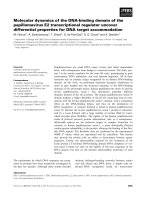
The Medicago root meristemFigure 1
The Medicago root meristem. A median longitudinal sec-
tion of the Medicago root stained with toluidine blue clearly
shows that basic-open meristem architecture of M. truncat-
ula, the zone of initials is not clearly divided into tiers; VC =
vascular cylinder, C = cortex, E = epidermis, and RC = root
cap; scale bar = 50 µm.
BMC Plant Biology 2008, 8:21 />Page 3 of 12
(page number not for citation purposes)
To characterise the transcriptomes of the meristematic
and non-meristematic root of the M. truncatula we
extracted RNA from the meristem and non-meristematic
zones from the roots of three independently grown sets of
plants four days after germination. The three biological
replicates were analysed using the Affymetrix Medicago
genome array. An average of 51% (26,9610 probe sets) of
the over 52, 000 plant gene probes of the Medicago
Genome Array GeneChip produced 'present' calls when
hybridised with biotin-labelled cRNA from M. truncatula
roots consistent with early reports [22]. Following nor-
malisation with GCRMA, we identified 324 transcripts
that are greater that 2.0 fold over-expressed in the meris-
tem and 363 that are over-expressed in the non-meristem.
The full data set has been deposited in the Gene Expres-
sion Omnibus database as accession GSE8115; the nor-
malised data set is available in additional file 1.
Although our meristem sample is comprised of multiple
cells types, including stem cells, elongating and differenti-
ating cells and some differentiated cell types, proteome
and transcriptome data suggests that the material contains
a significant proportion of stem cells. Previously we have
shown with proteomic analysis that the root meristem
accumulates significantly more actin and tubulin than
non-meristematic tissues, consistent with cell prolifera-
tion in the meristem [21]. The array data also shows that
the transcript of the ortholog of the M. sativa cyclin A2,
Medsa;cycA2;2 (Mtr.44839.1.S1_at) is expressed greater
than 2 fold in our meristematic root section. Cyclin A2 is
important in the transition to DNA synthesis and replica-
tion phases of the cell cycle and is destroyed as the cell
moves into mitosis, in M. sativa Medsa;cycA2;2 2 is
restricted to proliferating cells designated to meristem for-
mation during developmental programs; and expression
of the gene is directly activated by auxin [23,24]. Presence
of the cyclin A2 transcript at a high level therefore serves
as a good indicator of stem cell activity in our root meris-
tem section.
Array verification
Quantitative real-time RT-PCR was used to confirm the
level of expression of 10 transcripts from the array, see
Table 1. The transcripts analysed were chosen based on a
demonstrated or predicted role of orthologous genes in
plant stem cells, or due to general interest; the functional
significance of the transcripts validated by qRT-PCR is dis-
cussed in more detail below.
For all probe sets, the expression ratios displayed the same
pattern of expression as the array data. Low abundance
transcripts that were not differentially expressed at 2.0
fold (Mtr.32712.1.S1_at and Mtr.20966.1.S1_at) were
also shown to be significantly expressed by qRT-PCR.
Although the qRT-PCR and microarray expression ratios
were numerically different with qRT-PCR showing larger
fold changes in transcript expression, the data sets were
correlated with a Pearson correlation co-efficient of 0.704
(n = 10). Experimental reasons for these differences have
been reviewed by Morey et al [25]. These results help to
confirm the general accuracy of the microarray data we
present here.
Functional classification of differentially expressed probe
sets
The Medicago genome array does not incorporate the
entire M. truncatula genome, it was created based on an
incomplete genome sequence and a ESTs from the Medi-
cago truncatula Gene Index (MtGI). Over the course of the
experiment we have noted the inclusion of probe sets for
International Medicago Genome Annotation Group
(IMGAG) gene predictions and the corresponding EST
leading to a duplication of data, and the absence of some
consensus ESTs from MtGI available at the time the chip
was made (data not shown). Annotation of the probe sets
on the Genome array also varies widely in quality.
To interpret gene expression results, we used GeneBins to
assign a relationship the genes differentially expressed
Table 1: Comparison of qRT-PCR and microarray results for selected genes
Probe ID Annotation Microarray (log
2
)qRT-PCR (log
2
)
Mtr.16722.1.S1_at DVL-like 1.84 5.80
Mtr.20966.1.S1_at AT HOOK 0.69 3.48
Mtr.46508.1.S1_at PLATZ 1.28 4.74
Mtr.32712.1.S1_at LOB 0.52 3.26
Mtr.49764.1.S1_at MtPIN9 -1.43 -2.48
Mtr.49495.1.S1_at bHLH 1.25 4.52
Mtr.21627.1.S1_at AP2/EREBP 1.44 4.03
Mtr.24270.1.S1_s_at bHLH 1.52 5.18
Mtr.39218.1.S1_at GIF 1.22 4.45
Mtr.50542.1.S1_at GRF 1.32 6.40
The microarray data was validated using qRT-PCR. Values shown are ratios of the means of three independent measurements from microarray data
or qRT-PCR.
BMC Plant Biology 2008, 8:21 />Page 4 of 12
(page number not for citation purposes)
transcripts on the Medicago genome array to a hierarchical
functional classification modelled on KEGG ontology
[26,27]. This analysis showed that the metabolism of the
root meristem and non-meristem varies significantly
between the two sections, see Figure 2. About 28% percent
of differentially expressed probe sets could be assigned a
functional classification with GeneBins; of note 7% and
3.3% of transcripts differentially expressed are involved in
carbohydrate metabolism and the biosynthesis of second-
ary metabolites respectively. 25.5% of differentially
expressed transcripts have no homolog, however by far
the largest class of probe sets that had significantly altered
expression in our analysis were unclassified with a
homolog. This result led us to use other bioinformatics
strategies to annotate the probe sets on the genome array.
To further refine the functional classification and annota-
tion of metabolic probe sets on the Medicago genome
array we used PathExpress [28]. Using this database we
were able to identify statistically significant over-represen-
tation of metabolic pathways in the meristematic and
non-meristematic root, shown in Table 2. Four metabolic
pathways are significantly over-represented in the meris-
tem and 10 are over-represented in the non-meristematic
root.
We also annotated the chip by comparing the data set
with the Arabidopsis Gene Family Information database
maintained by the Arabidopsis Information Resource
[29]. As of April 2007 the database contained 996 gene
families and 8,331 genes. Using BLAST, we were able to
classify 3159 Medicago probe sets into these families.
Sixty-nine and 71 of the differentially expressed probe sets
from the meristem and non-meristem respectively were
classified in the gene families; no families were signifi-
cantly over-represented in either section (additional file
Classification of expression changes with GeneBinsFigure 2
Classification of expression changes with GeneBins. GeneBins classification of probe sets with changes in expression
that are significant at 2.0 fold.
BMC Plant Biology 2008, 8:21 />Page 5 of 12
(page number not for citation purposes)
2). Finally, transcription factors (TF) on the Genome array
were predicted by homology relationship based on the
Database of Arabidopsis Transcription Factors (DATF)
[30]. This analysis showed that 2932 probe sets on the
Genome array have sequence homology to described
plant TFs (additional file 3).
Carbohydrate metabolism and cell wall biosynthesis
The most notable metabolic difference between the mer-
istem and non-meristematic root is carbon metabolism,
carbon is fixed in the non-meristematic root and sugars
are metabolised in the meristem. PathExpress shows that
transcripts of enzymes from the pathway of carbon
metabolism are significantly over-expressed in the non-
meristematic root; they include glyceraldehyde-3-phos-
phate dehydrogenase (NADP+) (Mtr.22603.1.S1_at,
Mtr.52116.1.S1_at, Mtr.47901.1.S1_x_at), fructose 1,6-
diphosphate phosphatase (Mtr.37533.1.S1_at), sedohep-
tulose 1,7-diphosphatase (Mtr.40432.1.S1_at), RuBisCO
small subunit (Mtr.12203.1.S1_at, Mtr.19517.1.S1_at,
Mtr.12202.1.S1_at, Mtr.19516.1.S1_at), 5-phosphoribu-
lose kinase (Mtr.10464.1.S1_at) and phosphoenolpyru-
vate carboxylase (Mtr.8683.1.S1_at). PathExpress analysis
also shows that transcripts of sugar metabolism enzymes
are over-expressed in the meristem, they include ADP glu-
cose pyrophosphorylase (Mtr.22751.1.S1_at), cellulose
1,4-beta-cellobiosidase (Mtr.45092.1.S1_at), beta-glu-
cosidase (Mtr.35316.1.S1_at), cellulose synthase (UDP-
forming) (Mtr.5728.1.S1_at, Mtr.35544.1.S1_at), polyga-
lacturonase (Mtr.45925.1.S1_s_at, Mtr.18904.1.S1_at),
pectinesterase (Mtr.28556.1.S1_at, Mtr.4467.1.S1_at),
glucan endo-1,3-beta-D-glucosidase (Mtr.18873.1.S1_at,
Mtr.44762.1.S1_s_at, Mtr.13666.1.S1_at,
Mtr.44304.1.S1_at), diphosphoinositol-polyphosphate
diphosphatase (Mtr.43946.1.S1_at).
Beyond basic cellular energy needs, at least two processes
in the root meristem have significant energy require-
ments. Gravitropism requires the accumulation of starch
in the root cap, a component of our root meristem sam-
ple, in organelles known as statocytes. Arabidopsis plants
that lack or have reduced accumulation of starch in the
root have reduced response to gravity [31]; gravity signal-
ling by statocytes and other signal transduction pathways
leads the redistribution of auxin in the root cap in
response to gravity [32]. Another sink for sugar metabo-
lised in the meristem is cell wall biosynthesis and modifi-
cation, for a recent review of the biosynthesis of plant cell
wall polysaccharides see Lerouxel et al. [33]. PathExpress
analysis shows that enzymes implicated in the biosynthe-
sis of stilbene, coumarine and lignin are significantly
over-represented in both root sections; however in both
instances the enzymes implicated are multiple isoforms of
heme peroxidase, cytochrome P450 containing monoox-
ygenases and beta-glucosidase. These enzymes contain
common catalytic domains and could be involved in
numerous cellular processes where reactive oxygen species
are generated and detoxified, such as ascorbate and alda-
rate metabolism and pentose and glucuronate intercon-
versions where they also are over-represented in the non-
meristematic root in the PathExpress classification. More
specifically related to cell wall biosynthesis cellulose syn-
thase is up-regulated in the meristematic root. Cell wall
plasticity is also required in dividing and elongating cells
[34,35], we also find transcripts of cell wall modifying
pectinesterases, polygalacturonases and expansins
Table 2: Metabolic differences in meristem and non-meristematic root
Root meristem
Pathway E.C numbers in genome array pathway No. E.C. numbers expressed >2.0 fold P
Starch and sucrose metabolism 33 6 1.36E-03
Stilbene, coumarine and lignin biosynthesis 10 3 6.02E-03
Pentose and glucuronate interconversions 14 3 1.63E-02
Non-meristematic root
Carbon fixation 22 6 2.14E-03
Lipopolysaccharide biosynthesis 10 4 2.75E-03
Gamma-Hexachlorocyclohexane degradation 9 4 1.74E-03
1,4-Dichlorobenzene degradation 8 3 1.24E-02
Flavonoid biosynthesis 14 4 1.07E-02
Penicillins & cephalosporins biosynthesis 3 2 1.26E-02
Stilbene, coumarine & lignin biosynthesis 10 3 2.42E-02
Ascorbate and aldarate metabolism 10 3 2.42E-02
Histidine metabolism 18 4 2.69E-02
Indole and ipecac alkaloid biosynthesis 5 2 3.85E-02
Metabolic pathways significantly over-represented (p ≤ 0.05) amongst differentially expressed probe sets at 2.0 fold as determined by PathExpress.
BMC Plant Biology 2008, 8:21 />Page 6 of 12
(page number not for citation purposes)
(Mtr.20976.1.S1_at, Mtr.22752.1.S1_s_at,
Mtr.47780.1.S1_at, Mtr.9830.1.S1_at) are significantly
more abundant in the meristematic root.
Neither RAM, nor SAM are photosynthetic, thus their sta-
tus and carbohydrate sinks is notable. Pien et al [36] have
linked carbohydrate metabolism with the earliest phase of
commitment by meristem cells to form a leaf. They
showed that meristem cells express ADP glucose pyro-
phosphorylase transcripts and accumulate starch with an
increased frequency in the region of cells forming the leaf
primordium. Based on their data they also propose that
sugars may regulate the expression of genes within the
meristem which encode enzymes that can function to
influence sugar metabolism. Our meristem transcript data
shows that expression of ADP glucose pyrophosphorylase
over 2 fold and sucrose synthase over 1.5 fold. Data from
gus-sucrose synthase reporter in M. truncatula demon-
strates the expression of sucrose synthase in the root and
nodule meristems and in cells activated to divide through
association with rhizobia and endomycorrhiza [37]. The
role of sugar in the modification of gene expression and
its relationship with auxin in the RAM may be worthy of
further investigation.
Flavonoids
Flavonoids are important for some aspects of root and
nodule development in M. truncatula, the analysis of an
RNAi-knockdown of chalcone synthase, the enzyme that
catalyzes the first committed step of the flavonoid path-
way, showed that the plant can maintain active root and
lateral root meristems in the absence of endogenous fla-
vonoids but cannot initiate nodules [38]. This work also
showed that flavonoid-deficient roots have an increased
rate of polar auxin transport (PAT) and implicated flavo-
noids as a regulator of auxin transport, consistent with
their reported role as endogenous auxin transport inhibi-
tors [39,40].
Our data suggests a role for flavonoids and their deriva-
tives in the non-meristematic root, where PathExpress
shows that the flavonoid biosynthesis pathway is signifi-
cantly over-represented. Isoflavone reductase
(Mtr.24228.1.S1_at) is greater than 2.0 fold over-
expressed in the non-meristematic root, where lateral
roots are formed and symbiosis may be established with
rhizobia; we have also shown the significant accumula-
tion of this protein in the non-meristem [21]. Isoflavones
have been shown to inhibit root formation in vitro in M.
truncatula [19], their production is induced during nitro-
gen deficiency [41], and they are required for the estab-
lishment of symbiosis with rhizobia [42]. Flavonoid 3', 5'-
hydroxylase (Mtr.44207.1.S1_at) and dihydrokaempferol
4-reductase (Mtr.31382.1.S1_at) both contribute to the
production of anthocyanins and are also highly expressed
in the non-meristematic root. Analysis of the
anthocyanninless2 mutant of Arabidopsis implicates the tis-
sue-specific accumulation of anthocyanins in sub epider-
mal tissues of the root in the maintenance of root
organisation [43]. The relationship between anthocyanin
deposition and polar auxin transport in the root has not
been tested.
Hormones and cell to cell communication
In our analysis of plant gene families we could identify
probe sets that indicated that there are significant differ-
ences in cell to cell communication and response to hor-
mones in the meristem and non-meristematic root. Auxin
transport in the RAM is by a system of auxin efflux carrier
proteins from the PIN family, has been well described in
M. truncatula [44]. PIN proteins are localised in an asym-
metric distribution on either the basal or apical side of
cells where they control PAT [45], their expression and
reorganisation is also essential for creating localised auxin
gradients which are required for many aspects of plant
development. In the M. truncatula root, MtPIN2 is
expressed in the root, our data also showed the expression
of this gene (Mtr.45124.1.S1_at), specifically up-regulated
in the root meristem, consistent with the reported locali-
sation of the AtPIN2 protein, where its localisation directs
auxin flow from the root tip into the elongation zone and
is crucial in mediating the gravitropic response [46]. Our
array data, confirmed by qRT-PCR, also shows that
MtPIN9 (Mtr.49764.1.S1_at) is preferentially expressed in
the non-meristematic root. Multidrug resistance/P-glyco-
protein-type ABC transporters also function as auxin
efflux carriers, our data shows the over-expression of three
homologous transcripts (Mtr.23681.1.S1_x_at,
Mtr.23679.1.S1_x_at, Mtr.43342.1.S1_at) in the non-
meristem with strong C-terminal protein sequence simi-
larity with Arabidopsis Multidrug Resistance-Like1 (MDR1)
and MDR4, two transporters recently shown to mediate
acropetal and basipetal auxin transport proximal to the
meristem respectively [47].
Cytokinin also contributes to the establishment and
maintenance of the RAM, and recently it has been shown
that cytokinin controls the rate of meristematic cell differ-
entiation determining the size of the RAM through a two-
component receptor histidine kinase-transcription factor
signalling pathway [48]. In the RAM cytokinin is detected
by ARABIDOPSIS HISTIDINE KINASE 3, which leads to
the expression of ARABIDOPSIS RESPONSE REGULATOR
transcription factors. In our root meristem section we
were able to detect the accumulation of a probe set orthol-
ogous to ARABIDOPSIS HISTIDINE KINASE 5
(Mtr.30157.1.S1_at); AHK5 has been shown to be
expressed in the elongating root where it acts as a negative
regulator in the signaling pathway in which ethylene and
BMC Plant Biology 2008, 8:21 />Page 7 of 12
(page number not for citation purposes)
abscisic acid inhibit root elongation through ethylene
receptor ETR1 [49].
Receptor-like kinases (RLKs) have been implicated in
numerous developmental signalling pathways in plant
development. We see significant differential expression of
three RLKs in the meristem (Mtr.5784.1.S1_at,
Mtr.1137.1.S1_s_at, Mtr.413.1.S1_at) and one in the non-
meristem (Mtr.50901.1.S1_at). Mtr.1137.1.S1_s_at is an
ortholog of CLAVATA1, the leucine-rich RLK responsible
for specification of the SAM through interaction with the
CLAVATA3 peptide. A CLAVATA-like pathway has been
implicated in RAM maintenance, root specific RLKs have
been identified in Arabidopsis with the peptide
CLAVATA3/ESR-RELATED 19 (CLE19) a possible ligand,
but due to redundancy within this large family of kinases
the pathway has not yet been fully described [50,51].
In the microarray data we identified several putative pep-
tide hormone transcript highly expressed in the root mer-
istem. The probe set (Mtr.16722.1.S1_at), expression
confirmed by qRT-PCR (Table 1) has strong protein
sequence similarity to the Arabidopsis gene DEVIL 19, a
member of the DVL gene family. Some DVL peptides have
been in shown to inhibit cell proliferation during leaf
development [52,53], their receptor is unknown. We also
find transcripts homologous to Rapid Alkalization Factors
(RALF) expressed highly in the meristem
(Mtr.18300.1.S1_at and Mtr.35639.1.S1_s_at); RALFs
have been shown to act as peptide hormones in tobacco
and Arabidopsis [54]. These peptides may have a role in M.
truncatula meristem maintenance.
Transcription factors
Of the 2,957 probe sets on the genome array have
sequence homology to described plant TFs, 37 predicted
TFs were up-regulated in meristem and 18 TFs were up-
regulated at least 2 fold in non-meristematic cells (Addi-
tional file 3). Of the 64 predicted TF families in the DATF
database, only 21 were differentially expressed in meris-
tem and non-meristematic root (Table 3). Of these, nine
families were significantly over-represented (p ≤ 0.05)
within the up-regulated probe sets, no TF families were
over-represented in the non-meristem. The families up-
regulated in the meristem are the basic/helix-loop-helix
(bHLH), basic leucine zipper (bZIP), growth regulating
factor (GRF) and the GRF-interacting factors (GIF),
APETALA2 and ethylene-responsive element binding pro-
teins (AP2/EREBP), auxin-responsive protein/indoleace-
tic acid-induced protein (AUX/IAA), GATA factors (C2C2-
GATA), auxin-response factors (ARF) and plant AT-rich
sequence- and zinc-binding proteins (PLATZ). With the
exception of bHLH, bZIP and C2C2-GATA domain con-
taining TFs, the significantly up-regulated TF gene families
are plant specific. We confirmed the expression of several
TFs that significantly accumulate in the meristem using
qRT-PCR (Table 1).
Table 3: Transcription factors
Number of probe sets
Family On array > 2 fold over-expressed in meristem >2 fold over-expressed in non-meristem
AP2/EREBP 140 5
ARF 48 2
bHLH 277 10 1
bZIP 90 3 1
C2C2-DOF 274 1
C2C2-GATA 28 2
C3H 169 1
C2H2 199 1
CCAAT-HAP3 12 1 1
GARP-G2-like 48 1 1
GIF 3 1
GRAS 75 2
GRF 8 2
HB 104 1 2
HSF 72 2
MYB 209 1
NAC 318 1
PLATZ 8 1
WRKY 837 4 6
ZF-HD 13 1
Transcription factors were predicted by homology relationship based on the Database of Arabidopsis Transcription Factors and grouped by
families.
BMC Plant Biology 2008, 8:21 />Page 8 of 12
(page number not for citation purposes)
FiveAP2/EREPB domain containing TFs expressed in the
meristematic root, including BABY BOOM1 (BBM)
(Mtr.21627.1.S1_at) and two with described Arabidopsis
orthologs. Mtr.23155.1.S1_at is an ortholog to
PLETHORA1 and Mtr.45360.1.S1_at is an ortholog of Ara-
bidopsis ANTIGUMENTA-LIKE 5. Tandem AP2 domain
transcription factors are strongly associated with plant
development, and PLETHORA and BABY BOOM with
root development where they have recently been shown
to be dose-dependent regulators of root stem cell identity
and maintenance [55]. The accumulation of these tran-
scripts in the Medicago root meristem and absence of
expression in the differentiated root is consistent with
these findings.
Auxin is key regulator of plant gene expression and the
AUX/IAA and ARF TFs are important regulators of auxin
response. AUX/IAA TFs repress the expression of auxin
activated genes until they are degraded by the SCF
TIR1
E3
ubiquitin ligase complex in the presence of IAA. ARFs can
activate or repress transcription in the presence of auxin
by binding to auxin-response elements. Two AUX/IAA
transcripts are highly expressed in the meristem,
Mtr.22904.1.S1.at and Mtr.16803.1.S1.at that are orthol-
ogous to Arabidopsis IAA33 and IAA30 respectively; nei-
ther has a described functional role in roots, but IAA30 is
expressed in the root QC and during embryo maturation
[12,56]. Two ARFs are highly expressed in RAM of M. trun-
catula, Mtr.13650.1.S1_at and Mtr.39233.S1_at share sim-
ilarity with the open reading frames of Arabidopsis ARF8
and ARF10 respectively. In Arabidopsis ARF 8 has been
shown to be involved in controlling the level of free IAA
via a negative feedback loop by regulating the expression
of IAA conjugating GH3 enzymes [57]. ARF10 has been
shown to restrict the size of the stem cell niche in the distil
root causing the differentiation of root cap cells, it is reg-
ulated by both IAA and miR160 [58].
Transcription factors with no similarity to those with a
described role in meristems were also screened including
two basic helix-loop-helix domain containing genes
(Mtr.49495.1.S1_at, Mtr.24270.1.S1_s_at), a PLATZ
domain (Mtr.46508.1.S1_at) and AT HOOK domain
(Mtr.20966.1.S1_at) TFs; PLATZ and AT HOOK contain-
ing TFs have not previously been shown to be expressed in
plant stem cells. Quantitative RT-PCR confirmed that all
these TFs accumulate significantly in the meristem (Table
1).
Although no TF families are significantly over-represented
in the non-meristem, it is of interest that two GRAS
domain TFs are significantly expressed in this section. Sev-
eral GRAS domain containing TFs are known to have roles
in the root, the best characterized are the Arabidopsis GRAS
genes SCARECROW (SCR) which in combination with
SHORT ROOT are required for the specification of the QC
and endodermis [59], two GRAS domain containing TFs
have also been shown to be required for the establishment
of nodulation in Lotus japonicus [60]. Our analysis showed
the accumulation of GRAS transcript (Mtr.1484.1.S1_at)
in the non-meristem. It is an ortholog of Arabidopsis gene
LATERAL SUPPRESSOR (LAS); LAS has been show to sup-
press the formation of auxiliary meristems in Arabidopsis
shoots, the mRNA was shown to also accumulate in roots
but the effect of the mutation on lateral root development
was not described [61]. It may have a role in inhibition of
lateral root initiation.
Conclusion
We have described differences between the root meristem
and non-meristematic transcriptomes. Notably they
include significant variations in carbon and flavonoid
metabolism, auxin and cytokinin signalling, cell to cell
communication and gene regulation. This data will facili-
tate the mapping of regulatory and metabolic networks
involved in root meristem establishment and mainte-
nance, and may lead to a better understanding of root
stem cells in M. truncatula and other species with open
meristem organisation where different mechanisms must
operate to control meristem size and cell fate than those
that operate in Arabidopsis.
Methods
Plant material
Seeds of M. truncatula accession A17 were scarified, sur-
face-sterilised with 6% hypochlorite solution and washed
7 times with sterile distilled water. Seeds were germinated
on nitrogen-free Fåhraeus medium on Petri plates in the
dark for 24 to 30 hours. To provide intact primary roots
for sectioning, germinated seeds that lacked any visible
signs of microbial contamination were transferred to new
Petri plates, 14 to 16 seedlings per plate, and grown for a
further 3 days in a growth chamber until the roots had
reached a length of 3 to 4 cm and before lateral roots
emerged. At least 150 plants were required per RNA
extraction.
Plates were kept vertically and the bottom half of each
plate was sealed with Nescofilm R. Light was kept from
the roots by the insertion of a black sheet between the
plates during incubation. An aluminium foil spacer was
placed under the lid of the Petri dish to allow gas
exchange. Plates were incubated in a growth chamber at
20°C over a 16 hour photoperiod and a photon flux den-
sity of 100 mmol m
-2
s
-1
and 86% relative humidity.
To compare meristematic and non-meristematic root tis-
sues, root sections were harvested from 3 day old plants.
Tissue 3 mm from the root tip which contains meristem-
atic cells and a further 1 cm section from the root contain-
BMC Plant Biology 2008, 8:21 />Page 9 of 12
(page number not for citation purposes)
ing non-meristematic cells were collected. All harvested
plant materials were immediately frozen in liquid nitro-
gen and stored at -80°C.
For structural analysis, roots were fixed in phosphate
buffer and glutaraldehyde, taken through an ethanol
dehydration series then embedded in araldite [62]. Sec-
tions 1.5 µM thick were cut using a Leica Ultracut, stained
with toluidine blue and viewed using a Zeiss Axioskop.
RNA isolation, hybridization and data pre-processing
Total RNA was extracted and purified from plant tissues
using the Qiagen RNeasy plant mini kit (Qiagen, Valen-
cia, CA, USA). Total RNA was quantified using a Nano-
Drop ND-1000 Spectrophotometer; RNA with an
absorbance A
260
/A
280
ratio > 2.0 was quality tested using
the Agilent 2100 Bioanalyzer.
Preparation of cRNA, hybridization, and scanning of the
Test3 arrays and Medicago GeneChip
®
were performed
according to the manufacturer's protocol (Affymetrix,
Santa Clara, CA, USA) (at the Biomolecular Resource
Facility, JCSMR, ANU). Briefly, double-stranded cDNA
was synthesized from 5 to 8 µg of each RNA sample via
oligo T
7
-(dT)
24
primer-mediated reverse transcription.
Biotin-labelled cRNA was generated using the Enzo BioAr-
ray kit (Affymetrix), purified using RNeasy spin columns
(Qiagen), and then quantified by spectrophotometer. Fif-
teen to 20 µg of each biotin-labelled fragmented cRNA
sample was used to prepare 300 µL of hybridization mix-
ture. Aliquots of each sample (100 µL) were hybridized
onto Test3 arrays to check the quality of the samples prior
to hybridization (200 µL) onto the Medicago genome
arrays. The arrays were washed with optimized wash pro-
tocols, stained with strepdavidin/phycoerythrin followed
by antibody amplification, and scanned with the Agilent
GeneArray Scanner (Affymetrix).
To remove certain systematic biases from the microarray,
the raw Affymetrix data (.cel files) were normalized with
the GCRMA (GC content – Robust Multi-Array Average)
algorithm (ver. 2.2.0) including quantile normalization
and variance stabilisation [63], using the affy package of
the bioconductor software [64]. The normalized average
of the replicates was then log transformed in base 2 to
reduce the proportional relationship between random
error and signal intensity. Differentially expressed probe
sets were identified by evaluating the log
2
ratio between
the two conditions associated to a standard t-test [65]. All
probe sets that differed by more than a two-fold difference
with a t-test p ≤ 0.05 were considered to be differentially
expressed.
Genome array data analysis
Functional categories significantly associated (p ≤ 0.05,
adjusted using the Bonferroni correction) with the up-
and down-regulated sequences were identified using
GeneBins, a database that provides a hierarchical func-
tional classification modelled on the KEGG ontology [66]
of probe set sequences represented on Affymetrix arrays
[67]. We used PathExpress [68], a web-based tool based
on the KEGG Ligand database [69], to detect whether
probe sets associated with a metabolic pathway or sub-
pathway were statistically over-represented in the differ-
entially expressed sets of sequences (p ≤ 0.05).
In addition, probe sets of the Affymetrix Medicago genome
array were assigned to gene families described in the TAIR
database (Rhee et al., 2003) and to transcription factor
families provided by the Database of Arabidopsis Tran-
scription Factors [30] based on their sequence similarity
with Arabidopsis thaliana proteins. BLASTXx [70] was used
to find the best match (E ≤ 10
-8
) for the sequences repre-
senting each probe set (i.e. sequences derived from the
most 5' to the most 3' probe in the public UniGene clus-
ter). The differentially expressed sets of sequences were
compared to the composition of each gene family to iden-
tify if a certain category was statistically over-represented.
For each test, a P-value, representing the probability that
the intersection of the list of up- or down-regulated probe
sets with the list of probe sets belonging to the given gene
family occurs by chance, was calculated using the hyper-
geometric distribution [71].
Sequences of interest were analysed using BLAST and mul-
tiple sequence alignments to identify genes and proteins
with sequence similarity from Arabidopsis. To identify
orthologs in Arabidopsis AffyTrees was used [72,73],
AffyTrees automatically detects sequence orthologs based
on phylogenetic trees.
Quantitative Real-Time PCR
Total RNA was isolated from three biological repeats of
tissue harvested from M. truncatula as described above
using the Qiagen RNeasy MINI kit (Qiagen). The RNeasy
kit protocol was modified to incorporate a DNase treat-
ment using the DNase spin columns (Qiagen). cDNA syn-
thesis was performed with SuperScript™ III reverse
transcriptase (Invitrogen) using 2 µg total RNA for each
sample using oligo (dT18) primers. For the no reverse
transcriptase control, water was added instead of Super-
Script III. For the real-time reverse transcription polymer-
ase chain reaction (RT-PCR), gene specific primers were
designed using Primer Express software (Applied Biosys-
tems, Foster City, CA, USA) and ordered from Sigma
Genosys. The PCR was carried out in a total volume of 10
µL containing 0.3 µM of each primer, 1 × SYBR green PCR
master mix (Applied Biosystems). Reactions were ampli-
BMC Plant Biology 2008, 8:21 />Page 10 of 12
(page number not for citation purposes)
fied as follows: 95°C for 10 min, then 40 cycles of 95°C
for 15 sec, 60°C for 1.5 min. Amplifications were per-
formed in 384-well clear optical reaction plates (Applied
Biosystems) with an ABI PRISM 7900 Sequence Detection
System (at the Biomolecular Resource Facility, JCSMR,
ANU) using version SDS 2.2.2 software (Applied Biosys-
tems) to analyse raw data. The absence of genomic DNA
and non-specific by-products of the PCR amplification
was confirmed by analysis of dissociation curves and aga-
rose gel electrophoresis using 3% agarose gels stained
with 0.5 µg mL
-1
ethidium bromide. Normalisation was
done as described by Searle et al. [74]; against the
MtUBQ10 gene by calculating differences between the C
T
of the target gene and the C
T
of Ubiquitin 10 and relative
gene expression levels were calculated.
The transcripts whose expression levels were verified by
quantitative RT-PCR were as follows:
Mtr.20966.1.S1_at (AT HOOK), FP, 5'-TCG AGT AAT
CGG AGG TGC TGT T-3', RP, 5'-ATG AAG CTC CCC ACT
ACA ATT TG-3'.
Mtr.16722.1.S1_at (DVL like), FP, 5'-TCA AAA CTT GAA
GTA CAA GCA AGG A-3', RP, 5'-CAC AAC GAC GAA CGA
TGT AGA GA-3'.
Mtr.46508.1.S1_at (PLATZ), FP, 5'-TTG CAC TAG TAT
TTG TCC TCA TTG C-3', RP, 5'-TGA TAA ACA TAA CGA
CGA ACT TGA AGA-3'.
Mtr.32712.1.S1_at (LOB), FP, 5'-TTG CAC TAG TAT TTG
TCC TCA TTG C-3', RP, 5'-AGG GCA TTC CTC AGC ACA
TC-3'.
Mtr.49764.1.S1_at (MtPIN9), FP, 5'-CCA CTC TTC GCC
TTC GAG TT-3', RP, 5'-TGT CCG CGC CTA TAA ATA AGA
AGT-3'.
Mtr.49495.1.S1_at (bHLH), FP, 5'-GTC TCC AAG TTG
CAG CAA CTT CT-3', RP, 5'-GCA ATA CCC TCG AAG CTG
AAA-3'.
Mtr.21627.1.S1_at (AP2/EREBP), FP, 5'-TTG TTG CAT
GAA TAG ATG ATT TGA GA-3', RP, 5'-CCT TCT TCA AGA
TAC ATG CCA ATG-3'.
Mtr.24270.1.S1_s_at (bHLH), FP, 5'-GAC CAA AGC TGC
CAT AGC TGA T-3', RP, 5'-GTC CTG GTC TTG TCC TAG
TGA GAA TT-3'.
Mtr.39218.1.S1_at (GIF), FP, 5'-GAG GAA GGG ACA
CGC AGT TC-3', RP, 5'-TCT TGT CTC TCA CTC TGC AAC
GTT-3'.
Mtr.50542.1.S1_at (GFR), FP, 5'-AGG CAC TGA CAT CAA
GTC AAC AA-3', RP, 5'-CTA GCC AGG AAT CTG TGT TCT
TTG-3'.
The internal control gene was UBQ10 (TC100142), FP, 5'-
GAA CTT GTT GCA TGG GTC TTG A-3', RP, 5'-CAT TAA
GTT TGA CAA AGA GAA AGA GAC AGA-3'.
qRT-PCR for each gene was done on three biological rep-
licates with duplicates for each biological replicate and no
RT control. The relative transcript level was determined
for each sample, normalised using the UBQ10 cDNA
level, and averaged over three replicates and log trans-
formed in base 2 to reduce the proportional relationship
between random error and signal intensity. Significant
variation from the internal control was determined using
a t-test where p ≤ 0.05 was considered to be differentially
expressed.
Authors' contributions
PH conducted all experiments and drafted the manu-
script. NG and GFW performed statistical and bioinfor-
matics analysis. BGR and NI participated in the design of
the study and assisted with manuscript preparation.
Additional material
Acknowledgements
This research is supported by a grant from the Australian Research Council
Centre of Excellence Program (CE0348212). PH was supported by an Aus-
tralian Postgraduate Award. We thank Lily Shen from the ANU Electron
Microscopy Unit for assistance with microscopy.
Additional file 1
Microarry expression ratios for the M. truncatula root meristem (RM)
and non-meristem (NMR). All quantitative data is expressed as log
2
(mer-
istem:non-meristem) expression ratios.
Click here for file
[ />2229-8-21-S1.txt]
Additional file 2
Gene family classification for transcripts
≥
2.0 fold differentially expressed.
Click here for file
[ />2229-8-21-S2.xls]
Additional file 3
Transcription factors
≥
2.0 fold differentially expressed, as predicted by
homology relationship based on members of Database of Arabidopsis
Transcription Factors.
Click here for file
[ />2229-8-21-S3.xls]
BMC Plant Biology 2008, 8:21 />Page 11 of 12
(page number not for citation purposes)
References
1. Scheres B, Wolkenfelt H, Willemsen V, Terlouw M, Lawson E, Dean
C, Weisbeek P: Embryonic origin of the Arabidopsis primary
root and root meristem initials. Development 1994,
120(92475-2487 [ />9/2475].
2. Weigel D, Jurgens G: Stem cells that make stems. Nature 2002,
415(6873):751-754.
3. van den Berg C, Willemsen V, Hage W, Weisbeek P, Scheres B: Cell
Fate in the Arabidopsis Root-Meristem Determined by
Directional Signaling. Nature 1995, 378:62-65.
4. van den Berg C, Weisbeek P, Scheres B: Cell fate and cell differ-
entiation status in the Arabidopsis root. Planta 1998,
205(4):483-491.
5. van den Berg C, Willemsen V, Hendriks G, Weisbeek P, Scheres B:
Short-range control of cell differentiation in the Arabidopsis
root meristem. Nature 1997, 390:287-289.
6. Sabatini S, Heidstra R, Wildwater M, Scheres B: SCARECROW is
involved in positioning the stem cell niche in the Arabidopsis
root meristem. Genes Dev 2003, 17(3):354-358.
7. Blilou I, Xu J, Wildwater M, Willemsen V, Paponov I, Friml J, Heidstra
R, Aida M, Palme K, Scheres B: The PIN auxin efflux facilitator
network controls growth and patterning in Arabidopsis
roots. Nature 2005, 433(7021):39-44.
8. Friml J, Vieten A, Sauer M, Weijers D, Schwarz H, Hamann T, Offringa
R, Jurgens G: Efflux-dependent auxin gradients establish the
apical-basal axis of Arabidopsis. Nature 2003,
426(6963):147-153.
9. Sabatini S, Beis D, Wolkenfelt H, Murfett J, Guilfoyle T, Malamy J, Ben-
fey P, Leyser O, Bechtold N, Weisbeek P, Scheres B: An auxin-
dependent distal organizer of pattern and polarity in the
Arabidopsis root. Cell 1999, 99(5):463-472.
10. Werner T, Motyka V, Laucou V, Smets R, Van Onckelen H, Schmulling
T: Cytokinin-Deficient Transgenic Arabidopsis Plants Show
Multiple Developmental Alterations Indicating Opposite
Functions of Cytokinins in the Regulation of Shoot and Root
Meristem Activity. Plant Cell 2003, 15(11):2532-2550.
11. Beemster GTS, Baskin TI: STUNTED PLANT 1 Mediates Effects
of Cytokinin, But Not of Auxin, on Cell Division and Expan-
sion in the Root of Arabidopsis. Plant Physiol 2000,
124(4):1718-1727.
12. Nawy T, Lee JY, Colinas J, Wang JY, Thongrod SC, Malamy JE, Birn-
baum K, Benfey PN: Transcriptional profile of the Arabidopsis
root quiescent center. Plant Cell 2005, 17(7):1908-1925.
13. Birnbaum K, Shasha DE, Wang JY, Jung JW, Lambert GM, Galbraith
DW, Benfey PN: A gene expression map of the Arabidopsis
root. Science 2003, 302(5652):1956-1960.
14. Brady SM, Orlando DA, Lee JY, Wang JY, Koch J, Dinneny JR, Mace
D, Ohler U, Benfey PN: A High-Resolution Root Spatiotempo-
ral Map Reveals Dominant Expression Patterns. Science 2007,
318(5851):801-806.
15. Jiang K, Zhang SB, Lee S, Tsai G, Kim K, Huang HY, Chilcott C, Zhu
T, Feldman LJ: Transcription profile analyses identify genes and
pathways central to root cap functions in maize. Plant Molec-
ular Biology 2006, 60(3):343-363.
16. Groot EP, Doyle JA, Nichol SA, Rost TL: Phylogenetic distribu-
tion and evolution of root apical meristem organization in
dicotyledonous angiosperms. International Journal of Plant Sciences
2004, 165(1):97-105.
17. Hamamoto L, Hawes MC, Rost TL: The Production and Release
of Living Root Cap Border Cells is a Function of Root Apical
Meristem Type in Dicotyledonous Angiosperm Plants. Ann
Bot 2006, 97(5):917-923.
18. Zhu Y, Pierson III LS, Hawes MC: Induction of Microbial Genes
for Pathogenesis and Symbiosis by Chemicals from Root
Border Cells. Plant Physiol 1997, 115(4):1691-1698.
19. Imin N, Nizamidin M, Wu T, Rolfe BG: Factors involved in root
formation in Medicago truncatula. Journal of Experimental Botany
2007, 58(3):439-451.
20. Rose RJ, Wang XD, Nolan KE, Rolfe BG: Root meristems in Med-
icago truncatula tissue culture arise from vascular-derived
procambial-like cells in a process regulated by ethylene. J Exp
Bot 2006, 57(10):2227-2235.
21. Holmes P, Farquharson R, Hall PJ, Rolfe BG: Proteomic Analysis of
Root Meristems and the Effects of Acetohydroxyacid Syn-
thase-Inhibiting Herbicides in the Root of Medicago Trunca-
tula. J Proteome Res 2006, 5(9):2309-2316.
22. Tesfaye M, Silverstein KAT, Bucciarelli B, Samac DA, Vance CP: The
Affymetrix Medicago GeneChip® array is applicable for
transcript analysis of alfalfa (Medicago sativa). Funct Plant Biol
2006, 33(8):783-788.
23. Kondorosi E, Redondo-Nieto M, Kondorosi A: Ubiquitin-Medi-
ated Proteolysis. To Be in the Right Place at the Right
Moment during Nodule Development. Plant Physiol 2005,
137(4):1197-1204.
24. Roudier F, Fedorova E, Lebris M, Lecomte P, Gyorgyey J, Vaubert D,
Horvath G, Abad P, Kondorosi A, Kondorosi E: The Medicago
Species A2-Type Cyclin Is Auxin Regulated and Involved in
Meristem Formation But Dispensable for Endoreduplica-
tion-Associated Developmental Programs. Plant Physiol 2003,
131(3):1091-1103.
25. Morey JS, Ryan JC, Dolah FMV: Microarray validation: factors
influencing correlation between oligonucleotide microar-
rays and real-time PCR. Biol Proced Online 2006, 8:175-193.
26. Goffard N, Weiller G: GeneBins: A database for classifying
gene expression data. [ />GeneBins/].
27. Thimm O, Blasing O, Gibon Y, Nagel A, Meyer S, Kruger P, Selbig J,
Muller L, Rhee S, Stitt M: MAPMAN: a user-driven tool to dis-
play genomics data sets onto diagrams of metabolic path-
ways and other biological processes. Plant Journal 2004,
37:914-939.
28. Goffard N, Weiller G: PathExpress: A web-based tool to iden-
tify relevant pathways in gene expression data. [http://bioin
foserver.rsbs.anu.edu.au/utils/PathExpress/].
29. TAIR: Arabidopsis Gene Family Information. [
bidopsis.org/browse/genefamily/index.jsp].
30. Guo A, He K, Liu D, Bai S, Gu X, Wei L, Luo J: DATF: a database
of Arabidopsis transcription factors. Bioinformatics 2005,
21(10):2568-2569.
31. Kiss JZ, Guisinger MM, Miller AJ, Stackhouse KS: Reduced Gravit-
ropism in Hypocotyls of Starch-Deficient Mutants of Arabi-
dopsis. Plant Cell Physiol 1997, 38(5):518-525.
32. Perrin RM, Young LS, Narayana Murthy UM, Harrison BR, Wang Y,
Will JL, Masson PH: Gravity Signal Transduction in Primary
Roots. Ann Bot 2005, 96(5):737-743.
33. Lerouxel O, Cavalier DM, Liepman AH, Keegstra K: Biosynthesis of
plant cell wall polysaccharides a complex process. Current
Opinion in Plant Biology 2006, 9(6):621-630.
34. Lamport DTA: Life behind cell walls: paradigm lost, paradigm
regained. Cellular and Molecular Life Sciences 2001,
58(10):1363-1385.
35. Blamey FPC: A role for pectin in the control of cell expansion.
Soil Science and Plant Nutrition 2003, 49(6):775-783.
36. Pien S, Wyrzykowska J, Fleming AJ: Novel marker genes for early
leaf development indicate spatial regulation of carbohydrate
metabolism within the epical meristem. Plant Journal 2001,
25(6):663-674.
37. Hohnjec N, Perlick AM, Puhler A, Kuster H: The Medicago trun-
catula sucrose synthase gene MtSucS1 is activated both in
the infected region of root nodules and in the cortex of roots
colonized by arbuscular mycorrhizal fungi. Molecular Plant-
Microbe Interactions 2003, 16(10):903-915.
38. Wasson AP, Pellerone FI, Mathesius U: Silencing the flavonoid
pathway in Medicago truncatula inhibits root nodule forma-
tion and prevents auxin transport regulation by rhizobia.
Plant Cell 2006, 18(7):1617-1629.
39. van Noorden GE, Kerim T, Goffard N, Wiblin R, Pellerone FI, Rolfe
BG, Mathesius U: Overlap of Proteome Changes in Medicago
truncatula in Response to Auxin and Sinorhizobium meliloti.
Plant Physiol 2007, 144(2):1115-1131.
40. Peer WA, Bandyopadhyay A, Blakeslee JJ, Makam SN, Chen RJ, Mas-
son PH, Murphy AS: Variation in Expression and Protein Local-
ization of the PIN Family of Auxin Efflux Facilitator Proteins
in Flavonoid Mutants with Altered Auxin Transport in Ara-
bidopsis thaliana. Plant Cell 2004, 16(7):1898-1911.
41. Coronado C, Zuanazzi JAS, Sallaud C, Quirion JC, Esnault R, Husson
HP, Kondorosi A, Ratet P: Alfalfa Root Flavonoid Production Is
Nitrogen Regulated. Plant Physiol 1995, 108(2):533-542.
42. Subramanian S, Stacey G, Yu O: Endogenous isoflavones are
essential for the establishment of symbiosis between soy-
Publish with BioMed Central and every
scientist can read your work free of charge
"BioMed Central will be the most significant development for
disseminating the results of biomedical research in our lifetime."
Sir Paul Nurse, Cancer Research UK
Your research papers will be:
available free of charge to the entire biomedical community
peer reviewed and published immediately upon acceptance
cited in PubMed and archived on PubMed Central
yours — you keep the copyright
Submit your manuscript here:
/>BioMedcentral
BMC Plant Biology 2008, 8:21 />Page 12 of 12
(page number not for citation purposes)
bean and Bradyrhizobium japonicum. The Plant Journal 2006,
48(2):261-273.
43. Kubo H, Peeters AJM, Aarts MGM, Pereira A, Koornneef M:
ANTHOCYANINLESS2, a Homeobox Gene Affecting
Anthocyanin Distribution and Root Development in Arabi-
dopsis. Plant Cell 1999, 11(7):1217-1226.
44. Schnabel EL, Frugoli J: The PIN and LAX families of auxin trans-
port genes in Medicago truncatula. Molecular Genetics and
Genomics 2004, 272(4):420-432.
45. Gälweiler L, Guan C, Müller A, Wisman E, Mendgen K, Yephremov A,
Palme K: Regulation of Polar Auxin Transport by AtPIN1 in
Arabidopsis Vascular Tissue. Science 1998,
282(5397):2226-2230.
46. Abas L, Benjamins R, Malenica N, Paciorek T, Wisniewska J,
Moulinier-Anzola JC, Sieberer T, Friml J, Luschnig C: Intracellular
trafficking and proteolysis of the Arabidopsis auxin-efflux
facilitator PIN2 are involved in root gravitropism. Nature Cell
Biology 2006, 8(3):249-256.
47. Lewis DR, Miller ND, Splitt BL, Wu G, Spalding EP: Separating the
Roles of Acropetal and Basipetal Auxin Transport on Grav-
itropism with Mutations in Two Arabidopsis Multidrug
Resistance-Like ABC Transporter Genes. Plant Cell 2007,
19:1838-1850.
48. Dello Ioio R, Linhares FS, Scacchi E, Casamitjana-Martinez E, Heidstra
R, Costantino P, Sabatini S: Cytokinins determine Arabidopsis
root-meristem size by controlling cell differentiation. Current
Biology 2007, 17(8):678-682.
49. Iwama A, Yamashino T, Tanaka Y, Sakakibara H, Kakimoto T, Sato S,
Kato T, Tabata S, Nagatani A, Mizuno T: AHK5 Histidine Kinase
Regulates Root Elongation Through an ETR1-Dependent
Abscisic Acid and Ethylene Signaling Pathway in Arabidopsis
thaliana. Plant Cell Physiology 2007, 48(2):375-380.
50. Casamitjana-Martinez E: Receptor Kinase Signaling in Arabidop-
sis Root Meristem Maintenance. In Department of Developmental
Genetics Volume PhD. Utrecht , University of Utrecht; 2003:103.
51. Casamitjana-Martinez E, Hofhuis HF, Xu J, Liu CM, Heidstra R,
Scheres B: Root-Specific CLE19 Overexpression and the sol1/
2: Suppressors Implicate a CLV-like Pathway in the Control
of Arabidopsis Root Meristem Maintenance. Current Biology
2003, 13(16):1435-1441.
52. Narita NN, Moore S, Horiguchi G, Kubo M, Demura T, Fukuda H,
Goodrich J, Tsukaya H: Overexpression of a novel small peptide
ROTUNDIFOLIA4 decreases cell proliferation and alters
leaf shape in Arabidopsis thaliana. Plant Journal 2004,
38(4):699-713.
53. Wen J, Lease KA, Walker JC: DVL, a novel class of small
polypeptides: overexpression alters Arabidopsis develop-
ment. Plant J 2004, 37(5):668-677.
54. Pearce G, Moura DS, Stratmann J, Ryan CA Jr: RALF, a 5-kDa ubiq-
uitous polypeptide in plants, arrests root growth and devel-
opment. P Nat Acad Sci USA 2001, 98(22):12843-12847.
55. Galinha C, Hofhuis H, Luijten M, Willemsen V, Blilou I, Heidstra R,
Scheres B: PLETHORA proteins as dose-dependent master
regulators of Arabidopsis root development. In Nature Volume
449. Issue 7165 Nature Publishing Group; 2007:1053-1057.
56. Braybrook SA, Stone SL, Park S, Bui AQ, Le BH, Fischer RL, Goldberg
RB, Harada JJ: Genes directly regulated by LEAFY
COTYLEDON2 provide insight into the control of embryo
maturation and somatic embryogenesis. P Nat Acad Sci USA
2006, 103(9):3468-3473.
57. Tian C, Muto H, Higuchi K, Matamura T, Tatematsu K, Koshiba T,
Yamamoto KT: Disruption and overexpression of auxin
response factor 8 gene of Arabidopsis affect hypocotyl elon-
gation and root growth habit, indicating its possible involve-
ment in auxin homeostasis in light condition. Plant Journal
2004, 40(3):333-343.
58. Wang JW, Wang LJ, Mao YB, Cai WJ, Xue HW, Chen XY: Control
of Root Cap Formation by MicroRNA-Targeted Auxin
Response Factors in Arabidopsis. Plant Cell 2005,
17(8):2204-2216.
59. Cui H, Levesque MP, Vernoux T, Jung JW, Paquette AJ, Gallagher KL,
Wang JY, Blilou I, Scheres B, Benfey PN: An Evolutionarily Con-
served Mechanism Delimiting SHR Movement Defines a Sin-
gle Layer of Endodermis in Plants. Science 2007,
316(5823):421-425.
60. Heckmann AB, Lombardo F, Miwa H, Perry JA, Bunnewell S, Parniske
M, Wang TL, Downie JA: Lotus japonicus nodulation requires
two GRAS domain regulators, one of which is functionally
conserved in a non-legume. Plant Physiology 2006,
142(4):1739-1750.
61. Greb T, Clarenz O, Schafer E, Muller D, Herrero R, Schmitz G,
Theres K: Molecular analysis of the LATERAL SUPPRESSOR
gene in Arabidopsis reveals a conserved control mechanism
for axillary meristem formation. Genes Dev 2003,
17(9):1175-1187.
62. Hayat MA: Basic Techniques for Transmission Electron Micro-
scopy . Academic Press; 1986.
63. Wu Z, Irizarry RA, Gentleman R, Murillo FM, Spencer F: A Model
Based Background Adjustment for Oligonucleotide Expres-
sion Arrays. Technical Report John Hopkins University, Department of
Biostatistics Working Papers, Baltimore, MD; 2004.
64. Gentleman RC, Carey VJ, Bates DM, Bolstad B, Dettling M, Dudoit S,
Ellis B, Gautier L, Ge Y, Gentry J, Hornik K, Hothorn T, Huber W,
Iacus S, Irizarry R, Leisch F, Li C, Maechler M, Rossini AJ, Sawitzki G,
Smith C, Smyth G, Tierney L, Yang JY, Zhang J: Bioconductor: open
software development for computational biology and bioin-
formatics. Genome Biol 2004, 5(10):R80.
65. Callow MJ, Dudoit S, Gong EL, Speed TP, Rubin EM: Microarray
expression profiling identifies genes with altered expression
in HDL-deficient mice. Genome Research 2000, 10:2022-2029.
66. Kanehisa M, Goto S, Kawashima S, Okuno Y, Hattori M: The KEGG
resource for deciphering the genome. Nucleic Acids Res 2004,
32(Database issue):D277-80.
67. Goffard N, Weiller G: GeneBins: a database for classifying gene
expression data, with application to plant genome arrays.
BMC Bioinformatics 2007, 8:87.
68. Goffard N, Weiller G: PathExpress: a web-based tool to iden-
tify relevant pathways in gene expression data . Nucl Acids Res
2007.
69. Goto S, Okuno Y, Hattori M, Nishioka T, Kanehisa M: LIGAND:
database of chemical compounds and reactions in biological
pathways. Nucleic Acids Res 2002, 30(1):402-404.
70. Altschul SF, Gish W, Miller W, Myers EW, Lipman DJ: Basic local
alignment search tool. J Mol Biol 1990, 215(3):403-410.
71. Cho RJ, Huang M, Campbell MJ, Dong H, Steinmetz L, Sapinoso L,
Hampton G, Elledge SJ, Davis RW, Lockhart DJ: Transcriptional
regulation and function during the human cell cycle. Nat
Genet 2001, 27(1):48-54.
72. Frickey T, Benedito VA, Udvardi M, Weiller G: AffyTrees: facilitat-
ing comparative analysis of Affymetrix plant microarray
chips. Plant Physiol 2008, 146:377-386.
73. Frickey T, Weiller G: AffyTrees. [http://bioinfos
erver.rsbs.anu.edu.au/utils/affytrees/].
74. Searle IR, Men AE, Laniya TS, Buzas DM, Iturbe-Ormaetxe I, Carroll
BJ, Gresshoff PM: Long-distance signaling in nodulation
directed by a CLAVATA1-like receptor kinase. Science 2003,
299(5603):109-112.