Pediatric Chest Imaging - part 3 pps
Bạn đang xem bản rút gọn của tài liệu. Xem và tải ngay bản đầy đủ của tài liệu tại đây (213.65 KB, 2 trang )
distress usually proceed to surgery. Older children
and adults with recurrent pneumonia are also re-
commended for surgery. The management of those
patients with a radiologic CCAM who remain asymp-
tomatic is less clear-cut. Certainly, there is a risk of
infection developing in a CCAM and there are also
several case reports of malignancy arising in CCAM.
Bronchoalveolar carcinoma, pleuropulmonary blas-
toma, rhabdomyosarcoma, and bronchogenic carci-
noma have all been reported [6,15,21 –26]. Some
authors advocate surgery in these patients, to eradi-
cate the risk of future infection or tumor. Other
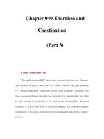
Fig. 4. (A) Type II congenital cystic adenomatoid malformation in an asymptomatic neonate (antenatal diagnosis). Chest
radiograph shows a hazy opacity in the right lower lobe and upward bowing of the minor fissure. (B) CT chest (lung windows)
confirms the presence of several small cysts in the right lower lobe. This infant was managed conservatively.
Fig. 3. (A) Type I congenital cystic adenomatoid malformation in a 12-month-old boy who presented with shortness of breath.
Chest radiograph shows a hyperlucent right hemithorax, with contralateral shift of the heart and mediastinal structures. Sparse
lung markings are seen in the right hemithorax. (From Donnelly LF. Chest. In: Fundamentals of pediatric radiology.
Philadelphia: WB Saunders; 2001. p. 38.) (B) CT scan of the chest (lung windows) demonstrates a large cyst filling the right
hemithorax. The compressed right middle lobe is seen behind the sternum.
paterson306
Technique of Pediatric Thoracic CT Angiography
Donald P. Frush, MD
Division of Pediatric Radiology, Department of Radiology, Duke University Health System,
1905 McGovern-Davison Children’s Health Center, Box 3808, Erwin Road, Durham, NC 27710, USA
One of the principle applications derived from the
evolution of multidetector row CT (MDCT), initially
seen with 16-slice and currently up to 64-slice CT, is
CT angiography. The ease, safety, and quality of the
examinations compared with traditional angiography
were quickly recognized, and the value of CT
angiography firmly established. For a variety of
reasons, the earliest MDCT angiography with single-
slice technology was problematic for the pediatric
population [1–4]. Some of these problems included
breathing artifact in children who could not hold their
breath, small volumes of contrast material, relatively
slow and inconsistent rates of injection, and small
cardiovascular structures [4]. Although these same
issues currently exist with pediatric CT angiography,
much faster scanning and isotropic display with
submillimeter image thickness have, to a large extent,
minimized the impact of these factors. Nevertheless,
it is still important to understand the special consid-
erations with pediatric CT angiography [5]. In trying
to make a potentially complex technique relatively
simple and practical, the following material is divided
into two parts: study preparation and study perform-
ance. The format is essentially step-by-step (Box 1),
with the supporting technical information either cited
or included in tables. Despite the fact this material
somewhat betrays the traditional academic format,
a greater benefit is served: excellent CT angiography
is possible in even the most problematic of pediat-
ric cases.
Planning the pediatric CT angiogram
Determine that CT angiography is the appropriate
examination
In addition to CT angiography, considerations for
thoracic cardiovascular structur al and functional
assessment include echocardiography, MR angiogra-
phy and venography, and conventional angiography.
CT angiography is advantageous in that it provides a
more global assessment of cardiovascular structures
and adjacent structures, such as the lung and airway.
The examination is also relatively quick to perform,
with times that can approach 1 second given 64-slice
technology. Sedation is rarely necessary compared
with MR imaging and echocardiography, and the
examination quality is more consistent (operator in-
dependent). CT angiography is a relatively non-
invasive procedure, compared with angiography. In
addition, monitoring and direct observation of the
patient are easier with CT angiography than with MR
imaging. Contraindications for MR imaging vascular
assessment including pacemakers and recent surgical
procedures with some metallic materials are not
present with CT a ngiography. Moreover, metal
artifact is much less an issue with CT angiography
than with MR angiography. For a more in-depth dis-
cussion of the relative merits and disadvantages with
CT angiography and MR angiography, the reader is
referred to a recent series of reviews [5 –8].
There are disadvantages with CT angiography. CT
angiography requires administration of intravenous
(IV) contrast media. Adverse reactions, however, are
singularly unusual in children. In addition, nephro-
toxicity from contrast media in children is much less
0033-8389/05/$ – see front matter D 2005 Elsevier Inc. All rights reserved.
doi:10.1016/j.rcl.2004.09.013 radiologic.theclinics.com
E-mail address:
Radiol Clin N Am 43 (2005) 419 – 433