Nanotechnology Health and Environmental Risks - Chapter 4 doc
Bạn đang xem bản rút gọn của tài liệu. Xem và tải ngay bản đầy đủ của tài liệu tại đây (563.64 KB, 14 trang )
63
4
The State of the Science — Human Health,
Toxicology, and Nanotechnological Risk
Brenda E. Barry
At present, considerable uncertainty exists regarding risks from nanoscale
materials and the products that incorporate them. This chapter gives an
introduction to some of the current science and its implications regarding
the effects of nanomaterials on human health. Although numerous studies
have been completed, they are not reviewed comprehensively here; rather,
this chapter gives an overview, focusing on carbon nanotubes as an example
of a category of nanomaterials, the types of heath effects observed, and the
complexities of toxicological studies with nanoscale materials.
The concerns about the potential toxicity of nanomaterials are based on
their unique surface, catalytic and magnetic properties, and how these prop-
erties may be expressed in biological systems and in the environment to
produce adverse effects. In one of the rst articles to broadly address the
impending issues related to nanotechnology, Colvin (2003) examined the
causes for concern regarding the potential biological and environmental
impacts of nanomaterials. Colvin’s discussion of these issues highlights a
main theme of this book — due to their unique composition and properties,
the key questions concerning nanomaterials are: (1) whether they present
new risks for health and the environment and, if so, (2) can the potential ben-
ets of nanotechnology be realized while minimizing the potential risks?
CONTENTS
4.1 Mechanisms of Toxicity
65
4.2 Types of Toxicological Studies 68
4.3 Findings 70
4.3.1 Pulmonary Toxicity Studies 70
4.3.2 In Vitro Studies 71
4.3.2.1 Dermal In Vitro Toxicity Studies 72
4.4 Future Directions 73
References 74
53639.indb 63 3/28/08 2:32:25 PM
© 2008 by Taylor & Francis Group, LLC
64 Nanotechnology: Health and Environmental Risks
The majority of scientic studies examining the potential toxic effects of
nanomaterials have been completed within the past ve years. Interestingly,
the results to date suggest that the behavior and effects of nanomaterials
are not always directly predictable from the results of previous studies with
other types of nanoscale materials. It is becoming increasingly apparent
that although they are composed of the same basic elements, at the atomic
or quantum level, nanomaterials have different properties and behave dif-
ferently from their bulk counterparts. For example, at the nano-scale, clus-
ters of gold atoms appear red (Kulinowski 2004). Similarly, although both
graphite and carbon nanotubes (CNT) are composed solely of carbon atoms,
the results from different in vivo and in vitro test systems indicate that the
properties of graphite do not accurately predict the properties of CNT. To
address the specic scientic questions raised by nanomaterials, a new area
of toxicology, termed nanotoxicology (Donaldson et al. 2006; Oberdörster et
al. 2005a), has emerged. Nanotoxicology can be dened simply as the sci-
ence that deals with the effects of nanostructures and nanodevices on living
organisms.
One of the rst steps in understanding the potential toxic effects of nano-
materials is to understand their specic characteristics. Because chemical
engineers have developed several different methods for producing a wide
variety of nanomaterials, a categorization scheme for nanomaterials, such as
the one developed by the EPA (2007), provides a useful approach for group-
ing the different types according to their composition or characteristics. The
EPA scheme proposes four major types of nanomaterials: (1) carbon-based,
which includes CNT and fullerenes; (2) metal-based, which includes quan-
tum dots, nanocrystals that can act as semiconductors, and metal oxides; (3)
dendrimers, which are nano-sized polymers built from branched units; and
(4) composites in which nanomaterials are combined with other nanoma-
terials or larger, bulk-type materials. Additional types of information are
also useful for characterizing and understanding the potential toxicity of
these different categories of nanomaterials. Some key parameters include the
number or concentration of the specic nanomaterials; the size character-
istics, including the length-to-width or aspect ratio; their surface area; and
their chemical composition.
An overall concern about the potential toxicity of all types of nanomateri-
als is their large surface area relative to their size (Oberdörster et al. 2005a).
This feature, which results in many of the benecial aspects of nanomateri-
als, has also been linked to their increased biological reactivity. Oberdörster
and colleagues (2005a) also comment on evidence that due to their small size,
inhaled nanomaterials can pass through the cells of the respiratory system
into the vascular system, and from there move to sites beyond the original
site of deposition in the organism. Similarly, a study by Kim and colleagues
(2006) reported that following injection of nanoparticles into the abdominal
area of mice, the particles penetrated the blood–brain barrier, yet did not
appear to affect brain function or produce toxicity.
53639.indb 64 3/28/08 2:32:25 PM
© 2008 by Taylor & Francis Group, LLC
The State of the Science 65
A basic concept of toxicology is that the dose makes the poison (Klaas-
sen 2001). Even materials essential to life itself, such as oxygen and water,
can cause death in organisms if provided in excess. Examples include inges-
tion of excess water that can produce an imbalance in the ionic composition
within cells, termed hyponatremia, which can result in brain swelling and
possibly death (Cotran et al. 1999). Similarly, inhalation of high concentra-
tions of oxygen, such as in a clinical setting to treat lung damage, can result
in the production of reactive oxygen species (ROS) in the lung tissues (Cotran
et al. 1999). ROS are reactive, unstable forms of oxygen that can damage and
kill these tissues, an effect called oxygen toxicity. The point here is that the
type of nanomaterials as well as the exposure amount, or dose, that may pro-
duce adverse effects in organisms and the environment are an active area of
nanotoxicology research and are not yet well understood.
Nanotoxicology has drawn together toxicologists from a variety of disci-
pline areas to apply their previous knowledge and expertise to questions
about the potential toxic effects of nanomaterials. They include inhalation
toxicologists with backgrounds in particle toxicology, who have studied the
adverse effects of nano-scale particles emitted as air pollutants from station-
ary industrial sources, such as smokestacks, as well as from mobile sources,
such as motor vehicles (Oberdörster et al. 2005a; Nel et al. 2006). Fiber toxi-
cologists with backgrounds in the toxic effects of natural mineral bers, such
as asbestos; synthetic vitreous bers, such as berglass; and other brous
materials are interested in studying the potential effects of carbon nano-
tubes (CNT) based on the similar aspect ratios and the durability of CNT
(Donaldson et al. 2006; Borm and Kreyling 2004; Mossman et al. 2007). Simi-
larly, dermal toxicologists are interested in learning whether the small size
of nanomaterials increases their potential to penetrate the skin layers and
to produce changes in the dermal cells and tissues, and how these changes
compare with dermal exposures to other types of materials (Monteiro-
Riviere and Inman 2006).
4.1 Mechanisms of Toxicity
A toxicologist evaluates a number of factors to understand a potential toxic
effect. One important determinant factor is the likely route of exposure for
the material of interest. The pathways for exposure to nanomaterials as well
as any other material include inhalation, dermal contact, and ingestion. In
some cases, ingestion can occur following dermal contact, when the material
sticks to the skin and is later transferred to the mouth. For nanomaterials,
the eyes may also be an area of concern, when nanomaterials on the skin are
transferred by hand contact with the eyes.
The exposure dose that an organism receives depends on the concentra-
tion of the material of interest, including a nanomaterial, and the duration
53639.indb 65 3/28/08 2:32:25 PM
© 2008 by Taylor & Francis Group, LLC
66 Nanotechnology: Health and Environmental Risks
and frequency of the exposure. Following an exposure, the fate of that mate-
rial in an organism is a product of several different processes, including its
absorption, distribution, metabolism, or breakdown in the organism, and
how effectively it is subsequently eliminated from the system. For many
materials, the site or sites where a toxic material causes damage, called a tar-
get tissue, may be identied. This target tissue may be specically affected
by exposure to the toxic material, perhaps due to buildup of the material or
the particular sensitivity of that tissue or area to the material. It is impor-
tant to keep in mind that the target tissue may not always be at the initial
deposition site, because the material or one of its breakdown products could
be transported to another location in the organism that becomes the target
tissue.
The mechanisms by which a compound or material, such as a nanomaterial,
produces a toxic effect can be grouped into several broad categories because
cells, tissues, or an organism have a relatively limited number of ways to
respond to an exposure. The material of interest may cause direct irritation
that produces a reaction at the site of contact. Alternatively, the material may
produce oxidative stress due to the generation of ROS. As mentioned previ-
ously, ROS are unstable forms of oxygen; they can cause cell injury by inter-
acting with cell membranes, breaking them, and causing the cell contents to
leak. Both of these events can result in the release of a number of different
protein factors — including cytokines, chemokines, and cell growth factors
— that can initiate more complex reactions involving immune and inam-
matory cells, the release of additional factors, and more reactive processes
occurring at the site of initial injury. This overall process is called inamma-
tion, a protective response by the organism that is designed to rid it of the
foreign material that is the cause of the injury (Cotran et al. 1999). The most
severe response to a toxic material is cancer, which results in uncontrolled
cell growth at the site of damage.
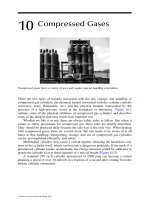
A recent review by Donaldson and colleagues (2006) discusses a number
of different features of one type of nanomaterial, specically CNT, that may
affect potential mechanisms of toxicity, particularly related to pulmonary
toxicology. Drawing upon the authors’ previous extensive experience in
particle and ber toxicology, they suggest that previous studies in this eld
can provide a basis for understanding the effects of nanoscale particles and
bers. They comment that if CNT are longer than 20 µm, they would likely
cause the same type of pathological damage as mineral bers, such as asbes-
tos, and synthetic vitreous bers, such as berglass. The damage can include
inammatory responses, as previously described, and possibly cancer. They
also note that several classes of impurities, such as small amounts of metals,
organic residual matter, and support materials, may be present in CNT sam-
ples following the production processes. As observed following exposures
to different types of bers, pro-inammatory effects produced by CNTs may
be caused by their length, their reactive surfaces, or the release of metal ions
that may be toxic to the cells or tissues (Figure 4.1).
These processes can cause
53639.indb 66 3/28/08 2:32:26 PM
© 2008 by Taylor & Francis Group, LLC
The State of the Science 67
oxidative stress to the affected cells and tissues, similar to the toxic effects
previously reported for mineral and synthetic vitreous bers.
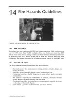
The Five D’s of particle toxicology (Figure 4.2) can provide important per
-
spectives for consideration of the toxic effects of nanomaterials (Borm and
Kreyling 2004). Although developed primarily for inhalation toxicology, the
FIGURE 4.1
Carbon nanotube characteristics and potential adverse effects. SWCNT — single-walled
carbonnanotube. MWCNT — multi-walled carbon nanotubes. Figure adapted from
Donaldson et al. (2006). (See color insert following page 76.)
FIGURE 4.2
The ve Ds of particle toxicology for nanomaterials. Adapted from Borm and Kreyling
(2004).
53639.indb 67 3/28/08 2:32:27 PM
© 2008 by Taylor & Francis Group, LLC
68 Nanotechnology: Health and Environmental Risks
ve D’s — dose, deposition, dimension, durability, and defense — are rel-
evant characteristics for examining the responses to nanomaterials in other
types of toxicological studies. These characteristics are particularly appro-
priate in light of the noted stable properties of nanomaterials. Certainly dose
is a critical factor, as discussed previously, as well as the site of nanomaterial
deposition, because this impacts the cells and tissues in direct contact with
the nanomaterials. The dimension and durability properties of nanomateri-
als are specically relevant for CNT, whether single-walled or multi-walled.
Some investigators have suggested that the durability and dimensions of
CNT resemble those of asbestos bers, raising concerns about the persis-
tence of these nanomaterials in biological systems once they have entered
the organism, termed biopersistence. These concerns become increasingly
important as chemical engineers continue to rene methods for producing
longer CNT many microns in length, such that they have both the dimen-
sions and durability of asbestos bers.
In his recent review article, Hardman (2006) discussed the toxicity of quan-
tum dots (QDs), which are semiconductor nanocrystals that have unique
optical and electrical properties. Based on his review, he concluded that QDs
cannot be viewed as a uniform group of substances with a specic toxic-
ity. As noted for other nanomaterials, the specic properties of QDs are of
interest and how these may affect their potential toxicity must be evaluated.
Because bioconjugated QDs — that is, QDs linked with biological materials,
such as proteins and antibodies — are under consideration for biomedical
applications as tools for site-specic gene and drug delivery, as well as in vivo
biomedical imaging, the potential human health and environmental risks of
their use must be considered carefully.
4.2 Types of Toxicological Studies
Oberdörster and colleagues (2005b) have proposed a screening strategy
for evaluating the toxicity of nanomaterials that includes a comprehensive
array of in vitro and in vivo assays and a two-tier approach for in vivo stud-
ies, described in Section 5.6. This strategy employs traditional toxicology
and assay techniques to understand the potential toxicity of nanomaterials
under dened test conditions. The different types of testing systems and
their advantages and disadvantages will now be considered.
In vivo models use whole animals to study the effects of exposures to nano-
materials. One model is intratracheal instillation, in which a nanomaterial
suspended in a uid is injected directly into the trachea and to the lungs of
an anesthetized experimental animal. A concern with this model is that it
delivers the nanomaterial as a one-time, concentrated amount of material,
termed a bolus, into the lungs, in contrast with the more natural inhala-
tion mode of entry, in which small amounts of a material are progressively
53639.indb 68 3/28/08 2:32:27 PM
© 2008 by Taylor & Francis Group, LLC
The State of the Science 69
delivered to the lungs with each breath. The one-time delivery of a large
amount of the nanomaterial may produce effects more related to the deliv-
ery method than the material. Pharyngeal aspiration is an approach that
attempts a more physiologically natural mode of entry of nanomaterials into
the lungs. A small amount of the nanomaterial solution is placed on the back
of the tongue of an anesthetized animal; with its next breath, the animal
aspirates the nanomaterial solution into its lungs.
Another potential in vivo exposure approach is the use of inhalation cham-
bers, in which test animals are exposed to a measured concentration of an
aerosolized nanomaterial for a specied exposure period. This approach can
be costly because a large amount of nanomaterial is needed to generate the
aerosol and this may be expensive. In addition, the physiochemical prop-
erties of nanomaterials can complicate generation of the aerosol as well as
maintenance of the desired aerosol characteristics in the chamber, due to the
tendency of the nanomaterials to agglomerate due to static forces. Although
results from inhalation chamber studies with nanomaterials have yet to be
reported, such studies are either in the planning stages or underway.
In vitro approaches allow the study of the mechanisms of action and bio-
logical effects of nanomaterials on cells and tissues under controlled condi-
tions. Such studies can include the use of cells derived from a variety of
sources, such as lung or skin, that have been grown in media on plate sur-
faces or in test tubes, to which nanomaterials can be added. Other types of
in vitro exposure systems can utilize sections of selected tissues obtained
from animals or humans. Examples of these “test tube” assays include ow-
through diffusion cell studies (Ryman-Rasmussen et al. 2006) and skin ex-
ion model studies (Rouse et al. 2007).
The disadvantages of in vitro test systems require consideration when
interpreting study results because the effects observed in vitro are difcult to
compare to possible effects that may occur in the naturally more complex in
vivo systems. These systems include defense systems, as well as feedback and
immune response mechanisms designed to deal with foreign matter in the
body. For example, immune and inammatory cells, which can contribute a
variety of cell mediators to a toxic response in vivo, are absent. In addition, in
vitro systems do not have the normal clearance or dissolution mechanisms
that usually operate in vivo, which may reduce the amount of available nano-
material and the observed effects. Such factors can complicate extrapolating
the effects of a delivered in vitro test dose to an in vivo exposure dose.
Teeguarden and colleagues (2007) reviewed aspects of pharmacokinetics,
an approach used in pharmacology to determine the fate of materials, such
as drugs, in an organism, and how this approach may affect interpretation
of cell dose of nanomaterials under in vitro conditions. Based on the specic
properties, nanoscale particles can diffuse, settle, and agglomerate in the
culture media; as a result, simple representatives (surrogates) of dose, such
as the amount of a nanomaterial directly added to the in vitro test system,
may be an inappropriate reference marker for evaluating uptake of nanoma-
terials and responses of the cells in in vitro test systems. The authors propose
53639.indb 69 3/28/08 2:32:28 PM
© 2008 by Taylor & Francis Group, LLC
70 Nanotechnology: Health and Environmental Risks
that use of pharmacokinetics and principles of dosimetry (the relationship
between dose and observed response) can improve the validity of nanomate-
rial in vitro toxicity assessments.
For both in vivo and in vitro toxicity studies, the validity of using specic
assays to evaluate the parameter of interest should also be veried, to ensure
that the results are relevant and that false positives are not produced. An
example of the latter point is the colorimetric MTT assay routinely used for
evaluation of in vitro cell viability. It is based on the reduction of yellow 3-
(4,5-dimethylthiazol-2-yl)-2,5-diphenyltetrazolium bromide to purple MTT-
formazan due to the release of enzymes from damaged cells. Wörle-Knirsch
and colleagues (2006) reported that in in vitro assays, single-walled carbon
nanotubes (SWCNT) could directly interact with MTT to give a positive pur-
ple result that was not related to cell enzyme release. The positive colorimet-
ric result indicating cell damage due to SWCNT exposure was not evident in
results from the WST cell viability assay, another test that is commonly used
to determine whether cells are damaged or killed by a treatment. This means
the MTT assay is not a valid test for SWCNT.
Another factor that currently complicates interpretation of results from
both in vivo and in vitro studies is a lack of nanomaterial reference standards.
At present, considerable variability can exist within the same type of nano-
material, such as CNT, depending on who manufactured it, as well as if and
how the nanomaterial was chemically treated after synthesis. As an example,
a sample of CNT can contain variable amounts of metals as contaminants
from the manufacturing process. The presence of these metals may affect
the responses of the cell or tissues because of the toxic effects attributable to
the metals. The lack of nanomaterial reference standards can also confound
comparison of the results of toxicological studies by different investigators
using the same category of nanomaterial, but which were manufactured dif-
ferently and with a different composition.
4.3 Findings
4.3.1 Pulmonary Toxicity Studies
Pulmonary toxicity studies comprise a sizeable segment of the recent and
current research designed to understand the toxic effects of nanomaterials.
The practical basis for this research is the potential for inhalation of nano-
materials, particularly in regard to worker exposures through handling and
managing nanomaterials. Results from several in vivo studies reported within
the past few years have provided some of the rst evidence that exposures
to nanomaterials could cause injury in the lungs of experimental animals.
The in vivo studies reported to date have primarily focused on the effects of
53639.indb 70 3/28/08 2:32:28 PM
© 2008 by Taylor & Francis Group, LLC
The State of the Science 71
exposures to metal oxides and to carbon-based particles such as SWCNT and
multi-walled CNT (MWCNT).
Studies by Lam et al. (2003) and Warheit et al. (2004) used intratracheal
instillation as the method to deliver SWCNT to the lungs of rats and mice,
respectively. From their short-term (acute) toxicity study, Lam et al. (2003)
reported that the instillation of SWCNT produced granulomas, small nodules
of cells that may include macrophages, lymphocytes, and a variety of inam-
matory cells in the lung tissues and that their appearance increased with
the dose of SWCNT, suggesting it was dose-dependent. Based on their 2004
study, Warheit and colleagues also reported the presence of granulomas in a
number of areas in the lung tissues of exposed rats, but their appearance was
not dependent on dose. Unexpectedly, the reported changes also occurred in
the absence of increases in markers of inammation and cell division within
uids obtained when the lungs of the experimental animals were rinsed
with saline. These markers include cell enzymes normally found only inside
cells, and are indicators of dividing cells, both of which are usually detected
in the lung uid following these types of studies. Subsequent studies that
also used intratracheal instillation of CNT as the treatment method (Muller
et al. 2005; Grubeck-Jaworska 2006) indicated inammatory changes and the
appearance of scar-like, or brotic, areas in the lungs of exposed animals.
Shvedova and colleagues (2005) used pharyngeal aspiration to deliver
SWCNT to the lungs of mice. They reported that their treatment produced
not only a strong inammatory reaction shortly after treatment but also pro-
gressive and dose-dependent development of brotic changes in the lung
tissues. Surprisingly, this brotic reaction occurred in the absence of signs
of persistent inammation and at sites distant from the SWCNT deposition
sites. More recently, this team demonstrated that inammatory effects were
mitigated when exposed mice were also given vitamin E, an antioxidant
(Shvedova et al. 2007).
The results of all of the studies briey reviewed here suggest that the
SWCNT may be capable of producing brotic alterations in the lungs similar
to those reported following exposures to other types of brous materials.
However, as discussed in Chapter 3, there are numerous uncertainties in the
dosing of these studies that affect their interpretation. In particular, the pres-
ence of iron contamination and the sheer number of nanotubes used in the
experiments make interpretation of these ndings to real world exposures
difcult. Studies are underway at the U.S. National Toxicology Program to
develop experimental protocols for SWCNT by inhalation (NTP 2007).
4.3.2 In Vitro Studies
Numerous in vitro studies have been conducted using a variety of nanomate-
rials and cell types to understand the mechanisms and potential toxic effects
concerning exposures to nano-scale materials. In 2004, Sayes and colleagues
reported that the cell toxicity of water-soluble fullerenes was a function of
the nature of their surface, and that fullerene toxicity was caused by lipid
53639.indb 71 3/28/08 2:32:28 PM
© 2008 by Taylor & Francis Group, LLC
72 Nanotechnology: Health and Environmental Risks
peroxidation of cell membranes due to generation of ROS. Using alveolar
macrophages (the respiratory defense cells present in the air spaces in the
lungs), Jia and co-workers (2005) evaluated several types of nanomaterials
and reported that in their cell assay system, SWCNT were more toxic than
fullerenes. Bottini and colleagues (2006) observed that MWCNT oxidized
by treatment with a strong acid were more toxic than untreated, or pristine,
MWCNT; while Brunner and co-workers (2006) concluded that solubility was
a strong inuence in the cell toxicity observed in their assays following cell
exposures to silica, asbestos, and several different nano-scale materials. Lim-
bach and colleagues (2007) quantied oxidative stress through the release
of ROS from human lung epithelial cells treated with nano-scale silica par-
ticles that contained a variety of metals. They reported that the nanoparticles
could act like Trojan horses carrying the metals inside the cells and that the
specic chemical composition of the particles was the most inuential factor
for causing the oxidative stress.
In vitro studies have also demonstrated that alteration of the nanomaterial
surface by the addition of functional groups can modify the toxic proper-
ties of nanomaterials. Sayes and colleagues (2004; 2006) reported that attach-
ment of different chemical groups to the surface of CNT and fullerenes could
change their properties and decrease their toxicity.
4.3.2.1 Dermal In Vitro Toxicity Studies
Investigators have increasingly focused on skin, or dermal, contact as an
important route of exposure to nanomaterials. In one of the rst occupa-
tional studies attempting to understand potential exposures to nanomateri-
als under actual worker conditions, Maynard and colleagues (2004) obtained
measurements for aerosol concentrations of SWCNT and evaluated potential
for dermal exposures. They reported that aerosol concentrations of SWCNT
were low and that energetic processes would likely be needed to increase
airborne concentrations. It is important to note the study was conducted in
a simulated work environment and therefore may not reect conditions in a
manufacturing facility. Maynard et al. (2004) also observed that the gloves of
workers were contaminated with SWCNT, indicating the importance of der-
mal contact as a source of worker exposures to nanomaterials. These ndings
have been followed by a number of in vitro studies to determine the poten-
tial effects of nanomaterial exposures on dermal cell systems and whether
nanomaterials behave similarly or dissimilarly to other types of nano-scale
materials, such as beryllium (Tinkle et al. 2003).
In a study using human epidermal keratinocytes (HEK) — cells in human
skin that produce the protein keratin — Shvedova and colleagues (2003)
reported that exposures to unrened SWCNT produced oxidative stress
and cellular toxicity in the HEK. They concluded that their ndings sug-
gested that exposures to unrened SWCNT may lead to dermal toxicity in
the skin of workers. A study by Monteiro-Riviere and colleagues (2005) also
using HEK determined that chemically unmodied MWCNT were taken up
53639.indb 72 3/28/08 2:32:28 PM
© 2008 by Taylor & Francis Group, LLC
The State of the Science 73
by the cells and that the nanomaterial exposures caused the release of pro-
inammatory cytokines. This suggests that, although the skin is normally a
good barrier to keep many materials from entering the body, nanomaterials,
due to their very small size, may be able to enter the skin and produce toxic
responses. This penetration capability may be a benecial aspect, if the nano-
material is a drug or a cosmetic treatment; however, it may not be benecial
if the nanomaterial entry results in a toxic response in the skin, or allows a
nanomaterial to enter the body and subsequently be transported to another
site where a toxic effect may occur.
In a study to examine the potential toxic effects of QDs on skin, Ryman-Ras-
mussen and co-workers (2006) reported that in their ow-through diffusion
system, QDs with different shapes, sizes, and surface coatings could penetrate
intact porcine skin at occupationally relevant concentrations. In a study using
a porcine skin exion model, Rouse and colleagues (2007) described dermal
penetration of fullerene nanoparticles and their presence within the spaces
between cells in a sub-layer of the skin called the stratum granulosum.
4.4 Future Directions
As illustrated in this section, both in vivo and in vitro systems can provide
useful information for understanding the mechanisms of toxicity as well as
the toxic responses of organisms, tissues, and cells following exposures to
nanomaterials. As noted earlier, a disadvantage of in vitro test systems for
evaluating nanomaterial toxicity is the difculty in correlating the ndings
with effects that may occur in the naturally more complex in vivo systems.
The type of nanomaterial, its chemical (or functionalization) treatment prior
to addition to the in vitro assay system, the types of cells used, the assay
system, and other factors can all contribute to the sometimes contradictory
results from different investigator groups.
In a recent study, Sayes and colleagues (2007) asked how well the results
from in vitro assays could predict the toxicity results produced in vivo for
several different types of nano-scale and ne-scale particles, including silica
and zinc oxide. Using a variety of in vitro assays and an intratracheal deliv-
ery method for their exposure systems, they noted little correlation between
the results from the in vitro and in vivo assays. They concluded that in vitro
cellular assay systems require further development, standardization, and
validation to provide useful and reliable screening data to assess the toxicity
of inhaled materials. This conclusion dovetails well with the future needs
described by Teeguarden and colleagues (2007) for development of high-
throughput in vitro assays that can reliably predict the toxicity of nanomateri-
als. Ultimately, the results from such an in vitro assay should also be relevant
to those effects that may occur in vivo. Such test systems will be invaluable
for efcient evaluation of the potential toxicity of the thousands of types of
53639.indb 73 3/28/08 2:32:28 PM
© 2008 by Taylor & Francis Group, LLC
74 Nanotechnology: Health and Environmental Risks
nanomaterials likely to be produced in the near future. This is because reli-
ance on a traditional toxicology battery of both in vitro and in vivo assays for
each of these nanomaterials would be both time and cost prohibitive.
This brief review of recent reports concerning the potential toxicity of
nanomaterials identies some of the variability and inconsistency in the
reported ndings using similar test systems and even the same category of
nanomaterials. Variability among the results is likely due to the fact that
toxicological assays for nanomaterials have only been conducted within the
past few years, and relatively few nanomaterials have been studied thor-
oughly. Some of these differences may be attributed to the current absence
of nanomaterial reference materials that could be used to standardize results
with different test systems and among different research laboratories that
conduct the testing. Nevertheless, the trend of current ndings for exposures
to several different types of nanomaterials is that they can produce toxic and
unexpected responses in the various test systems used to date.
References
Borm, P. J. A., and W. Kreyling. 2004. Toxicological hazards of inhaled nanoparticles
— Potential implications for drug delivery. Journal of Nanoscience and Nanotech-
nology 4:521–531.
Bottini, M., S. Bruckner, K. Nika, N. Bottini, S. Bellucci, A. Magrini, A. Begamaschi,
T. Mustelin. 2006. Multi-walled carbon nanotubes induce T lymphocyte apop-
tosis. Toxicology Letters 160:121–126.
Brunner, T. J., P. Wick, P. Manser, P. Spohn, R. N. Grass, L. K. Limbach, A. Bruinink,
W. J. Stark. 2006. In vitro cytotoxicity of oxide nanoparticles: Comparison to
asbestos, silica, and the effect of particle solubility. Environmental Science and
Technology 40:4374–4381.
Colvin, V. L. 2003. The potential environmental impact of engineered nanomaterials.
Nature Biotechnology 21(10): 1166–1170.
Cotran, R. S., V. Kumar, and S. L. Robbins. 1999. Robbins pathologic basis of disease.
F. J. Schoen (ed). Philadelphia, PA: W.B. Saunders Company.
Donaldson, K., R. Aitken, L. Tran, V. Stone, R. Dufn, G. Forrest, and A. Alexander.
2006. Carbon nanotubes: A review of their properties in relation to pulmonary
toxicology and workplace safety. Toxicological Sciences 92:5–22.
Grubek-Jaworska, H., P. Nejman, K. Czuminska, T. Przbylowski, A. Huczko, H.
Lange, M. Bystrzejewski, P. Baranowski, R. Chazan. 2006. Preliminary results
on the pathogenic effects of intratracheal exposure to one-dimensional nano-
carbons. Carbon 44:1057–1063.
Hardman, R. 2006. A toxicologic review of quantum dots: toxicity depends on
physicochemical and environmental factors. Environmental Health Perspectives
114:165–172.
Jacobs, M. N., G. T. Nolan, and S. R. Hood. December 2004. Lignins, bactericides and
organochlorine compounds activate the human pregnane X receptor (PXR).
Toxicol. Appl. Pharmacol. 209(2):123–133.
53639.indb 74 3/28/08 2:32:29 PM
© 2008 by Taylor & Francis Group, LLC
The State of the Science 75
Jia, G., H. Wang, L. Yan, X. Wang, R. Pei, T. Yan, Y. Zhao, X. Guo. 2004. Cytotoxicity of
carbon nanomaterials: Single-wall nanotube, multi-wall nanotube, and fuller-
ene. Environmental Science and Technology 39:1378–1383.
Kim, J. S., T-J Yoon, K. N. Yu, B. G. Kim, S. J. Park, H. W. Kim, K. H. Lee, S. B. Park, J K.
Lee, M. H. Cho. 2006. Toxicity and tissue distribution of magnetic nanoparticles
in mice. Toxicological Sciences 89:338–347.
Klaassen, C. D. 2001. Cassaret & Doull’s toxicology: The basic science of poisons. New
York: McGraw-Hill.
Kulinowski, K. 2004. Nanotechnology: From “wow” to “yuck”? Bulletin of Science,
Technology & Society 24:13–20.
Lam, C. W., J. T. James, R. McCluskey, R. L. Hunter. 2004. Pulmonary toxicity of sin-
gle-wall carbon nanotubes in mice 7 and 90 days after intratracheal instillation.
Toxicological Sciences 77:126–134.
Limbach, L.K., P. Wick, P. Manser, R. N. Grass, A. Bruinink, W. J. Stark. 2007. Expo-
sure of engineered nanoparticles to human lung epithelial cells: Inuence of
chemical composition and catalytic activity on oxidative stress. Environmental
Science and Technology 41:4158–4163.
Maynard, A. D., P. A. Baron, A. A. Shvedova, E. R. Kisin, V. Castranova. 2004. Expo-
sure to carbon nanotube material: aerosol release during the handling of unre-
ned single-walled carbon nanotube material. J. Toxicology and Environmental
Health A(67):87–107.
Monteiro-Riviere N. A., and A. O. Inman. 2006. Challenges for assessing carbon
nanomaterial toxicity to the skin. Carbon 44:1070–1078.
Monteiro-Riviere N. A., R. J. Nemanich, A. O. Inman, Y. Y. Wang, and J. E. Riviere.
2004. Multi-walled carbon nanotube interactions with human epidermal kera-
tinocytes. Toxicology Letters 155:377–384.
Mossman, B. T., P. J. Borm, V. Castranova, D. L. Costa, K. Donaldson, S. R. Kleeberger.
2007. Mechanisms of action of inhaled bers, particles and nanoparticles in the
lungs and cardiovascular diseases. Particle and Fibre Toxicology 4:4.
Muller, J., F. Huaux, N. Moreau, P. Mission, J. F. Heilzer, M. Delos, M. Arras, A. Fon-
seca, J. B. Nagy, D. Lison. 2004. Respiratory toxicity of multi-wall carbon nano-
tubes. Toxicology and Applied Pharmacology 207:221–231.
Nel A., T. Xia, L. Madler, and N. Li. 2006. Toxic potential of materials at the nanolevel.
Science 311:622–627.
NIOSH 2004. Draft NIOSH current intelligence bulletin: Evaluation of health hazard
and recommendations for occupational exposure to titanium dioxide. http://
www.cdc.gov.mill1.sjlibrary.org/niosh/review/public/tio2/pdfs/TIO2Draft.pdf.
NTP. 2007. NTP Nanotechnology safety initiative. />index.cfm?objectid=303069A0-F1F6-975E-72D33ECBD3E11363#mox.
Oberdörster, G., E. Oberdörster, and J. Oberdörster. 2005a. Nanotoxicology: An
emerging discipline evolving from studies of ultrane particles. Environmental
Health Perspectives 113:823–839.
Oberdörster, G., A. Maynard, K. Donaldson, V. Castranova, J. Fitzpatrick, K. Ausman,
J. Carter, B. Karn, W. Kreyling, D. Lai, S. Olin, N. Monteiro-Rivere, D. Warheit,
H. Yang. 2005b. Principles for characterizing the potential human health effects
from exposure to nanomaterials: Elements of a screening strategy. Particle and
Fibre Toxicology 2:8.
Rouse, J. G., J. Yang, J. P. Ryman-Rasmussen, A. R. Barron, N.A. Monteiro-Rivere.
2007. Effects of mechanical exion on the penetration of fullerene amino acid-
derivatized peptide nanoparticles through skin. Nano. Letters 7:155–160.
53639.indb 75 3/28/08 2:32:29 PM
© 2008 by Taylor & Francis Group, LLC
76 Nanotechnology: Health and Environmental Risks
Ryman-Rasmussen, J. P., J. E. Riviere, and N. A. Monteiro-Riviere. 2006. Penetration
of intact skin by quantum dots with diverse physicochemical properties. Toxi-
cological Sciences 91:159–164.
Sayes, C. M., J. D. Fortner, W. Guo, D. Lyon, A. M. Boyd, K. D. Ausman, Y. J. Tao, B.
Sitharaman, L. J. Wilson, J. B. Hughes, J. L. Wert, V. L. Colvin. 2004. The differ-
ential cytotoxicity of water-soluble fullerenes. Nano. Letters 4:1881–1887.
Sayes, C. M., F. Liang, J. L. Hudson, J. Mendez, W. Guo, J. M. Beach, V. C. Moore, C. D.
Doyle, J. L. West, W. E. Billips, K. D. Ausman, V. L. Colin. 2006. Functionaliza-
tion density dependence of single-walled carbon nanotubes cytotoxicity in vitro.
Nano. Letters 161:135–142.
Sayes, C. M., K. L. Reed, and D. B. Warheit. 2007. Assessing toxicity of ne and
nanoparticles: Comparing in vitro measurements to in vivo toxicity proles.
Toxicological Sciences 97:163–180.
Shvedova, A. A., V. Castranova, E. R. Kisin, D. Schwegler-Berry, A. Murray, V. Gan-
delsman, A. Maynard, P. Baron. 2003. Exposure to carbon nanotube material;
assessment of cytotoxicity using human keratinocyte cells. Journal of Toxicology
and Environmental Health A(66):1909–1926.
Shvedova, A. A., E. R. Kisin, R. Mercer, A. R. Murray, V. J. Johnson, A. I. Potapovich,
Y. Y. Tyurina, V. Gorelik, S. Arepalli, D. Schuegler-Barry, A. F. Hubbs, J. A. Anto-
nini, D. E. Evans, B K. Ku, D. Ramsey, A. Maynard, V. E. Kagan, V. Castranova,
P. Baron. 2005. Unusual inammatory and brogenic pulmonary responses to
single-walled carbon nanotubes in mice. American Journal of Physiology — Lung
Cellular and Molecular Physiology 289:L698–L708.
Shvedova, A. A., E. R. Kisin, A. R. Murray, O. Gorelik, S. Arepalli, V. Castranova,
S. H. Young, F. Gao, Y. Y. Tyurina, T. D. Oury, and V. E. Kagan. 2007. Vitamin
E deciency enhances pulmonary inammatory response and oxidative stress
induced by single-walled carbon nanotubes in C57BL/6 mice. Toxicol. Appl.
Pharmacol. 221(3):339–348.
Teeguarden, J. D., P. M. Hinderliter, G. Orr, B. D. Thrall, and J. G. Pounds. 2007. Phar-
macokinetics in vitro: dosimetry considerations for in vitro nanoparticle toxicity
assessments. Toxicological Sciences 95:300–312.
Tinkle, S. S., J. M. Antonini, B. A. Rich, J. R. Roberts, R. Salmen, K. DePress, E. J.
Adkins. 2003. Skin as a route of exposure and sensitization in chronic beryl-
lium disease. Environmental Health Perspectives 111:1202–1208.
U.S. Environmental Protection Agency. 2007. Nanotechnology White Paper.
EPA 100/B-07/001. Science Policy Council. U.S. Environmental Protection
Agency. Washington, D.C.
Warheit, D. B., B. R. Laurence, K. L. Reed, D. H. Roach, G. A. M. Reynolds, T. R.
Webb. 2004. Comparative pulmonary toxicity assessment of single-wall car-
bon nanotubes in rats. Toxicological Sciences 77:117–124.
Wörle-Knirsch, J. M., K. Pulskamp, and H. F. Krug. 2006. Oops…they did it again!
Carbon nanotubes hoax scientists in viability assays. Nano. Letters 6:1261–1268.
53639.indb 76 3/28/08 2:32:29 PM
© 2008 by Taylor & Francis Group, LLC