SOIL ORGANIC MATTER IN SUSTAINABLE AGRICULTURE - CHAPTER 6 pot
Bạn đang xem bản rút gọn của tài liệu. Xem và tải ngay bản đầy đủ của tài liệu tại đây (485.78 KB, 20 trang )
1294_C06.fm Page 179 Friday, April 23, 2004 11:19 AM
Fungi
6 Contributions ofMatter
to Soil Organic
in Agroecosystems
Kristine A. Nichols and Sara F. Wright
CONTENTS
Estimating Fungal Biomass ..........................................................................................................180
Saprophytic Fungi .........................................................................................................................180
Fungal Plant Pathogens ................................................................................................................181
Biotrophic Mutualistic Fungi .......................................................................................................182
Arbuscular Mycorrhizal Fungi ..............................................................................................182
Glomalin, a Glycoprotein Produced by AM Fungi ..............................................................184
Pools of Glomalin ..................................................................................................................184
Characterization of Glomalin ................................................................................................187
Glomalin, a Major Component of Soil Organic Matter .......................................................187
Quantities of Glomalin ..........................................................................................................187
Single-Species Pot Culture Experiments ..............................................................................189
Depth and Deposition Experiment ........................................................................................190
Glomalin and Aggregate Stability .........................................................................................191
Glomalin under Elevated CO2 ...............................................................................................194
Contribution of Soil Fungi to Organic Matter .............................................................................194
Managing Soil Fungi to Increase Soil Organic Matter ................................................................195
References .....................................................................................................................................196
Soil fungi are important agents of decomposition, pathogenicity, and plant and soil health (i.e.,
nutrient cycling, soil fertility, aggregate stability, and soil organic matter turnover). In agricultural
soils, there are at least 25,000 fungal species (Carlile and Watkinson, 1996a), accounting for ca.
70% of the microbial biomass (Paul and Clark, 1996). Fungal growth is a function of carbon
availability. Hyphal lengths often range from 3 to 300 m/g soil (Frey et al., 1999; Miller et al.,
1995; Olsson et al., 1999; Rillig et al., 1999). Most fungal organisms are found in the rhizosphere,
which is enriched in organic carbon from proteins, amino acids, organic acids, and sugars released
by roots; the mucopolysaccharide mucigel on the root; and sloughed root cap cells.
Fungal contributions to agroecosystem function are difficult to quantify because of the lack of
accurate methods to measure fungal biomass and activity. The benefits and limitations of some
typical quantification methods are discussed in this chapter. Three major groups of soil fungi are
important in agroecosystems: (1) saprophytes, (2) pathogens, and (3) mutualists. The mutualistic
arbuscular mycorrhizal (AM) fungi account for the majority of fungal biomass (Olsson et al., 1999;
Vieira et al., 2000) and are examined in some detail along with glomalin, a glycoproteinaceous
substance that coats AM hyphae. Glomalin might act as a hydrophobin, which are a class of
biomolecules that protect hyphae from nutrient loss (Wessels, 1997), form and stabilize soil
aggregates (Wright and Upadhyaya, 1998), and store soil carbon (Rillig et al., 2001b).
179
© 2004 by CRC Press LLC
1294_C06.fm Page 180 Friday, April 23, 2004 11:19 AM
180
Soil Organic Matter in Sustainable Agriculture
ESTIMATING FUNGAL BIOMASS
Microbial biomass is often used as an indicator of the microbial contribution to soil organic matter,
plant health, and understanding nutrient fluxes (i.e., the transport and storage of nutrients). Estimates
of fungal biomass are often included with bacterial biomass numbers in techniques based on the
amount of carbon released after chloroform fumigation followed by incubation or extraction (i.e.,
total microbial biomass). Chloroform fumigation methods are inherently more accurate for bacteria
than for fungi (Olsson et al., 1999; Paul and Clark, 1996; Vieira et al., 2000). Even with variations
in the incubation procedure, fungal cell walls and spores do not completely lyse (Horwath and
Paul, 1994; Paul and Clark, 1996).
Microscopic counts of hyphae and other fungal structures by the grid-line intersect method are
tedious and have many technical problems. Hyphal diameters range from 2 to 20 µm and can be
used to estimate biovolume or to classify different groups of fungi (Bottomley, 1994; Carlile and
Watkinson, 1996a; Miller et al., 1995). Various stains are applied to help visualize hyphae or to
determine viability, or both. However, viability stains, such as 4¢6 diamidino-2-phenyl indole (DAPI;
reacts with active DNA) or fluorescein diacetate (an indicator of cytoplasmic constituents, i.e.,
esterase), might be ineffective in determining the length of aseptate hyphae when nuclei and
cytoplasmic contents are not distributed evenly (Bottomley, 1994; Carlile and Watkinson, 1996a;
Paul and Clark, 1996). Nonspecific stains make hyphae more visible but do not correct for errors
due to (1) exclusion of spores or yeasts; (2) large differences in counts between individuals or
laboratories; (3) heterogeneous distribution of hyphae in the soil; and (4) differences in extraction
techniques, such as grinding soil in a mixer compared to shaking free hyphae from soil (Millner
and Wright, 2002; Rillig et al., 1999; Stahl et al., 1995). In extracting fungal hyphae from soil, a
balance must be achieved between homogenizing soil to effectively release hyphae and excessively
fragmenting hyphae (Bottomley, 1994). Inherent variability makes it difficult to determine differences between treatments unless numerous replicate samples are examined (Stahl et al., 1995).
Other methods for measuring fungal biomass quantify a specific substance, such as chitin or
ergosterol. The major limitations in these assays are that these substances (1) are not found in all
fungi, (2) might be present in other soil organisms, (3) vary in concentration in different fungal
species or physiological states, and (4) are not calibrated with fungal biomass (Bottomley, 1994;
Paul and Clark, 1996; Vieira et al., 2000). Chitin is found in the cell walls of most fungi, but is
missing in Oomycetes and is present in insects and mites. (Although Oomycetes have been reclassified into the Kingdom Chromista in the eight-kingdom system, in this chapter they are still
considered as part of Kingdom Fungi.) Ergosterol is also found in other organisms, such as algae
and protozoa, and can only be measured in living mycelium.
Seasonal fluctuations and substrate (i.e., carbon) availability influence fungal biomass (Carlile
and Watkinson, 1996a; Bottomley, 1994). For example, following an increase in soil moisture from
precipitation or irrigation, the germination and proliferation of fungi can increase as soluble carbon
compounds are released by plants, but this rapid growth declines when substrates become limited
(Carlile and Watkinson, 1996a; Klein et al., 1995). Therefore, it is important not only to take a
number of samples from a site but also to note the time of sampling, to sample a number of times
a year, or to do repeated sampling over a number of years at the same time. Sampling times should
be dictated not by the calendar but by climatic conditions and management events, such as sampling
at the same time in reference to precipitation, frost, planting, harvesting, or application of fertilizer,
herbicide, or pesticide (Bottomley, 1994).
SAPROPHYTIC FUNGI
Fungal saprophytes are the primary degraders of plant debris, whereas bacteria and select highly
specific fungi decompose animal material and microbes (Bird et al., 2002; Carlile and Watkinson,
1996a; Frey et al., 1999; Stevenson, 1994; Vieira et al., 2000). Because of their relatively benign
© 2004 by CRC Press LLC
1294_C06.fm Page 181 Friday, April 23, 2004 11:19 AM
Contributions of Fungi to Soil Organic Matter in Agroecosystems
181
role as decomposers, saprophytic fungi are often overlooked by agricultural scientists, but life on
this planet could not be maintained without these fungi recycling basic nutrients such as C, N, P,
and K, and we would have been buried hundreds of times over by undecomposed leaves, roots,
and other plant material (Carlile and Watkinson, 1996a; Klein et al., 1995). Although these fungi
play a vital role in nutrient cycling, they are mostly on surface residues and account for less than
1% of the total microbial biomass to a depth of 20 cm (Frey et al., 1999).
For the most part, saprophytic fungi are not plant species specific but rather substrate specific.
Substrates can be divided into several groups: (1) soluble, simple sugars, (2) insoluble sugars, and
(3) lignin and cellulose (Carlile and Watkinson, 1996a). Soluble-sugar-utilizing fungi are mostly
Zygomycetes, with a short lifespan consisting of rapid growth and sporulation. Insoluble sugars
are degraded primarily by Ascomycetes, which are ubiquitous in soil and often produce or tolerate
antibiotics to help them compete successfully for substrates. Lignin and cellulose degraders are
mostly slow-growing Basidiomycetes that usually use other substances as carbon energy sources
but contain enzymes that break lignin or cellulose down into substrates that are further processed
by other microorganisms (Carlile and Watkinson, 1996a).
FUNGAL PLANT PATHOGENS
Plant pathogens are important to agroecosystems because of economic losses resulting from fungal
infection. Fungal pathogens break down plant tissue, decrease yields, or produce animal toxins.
Both aboveground tissue (leaves, stems, and fruiting bodies) and belowground roots might become
infected by pathogens (Carlile and Watkinson, 1996b). Infection aboveground often causes widespread destruction, because spores can be dispersed by wind over long distances. Belowground
pathogen spread is slower, because propagules are disseminated in soil solution or by small animals.
These propagules exist as fungal spores or infected roots and can remain dormant for long periods
of time until a susceptible plant releases the organic C compounds that trigger germination (Carlile
and Watkinson, 1996b). Pathogens often enter the plant tissue through the younger parts such as
root hairs or through wounds. Some typical examples of root pathogenic fungi are Fusarium,
Phytophthora, Pythium, and Rhizoctonia.
Plants have several mechanisms to defend against fungal infection. Physical barriers such as
the mucigel on plant roots and the plant cell wall are the first lines of defense. Other defense
mechanisms include (1) the hypersensitivity response (the death of host tissue around the point of
infection to stop spread), (2) lignification of the cell wall, (3) synthesis of cellulose or callose, (4)
phytoalexin accumulation, (5) release of hydrolytic enzymes, (6) synthesis of proteinase inhibitors,
and (7) accumulation of hydroxyproline-rich glycoproteins (Carlile and Watkinson, 1996b).
Despite these defenses, conventional agricultural practices might help promote disease spread
through the use of monocultures or only a few crops in a rotation, introduction of nonnative crop
species, or the use of plants with gene-for-gene resistance instead of multiple-gene resistance. Crop
varieties with gene-for-gene resistance are not effective over the long term, especially when planted
across the whole field instead of being mixed with nonresistant varieties. In gene-for-gene resistance,
only a single gene in the plant is active in defense, for which pathogens might evolve mechanisms
to overcome. When multiple genes are employed in disease resistance, the pathogens are less likely
to compensate and become infective (Carlile and Watkinson, 1996b). An example of the devastating
results of a monoculture system with a nonnative crop species was the potato famine in Ireland
during the 1840s caused by the fungal pathogen Phytophthora infestans, which led to over one
million deaths from starvation and to mass emigration to the U.S. (Carlile and Watkinson, 1996b).
Agricultural practices, especially sustainable agricultural practices, can control fungal pathogens by (1) using cultivation to bury propagules away from new roots; (2) eliminating monoculture
systems by increasing the number of crops in a rotation, or using buffer strips, shelterbelts, or
interrow crops; (3) using resistant cover crops or fallow periods to limit propagule survival; (4)
using fungicides or biocontrol methods [such as composting, mycoparasites (i.e., fungi parasitic to
© 2004 by CRC Press LLC
1294_C06.fm Page 182 Friday, April 23, 2004 11:19 AM
182
Soil Organic Matter in Sustainable Agriculture
other fungi) or microbial competitors]; or (5) growing crops with multiple-gene pathogen resistance
or limiting the number of gene-for-gene-resistant plants in a field (Carlile and Watkinson, 1996b).
Increasing plant diversity through additional crops in a rotation system or using cover crops, buffer
strips, or shelterbelts reduces or eliminates pathogens, because unlike saprophytes and most mutualists, fungal plant pathogens are usually host specific.
BIOTROPHIC MUTUALISTIC FUNGI
In mutualistic relationships, both plant host and fungal invader obtain benefits that outweigh the
inherent costs of the symbiosis. The fungi are carbon-limited and form associations with plants to
acquire photosynthetic carbon. Some of these fungi might be saprophytic (e.g., many ectomycorrhizal species or endomycorrhizal species after first germinating) or pathogenic under some conditions, but for the most part the mutualistic relationship is the norm. Plant host biomass increases
because of low-cost acquisition of nutrients, especially highly immobile nutrients such as P and
Zn. Better nutrition can enhance drought tolerance and disease resistance (Bolan, 1991; Hooker
and Black, 1995; Paul and Clark, 1996).
The fungal symbiont causes physiological changes in the plant host. Disease resistance increases
when the fungus triggers changes in plant cell wall chemistry or a hypersensitivity response to
slow or eliminate infection. Plants stimulate colonization by the mutualistic fungus through
increased root exudation, which stimulates spore germination and germ tube growth; increased root
branching, which provides a greater surface area for colonization; and changes in the permeability
of the cell membrane to promote colonization (Carlile and Watkinson, 1996b).
ARBUSCULAR MYCORRHIZAL FUNGI
Of the four major types of mycorrhizal fungi [orchidaceous, ericoid (etcoendo-), ectomycorrhizal,
and endomycorrhizal], the endomycorrhizal (AM) fungi are the most abundant and ubiquitous in
agroecosystems (Millner and Wright, 2002; Olsson et al., 1999). AM fungi account for 5 to 50%
of the total microbial biomass (Olsson et al., 1999) and are associated with ca. 70% of the vascular
plant species (Trappe, 1987), including almost all crop plants. Exceptions are some members of
the Brassicaceae (formerly the Cruciferae), namely broccoli, cauliflower, crambe, and canola.
Brassicaceae is traditionally regarded as a nonmycorrhizal family. However, AM fungal colonization
has been reported in ca. 33% of the plant species examined in this family (Harley and Harley, 1987).
Endomycorrhizal hyphae might colonize up to 80% of plant host root length (Millner and
Wright, 2002), penetrating the plant cell wall and forming branched structures, called arbuscules,
where nutrients and carbon are exchanged. Intraradical colonization includes hyphae, spores,
arbuscules, and vesicles (storage structures). Colonization can be easily measured and used to
indicate fungal activity (Giovannetti and Mosse, 1980), but accounts for a small amount of AM
biomass (Olsson et al., 1999). Extraradical hyphae and spores account for 80 to 90% of the AM
fungal biomass (Olsson et al., 1999). However, it requires some expertise to correctly differentiate
AM hyphae from other fungal hyphae when measuring extraradical hyphal length (Steinberg and
Rillig, 2003).
About 12 to 30% of plant photosynthetic carbon is translocated belowground in the form of
sugars that support fungal growth and development (Paul and Clark, 1996; Tinker et al., 1994).
These sugars are rapidly converted into sugar alcohols to maintain C flow to the fungus (Tinker et
al., 1994). Carbon cost to the plant is balanced by access to a greater volume of soil through fungal
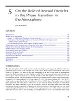
hyphae. Hyphae have a much larger surface area to volume ratio than do root hairs and fan out up
to 8 cm beyond nutrient depletion zones around roots (Douds and Millner, 1999; Millner and
Wright, 2002; Figure 6.1). This allows AM fungi to scavenge even highly immobile nutrients such
as phosphate. Also, the fungal cell membrane is capable of concentrating solutes against a gradient
(Bolan et al., 1991; George et al., 1992). The high carbon cost of P uptake is compensated for by
© 2004 by CRC Press LLC
1294_C06.fm Page 183 Friday, April 23, 2004 11:19 AM
Contributions of Fungi to Soil Organic Matter in Agroecosystems
183
Aggregate
Spore
Arbuscule
Stele
Root Hairs
FIGURE 6.1 Hyphae of arbuscular mycorrhizal fungi can access much more of the soil than can roots and
root hairs and form a framework on which aggregates can form.
an increase in photosynthetic capability of the host through increased leaf surface area and photosynthetic efficiency (Bolan et al., 1991; George et al., 1992). Mycorrhiza is the most efficient
mechanism for P acquisition, especially under stress conditions.
To varying degrees, mycorrhizal fungi can also provide other benefits, such as more efficient
uptake of N, the micronutrients Fe, Cu, and Zn (Clark and Zeto, 1996; Pawlowska et al., 2000),
and water; disease suppression; protection from heavy metal toxicity; and improved soil structure.
The mycorrhizal relationship reduces the growth of plant pathogens, especially fungal pathogens,
by increasing host resistance (triggering a defense response), altering root exudations to stimulate
the growth of microbes antagonistic to pathogens, competing for photosynthetic carbon, and
reducing the number of infection sites (Borowicz, 2001). The type of pathogen (nematode or fungal),
pathogen species, mode of action (necrotrophic or wilt for fungal pathogens and migratory or
sedentary for nematodes), and pathogen density help determine the severity of disease (Borowicz,
2001). As with other benefits in the mycorrhizal relationship, the magnitude and direction effects
of AM fungi on disease resistance depend on host genotype, AM species and isolate, timing of
AM colonization, other soil organisms, and abiotic factors.
Mycorrhizal host plants have been found at many heavy-metal contaminated sites, but the fungi
typically are not examined (Pawlowska et al., 2000). In pot culture experiments, it has been shown
that mycorrhizal fungi can take up toxic heavy metals, such as Cd and Pb, in addition to
© 2004 by CRC Press LLC
1294_C06.fm Page 184 Friday, April 23, 2004 11:19 AM
184
Soil Organic Matter in Sustainable Agriculture
micronutrients (Gonzalez-Chavez et al., 2002; Diaz et al., 1996). Metal uptake depends on soil
fertility, metal concentration, pH, the host plant, and AM species, and might interfere with P nutrition
in the host plant (Gonzalez-Chavez et al., 2002; Diaz et al., 1996).
In addition to improving plant health, fungal hyphae improve soil structure by helping form
water-stable soil aggregates (Miller and Jastrow, 1990; Rillig and Steinberg, 2002; Tisdall et al.,
1997). Mycorrhizal fungi also improve rhizosphere health by stimulating root exudation, which
promotes the growth of other soil microbes (Borowicz, 2001; Paul and Clark, 1996). Many excellent
books and review articles have been published on AM fungi and agroecosystems (Bolan et al.,
1991; Douds and Millner, 1999; George et al., 1992; Zak and McMichael, 2001).
GLOMALIN,
A
GLYCOPROTEIN PRODUCED
BY
AM FUNGI
The identification of glomalin, a glycoprotein produced by AM fungi, has led to a reevaluation of
fungal contributions to SOM and aggregate stability. Glomalin was identified at the United States
Department of Agriculture (USDA) in 1993 during work to produce monoclonal antibodies reactive
with AM fungi. One of these antibodies reacted with a substance on the hyphae of a number of
AM species (Wright et al., 1996). This substance was named glomalin after Glomales, the order
to which AM fungi belong. Several other typical soil fungi, such as Rhizoctonia, Gaeumannomyces,
Endogone, Mucor, and Phytophthora, were tested for cross-reactivity with the antibody against
glomalin, but were not immunoreactive (Wright et al., 1996). The glomalin fraction is operationally
defined by its extraction procedure, but is further characterized by total and immunoreactive protein
assays (Wright et al., 1996). Glomalin is found in abundance in both native and agricultural soils
(2–14 mg/g soil and 2–5 mg/g soil, respectively; Wright and Upadhyaya, 1998; Wright et al., 1999)
and appears to be as ubiquitous as AM fungi themselves (Carlile and Watkinson, 1996b; Olsson
et al., 1999; Wright and Upadhyaya, 1998; Wright, unpublished data).
Glomalin was revealed on AM fungal hyphae by using an indirect immunofluorescence
procedure that employs the antibody against glomalin and a second antibody tagged with a
fluorescein isothiocyanate (FITC) molecule (Wright, 2000). Evidence that glomalin is produced
by AM fungi and not plant roots was obtained early in the investigation of the reaction of the
monoclonal antibody against glomalin. Colonized and uncolonized roots were submitted for
evaluation of the technique by J.B. Morton (West Virginia University) in a blind experiment.
Colonization was correctly identified by immunofluorescence only on the roots that were later
described as having been inoculated. Immunofluorescence was absent on the roots later described
as uninoculated controls (Wright, unpublished data). In more recent work with an axenic culture
of transformed carrot roots, glomalin was extracted from hyphae in a root-free zone (Rillig and
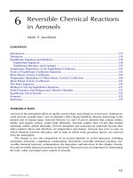
Steinberg, 2002). Glomalin is also routinely extracted from hyphae up to 7 cm away from roots
in pot cultures wherein hyphae is separated from roots by a 38-mm nylon mesh bag (Wright and
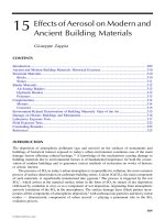
Upadhyaya, 1999; Figure 6.2). Immunofluorescence assays show that glomalin coats AM fungal
hyphae (Figure 6.3A to C); sloughs from hyphae onto colonized roots, organic matter, soil
particles, horticultural or nylon mesh (Figure 6.3D), and glass beads (Figure 3E); and is found
on arbuscules (green) within autofluorescing (yellow) root cells (Figure 6.3F; Wright et al., 1996;
Wright and Upadhyaya, 1999; Wright, 2000).
POOLS
OF
GLOMALIN
Glomalin consists of four major pools: (1) easily extractable glomalin (EEG), (2) total glomalin
(TG), (3) recalcitrant glomalin (RG), and (4) scum. The EEG pool is extracted with 20 mM citrate,
pH 7.0, for 0.5 h (Wright and Upadhyaya, 1998). Total glomalin is extracted with 50 mM citrate,
pH 8.0, in 1-h intervals (Wright and Upadhyaya, 1998), and recalcitrant glomalin is soluble only
in 50 mM citrate, pH 8.0, at 121oC after harsh treatment of the soil (i.e., treatment with dilute acid
for 1 h followed by three 16- to 18-h extractions in alkaline solutions; Nichols, 2003). When mature
© 2004 by CRC Press LLC
1294_C06.fm Page 185 Friday, April 23, 2004 11:19 AM
Contributions of Fungi to Soil Organic Matter in Agroecosystems
185
38 µm Nylon
Mesh Bag
Root
Compartment
Hyphal
Compartment
FIGURE 6.2 Single-species arbuscular mycorrhizal fungal cultures can be grown with different plant hosts
to examine glomalin accumulation in sterile sand, or, as in this case, sand and crushed coal medium where
the roots are contained in the root compartment within the nylon mesh bag, and the fungal hyphae, which
grows through the mesh and into the surrounding media in the hyphal compartment, can be examined under
single-species conditions.
sand-based pot cultures are submerged in water, an unattached fraction of glomalin forms tancolored foam on the surface of water. This scum is apparently a sloughed component of glomalin
and is very hydrophobic. We speculate that scum floats on soil water until it attaches to soil or
organic matter particles, but the chemistry of this interaction is not currently defined. Our lab
postulates that hydrophobic or cationic interactions, or both, might be the mechanisms by which
glomalin becomes deposited on soil or organic particles and mesh or glass beads (Wright and
Upadhyaya, 1996; Nichols and Wright, unpublished data). Glomalin contains high concentrations
of iron (2 to 12%), and recently it has been speculated that Al- and Fe-hydroxyls are involved in
aggregate formation by bridging organic matter to clay particles (Bird et al., 2002; Chenu et al.,
2000). It appears that glomalin can move in and out of these operationally defined pools (i.e., EEG
becomes scum and scum becomes TG). Steinberg and Rillig (2003) found that during an incubation
experiment EEG increased while TG decreased. They speculated that partial degradation decreases
sorption of glomalin to soil particles, which might increase the solubility and amount in the EEG
pool.
Glomalin concentration in these pools is measured by a Bradford total protein assay (i.e., TG
and EEG), immunoreactive protein (i.e., IRTG and IREEG) assays (Wright et al., 1996), or as
gravimetric or carbon weight. The Bradford protein assay is nonspecific and detects any proteinaceous material. Bradford concentrations are based on comparison with a bovine serum albumin
(BSA) standard curve. The immunoreactive protein assay (ELISA) uses the monoclonal antibody
specific for glomalin, but certain artificial conditions might reduce immunoreactivity. The ELISA
values are determined by comparison to 100% immunoreactive glomalin extracted from hyphae or
soil (Wright et al., 1996). The total protein assay measures concentrations from 1.25 to 5.0 mg,
whereas ELISA measures concentrations from 0.005 to 0.04 mg (Wright and Upadhyaya, 1999).
Because the range of Bradford values is ~100 times higher than that for ELISA, it can support
values of more than 100%. Both gravimetric and carbon weight have been used to quantify glomalin
partially purified by acid precipitation and dialysis against water (Nichols, 2003; Wright et al.,
1996). These weights are not based on structural components of glomalin but are rather direct
measurements on lyophilized material.
Comparisons of the total and immunoreactive pools of glomalin extracted from soil or pot
culture show that not all the extracted material is immunoreactive. Reduction in immunoreactivity
can be due to exposure to conditions that affect the site of binding of the antibody. The reactive
site for a monoclonal antibody is very specific (Goding, 1986), and some reactivity is lost probably
© 2004 by CRC Press LLC
1294_C06.fm Page 186 Friday, April 23, 2004 11:19 AM
186
Soil Organic Matter in Sustainable Agriculture
FIGURE 6.3 Arbuscular mycorrhizal fungi can be cultured in hydroponic pot cultures with a mycorrhizal
host plant, and glomalin can be examined by an immunofluorescence assay with a monoclonal antibody against
glomalin (seen as bright spots). Glomalin has been found coating and sloughing from hyphae of Acaulospora
morrowiae (CL551) (A), on a Gigaspora rosea (FL224) hyphal mat adhering to a horticultural mesh (B), on
Glomus intraradices hyphae grown in liquid cultures media by Dr. Yair Shachar-Hill at the New Mexico State
University (C), deposited on and around a hole in a horticultural mesh by Gi. rosea (FL224) (D), on a glass
bead by A. morrowiae (CL551) (E), and on arbuscules of G. etunicatum (BR220) in a corn root (F).
because of conformational changes by exposure to high heat (121°C) for a long time period (at
least 0.5 to 1.0 h) during extraction (Wright and Upadhyaya, 1999; Wright, unpublished data). In
the soil, organic matter, metals (such as iron), clays, and other substances might bind to glomalin,
causing conformational changes or masking the reactive site and thereby interfering with immunoreactivity. Also, conformational changes can occur in the molecule because of hydrophobic
interactions when it sloughed from the hyphae and is in the scum pool. Degradation is a factor in
soil extracts and might result in a decline in immunoreactivity (Wright and Upadhyaya, 1999).
Differences in immunoreactivity and extraction techniques are used to further describe some of the
© 2004 by CRC Press LLC
1294_C06.fm Page 187 Friday, April 23, 2004 11:19 AM
Contributions of Fungi to Soil Organic Matter in Agroecosystems
187
glomalin pools, such as the highly immunoreactive EEG (IREEG), lower immunoreactive TG
(IRTG; Wright and Upadhyaya, 1996), and very low immunoreactive RG (IRRG; Nichols, 2003).
CHARACTERIZATION
OF
GLOMALIN
Glomalin extracted from soil is very similar to that extracted from single-species pot cultures.
Samples have been examined by SDS-PAGE (Nichols, 2003; Rillig et al., 2001b; Wright et al.,
1996; Wright and Upadhyaya, 1996); nuclear magnetic resonance (NMR) (Nichols, 2003; Rillig
et al., 2001b; Nichols and Wright, unpublished data); carbohydrate analyses by a colorimetric assay;
gas chromatography–mass spectroscopy (GC-MS) and capillary electrophoresis (CE; Wright et al.,
1998; Nichols and Wright, unpublished data); and C, H, N analysis by combustion (Nichols, 2003;
Rillig et al., 2001b). There are minor variations in elemental constituents of glomalin among
samples, but the structural group assays (NMR, GC-MS, and CE) and SDS-PAGE demonstrate that
glomalin extracted from soil is similar to that from hyphae.
Rillig et al. (2003) and Steinberg and Rillig (2003) examined decomposition of glomalin
following soil incubation. One of the incubation studies (Steinberg and Rillig, 2003) showed that
hyphal length declined by 60% after 150 d of incubation, whereas the TG of glomalin declined by
25%, the IRTG disappeared almost completely, the EEG did not change, but the IREEG increased
fivefold. In the other study (Rillig et al., 2003), the TG declined by 48–81% and the EEG declined
by 51–88% after 413 d of incubation. By 14C data, Rillig et al. (1999) calculated a turnover time
for glomalin of 6 to 42 years. These recent incubation studies suggest that a long-lived, recalcitrant
glomalin fraction exists with a much longer turnover time.
Experiments to identify structural units of glomalin are currently underway. Information
obtained to date shows that glomalin is composed of proteinaceous, carbohydrate, and aliphatic
(potentially polymerized) components and binds multivalent cations (i.e., Fe and Al; Nichols, 2003;
Wright and Anderson, 2000; Nichols and Wright, unpublished data). The protein component appears
to be 30 to 40% of the molecular structure, measured by comparisons of gravimetric and protein
weight and preliminary amino acid measurements. The carbohydrate component is 3 to 6% according to a colorimetric assay, which measures oligosaccharide concentration. Aliphatic groups comprise 20 to 70% according to mass balance and NMR spectroscopy. Glomalin has 2 to 12% iron
based on acid hydrolysis and atomic adsorption measurements.
GLOMALIN,
A
MAJOR COMPONENT
OF
SOIL ORGANIC MATTER
A study was conducted to compare concentrations of glomalin to humic acid (HA), fulvic acid
(FA), and particulate organic matter (POM) in eight undisturbed soils in the U.S. All the fractions
have been operationally defined by extraction techniques. The appropriate extraction method was
used to remove each fraction: (1) alkaline extraction of HA and FA followed by acidic separation,
(2) citrate extraction of glomalin, and (3) density separation of POM. Quantities were measured
by using gravimetric and carbon weights and comparing total and immunoreactive protein concentration. The protein values also were used to correct for coextraction of glomalin in HA. The study
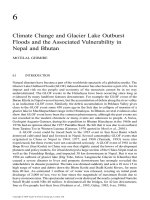
showed that glomalin represents a major fraction of soil organic carbon (SOC; 22 to 27%) and the
extractable part of the material previously identified as HA and humin contains glomalin (Figure
6.4; Nichols, 2003; Nichols and Wright, unpublished data).
QUANTITIES
OF
GLOMALIN
Soils from a variety of ecosystems throughout the U.S. and the world have been extracted for
glomalin, with TG concentrations ranging from 2 to 14 mg/g in most soils (Wright and Upadhyaya,
1998). High glomalin (TG) amounts have been found in undisturbed, volcanic soils from Japan
and Hawaii (19 mg/g soil and >60 mg/g soil, respectively; Rillig et al., 2001b; Wright, unpublished
data) and a humoferric podzol oak forest soil in Ireland (69 mg/g soil; Nichols and Wright,
© 2004 by CRC Press LLC
1294_C06.fm Page 188 Friday, April 23, 2004 11:19 AM
188
Soil Organic Matter in Sustainable Agriculture
HA
3.99
FA
1.70
Humin
10.88
POM
3.40
A
HA
1.68
Humin
8.15
Glomalin
5.52
FA
1.70
POM
2.92
B
FIGURE 6.4 The major fractions of soil organic carbon (SOC) have historically been (A) humic acid (HA),
fulvic acid (FA), humin (or humus), and particulate organic matter (POM), but with the identification of
glomalin and its separation from humic components (B), a sizable amount of SOC has been found in this
fraction. Units are mg C in the fraction per g soil extracted. (Adapted from Nichols, K.A. 2003. Ph.D. thesis,
University of Maryland, College Park. With permission.)
unpublished data). Typically, acidic soils have higher glomalin concentrations than do calcareous
soils (Wright and Upadhyaya, 1996; M. Haddad, personal communication), as do undisturbed soils
compared with agricultural soils (Wright and Upadhyaya, 1998; Nichols, 2003). Acidic soils have
lower decomposition rates and more soluble metals (such as Fe and Al), which might increase
glomalin concentrations by interactions with the molecules that inhibit degradation. Undisturbed
soils have lower decomposition rates than agricultural soils and a greater presence of AM fungi
because undisturbed soils have no P inputs from fertilizers and tillage has not disrupted hyphal
networks.
© 2004 by CRC Press LLC
1294_C06.fm Page 189 Friday, April 23, 2004 11:19 AM
Contributions of Fungi to Soil Organic Matter in Agroecosystems
189
Glomalin concentrations have been measured in a number of agricultural soils with different
tillage treatments; crop rotations; and fertilizer, herbicide, and pesticide amendments. In most of
these systems, there was a correlation between aggregate stability and glomalin concentration, with
no-till or minimum-till systems having the highest values (Figure 6.5; Rillig et al., 2002; Wright
and Anderson, 2000; Wright et al., 1999; Nichols and Wright, unpublished data). At a site in
Maryland, different management systems [(1) no-till, synthetic amendments (NT); (2) conventional
tillage, synthetic amendments (CT); and (3) minimum tillage, organic amendments (MT)] in the
same field were examined to determine how management affects glomalin-C and POM-C. The MT
treatment had the highest content of glomalin-C, POM-C, and aggregate stability, whereas the NT
and CT systems did not differ (Figure 6.6; Nichols, Wright, and Cavigelli, unpublished data).
SINGLE-SPECIES POT CULTURE EXPERIMENTS
Glomalin has been extracted from AM hyphae grown in single-species pot cultures with sterile
sand media and crushed coal where roots are contained in a 38-µm nylon mesh bag that only hyphae
can penetrate (Figure 6.2). Plants were watered with low-P nutrient solution (Millner and Kitt,
1992) and grown under metal halide and sodium vapor lights in a growth chamber or sodium vapor
lights in the greenhouse. Several different pot culture experiments have been conducted to measure
glomalin concentrations. In total, nine different species from four of five genera of AM fungi have
been grown with up to five different host plants (i.e., corn, clover, sudangrass, sorghum, fescue).
All these AM species produced glomalin in amounts that vary with culture conditions (i.e., light
intensity, media, etc.) and plant host. Glomalin concentrations (TG) in the outer hyphae chamber
typically range from 2 to 13 mg/pot, with values of 2 to 20 mg/g hyphae (Nichols and Wright,
unpublished data) and 5 to 40 mg glomalin/cm2 on horticultural mesh strips inserted into sand in
the hyphae chamber (Wright and Upadhyaya, 1999).
In one experiment, TG was extracted from five AM species (Glomus etunicatum, G. viscosum,
G. caledonium, Gigaspora rosea, and Gi. gigantea) produced on corn (Zea mays) and crimson
clover (Trifolium incarnatum L.). For three of the fungi (G. etunicatum, G. viscosum, and Gi.
gigantea), two INVAM isolates that had been collected from the same state or country but from a
60
TG
3
50
Stability
2.5
40
2
30
1.5
20
1
Aggregate stability (%)
Total glomalin (mg g−1 aggregates)
3.5
10
0.5
0
0
NT − 1y
NT − 2y
NT − 3y
Grass
FIGURE 6.5 Total glomalin concentration (mg/g aggregates) and aggregate stability (%) in 0- to 5-cm soil
samples of plots in transition from plow tillage (PT) to no-till (NT) in 1-, 2-, and 3-year increments and a
continuous grass (Grass) for ca. 15 years. (Adapted from Wright, S.F. et al. 1999. Soil Sci. Soc. Am. J. 63:
1825–1829. With permission.)
© 2004 by CRC Press LLC
1294_C06.fm Page 190 Friday, April 23, 2004 11:19 AM
190
Soil Organic Matter in Sustainable Agriculture
1.8
90
Glomatin
POM
Stability
1.6
80
1.2
60
1
50
0.8
40
0.6
30
0.4
20
0.2
Carbon (mg g−1soil)
70
10
0
Aggregate stability (%)
1.4
0
NT
CT
MT
FIGURE 6.6 Carbon in glomalin (glomalin-C, mg C glomalin/g soil) or particulate organic matter (POM)
(POM-C, mg C POM/g soil) extracted from soil (1 g) with 50 mM citrate or by using density separation,
respectively. Soil samples were collected from a Maryland field site under three different management
treatments: (1) no-till, synthetic amendments (NT); (2) conventional tillage, synthetic amendments (CT); and
(3) minimum tillage, organic amendments (MT).
different site were used. Glomalin concentrations (TG) in the hyphal compartment ranged from
0.5 to 2.7 mg/pot and in the root compartment from 1.2 to 13.8 mg/g hyphae (Table 6.1). Glomus
viscosum had the lowest total protein values in both the hyphae and root chambers, but the % IR
values in the root chamber were among the highest for both isolates and both hosts. One isolate
of G. etunicatum (BR220) had total protein values almost twice that of the other isolates, but this
was not true of the other G. etunicatum isolate (BR211). Although these plants were grown under
the same conditions, overall variations existed among isolates, species, and hosts that do not follow
any common trend.
DEPTH
AND
DEPOSITION EXPERIMENT
A pot culture experiment was performed to determine the amount of glomalin produced by
Gigaspora rosea (FL 224) colonizing single crimson clover (Trifolium incarnatum L.) plants. Figure
6.7 shows the experimental setup. Roots were confined within a nylon mesh bag, and hyphae grew
into a root-free zone. Plants were supplied with a low-P nutrient solution (Millner and Kitt, 1992)
and grown under sodium vapor lights. The following measurements were made in 8-cm-deep
increments of the hyphae chamber after 3 months of plant growth: (1) glomalin on hyphae, (2)
glomalin deposited on the horticultural mesh, (3) unattached glomalin (i.e., scum), and (4) percent
colonization of roots. Hyphae and scum were obtained by immersing the sand from each depth
increment in water, shaking the sand vigorously, and decanting the water into stacked sieves (150,
150, and 53 µm top to bottom). This was repeated four times. Hyphae and unattached glomalin on
the 150 and 53 µm sieves were washed into petri dishes. Scum floats on water and was separated
from hyphae by pipetting. The horticultural mesh was cut into small pieces for processing. Hyphae,
scum, and horticultural mesh were extracted in 20 mM citrate, pH 7.0 at 121°C for 1 h. Glomalin
was quantified by the Bradford assay (Wright and Upadhyaya, 1996). Percent colonization was
determined by the method of Giovannetti and Mosse (1980).
© 2004 by CRC Press LLC
1294_C06.fm Page 191 Friday, April 23, 2004 11:19 AM
Contributions of Fungi to Soil Organic Matter in Agroecosystems
191
TABLE 6.1
Glomalin Extracted from Single-Species Pot Cultures of One or More Isolates of Five
Arbuscular Mycorrhizal Fungal Species on Two Plant Hosts and Measured as Total
Protein (TP) and Percentage Immunoreactive Protein (% IR)
Hyphae Chamber
a
Root Chamber
b
% IRb
44
Species
G. etunicatum
Isolate
BR211
Host
Clover
TP (mg/pot)
1.90
% IR
52
G. etunicatum
BR220
Clover
1.11
58
11.82
12
Gi. rosea
FL224
Clover
2.05
52
5.25
12
Gi. gigantea
MA401C
Clover
1.63
48
5.45
46
Gi. gigantea
MA453A
Clover
1.28
50
4.08
47
G. viscosum
MD215
Clover
0.99
31
1.98
156
G. viscosum
MD216
Clover
1.17
34
1.55
116
G. caledonium
UK301
Clover
1.44
35
4.53
28
G. etunicatum
BR211
Corn
1.43
69
2.82
94
G. etunicatum
BR220
Corn
1.70
57
13.80
22
Gi. rosea
FL224
Corn
2.46
40
2.13
140
Gi. gigantea
MA401C
Corn
1.47
39
7.58
105
Gi. gigantea
MA453A
Corn
2.74
40
7.56
153
G. viscosum
MD215
Corn
0.47
92
1.22
94
G. viscosum
MD216
Corn
0.83
45
1.44
191
G. caledonium
UK301
Corn
1.13
62
7.16
185
TP (mg/pot)
6.57
a
Species from two genera Glomus (G.) and Gigaspora (Gi.).
% IR = (immunoreactive protein/total protein) ¥ 100. Immunoreactive protein values were determined by
comparison to 100% immunoreactive glomalin extracted from an undisturbed prairie soil. The scale for the standard
curve used to measure immunoreactive protein concentrations was ca. 100 times less than the scale for the total
protein standard curve. This gives very precise values for immunoreactive protein concentrations and might result
in % IR values higher than 100, especially when measuring microgram quantities.
Note: n = 3 pots per isolate. In these pot cultures, the hyphae chamber was separated from the root chamber by a
38-mm nylon mesh bag that roots cannot penetrate.
b
Glomalin production varied greatly for three plants (Figure 6.8). This might have been due to
factors controlled by individual plants, differences in light intensity, or a combination of factors,
which will require further investigation.
Figure 6.9 examines more closely the distribution of glomalin produced by Plant 2. Hyphae
grew into the root-free zone in the top half of the pot (Figure 6.9) and apparently released glomalin
that was unattached to hyphae or adhered to the horticultural mesh. Movement of glomalin unattached to hyphae through coarse sand is suggested, because unattached and mesh-trapped glomalin
were measured in the absence of detectable amounts of hyphae in the lower half of the pot.
Sloughing of glomalin from hyphae and attachment to soil particles is also suggested by the results
of this experiment.
GLOMALIN
AND
AGGREGATE STABILITY
Loss of topsoil due to erosion is a serious consideration in agroecosystems. Pimentel et al. (1995)
estimated that during the past 40 years nearly one third of the world’s arable land was lost to
erosion, with a current rate of 10 million ha/year. Soil aggregates are important to (1) maintain soil
© 2004 by CRC Press LLC
1294_C06.fm Page 192 Friday, April 23, 2004 11:19 AM
192
Soil Organic Matter in Sustainable Agriculture
Clover
Plant
cm
Root Chamber
8
Hyphae Chamber
16
Plastic Horticultural
Mesh
24
32
FIGURE 6.7 Configuration of pots used to determine glomalin production and deposition. A single red clover
plant was grown inside an 8-cm-diameter 38-µm nylon bag filled with coarse sand in a 25-cm-wide by 40cm-deep pot. Outside the nylon bag, the pot was filled with coarse sand. Horticultural mesh disks were placed
at 8-cm-deep intervals within the root-free hyphae chamber.
0 – 8 cm
8 – 16 cm
16 – 24 cm
24 – 32 cm
Glomalin (mg)
1.5
1.0
0.5
0.0
Plant # 1
Plant # 2
Plant # 3
FIGURE 6.8 Glomalin production by three individual clover plants. Production at each depth is the sum of
glomalin on hyphae, attached to the horticultural mesh, and unattached scum.
porosity, which provides aeration and water infiltration rates favorable for plant and microbial
growth, (2) increase stability against wind and water erosion, and (3) store carbon by protecting
organic matter from microbial decomposition (Bird et al., 2002; Hassink and Whitmore, 1997).
Because both aggregate stability and SOM decline on cultivation, it is possible that SOM (i.e.,
POM, humic substances, microbially produced molecules, and fungal hyphae) plays a role in
aggregate formation, but the exact mechanism is not understood. Aggregate formation is a complex
process of physical and chemical interactions.
Electron microscopy shows that aggregates are a conglomeration of soil minerals (clay particles,
fine sand, and silt), small plant or microbial debris, bacteria, free amorphous organic matter, and
organic matter strongly associated with clay coatings (Chenu et al., 2000). Fungal hyphae can
initiate aggregate formation by providing the framework on which organic mater collects (Miller
and Jastrow, 1990; Tisdall et al., 1997). Chemical processes then contribute to aggregate formation
and stability by gluing with polysaccharides, coating with hydrophobic polymers, binding mineral
© 2004 by CRC Press LLC
1294_C06.fm Page 193 Friday, April 23, 2004 11:19 AM
Contributions of Fungi to Soil Organic Matter in Agroecosystems
193
0.6
Hyphae
Unattached
Attached to mesh
0.5
Glomalin (mg)
0.4
0.3
0.2
0.1
0.0
0–8 cm
8–16 cm
16–24 cm
24–32 cm
Depth
FIGURE 6.9 Glomalin production on a clover plant by depth. Percent colonization of roots at each depth
increment is shown above the bar.
particles with organic polymers, and bridging organic matter and clay particles by polyvalent cations
(Degens, 1997; Piccolo and Mbagwu, 1999; Chenu et al., 2000). Drying and wetting actions,
shrinking and swelling of clays, freeze–thaw cycles, compaction, and enmeshing by fungal hyphae
and fine roots physically stabilize aggregates (Chaney and Swift, 1986a, 1986b; Degens, 1997).
Soil aggregates can be disrupted by rainfall because of slaking, differential swelling of clays,
mechanical dispersion by the kinetic energy of raindrops, and physiochemical dispersion without
the protection of hydrophobic coatings. The molecules involved in aggregate formation increase
water stability and long-term survival of aggregates, because attractive forces between these molecules are much stronger internally than externally (Degens, 1997; Piccolo and Mbagwu, 1999;
Chenu et al., 2000). In reduced or no-till systems, Chaney and Swift (1986a) found that the stubble
and mulch litter promote aggregate formation, because fungal decomposition of organic matter
produces gluing agents such as polysaccharides and mucigels. Caesar-TonThat and Cochran (2002)
found that ligninolytic basidomycetes produce large quantities of polysaccharides, glycolipids, or
glycoproteins that bind to and stabilize soil particles in water-stable aggregates. However, many of
the polysaccharides produced by microbial degradation glue aggregates together quickly but are
water-soluble and ephemeral and do not to contribute to the long-term stability of aggregates
(Chaney and Swift, 1986a; Six et al., 2001). Soil organic matter containing high concentrations of
aliphatic groups, such as HA, can increase aggregate stability and the long-term stabilization of
organic materials (Piccolo and Mbagwu, 1999). These aliphatic hydrophobic groups and polymers
are the major contributors to the water stability of aggregates. They increase the contact angle for
water penetration, which restricts infiltration and slaking, lowers wettability, and increases the
internal cohesion of aggregates (Chenu et al., 2000). Complexes between organic matter and
amorphous Fe and Al compounds also decrease the wettability of aggregates (Chenu et al., 2000).
Glomalin contributes to the stabilization of aggregates because it sloughs off hyphae onto
surrounding organic matter (Figure 6.10); binds to clays, probably via cation bridging by iron; and
has hydrophobic character owing to a number of aliphatic groups (Nichols, 2003; Wright and
Upadhyaya, 1999). This is demonstrated in a number of experiments in which total, and, especially,
immunoreactive concentration, of glomalin are positively correlated with percent water-stable soil
aggregates in both agricultural and native soils (Figure 6.5 and Figure 6.6; Bird et al., 2002; Rillig
et al., in press; Wright and Anderson, 2000; Wright and Upadhyaya, 1998; Wright et al., 1999).
© 2004 by CRC Press LLC
1294_C06.fm Page 194 Friday, April 23, 2004 11:19 AM
194
Soil Organic Matter in Sustainable Agriculture
FIGURE 6.10 Glomalin and arbuscular mycorrhizal hyphae on the surface of a 1- to 2-mm aggregate separated
from a Philippine soil provided by Dr. Angela Almendras, Department of Agronomy and Soil Science, ViSCA.
Glomalin is indicated by the bright spots, which are illuminated by an immunofluorescence assay using a
monoclonal antibody against glomalin.
Figure 6.5 shows that both glomalin and aggregate stability can be used to quantify changes that
occur in the soil, with a transition from continuous plow tillage to no till over a short period of
time (1 to 3 years). Even though glomalin and aggregate stability increased significantly after only
2 years of no-till management, the 15 years of undisturbed grass site had much higher glomalin
concentrations (TG) and aggregate stability than any other treatment. This study indicates that
glomalin, POM, and aggregate stability all continue to increase with time within reduced tillage
systems.
GLOMALIN
UNDER
ELEVATED CO2
Several studies were conducted to compare glomalin concentrations to aggregate stability under
elevated CO2 conditions. In a native grassland ecosystem in northern California, TG and IRTG
concentrations increased with higher CO2 concentrations, along with hyphal length at one site,
and aggregate stability in 1–2 mm and 0.25–1 mm aggregate size fractions (Rillig et al., 1999).
Long-term exposure to elevated atmospheric CO2 conditions from a natural CO2 spring in New
Zealand resulted in a linear increase in percent root colonization by AM fungi, soil hyphal length,
TG, and EEG along a CO2 gradient (Rillig et al., 2000). In a sorghum field, aggregate stability,
hyphal length, and EEG increased with elevated CO2 (Rillig et al., 2001a). In both the grasslands
(Rillig et al., 1999) and sorghum field (Rillig et al., 2001a), aggregate stability was correlated
with glomalin concentrations. These studies show that under elevated CO2 conditions, photosynthetic carbon is allocated belowground and glomalin might provide a significant sink to trap
carbon in the soil.
CONTRIBUTION OF SOIL FUNGI TO ORGANIC MATTER
Although hundreds of meters of hyphae can be found, fungal biomass is typically underestimated
and the contribution of fungi to SOM on a mass basis is not quantified at present. Stevenson (1994)
estimates that fungal numbers are 10 to 20 million/g of soil, whereas bacteria numbers are >1
billion/g of soil. Olsson et al. (1999) estimated the hyphal dry weight of AM fungi to be 0.03 to
© 2004 by CRC Press LLC
1294_C06.fm Page 195 Friday, April 23, 2004 11:19 AM
Contributions of Fungi to Soil Organic Matter in Agroecosystems
195
0.35 mg/g soil according to phospholipid fatty acid analysis. Some of the largest organisms in the
world are slow-growing soil fungi; for example, the basidiomycete Armillaria bulbosa has been
found in the soil that covers 15 ha, weighs >10,000 kg, and is over 1500 years old (Paul and Clark,
1996).
Soil fungi contribute to the formation and function of SOM. Upon decomposition by saprophytic
fungi, elements in POM, such as nitrogen and phosphorus, are transformed from unavailable organic
compounds to available inorganic nutrient sources. The degraded plant material then becomes part
of the humic fraction of soil. In addition, AM fungi and glomalin help stabilize SOM by contributing
to aggregate formation.
Overall, the diversity of soil organisms is reduced by agricultural practices and mineralization rates increased, making C the limiting nutrient for soil fungi. Fungal biomass typically
responds positively to no-till management; for example, Frey et al. (1999) found that fungal
hyphae length was 2 to 2.5 times higher in no-till than conventionally tilled systems. Fungi are
favored in no-till systems because (1) hyphal networks can be maintained, (2) fungi can bridge
the soil–residue interface and utilize spatially separated nutrients, especially C and N; and (3)
fungi can maintain activity, even in dry locations or across air-filled pores. With the identification
of glomalin and the correlations between the immunoreactive fraction of glomalin and aggregate
stability, this glycoprotein is proving useful as an indicator of soil quality and mycorrhizal
input. As more information comes to light about the structure of this molecule and its different
pools, the role of glomalin in agroecosystems will become more defined. However, from what
is already known, glomalin is a major component of the SOM and important to sustainable
agroecosystem functioning.
MANAGING SOIL FUNGI TO INCREASE SOIL ORGANIC MATTER
A sustainable agroecosystem is one in which the system’s internal mechanisms and resources can
maintain productivity, recover quickly from disturbances (such as tillage), and keep pests and
disease at tolerable levels with only minimal external inputs. Agricultural soils contain unnaturally
high amounts of P, N, and K from fertilizers, are physically disrupted by tillage, and often are
vegetated by only one or two plant species (Gliessman, 2000; Muramoto et al., 2000). To decrease
pathogenic fungi and enhance the biomass, diversity, and function of mutualistic fungi, one or more
of the following management options can be implemented (Carlile and Watkinson, 1996a; Horwath
and Paul, 1994; Stenberg, 1999; Wright and Anderson, 2000):
1. Reduced fertilizer inputs (especially high-P fertilizers) will increase the need for mutualistic fungi to scavenge immobile nutrients.
2. Conservation or no-tillage systems will prevent disruption of hyphal networks.
3. Increasing the number of crops in the rotation, planting interrow crops, or using buffer
strips or shelterbelts will increase aboveground diversity and decrease hosts for pathogenic fungi.
4. Planting cover crops instead of having fallow periods will maintain the presence of living
roots as hosts for mutualistic fungi.
5. Use of biocontrol measures for weeds and pests will reduce the loss of beneficial fungi
by fungicides and other pesticides.
These strategies can be used in agroecosystems to manage saprophytic, pathogenic, and mutualistic
soil fungi in order to allow greater crop production at lower costs.
© 2004 by CRC Press LLC
1294_C06.fm Page 196 Friday, April 23, 2004 11:19 AM
196
Soil Organic Matter in Sustainable Agriculture
REFERENCES
Abbott, L.K., and A.D. Robson. 1994. The impact of agricultural practices on mycorrhizal fungi. In C.E.
Pankhurst et al. (Eds.), Soil Biota: Management in Sustainable Farming. CSIRO Melbourne, Australia.
Bird, S.B., J.E. Herrick, M.M. Wander, and S.F. Wright. 2002. Spatial heterogeneity of aggregate stability
and soil carbon in semi-arid rangeland. Environ. Pollut. 116: 445–455.
Bolan, N.S. 1991. A critical review on the role of mycorrhizal fungi in the uptake of phosphorus by plants.
Plant Soil 134: 189–207.
Borowicz, V.A. 2001. Do arbuscular mycorrhizal fungi alter plant-pathogen relations? Ecology 82(11):
3057–3068.
Bottomley, P.J. 1994. Light microscopic methods for studying soil microorganisms. In R.W. Weaver et al.
(Eds.), Methods of Soil Analysis. Part 2. Microbiological and Biochemical Properties. SSSA Book
Series, No. 5, Soil Science Society of America, Madison, WI, chap. 6.
Caesar-TonThat, T.-C., and V.L. Cochran. 2000. Soil aggregate stabilization by a saprophytic lignin-decomposing basidiomycete fungus. I. Microbiological aspects. Biol. Fertil. Soils 32: 374–380.
Carlile, M.J., and S.C. Watkinson. 1996a. Saprotrophs and ecosystems. In M.J. Carlile and S.C. Watkinson
(Eds.), The Fungi. Academic Press, New York, chap. 6.
Carlile, M.J., and S.C. Watkinson. 1996b. Parasites and mutualistic symbionts. In M.J. Carlile and S.C.
Watkinson (Eds.), The Fungi. Academic Press, New York, chap. 7.
Chaney, K., and R.S. Swift. 1986a. Studies on aggregate stability. I. Reformation of soil aggregates. J. Soil
Sci. 37: 329–335.
Chaney, K., and R.S. Swift. 1986b. Studies on aggregate stability. II. The effect of humic substances on the
stability of re-formed soil aggregates. J. Soil Sci. 37: 337–343.
Chenu, C., Y. Le Bissonnais, and D. Arrouays. 2000. Organic matter influence on clay wettability and soil
aggregate stability. Soil Sci. Soc. Am. J. 64: 1479–1486.
Clark, R.B., and S.K. Zeto. 1996. Iron acquisition by mycorrhizal maize grown on alkaline soil. J. Plant Nutr.
19(2): 247–264.
Degens, B.P. 1997. Macro-aggregation of soils by biological bonding and binding mechanisms and the factors
affecting these: A review. Aust. J. Soil Res. 35: 431–459.
Diaz, G., C. Azcon-Aguilar, and M. Honrubia. 1996. Influence of arbuscular mycorrhizae on heavy metal (Zn
and Pb) uptake and growth of Lygeum spartum and Anthyllis cytisoides. Plant Soil 180: 241–249.
Douds, D.D., and P.D. Millner. 1999. Biodiversity of arbuscular mycorrhizal fungi in agroecosystems. Agric.
Ecosyst. Environ. 74: 77–93.
Frey, S.D., E.T. Elliott, and K. Paustian. 1999. Bacterial and fungal abundance and biomass in conventional
and no-tillage agroecosystems along two climatic gradients. Soil Biol. Biochem. 31: 573–585.
George, E., K. Haussler, S.K. Kothari, X.-L. Ki, and H. Marschner. 1992. Contribution of mycorrhizal hyphae
to nutrient and water uptake by plants. In D.J. Read et al. (Eds.), Mycorrhizas in Ecosystems. CAB
International, Cambridge, U.K.
Giovannetti, M, and B. Mosse. 1980. An evaluation of techniques for measuring vesicular arbuscular mycorrhizal infection in roots. New Phytol. 84: 489–500.
Gliessman, S.R. 2000. The ecological foundations of agroecosystem sustainability. In S.R. Gliessman (Ed.),
Agroecosystem Sustainability: Developing Practical Strategies. CRC Press, Boca Raton, FL.
Goding, J.W. 1986. Monoclonal Antibodies: Principles and Practice. Academic Press, San Diego, 315 pp.
Gonzales-Chavez, C., J. D’Haen, J. Vangronsveld, and J.C. Dodd. 2002. Copper sorption and accumulation
by the extraradical mycelium of different Glomus spp. (arbuscular mycorrhizal fungi) isolated from
the same polluted soil. Plant Soil 240: 287–297.
Harley, J.L., and E.L. Harley. 1987. A check-list of mycorrhiza in the British flora: Addenda, errata, and index.
New Phytol. 107: 741–749, 13–89.
Hassink, J., and A.P. Whitmore. 1997. A model of the physical protection of organic matter in soils. Soil Sci.
Soc. Am. J. 61: 131–139.
Hooker, J.E., and K.E. Black. 1995. Arbuscular mycorrhizal fungi as components of sustainable soil-plant
systems. Crit. Rev. Biotechnol. 15 (3/4): 201–212.
Horwath, W.R., and E.A. Paul. 1994. Microbial biomass. In R.W. Weaver et al. (Eds.), Methods of Soil Analysis.
Part 2. Microbiological and Biochemical Properties. SSSA Book Series No. 5, Soil Science Society
of America, Madison, WI, chap. 36.
© 2004 by CRC Press LLC
1294_C06.fm Page 197 Friday, April 23, 2004 11:19 AM
Contributions of Fungi to Soil Organic Matter in Agroecosystems
197
Klein, D.A., T. McLendon, M.W. Paschke, and E.F. Redente. 1995. Saprophytic fungal-bacterial biomass
variations in successional communities of a semi-arid steppe ecosystem. Biol. Fertil. Soils 19: 253–256.
Miller, R.M., and J.D. Jastrow. 1990. Hierarchy of root and mycorrhizal fungal interactions with soil aggregation. Soil Biol. Biochem. 22(5): 579–584.
Miller, R.M., D.R. Reinhardt, and J.D. Jastrow. 1995. External hyphal production of vesicular-arbuscular
mycorrhizal fungi in pasture and tallgrass prairie communities. Oecologia 103: 17–23.
Millner, P.D., and D.G. Kitt. 1992. The Beltsville method for soilless production of vesicular-arbuscular
mycorrhizal fungi. Mycorrhiza 2:9–15.
Millner, P.D., and S.F. Wright. 2002. Tools for support of ecological research on arbuscular mycorrhizal fungi.
Symbiosis 33: 101–123.
Muramoto, J., E.C. Ellis, Z. Li, R.M. Machado, and S.R. Gliessman. 2000. Field-scale nutrient cycling and
sustainability: Comparing natural and agricultural ecosystems. In S.R. Gliessman (Ed.), Agroecosystem
Sustainability: Developing Practical Strategies. CRC Press, Boca Raton, FL.
Nichols, K.A. 2003. Characterization of glomalin: A glycoprotein produced by arbuscular mycorrhizal fungi.
Ph.D. thesis, University of Maryland, College Park.
Olsson, P.A., I. Thingstrup, I. Jakobsen, and E. Baath. 1999. Estimation of the biomass of arbuscular mycorrhizal fungi in linseed field. Soil Biol. Biochem. 31(13): 1879–1887.
Paul, E.A., and F.E. Clark. 1996. Soil Microbiology and Biochemistry, 2nd ed. Academic Press, New York.
Pawlowska, T.E., R.L. Chaney, M. Chin, and I. Charvat. 2000. Effects of metal phytoextraction practices on
the indigenous community of arbuscular mycorrhizal fungi at a metal-contaminated landfill. Appl.
Environ. Microbiol. 66(6): 2526–2530.
Piccolo, A., and J.S.C. Mbagwu. 1999. Role of hydrophobic components of soil organic matter in soil aggregate
stability. Soil Sci. Soc. Am. J. 63: 1801–1810.
Pimentel, D., C. Harvey, P. Resosudarmo, K. Sinclair, D. Kurz, M. McNair, S. Crist, L. Shpritz, L. Fitton, R.
Saffouri, and R. Blair. 1995. Environmental and economic costs of soil erosion and conservation
benefits. Science 267: 1117–1123.
Rillig, M.C., G.Y. Hernandez, and P.C.D. Newton. 2000. Arbuscular mycorrhizae respond to elevated atmospheric CO2 after long-term exposure: Evidence from a CO2 spring in New Zealand supports the
resource balance model. Ecol. Lett. 3: 475–478.
Rillig, M.C., P.W. Ramsey, S. Morris, and E.A. Paul. 2003. Glomalin, an arbuscular-mycorrhizal fungal soil
protein, responds to land-use change. Plant Soil 253: 293–299.
Rillig, M.C., and P.D. Steinberg. 2002. Glomalin production by an arbuscular mycorrhizal fungus: A mechanism of habitat modification? Soil Biol. Biochem. 34: 1371–1374.
Rillig, M.C., S.F. Wright, M.F. Allen, and C.B. Field. 1999. Rise in carbon dioxide changes soil structure.
Nature 400: 628.
Rillig, M.C., S.F. Wright, and V.T. Eviner. 2002. The role of arbuscular mycorrhizal fungi and glomalin in
soil aggregation: Comparing effects of five plant species. Plant Soil 238: 325–333.
Rillig, M.C., S.F. Wright, B.A. Kimball, P.J. Pinter, G.W. Wall, M.J. Ottman, and S.W. Leavitt. 2001a. Elevated
carbon dioxide and irrigation effects on water stable aggregates in a Sorghum field: A possible role
for arbuscular mycorrhizal fungi. Glob. Change Biol. 7: 333–337.
Rillig, M.C., S.F. Wright, K.A. Nichols, W.F Schmidt, and M.S. Torn. 2001b. Large contribution of arbuscular
mycorrhizal fungi to soil carbon pools in tropical forest soils. Plant Soil 233: 167–177.
Six, J., A. Carpenter, C. van Kessel, R. Merck, D. Harris, W.R. Horwath, and A. Lüscher. 2001. Impact of
elevated CO2 on soil organic matter dynamics as related to changes in aggregate turnover and residue
quality. Plant Soil 234: 27–36.
Stahl, P.D, T.B. Parkin, and N.S. Eash. 1995. Sources of error in direct microscopic methods for estimation
of fungal biomass in soil. Soil Biol. Biochem. 27(8): 1091–1097.
Steinberg, P.D., and M.C. Rillig. 2003. Differential decomposition of arbuscular mycorrhizal fungal hyphae
and glomalin. Soil Biol. Biochem. 35: 191–194.
Stenberg, B. 1999. Monitoring soil quality of arable land: microbiological indicators. Acta Agric. Scand., B:
Soil and Plant Sci. 49: 1–24.
Stevenson, F.J. 1994. Humus Chemistry: Genesis, Composition, Reactions, 2nd ed. John Wiley & Sons, New
York.
Tinker, P.B., D.M. Durall, and M.D. Jones. 1994. Carbon use efficiency in mycorrhizas: Theory and sample
calculations. New Phytol. 128: 115–122.
© 2004 by CRC Press LLC
1294_C06.fm Page 198 Friday, April 23, 2004 11:19 AM
198
Soil Organic Matter in Sustainable Agriculture
Trappe, J.M. 1987. Phylogenetic and ecological aspects of mycotrophy in the angiosperms from an evolutionary
standpoint. In G.R. Safir (Ed.), Ecophysiology of VA Mycorrhizal Plants. CRC Press, Boca Raton,
FL, pp. 5–25.
Tisdall, J.M., S.E. Smith, and P. Rengasamy. 1997. Aggregation of soil by fungal hyphae. Aust. J. Soil Res.
35: 55–60.
Vieira, R.F., C.M.M.S. Silva, A.H.N. Maia, E.F. Fay, and K.C. Coelho. 2000. An appraisal of five methods
for the measurement of the fungal population in soil treated with chlorothalonil. Pest Manage. Sci.
56: 431–440.
Wessels, J.G.H. 1997. Hydrophobins: Proteins that change the nature of the fungal surface. Adv. Microb.
Physiol. 38: 1–45.
Wright, S.F. 2000. A fluorescent antibody assay for hyphae and glomalin from arbuscular mycorrhizal fungi.
Plant Soil 226: 171–177.
Wright, S.F., and R.L. Anderson. 2000. Aggregate stability and glomalin in alternative crop rotations for the
central Great Plains. Biol. Fertil. Soils 31: 249–253.
Wright, S.F., M. Franke-Snyder, J.B. Morton, and A. Upadhyaya. 1996. Time-course study and partial
characterization of a protein on arbuscular mycorrhizal hyphae during active colonization of roots.
Plant Soil 181: 193–203.
Wright, S.F., J.L. Starr, and I.C. Paltineanu. 1999. Changes in aggregate stability and concentration of glomalin
during tillage management transition. Soil Sci. Soc. Am. J. 63: 1825–1829.
Wright, S.F., and A. Upadhyaya. 1996. Extraction of an abundant and unusual protein from soil and comparison
with hyphal protein of arbuscular mycorrhizal fungi. Soil Sci. 161: 575–585.
Wright, S.F., and A. Upadhyaya. 1998. A survey of soils for aggregate stability and glomalin, a glycoprotein
produced by hyphae of arbuscular mycorrhizal fungi. Plant Soil 198: 97–107.
Wright, S.F., and A. Upadhyaya. 1999. Quantification of arbuscular mycorrhizal fungi activity by the glomalin
concentration on hyphal traps. Mycorrhiza 8: 283–285.
Wright, S.F., A. Upadhyaya, and J.S. Buyer. 1998. Comparison of N-linked oligosaccharides of glomalin from
arbuscular mycorrhizal fungi and soils by capillary electrophoresis. Soil Biol. Biochem. 30(13):
1853–1857.
Zak, J.C., and B. McMichael. 2001. Agroecology of arbuscular mycorrhizal activity. In M. Shiyomi and H.
Koizumi (Eds.), Structure and Function in Agroecosystem Design and Management. CRC Press, Boca
Raton, FL.
© 2004 by CRC Press LLC