Báo cáo y học: " Effects of PPARg ligands on TGF-b1-induced epithelial-mesenchymal transition in alveolar epithelial cells" ppsx
Bạn đang xem bản rút gọn của tài liệu. Xem và tải ngay bản đầy đủ của tài liệu tại đây (4.5 MB, 13 trang )
RESEA R C H Open Access
Effects of PPARg ligands on TGF-b1-induced
epithelial-mesenchymal transition in alveolar
epithelial cells
Xiahui Tan
1,2
, Hayat Dagher
1
, Craig A Hutton
2
, Jane E Bourke
1*
Abstract
Background: Transforming growth factor b1 (TGF-b1)-mediated epithelial mesenchymal transition (EMT) of alveolar
epithelial cells (AEC) may contribute to lung fibrosis. Since PPARg ligands have been shown to inhibit fibroblast
activation by TGF-b1, we assessed the ability of the thiazolidinediones rosiglitazone (RGZ) and ciglitazone (CGZ) to
regulate TGF-b1-mediated EMT of A549 cells, assessing changes in cell morphology, and expression of cell
adhesion molecules E-cadherin (epithelial cell marker) and N-cadherin (mesenchymal cell marker), and collagen
1a1 (COL1A1), CTGF and MM P-2 mRNA.
Methods: Serum-deprived A549 cells (human AEC cell line) were pre-incubated with RGZ and CGZ (1 - 30 μM) in
the absence or presence of the PPAR g antagonist GW9662 (10 μM) before TGFb-1 (0.075-7.5 ng/ml) treatment for
up to 72 hrs. Changes in E-cadherin, N-cadherin and phosphorylated Smad2 and Smad3 levels were analysed by
Western blot, and changes in mRNA levels including COL1A1 assessed by RT-PCR.
Results: TGFb-1 (2.5 ng/ml)-induced reductions in E-cadherin expression were associated with a loss of epithelial
morphology and cell-cell contact. Concomitant increases in N-cadherin, MMP-2, CTGF and COL1A1 were evident in
predominantly elongated fibroblast-like cells. Neither RGZ nor CGZ prevented TGFb1-induced changes in cell
morphology, and PPARg-dependent inhibitory effects of both ligands on changes in E-cadherin were only evident
at submaximal TGF-b1 (0.25 ng/ml). However, both RGZ and CGZ inhibited the marked elevation of N-cadherin
and COL1A1 induced by TGF-b1 (2.5 ng/ml), with effects on COL1A1 prevented by GW9662. Phosphorylation of
Smad2 and Smad3 by TGF-b1 was not inhibited by RGZ or CGZ.
Conclusions: RGZ and CGZ inhibited profibrotic changes in TGF-b1-stimulated A549 cells independently of
inhibition of Smad phosphorylation. Their inhibitory effects on changes in collagen I and E-cadherin, but not N-
cadherin or CTGF, appeared to be PPARg-dependent. Further studies are required to unravel additional
mechanisms of inhibition of TGF-b1 signalling by thiazolidinediones and their implications for the contribution of
EMT to lung fibrosis.
Background
In idiopathic pulmonary fibrosis (I PF), th e most promi -
nent change in lung architecture is the appearance of
fibro blast foci in the lung interstitium. Diminution of the
alveolar epithelial lining is accompanied by accumulation
of fibroblast-like mesenchymal cells that secrete excessive
extracellular matrix proteins such as fibri llar collagen I
and III [1]. Current treatment for IPF include s glucocor-
ticoids, which show poor efficacy and do not prevent
disease progression or reduce the high mortality rates
within 5 years of diagnosis [1-3]. It is critical therefore to
identify novel therapeutic agents that target foci develop-
ment to address this area of pressing medical need.
The exact origin of increased fibroblast-like cells within
foci is not known but these cells possess myofibroblast-
like properties evidenced by expression of a-smooth
muscle actin (aSMA) [4,5]. In IPF, myofibrob lasts are
regarded as the main perpetrators of fibrosis as they
appear to be the major source of ECM proteins such as
fibrillar collagen I [6,7]. Initially, it was thought that myo-
fibroblasts develop from the diffe rentiation of resident
* Correspondence:
1
Department of Pharmacology, University of Melbourne, Victoria, Australia
Tan et al. Respiratory Research 2010, 11:21
/>© 2010 Tan et al; licensee BioMed Central Ltd. This is an Open Access article distributed under the terms of the Creative Commons
Attribution License ( which permits unrestri cted use, distribution, and re production in
any medium, provided the original work is p roperly cited.
parenchymal fibroblasts in response to profibrotic cyto-
kines, such as transforming growth factor-b1(TGF-b1)
[8-11]. Recently however, immunostaining of lung biop-
sies from IPF patients has revealed fibroblast-like cells
expressing the surfactant protein C (SP-C) normally
synthesised and secreted by type II a lveolar epithelial
cell s (AECII) [4]. This suggests that in addition to regen-
erating damaged alveolar epithelial lining [12], AECII
may undergo epithelial-mesenchymal transition (EMT)
to contribute to foci development in the disease context.
TGF-b1 has been identif ied as a pote nt stimulus for
EMT, whereby epithelial cells acquire hyperplasticity
and develop a mesenchymal-cell phenotype [5,13-15].
TGF-b1 treatment has been shown to alter the cell mor-
phology of the human AECII derived A549 cell line
from cobblestone-shaped to a fibroblastoid appearance
[13,15]. Phenotypic markers associated with EMT
included diminished expression of E-cadherin, a cell
anchoring protein expressed specifically by epithelial
cells, and elevated expression of N-cadherin, normally
present at relatively higher levels in fibroblasts [8,10].
These alterations were accompanied by increased secre-
tion of the gelatinase matrix metalloproteinase-2 (MMP-
2) [13,14], increased cell motility [15,16] and de novo
synthesis of fibrillar collagen I a nd III [13]. Given the
established actions of TGF-b1 on fibroblast differentia-
tion, EMT and collagen synthesis, studies have investi-
gated potential therapeutic benefits of targeting
profibrotic effects of TGF-b1.
Peroxisome proliferator-activated receptor g (PPARg)
is a nuclear hormo ne receptor activated by the thiazoli-
dinedione class of anti-diabetic drugs that may have a
role in the regulation of both inflammation and fibrosis
in the lung [17,18]. In cultured lung fibroblasts from
subjects with and without IPF, the PPARg ligands rosi-
glitazone (RGZ), troglitazone (TGZ) and ciglitazone
(CGZ) prevented TGF-b1-mediated increases in aSMA
expression and fibrillar collagen synthesis [8,9]. Similar
findings have been observed in skin fibroblasts [19],
with PPARg ligands inhibiting TGF-b1 signalling via the
Smad pathway. Additional in vivo studies revealed that
treatment with TGZ and CGZ protected against bleo-
mycin-induced lung fibrosis in mice [9], a model in
which glucocorticoids are ineffective [20].
To date, the ability of PPARg ligands to antagonize
TGF-b1-mediated changes associated with EMT in
human AECII has not been explored. In the current study,
we use a well-validated model of EMT in the A549 human
alveolar cell line in which a suite of morphological, pheno-
typic and functional markers and outcomes have been
well characterised [13]. Our results demonstrate that RGZ
and CGZ inhibit several TGF-b1-induced changes in mar-
kers of EMT and lung fibrosis, including cadherin proteins
and collag en gene expression, to provide further support
for the antifibrotic potential of the thiazolidinedione class
of compounds.
Methods
Materials
Recombinant human TGF-b1 was purchased from R&D
systems (Minneapolis, MN). RGZ, CGZ and GW9662
and rabbit polyclonal antibody against human PPAR g
were from Cayman Chemical Corporation (Ann Arbor,
MI) and sheep polyclonal Texas-red conjugated anti-
rabbit antibody was from Abcam (Cambridge, UK).
Mouse monoclonal antibodies against human E-cadherin
and N-cadherin were from BD Transduction Laboratory
(Oxford, UK), against human b-actin from Abcam
(Cambridge, UK) and against human a SMA from Sigma
Aldrich (St. Louis, MO). Rabbit monoclonal antibodies
against human Smad2 and Smad3, and phosphorylated
Smad2 and Smad3 were from Cell Signaling Technology
(Beverly, MA). Sheep HRP-conjugated polyclonal anti-
mouse and anti-rabbit antibodies were from Chemicon
International (Temecula, CA). Quantitect primers for
the human genes COL1A1, COL3A1 and 18s rRNA
were from Qiagen (Hilden, Germany) and the primer
sequences for aSMA (ACTA2) and MMP-2 were pre-
viously reported in [14] and were synthesised by Gene-
works (Adelaide, Australia). Phosphate buffered saline
(PBS) was from Oxoid (Basingstoke, UK), with all other
chemicals and reagents from Sigma Aldrich.
Cell Culture and Drug Treatment
Human type II alveolar epi thelial cell line A549 (ATCC,
Manassas, VA) was maintained in high glucose-DMEM
(Invitrogen, Carlsbad, CA) containing 10% (v/v) FBS, 15
mM HEPES, 20 mM sodium bicarbonate, and 2 mM L-
glutamine at 37°C in a humidified 5% CO
2
atmosphere.
Cellswereseededatadensityof5×10
4
cells per well
onto 6-well plates, or at 2 × 10
3
cells per well onto 8-
well Lab-Tek Chamber slides which were pre-coated
with 0.1 mg/ml (w/v) poly-L-lysine solution. After over-
night attachment, cells were maintained in serum-free
DMEM containing 0.25% (w/v) bovine serum albumin
(BSA) for 24 hr.
PPARg ligandswerepreparedas30mMstocksin
dimethylsulfoxide (DMSO), with 0.1% DMSO used as
vehicle control for treatment of cells. Cells were prein-
cubated with PPARg agonists RGZ or CGZ (1-30 μM)
in the absence or presence of the PPARg antagonist
GW9662 (10 μM) [21] for 60 min prior to stimulation
with TGF-b1 (0.75-7.5 ng/ml) for 2-24 hr for immuno-
cytochemical studies, 24 hr for mRNA analysis or up to
72 hr for protein analysis. These drugs did not affect
A549 cell viability as measured by Trypan blue staining.
The concentration of GW9662 used has been shown to
have no effect on basal PPRE promoter activity but to
Tan et al. Respiratory Research 2010, 11:21
/>Page 2 of 13
suppress RGZ-induced activation of the PPRE reporter
[22,23] and the antiproliferative effects of RGZ in
human cultured airway smooth muscle [24].
Immunocytochemistry
Cells on chamber slides were washed with PBS, fixed in
ice-cold methanol for 10 min, and then blocked with 2%
BSA in PBS for 1 hr at room temperature. Cells were
then incubated with primary PPARg antibody (1:100) for
2 hr at room temperature. After washing, antibody bind-
ing was detected by incubating with sheep polyclonal
Texas-red conjugated anti-rabbit antibody (1:100) for 1
hr. Cell nuclei were visualised by staining with 1 μg/ml
(w/v) Hoescht-33258 dye for 20 min at room tempera-
ture. Images were colle cted with a Carl Zeiss Axioscope
microscope system at 20× magnification.
Preparation of cytoplasmic and nuclear cell extracts
A549 cells were washed with PBS, then treated with
whole cell lysis buffer (100 mM NaCl, 0.5% w/v 10 mM
Tris-HCl (pH = 7.5), 0.5% w/v sodium deoxycholate, 2
mM sodium EDTA and 1% v/v Triton-X) supplemented
with 1% v/v protease inhibitor cocktai l and phosphatase
inhibitor cocktail 2. Cells were lysed for 20 min on ice
before centrifugation and collection of supernatants.
To separate cytosolic and nuclear proteins, cells were
treated for 10 min on ice with cytosolic lysis buffer (10
mM HEPES, 10 mM KCl, 10 mM EDTA, 1 mM DTT)
supplemented with 1% v/v protease inhibitor cocktail
and 0.4% v/v IGEPAL. After centrifugation, the superna-
tant containing the cytosolic fraction was collected and
the remaining pellet treated with lysis buffer for cell
nuclei (10 mM HEPES, 400 mM NaCl, 1 mM EDTA,
10% v/v glycerol, pH 7.9) supplemented with 1% v/v
protease inhibitor cocktail and 1 mM dithiothreitol.
Sampl es were homogeni sed by vortexing and then incu-
bated at 200 rpm on ice for 2 h on a rocking platform.
All protein supernatant samples were collected follow-
ing centrifugation at 13,000 g (5 min, 4°C) and stored at
-20°C. Total protein concentration was measured via a
spectrophotometer (Thermoplus, Finland) using the
bicinchoninic acid (BCA) protein assay kit (Bio-Rad,
Hercules, CA) with BSA utilised as the protein standard.
SDS-PAGE and Western blot
20 μg of total protein from each sample was separated
on 10% polyacrylamide gels (Bio-Rad). After electro-
phoresis, separated proteins were transferred onto
Hybond Nitrocellulose membranes (Amersham, Buckin-
ghamsh ire, UK). Membranes were then blocked for 1 hr
at room temperature with 5% w/v skimmed milk solu-
tion or 2% w/v BSA in TBST. Membranes were then
probed with primary antibodies (1:10,000 for b-actin,
1:1000 for other antibodies) for 18 hr at 4°C. After
washing with TBST, membranes were probed with
either sheep anti-mouse or anti-rabbit HRP-conjugated
secondary antibodies for 1 hr at room temperature.
Immunoblots were visualised by enhanced chemilumi-
nescence (GE Healthcare, Chalfont St Giles, UK) and
band densities quantified using ImageQuant TL software
after image acquisition with an ImageQuant 350 (GE
Healthcare). Results were expressed relative to b-actin
band density used as a loading control.
Real Time Quantitative Reverse Transcription-PCR
Total RNA was extracted using the RNeasy Kit (Qia-
gen). For complementary DNA (cDNA) synthesis 1 μg
of total RNA was reverse transcribed using Superscript
III (Invitrogen). cDNA equivalent to 10 ng of total RNA
was used for all PCR reactions in the presence of 100
nM of forward and reverse primers (refer to Materials
section). All PCR reactions were performed using Plati-
num SYBR Green qPCR SuperMix-UDG (Invitrogen) in
an ABI Prism 7900 HT sequence detection system
(Applied Biosystems, Foster City, CA). For normaliza-
tion of all RT-PCR data, 18s rRNA expression was used
as a reference gene. Relative transcript abundance of
COL1A1, MMP-2 and CTGF were expressed in ΔCt
values (ΔCt = Ct
reference
-Ct
target
). Relative fold changes
in transcript levels compared to basal levels were calcu-
lated as 2
ΔΔCt
(ΔΔCt = ΔCt
treatment
-ΔCt
basal
).
Statistical Analysis
Data were expressed as mean response ± S.E.M. for n
repeated experiments and analysed using Graph Pad
Prism version 5.0 software. The effects of TGF-b1on
cadherin expressi on were analysed by one-way ANOVA
on raw densitometric data normalised for b -actin as
loading control. The effects of PPARg ligands in the pre-
sence and absence of antagonist on TGF-b1-mediated
changes of EMT markers were tested by one-way
repeated measures ANOVA followed by Bonferroni post
hoc test for multiple comparisons with data normalised
to control levels in the absence of TGF-b1. Effects were
considered to be statistically significant when P < 0.05.
Results
PPARg expression in A549 cells
Immunocytochemical staining for PPARg was performed
in serum-deprived A549 cells. Under basal conditions,
PPARg w as det ected in both cytoplasm and nuclei, with
higher levels of expression evident in the cytosol (Figure
1A). However, in cells treated with the PPARg ligand
RGZ, both perinuclear and nuclear staining for PPARg
was increased within 2 hr with maximal effec ts observed
at 24 hr (Figure 1A). Changes in receptor localization in
thepresenceofRGZwerepreventedbythePPARg
antagonist GW9662 (Figure 1A). RGZ-induced nuclear
Tan et al. Respiratory Research 2010, 11:21
/>Page 3 of 13
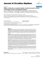
Figure 1 PPARg expression and localization in A549 cells. (A) Immunocytochemistry for PPARg in A549 cells. Cells were grown to confluence
on chamber slides, serum deprived for 24 hr and then treated with either vehicle (0.1% DMSO, left) for basal, 10 μM GW9662, 10 μM RGZ or
10 μM RGZ + 10 μM GW9662 for 24 hr. PPARg localization in the cells was detected by immunocytochemistry (left column) and the nuclei
visualised by Hoechst staining (right column). (B) Western blot for PPARg expression in the cytosol and nuclei of serum-deprived A549 cells in
the absence and presence of RGZ (10 μM) for 2 to 24 hr. Basal levels were measured at 2 hr after vehicle (0.1% DMSO) treatment. (C) Western
blot for PPARg expression in A549 cells in the absence and presence of TGF-b1 (72 hrs). All images and blots are representative of 3 separate
experiments. In the ICC images bar = 100 μm.
Tan et al. Respiratory Research 2010, 11:21
/>Page 4 of 13
translocation was confirmed by Western blotting showing
increased levels of PPARg in nuclear extracts at 2, 4 and
24 hrs (Figure 1B). Cellular PPARg protein levels were not
regulated by treatment with TGF-b1(Figure1C).
Having verified PPARg receptor expression and the abil-
ity of RGZ to cause receptor translocation in these cells,
we next investigated whether thiazolidinediones could reg-
ulate TGF-b1-mediated changes in markers of EMT.
Thiazolidinediones do not regulate basal E-cadherin and
N-cadherin expression
Under basal conditions, A549 cells expressed relatively
higher levels of E-cadherin than N-cadherin, consistent
with their epithelial phenotype. Treatment with RGZ
and CGZ at concentrations up to 30 μMdidnotaffect
basal levels of either cadherin (Figure 2A, B).
Thiazolidinediones inhibit TGF-b1-induced changes in
E-cadherin expression via PPARg
TGF-b1 treatment of A549 cells for 72 hr reduced basal
expression of E-cadherin in a concentration-dependent
manner (Figure 3A). The 60% reduction in E-cadherin
levels in the presence of 0.25 ng/ml TGF-b1was
partially inhibited by both RGZ and CGZ at concentra-
tions up to 10 μM but this effect was not maintained at
30 μM (Figure 3B, D). The effectiveness of RGZ was
also reduced when cells were stimulated with 0.75 or
2.5 ng/ml TGF-b1 (Figure 3B), and CGZ was unable to
prevent the maximal decrease in E-cadherin at these
TGF-b1 concentrations (data not shown). The PPARg
antagonist GW9662 alone did not affect the reduction
of E-cadherin expression induced by 0.25 ng/ml TGF-b1
but prevented the partial inhibitory effects of both
PPARg ligands (Figure 3C, D, E).
Thiazolidinediones inhibit TGF-b1-induced changes in
N-cadherin expression independent of PPARg
Low basal expression of N-cadherin in A549 cells was
increased approximately 3-fold by TGF-b1(Figure4A).
In contrast, basal expression of aSMA was not affected
by TGF-b1 at concentrations up to 7.5 ng/ml (data not
shown). Both RGZ and CGZ partially inhibited the max-
imum increase in N-cadherin expression in response to
2.5 ng/ml TGF-b1(Figure4B).Theinhibitoryeffectsof
RGZ and CGZ were maintained in the presence of
GW9662 (RGZ Figure 4B, CGZ data not shown).
Figure 2 Regulation of basal E-cadherin and N-cadherin expression by PPARg ligands. (A) Effect of TGF-b1 or PPARg ligands on expression
of epithelial marker E-cad (n = 4). (B) Effect of TGF-b1 or PPARg ligands on expression of mesenchymal marker N-cad (n = 4). Cell lysates were
prepared from A549 cells pre-incubated with vehicle, TGF-b1 (2.5 ng/ml), or RGZ or CGZ at the concentrations indicated for 72 hr. Densitometric
analysis values for band intensities from each Western blot were normalised to b-actin and expressed as a percentage of basal levels. Each point
represents mean ± s.e.m.
Tan et al. Respiratory Research 2010, 11:21
/>Page 5 of 13
Figure 3 Regulation of TGF-b1-induced E-cadherin expression by PPARg ligands. (A) Effect of TGF-b1 on expression of epithelial marker
E-cad (n = 4). (B) Effect of RGZ on regulation of E-cad expression by TGF-b1 (n = 6). (C, D) Effect of the PPARg antagonist GW9662 (10 μM) on
regulation of TGF-b1-mediated E-cad expression by RGZ (n = 4) and CGZ (n = 4). (E) Western blot showing the effect of GW9662 on RGZ-
mediated inhibition of TGF-b1 effect on E-cad expression (representative of n = 4 experiments). Cell lysates were prepared from A549 cells pre-
incubated with vehicle, RGZ or CGZ with or without GW9662 for 1 hr, stimulated with TGF-b1 at the concentrations indicated for 72 hr.
Densitometric analysis values for band intensities from each Western blot were normalised to b-actin and expressed as a percentage of basal
levels. Each point represents mean ± s.e.m. * P < 0.05, compared with basal (A) TGF-b1 (B-D).
Tan et al. Respiratory Research 2010, 11:21
/>Page 6 of 13
Thiazolidinediones inhibit TGF-b1-induced changes in
collagen I and CTGF mRNA
Treatment of A549 cells with 2.5 ng/ml TGF-b1for24
hr caused 71 ± 11 fold (n = 5, P < 0.01) and 190 ± 59
fold (n = 4, P < 0.01 ) increases in COL1A1 and MMP-2
mRNA levels respectively (Figure 5A, D). The increase
in CTGF mRNA level was more modest (11 ± 2 fold,
n = 7, P < 0.05, Figure 5C). Basal levels of collagen III
or aSMAmRNAwerenotsignificantly upregulated by
TGF-b1 (data not shown).
The TGF-b1-induced increase in COL1A1 mRNA was
attenuated by both RGZ and CGZ to a similar extent,
although RGZ was markedly more potent (Figure 5A).
Both RGZ and CGZ partially inhibited the increase in
CTGF (Figure 5C). The effects of both PPARg ligands on
COL1A1 but not CTGF mRNA were abolished in the pre-
sence of GW9662 (Figure 5B, C). In contrast, the increase
in MMP-2 m RNA fol lowing TGF-b1 treatment was not
inhibited by RGZ or CGZ (Figure 5D, n = 7, P < 0.05).
Thiazolidinediones do not prevent TGF-b1-induced
changes in cell morphology
Inthepresenceof2.5ng/mlTGF-b1, the morphology
of A549 cells changed from the classical cobblestone
appearance of alve olar epithelial cells to predominantly
elongated fibroblast-like cells(Figure6).Thesechanges
were not evident following treatment with 0.25 ng/ml
TGF-b1. Pre-treatment with RGZ or CGZ did not affect
cell morphology in the absence or presence of TGF-b1
(Figure 6, CGZ not shown).
Thiazolidinediones do not affect TGF-b1-mediated
phosphorylation of Smad2 and Smad3
Under basal conditions in the absence of TGF-b1, A549
cells expressed Smad2 and Smad3 proteins, and treat-
ment with RGZ or CGZ had no effect (Figure 7). The
phosphorylated forms of these proteins could be
detected as early as 15 min after TGF-b1 stimulation
(data not shown), with maximum phosphorylation after
1 hr. Pretreatment with RGZ or CGZ did not prevent
the increase in levels of phosphorylated Smad proteins
in response to TGF-b1 (Figure 7).
Discussion
In this study, we show that the PPARg ligands RGZ and
CGZ inhibit TGF-b1-induced changes in E-cadherin, N-
cadherin, collagen I and CTGF expression in A549 cells
in the absence of regulatory effects on cell morphology.
RGZ was consistently more effective than CGZ in inhi-
biting PPARg dependent changes in markers of EMT, in
Figure 4 Regulation of TGF-b1-induce d N-cadherin expression by PPARg ligands. (A) Effect of TGF-b1 on expression of mesenchymal
marker N-cad (n = 4). (B) Effect of PPARg ligands on regulation of N-cad expression by 2.5 ng/ml TGF-b1 (RGZ, n = 6; RGZ + GW9662, n = 6;
CGZ, n = 6). Cell lysates were prepared from A549 cells pre-incubated with vehicle, RGZ or CGZ with or without GW9662 for 1 hr, stimulated
with TGF-b1 at the concentrations indicated for 72 hr. Densitometric analysis values for band intensities from each Western blot were normalised
to b-actin and expressed as a percentage of basal levels. Each point represents mean ± S.E.M. * P < 0.05, ** P < 0.01 compared compared with
basal (A) or TGF-b1 (B).
Tan et al. Respiratory Research 2010, 11:21
/>Page 7 of 13
agreement with their relative binding affinities for
PPARg. However, since inhibitory effects occurred via
both PPARg-dependent and PPARg -indep endent path-
ways and were not associated with inhibition of Smad
phosphorylation, it is proposed that multiple mechan-
isms underlie potential antifibrotic actions of PPARg
ligands in these cells.
Recent studies provide evidence that TGF-b1-i nduced
EMT of AECII may contribute to the de n ovo
appearance of myofibroblasts in fibrotic lungs
[4,5,25-27]. Although these findings are not universal
[28], it has been shown that induction of lung fibrosis in
mice by overexpression of active TGF-b1 or bleomycin
treatment resulted in the accumulation of mesenchymal
cells with AECII origins adjacent to fibrotic lesions
[4,25]. Additionally, fibroblast-like cells in lung biopsies
from IPF patients expressed the AECII surfactant pro-
tein SP-C [4].
Figure 5 Regulation of TGF-b1-induced COL1A1, CTGF and MMP-2 mRNA levels by PPAR g li gands. (A) Effect of PPARg agonists on
regulation of COL1A1 mRNA levels by TGF-b1 (n = 5). (B) Effect of the PPARg antagonist GW9662 (10 μM) on regulation of TGF-b1-mediated
COL1A1 mRNA by RGZ (n = 6) and CGZ (n = 4). (C) and (D) Effect of PPARg ligands on regulation of CTGF and MMP-2 mRNA levels by TGF-b1
(n = 4,7). Total RNA was collected from A549 cells pre-incubated with vehicle, RGZ or CGZ with or without GW9662 for 1 hr and then stimulated
with 2.5 ng/ml TGF-b1 for 24 hr. Results were normalised to 18s rRNA levels and expressed as fold change from basal levels. Each point
represents mean ± S.E.M. * P < 0.05, ** P < 0.01, *** P < 0.001 compared with TGF-b1.
Tan et al. Respiratory Research 2010, 11:21
/>Page 8 of 13
Figure 6 Regulation of TGF-b1-induced changes in cell morphology by rosiglitazone. A549 cells were incubated with vehicle (0.1% DMSO),
TGF-b1 (0.25 ng/ml), TGF-b1 (2.5 ng/ml) or TGF-b1 (2.5 ng/ml) + RGZ (10 μM) for 72 hr and photographed at 100× magnification. The images
are representative of 4 separate experiments.
Figure 7 Regulation of TGF-b1-induced phosphorylation of Smad2 and Smad3 by PPARg ligands. Cell lysates were prepared from A549
cells pre-treated with vehicle, RGZ (10 μM) or CGZ (10 μM) for 1 hr and then treated with TGF-b1 at the indicated concentrations for 1 hr. The
Western blot is representative of 3 separate experiments.
Tan et al. Respiratory Research 2010, 11:21
/>Page 9 of 13
To explore potential regulation of EMT in AECII by
PPARg ligands, we have used A549 cells as a model of
human AECII. A549 cells possess many features of nor-
mal AECII cells [29] and have been used i n numerous
studies examining EMT [13-16,30-32]. We characterised
EMT in A549 cells by detecting changes in cell mor-
phology, E-cadherin and N-cadherin levels, and collage n
I, CTGF and MMP-2 gene expression. These markers
facilitate identification of cells along the spectrum of
transition from epithelial to mesenchymal phenotype.
Critically, A549 cells express PPARg, the receptor target
of the thiazolidinedione class of drugs which includes
RGZ and CGZ. In the current study, nuclear transloca-
tion of PPARg by RGZ provided evidence that PPARg
activat ion could potentially regulate cellular funct ions of
A549 cells, including EMT. This translocation was pre-
vented by the PPARg antagonist GW9662, supporting
the use of this pharmacological too l to explore the
PPARg-dependence of the actions of RGZ and CGZ.
In this study, treatment of cells with RGZ or CGZ in
the absence of TGF-b1 failed to elicit any detectable
changes in expression of cell adhesion molecules or
morphology. Direct regulation o f EMT by PPARg
ligands may vary depending on cellular origin, since
PPARg activation by RGZ has previously been shown to
promote EMT of gastrointestinal epithelial cells, charac-
terised by increa sed cell scattering and altered cell mor-
phology [33].
Consistent with previous findings in A549 cells, TGF-
b1 treatment caused cells to lose their polygonal appear-
ance and cell-c ell contacts leading to the acquisition of
elongated, spindle-shaped morphology consistent with
fibroblasts [13]. TGF-b1 also altered the expression of
cell adhesion molecules consistent with EMT as
reported previously [13,30]. Significant reductions in E-
cadherin, a protein expressed only by epithelial cells and
diminished during EMT [26], were evident even in the
absence of obvious morphological changes, suggesting
that a loss of >60% E-cadherin is required before cell
morphology is altered. Under these conditions, the par-
tial inhibition of the reduction in E-cadherin by both
RGZ and CGZ was PPARg dependent, since it was abol-
ished in the presence of the selective PPARg antagonist
GW9662. The modest inhibitory effect was not main-
tained at the highest concentrations of PPARg ligands
tested, but the mechanism for this loss of activity was
not explored.
The maximum effect of TGF-b1 elicited was a 90%
loss of E-cadherin expression, suggesting that cells that
have undergone EMT may still retain epithelial phenoty-
pic markers. In vivo studies have previously reported
retention of the AECII specific SP-C and pro SP-B pro-
teins in mesenchymal cells derived from EMT [4,5].
Thiazolidinedione treatment was unable to prevent this
maximal reduction in E-cadherin expression accompa-
nied by loss of epithelial morphology.
Low expression of N-cadherin and aSMA were
detected under basal conditions in A549 cells, despite
the classification of t hese markers as mesenchymal spe-
cific [34-36]. Similar findings have been described in
both A549 cells [13,31], and in RLE-6TN cells [37], a
rat AECII cell line reported to undergo EMT upon
TGF-b1 treatment [5,37,38]. In RLE-6TN cells, basal
expression of aSMA was attributed to constitutive acti-
vation of TGF-b1 type I receptor kinase (TGFbRI) [37],
and may also contribute to basal N-cadherin and aSMA
expression in A549 cells.
Following TGF-b1 treatment, concomitant increases in
N-cadherin accompanied reductions in E-cadherin
expression in A549 cells. Although both proteins med-
iate cell to cell attachment, cell adhesion by E-cadherin
is four times stronger than adhesion by N-cadherin [39].
In squamous car cinoma cells, N-cadherin has also been
shown to promote scattering and increased motility
[40]. It is likely then that the relative expression levels
of these molecules contributed to the loss of cell-cell
contact in A549 cells evident at higher concentrations
of TGF-b
1.
The maximum TGFb1-induced increase in N-cadherin
expression was approximately 3-fold higher than basal
levels. In con trast to their limited effects on the reduc-
tion in E-cadherin, both RGZ and CGZ were able to
decrease the elevati on in N-cadherin levels to a similar
extent following treatment with 2.5 ng/ml TGF-b1. In
addition, the inhibitory effects of PPARg ligands on the
increased N-cadherin expression were not attenuated by
GW9662, suggesting a separate PPARg-independent
pathway.
In addition to altered cadhe rin expression, A549 cells
stimulated with TGF-b1 displayed other features similar
to lung fibroblasts. Under basal conditions, A549 cells
do not synthesize fibrillar collagen I or CTGF, and only
express low levels of MMP-2 [13,14,41]. MMP-2 activity
is thought to be an important contributor to EMT by
facilitating basement membrane breakdown and migra-
tion of cells into the interstitium [42-44]. CTGF expres-
sion is induced by TGF-b1andhasprofibrotic
properties including stimulation of fibrillar collagen pro-
duction and myofibroblast accumulation [41,45]. As
confirmed in this study, stimulation with TGF-b1
caused marked increases in mRNA for COL1 A1, which
encodes the a1 chain of mature collagen I fibers, as well
as MMP-2 and CTGF. However, in contrast to a pre-
vious report [13], significant increases in COL3A1
mRNA were not detected.
Both RGZ and CGZ significantly reduced the induc-
tion of COL1A1 and CTGF, but not MMP-2 mRNA
levels by TGF-b1 in A549 cells. The higher potency of
Tan et al. Respiratory Research 2010, 11:21
/>Page 10 of 13
RGZ relativ e to CGZ to reduce COL1A1 was consistent
with their relative binding affinities for PPARg [46,47],
with PPARg dependence further supported by the abo li-
tion of their inhibitory effects in the presence of
GW9662. These findings are in agreement with studies
in skin and lung fibroblasts where PPARg ligands inhib-
ited fibrillar collagen I synthesis [8,9,19]. In this study
on A549 cells, the PPARg-dependent antifibrotic effects
of sub-micromolar concent rations of RGZ are of parti-
cular interest in the context of excessive collagen pro-
duction by myofibroblasts in lung fibrosis.
Overall, the current findings suggest that the inability of
PPARg ligands to prevent changes in A549 morphology
and cell-cell contact at high TGF -b1 concentrations may
be due to their limited capacity to exert inhibitory effects
on TGF-b1-induced changes in E-cadherin and MMP-2
expression. However, marked inhibitory effects of RGZ on
collagen and N-cadherin were maintained with maximal
TGF-b1 stimulation and appeared to be via PPARg-depen-
dent and PPARg-independent pathways respectively.
Although PPARg ligands may not prevent the acquisition
of a mesenchymal phenotype, the ir impact on collagen
synthesis may provide protection from the progression of
fibrosis. Ideally, these findings should be extrapolated to
primary human alveolar epithel ial cells to enable further
assessment of RGZ and related compounds in regulation
of TGF-b1-induced pro-fibrotic functions.
In addition to examination of PPAR g dependence,
further studies were conducted to assess potential
mechanisms whereby PPARg ligands regulate TGF-b1-
mediated changes associated with EMT. TGF-b1-
mediated EMT in A549 cells is thought to be dependent
on Smad2 and Smad3 phosphorylation [13,31,37,38],
since inhibition of Smad2 expression using siRNA pre-
vented the loss of E-cadherin expression induced by
TGF-b1 treatment [13]. Similar results were evident fol-
lowing induction of the intracellular Smad2 and Smad3
antagonist Smad7 by hepatocyte growth factor in RLE-
6TN cells [37].
Several studies have shown that PPARg activation can
directly interfere with Smad s ignaling by either i nhibit-
ing Smad2/3 phosphorylation or Smad2/3 nuclear trans-
location [48,49]. In this study, Smad phosphorylation in
response to TGF-b1 was not reduced by RGZ or CGZ.
Our findings may be explained by an alternative
mechanism identified in fibroblasts, whereby Smad-
dependent transcriptional responses were blocked by
PPARg without preventing Smad 2/3 activation [50]. In
this recent study, PPARg inhibited the interaction
between activated Smad2/3 and the transcriptional coac-
tivator and histone acetyltransferase p300 induced by
TGF-b1, and the accumulation of p300 on consen sus
Smad-binding DNA sequences and histone H4 hypera-
cetylation at the COL1A2 locus [50].
Alternative mechanisms for the potent PPARg-depen-
dent inhibitory effects observed for collagen I include
direct regulation of promoter activity by PPARg ligand-
receptor complexes. This possibility is supported by evi-
dence that constitutive COL1A2 promoter activation in
PPARg knockout mouse embryonic fibroblasts could be
normalised by recovery of PPARg expression [47]. In
addition PPARg activation is known to attenuate the sig-
naling of other transcriptio n factors such as Sp1 which
is essential for COL1A2 gene transcription in human
glomerular mesangial cells [51,52], and up regulation of
EGR-1 an early-immediate respo nse transcription factor
that is also responsible for TGF-b1-mediated fibrosis
[53].
Further studies are required to address the alternative
mechanisms of inhibition of Smad signaling which could
contribute to PPARg-dependent regulation of TGF-
b1
responses in A549 cells. In addition, PPARg-independent
effects of thiazolidinediones described in other cell types
[54,55] also remain to be explored in the context of the
effects of RGZ and CGZ on changes in expression of
phenotypic markers of EMT.
Conclusion
In the current study, treatment with PPARg ligands
markedly reduced TGF-b1-induced increases in col-
lagen I, CTGF and N-cadherin in A549 cells, with
inhibitory effects on changes in E-cadherin also evi-
dent. RGZ was generally more effective than CGZ, but
neither PPARg ligand inhibited the morphological
changes of these cells to become fibroblast-like in
appearance. The variable effects of the PPARg antago-
nist GW9662 on the inhibitory effects of RGZ and
CGZ implicate both PPARg-dependent and PPARg-
independent p athways in the regulation of TGF-b1-
mediated responses. Given the lack of effective therapy
to inhibit the progression oflungfibrosisandthepro-
posed contribution of EMT to this process, these find-
ings support further exploration of the antifibrotic
properties and mechanisms of action of PPARg ligands
in human alveolar epithelial cells to clarify their poten-
tial therapeutic benefit.
List of abbreviations
AECII: type II alveolar epithelial cells; TGF-b1: transforming growth factor-b1;
TGFbRI: TGF-b type I receptor kinase; COL1A1: collagen 1 a1 chain; CTGF:
connective tissue growth factor; RGZ: rosiglitazone; CGZ: ciglitazone; TGZ:
troglitazone; PPARg: peroxisome proliferator-activated receptor-g; IPF:
idiopathic pulmonary fibrosis; MMP-2: matrix metalloproteinase-2; EMT:
epithelial mesenchymal transition; aSMA: a-smooth muscle actin; RT-PCR:
reverse transcription-polymerase chain reaction; SP-C: surfactant protein C.
Acknowledgements
This work was supported by the National Health and Medical Research
Council [Grant 509239]; Asthma Foundation of Victoria; Contributing to
Australian Scholarship and Science (CASS) Foundation; and ANZ Medical
Tan et al. Respiratory Research 2010, 11:21
/>Page 11 of 13
Research and Technology in Victoria Fund. A549 cells were kindly supplied
by Professor Alastair Stewart (Dept. of Pharmacology, University of
Melbourne).
Author details
1
Department of Pharmacology, University of Melbourne, Victoria, Australia.
2
School of Chemistry and Bio21 Institute of Molecular Science and
Biotechnology, University of Melbourne, Victoria, Australia.
Authors’ contributions
XT, CH and JB conceived the study. XT and HD conducted the experiments
and the data was then analysed and interpreted by XT and JB. XT prepared
the draft manuscr ipt, which was edited by HD, CH and JB. All authors read
and approved the final manuscript.
Competing interests
The authors declare that they have no competing interests.
Received: 6 February 2009 Accepted: 23 February 2010
Published: 23 February 2010
References
1. ATS: American Thoracic Society. Idiopathic pulmonary fibrosis: diagnosis
and treatment. International consensus statement. American Thoracic
Society (ATS), and the European Respiratory Society (ERS). Am J Respir
Crit Care Med 2000, 161(2 Pt 1):646-664.
2. Selman M, Thannickal VJ, Pardo A, Zisman DA, Martinez FJ, Lynch JP:
Idiopathic pulmonary fibrosis: pathogenesis and therapeutic approaches.
Drugs 2004, 64(4):405-430.
3. Abdelaziz MM, Samman YS, Wali SO, Hamad MM: Treatment of idiopathic
pulmonary fibrosis: is there anything new?. Respirology 2005,
10(3):284-289.
4. Kim KK, Kugler MC, Wolters PJ, Robillard L, Galvez MG, Brumwell AN,
Sheppard D, Chapman HA: Alveolar epithelial cell mesenchymal transition
develops in vivo during pulmonary fibrosis and is regulated by the
extracellular matrix. Proc Natl Acad Sci USA 2006, 103(35):13180-13185.
5. Willis BC, Liebler JM, Luby-Phelps K, Nicholson AG, Crandall ED, du Bois RM,
Borok Z: Induction of epithelial-mesenchymal transition in alveolar
epithelial cells by transforming growth factor-beta1: potential role in
idiopathic pulmonary fibrosis. Am J Pathol 2005, 166(5):1321-1332.
6. Phan SH: The myofibroblast in pulmonary fibrosis. Chest 2002, 122(6
Suppl):286S-289S.
7. Zhang K, Rekhter MD, Gordon D, Phan SH: Myofibroblasts and their role
in lung collagen gene expression during pulmonary fibrosis. A
combined immunohistochemical and in situ hybridization study. Am J
Pathol 1994, 145(1):114-125.
8. Burgess HA, Daugherty LE, Thatcher TH, Lakatos HF, Ray DM, Redonnet M,
Phipps RP, Sime PJ: PPARgamma agonists inhibit TGF-beta induced
pulmonary myofibroblast differentiation and collagen production:
implications for therapy of lung fibrosis. Am J Physiol Lung Cell Mol Physiol
2005, 288(6):L1146-1153.
9. Milam JE, Keshamouni VG, Phan SH, Hu B, Gangireddy SR, Hogaboam CM,
Standiford TJ, Thannickal VJ, Reddy RC: PPAR-gamma agonists inhibit
profibrotic phenotypes in human lung fibroblasts and bleomycin-induced
pulmonary fibrosis. Am J Physiol Lung Cell Mol Physiol 2008, 294(5):L891-901.
10. Desmouliere A, Geinoz A, Gabbiani F, Gabbiani G: Transforming growth
factor-beta 1 induces alpha-smooth muscle actin expression in
granulation tissue myofibroblasts and in quiescent and growing
cultured fibroblasts. J Cell Biol 1993, 122(1):103-111.
11. Vaughan MB, Howard EW, Tomasek JJ: Transforming growth factor-beta1
promotes the morphological and functional differentiation of the
myofibroblast. Exp Cell Res 2000, 257(1):180-189.
12. Kasper M, Haroske G: Alterations in the alveolar epithelium after injury
leading to pulmonary fibrosis. Histol Histopathol 1996, 11(2):463-483.
13. Kasai H, Allen JT, Mason RM, Kamimura T, Zhang Z: TGF-beta1 induces
human alveolar epithelial to mesenchymal cell transition (EMT). Respir
Res 2005, 6:56.
14. Ranganathan P, Agrawal A, Bhushan R, Chavalmane AK, Kalathur RK,
Takahashi T, Kondaiah P: Expression profiling of genes regulated by TGF-
beta: differential regulation in normal and tumour cells. BMC Genomics
2007, 8:98.
15. Keshamouni VG, Michailidis G, Grasso CS, Anthwal S, Strahler JR, Walker A,
Arenberg DA, Reddy RC, Akulapalli S, Thannickal VJ, et al: Differential
protein expression profiling by iTRAQ-2DLC-MS/MS of lung cancer cells
undergoing epithelial-mesenchymal transition reveals a migratory/
invasive phenotype. J Proteome Res 2006, 5(5):1143-1154.
16. Yu H, Konigshoff M, Jayachandran A, Handley D, Seeger W, Kaminski N,
Eickelberg O: Transgelin is a direct target of TGF-beta/Smad3-dependent
epithelial cell migration in lung fibrosis. FASEB J 2008, 22(6):1778-1789.
17. Lakatos HF, Thatcher TH, Kottmann RM, Garcia TM, Phipps RP, Sime PJ: The
Role of PPARs in Lung Fibrosis. PPAR Res 2007, 2007:71323.
18. Ward JE, Tan X: Peroxisome proliferator activated receptor ligands as
regulators of airway inflammation and remodelling in chronic lung
disease. PPAR Res 2007, 2007:14983.
19. Ghosh AK, Bhattacharyya S, Lakos G, Chen SJ, Mori Y, Varga J: Disruption of
transforming growth factor beta signaling and profibrotic responses in
normal skin fibroblasts by peroxisome proliferator-activated receptor
gamma. Arthritis Rheum 2004, 50(4):1305-1318.
20. Langenbach SY, Wheaton BJ, Fernandes DJ, Jones C, Sutherland TE,
Wraith BC, Harris T, Schuliga MJ, McLean C, Stewart AG: Resistance of
fibrogenic responses to glucocorticoid and 2-methoxyestradiol in
bleomycin-induced lung fibrosis in mice. Can J Physiol Pharmacol 2007,
85(7):727-738.
21. Huang JT, Welch JS, Ricote M, Binder CJ, Willson TM, Kelly C, Witztum JL,
Funk CD, Conrad D, Glass CK: Interleukin-4-dependent production of
PPAR-gamma ligands in macrophages by 12/15-lipoxygenase. Nature
1999, 400(6742):378-382.
22. Allred CD, Kilgore MW: Selective activation of PPARgamma in breast,
colon, and lung cancer cell lines. Mol Cell Endocrinol 2005, 235(1-2):21-29.
23. Allred CD, Talbert DR, Southard RC, Wang X, Kilgore MW: PPARgamma1 as
a molecular target of eicosapentaenoic acid in human colon cancer (HT-
29) cells. J Nutr 2008, 138(2):250-256.
24. Ward JE, Gould H, Harris T, Bonacci JV, Stewart AG: PPARgamma ligands,
15-deoxy-Delta12,14-prostaglandin J2 and rosiglitazone regulate human
cultured airway smooth muscle proliferation through different
mechanisms. Br J Pharmacol 2004, 141(3):517-525.
25. Kim KK, Wei Y, Szekeres C, Kugler MC, Wolters PJ, Hill ML, Frank JA,
Brumwell AN, Wheeler SE, Kreidberg JA, et al: Epithelial cell alpha3beta1
integrin links beta-catenin and Smad signaling to promote
myofibroblast formation and pulmonary fibrosis. J Clin Invest 2009,
119(1):213-224.
26. Willis BC, Borok Z: TGF-beta-induced EMT: mechanisms and implications
for fibrotic lung disease. Am J Physiol Lung Cell Mol Physiol 2007,
293(3):
L525-534.
27. Willis BC, duBois RM, Borok Z: Epithelial origin of myofibroblasts during
fibrosis in the lung. Proc Am Thorac Soc 2006, 3(4):377-382.
28. Yamada M, Kuwano K, Maeyama T, Hamada N, Yoshimi M, Nakanishi Y,
Kasper M: Dual-immunohistochemistry provides little evidence for
epithelial-mesenchymal transition in pulmonary fibrosis. Histochem Cell
Biol 2008, 129(4):453-462.
29. Foster KA, Oster CG, Mayer MM, Avery ML, Audus KL: Characterization of
the A549 cell line as a type II pulmonary epithelial cell model for drug
metabolism. Exp Cell Res 1998, 243(2):359-366.
30. Ando S, Otani H, Yagi Y, Kawai K, Araki H, Fukuhara S, Inagaki C: Proteinase-
activated receptor 4 stimulation-induced epithelial-mesenchymal
transition in alveolar epithelial cells. Respir Res 2007, 8:31.
31. Shintani Y, Maeda M, Chaika N, Johnson KR, Wheelock MJ: Collagen I
promotes epithelial-to-mesenchymal transition in lung cancer cells via
transforming growth factor-beta signaling. Am J Respir Cell Mol Biol 2008,
38(1):95-104.
32. Illman SA, Lehti K, Keski-Oja J, Lohi J: Epilysin (MMP-28) induces TGF-beta
mediated epithelial to mesenchymal transition in lung carcinoma cells. J
Cell Sci 2006, 119(Pt 18):3856-3865.
33. Chen L, Necela BM, Su W, Yanagisawa M, Anastasiadis PZ, Fields A P,
Thompson EA: Peroxisome proliferator-activa ted receptor gamma
promotes epithelial to mesenchymal transformation by Rho GTPase-
depende nt activation of ERK1 /2. JBiolChem2006,
281(34):245 75-245 87.
34. Hatta K, Takeichi M: Expression of N-cadherin adhesion molecules
associated with early morphogenetic events in chick development.
Nature 1986, 320(6061):447-449.
Tan et al. Respiratory Research 2010, 11:21
/>Page 12 of 13
35. Matsuyoshi N, Imamura S: Multiple cadherins are expressed in human
fibroblasts. Biochem Biophys Res Commun 1997, 235(2):355-358.
36. Hinz B, Pittet P, Smith-Clerc J, Chaponnier C, Meister JJ: Myofibroblast
development is characterized by specific cell-cell adherens junctions.
Mol Biol Cell 2004, 15(9):4310-4320.
37. Shukla MN, Rose JL, Ray R, Lathrop KL, Ray A, Ray P: Hepatocyte Growth
Factor Inhibits Epithelial to Myofibroblast Transition in Lung Cells Via
Smad7. Am J Respir Cell Mol Biol 2008.
38. Xu GP, Li QQ, Cao XX, Chen Q, Zhao ZH, Diao ZQ, Xu ZD: The Effect of
TGF-beta1 and SMAD7 gene transfer on the phenotypic changes of rat
alveolar epithelial cells. Cell Mol Biol Lett 2007.
39. Chu YS, Eder O, Thomas WA, Simcha I, Pincet F, Ben-Ze’ev A, Perez E,
Thiery JP, Dufour S: Prototypical type I E-cadherin and type II cadherin-7
mediate very distinct adhesiveness through their extracellular domains.
J Biol Chem 2006, 281(5):2901-2910.
40. Islam S, Carey TE, Wolf GT, Wheelock MJ, Johnson KR: Expression of N-
cadherin by human squamous carcinoma cells induces a scattered
fibroblastic phenotype with disrupted cell-cell adhesion. J Cell Biol 1996,
135(6 Pt 1):1643-1654.
41. Bonniaud P, Margetts PJ, Kolb M, Haberberger T, Kelly M, Robertson J,
Gauldie J: Adenoviral gene transfer of connective tissue growth factor in
the lung induces transient fibrosis. Am J Respir Crit Care Med 2003,
168(7):770-778.
42. Birkedal-Hansen H: Proteolytic remodeling of extracellular matrix. Curr
Opin Cell Biol 1995, 7(5):728-735.
43. Lenz O, Elliot SJ, Stetler-Stevenson WG: Matrix metalloproteinases in renal
development and disease. J Am Soc Nephrol 2000, 11(3):574-581.
44. Yang J, Liu Y: Dissection of key events in tubular epithelial to
myofibroblast transition and its implications in renal interstitial fibrosis.
Am J Pathol 2001, 159(4):1465-1475.
45. Boes M, Dake BL, Booth BA, Erondu NE, Oh Y, Hwa V, Rosenfeld R, Bar RS:
Connective tissue growth factor (IGFBP-rP2) expression and regulation
in cultured bovine endothelial cells. Endocrinology 1999, 140(4):1575-1580.
46. Lehmann JM, Moore LB, Smith-Oliver TA, Wilkison WO, Willson TM,
Kliewer SA: An antidiabetic thiazolidinedione is a high affinity ligand for
peroxisome proliferator-activated receptor gamma (PPAR gamma). J Biol
Chem 1995, 270(22):12953-12956.
47. Willson TM, Cobb JE, Cowan DJ, Wiethe RW, Correa ID, Prakash SR, Beck KD,
Moore LB, Kliewer SA, Lehmann JM: The structure-activity relationship
between peroxisome proliferator-activated receptor gamma agonism
and the antihyperglycemic activity of thiazolidinediones. J Med Chem
1996, 39(3):665-668.
48. Fu M, Zhang J, Zhu X, Myles DE, Willson TM, Liu X, Chen YE: Peroxisome
proliferator-activated receptor gamma inhibits transforming growth
factor beta-induced connective tissue growth factor expression in
human aortic smooth muscle cells by interfering with Smad3. J Biol
Chem 2001, 276(49):45888-45894.
49. Saika S, Yamanaka O, Okada Y, Miyamoto T, Kitano A, Flanders KC,
Ohnishi Y, Nakajima Y, Kao WW, Ikeda K: Effect of overexpression of
PPARgamma on the healing process of corneal alkali burn in mice. Am J
Physiol Cell Physiol 2007, 293(1):C75-86.
50. Ghosh A, Bhattacharyya S, Wei J, Kim S, Barak Y, Mori Y, Varga J:
Peroxisome proliferator-activated receptor-{gamma} abrogates Smad-
dependent collagen stimulation by targeting the p300 transcriptional
coactivator. FASEB Journal 2009.
51. Poncelet AC, Schnaper HW: Sp1 and Smad proteins cooperate to mediate
transforming growth factor-beta 1-induced alpha 2(I) collagen
expression in human glomerular mesangial cells. J Biol Chem 2001,
276(10):6983-6992.
52. Necela BM, Su W, Thompson EA: Peroxisome proliferator-activated
receptor gamma down-regulates follistatin in intestinal epithelial cells
through SP1. J Biol Chem 2008, 283(44):29784-29794.
53. Wu M, Melichian DS, Chang E, Warner-Blankenship M, Ghosh AK, Varga J:
Rosiglitazone abrogates bleomycin-induced scleroderma and blocks
profibrotic responses through peroxisome proliferator-activated
receptor-gamma. Am J Pathol 2009, 174(2):519-533.
54. Cho H, Tai HH: Thiazolidinediones as a novel class of NAD(+)-dependent
15-hydroxyprostaglandin dehydrogenase inhibitors. Arch Biochem Biophys
2002, 405(2):247-251.
55. Feinstein DL, Spagnolo A, Akar C, Weinberg G, Murphy P, Gavrilyuk V, Dello
Russo C: Receptor-independent actions of PPAR thiazolidinedione
agonists: is mitochondrial function the key?. Biochem Pharmacol 2005,
70(2):177-188.
doi:10.1186/1465-9921-11-21
Cite this article as: Tan et al.: Effects of PPARg ligands on TGF-b1-
induced epithelial-mesenchymal transition in alveolar epithelial cells.
Respiratory Research 2010 11:21.
Submit your next manuscript to BioMed Central
and take full advantage of:
• Convenient online submission
• Thorough peer review
• No space constraints or color figure charges
• Immediate publication on acceptance
• Inclusion in PubMed, CAS, Scopus and Google Scholar
• Research which is freely available for redistribution
Submit your manuscript at
www.biomedcentral.com/submit
Tan et al. Respiratory Research 2010, 11:21
/>Page 13 of 13