Báo cáo y học: "Resolution of cell-mediated airways diseases Carl G Persson*1 and Lena Uller2" pps
Bạn đang xem bản rút gọn của tài liệu. Xem và tải ngay bản đầy đủ của tài liệu tại đây (1.2 MB, 12 trang )
Persson and Uller Respiratory Research 2010, 11:75
/>Open Access
REVIEW
© 2010 Persson and Uller; licensee BioMed Central Ltd. This is an Open Access article distributed under the terms of the Creative Com-
mons Attribution License ( which permits unrestricted use, distribution, and reproduc-
tion in any medium, provided the original work is properly cited.
Review
Resolution of cell-mediated airways diseases
Carl G Persson*
1
and Lena Uller
2
Abstract
"Inflammation resolution" has of late become a topical research area. Activation of resolution phase mechanisms,
involving select post-transcriptional regulons, transcription factors, 'autacoids', and cell phenotypes, is now considered
to resolve inflammatory diseases. Critical to this discourse on resolution is the elimination of inflammatory cells
through apoptosis and phagocytosis. For major inflammatory diseases such as asthma and COPD we propose an
alternative path to apoptosis for cell elimination. We argue that transepithelial migration of airway wall leukocytes,
followed by mucociliary clearance, efficiently and non-injuriously eliminates pro-inflammatory cells from diseased
airway tissues. First, it seems clear that numerous infiltrated granulocytes and lymphocytes can be speedily transmitted
into the airway lumen without harming the epithelial barrier. Then there are a wide range of 'unexpected' findings
demonstrating that clinical improvement of asthma and COPD is not only associated with decreasing numbers of
airway wall inflammatory cells but also with increasing numbers of these cells in the airway lumen. Finally, effects of
inhibition of transepithelial migration support the present hypothesis. Airway inflammatory processes have thus been
much aggravated when transepithelial exit of leukocytes has been inhibited. In conclusion, the present hypothesis
highlights risks involved in drug-induced inhibition of transepithelial migration of airway wall leukocytes. It helps
interpretation of common airway lumen data, and suggests approaches to treat cell-mediated airway inflammation.
Introduction
Mechanisms active in development of cell-mediated air-
ways disease such as asthma and chronic obstructive pul-
monary disease (COPD) may differ from mechanisms
involved in exacerbations of these diseases. Different
mechanisms again would be involved in resolution of
inflammation and healing of the diseased airways. A
major aspect of resolution is the elimination of inflamma-
tory cells from the diseased airway wall. This is accom-
plished, it is thought, by activation of a programmed cell
death (apoptosis) followed by 'silent' elimination through
phagocytosis of the apoptotic cells. Based on their poten-
tial to induce apoptosis of eosinophils and lymphocytes,
and increase phagocytosis of apoptotic leukocytes, the
mainstay airway anti-inflammatory drugs, glucocorti-
coids, are considered as pro-resolution drugs ([1], and
references cited therein). However, it appears that few in
vivo data have been publicised during the last two
decades in support of a significant role of leukocyte apop-
tosis in airways diseases, whether steroid treatment has
been involved or not. This limited support for a central
dogma on resolution may increasingly be realised by
authors involved in research on respiratory disorders:
Downey et al [2] recently observed that findings of
reduced neutrophil apoptosis in resolving exacerbations
of cystic fibrosis "seem counter intuitive as it should be
expected that neutrophil apoptosis should have increased
to aid resolution of infection and inflammation". On a
slightly different note Porter [3], examining transepithe-
lial migration of lymphocytes in vitro, stated that it is
widely assumed that the clearance of these cells from
inflamed airway tissues involves apoptosis thus "ignoring
a potentially very important exit across the bronchial epi-
thelial barrier". This exit has been named 'luminal entry'.
Analogous to the exit of cells across the venular endothe-
lial barrier it may also be called 'transepithelial egression',
'transepithelial migration', or 'transmigration'.
Here we discuss the possibility that transepithelial
migration of infiltrated airway wall leukocytes is impor-
tant for resolution of airway inflammation. The present
review is guided primarily by actual, independent in vivo
observations [4-6]. As such it may differ dramatically
from current mechanism-driven approaches by which in
vivo observations, too uncritically, may have to comply
with the accepted dogma. After introductory paragraphs
on development of the present hypothesis and on the rap-
* Correspondence:
1
Department of Clinical Pharmacology, Lund University Hospital, S-22185
Lund, Sweden
Full list of author information is available at the end of the article
Persson and Uller Respiratory Research 2010, 11:75
/>Page 2 of 12
idly growing interest in resolution of inflammation, we
discuss flaws in the studies that have suggested that apop-
tosis/phagocytosis are key drivers for inflammation reso-
lution in airways diseases. Then we provide a large
amount of circumstantial evidence in support of the
alternative concept of transepithelial migration/mucocili-
ary clearance as a means of inflammation resolution. Our
focus is on observations in patients with inflamed air-
ways. This approach is complemented by in vivo data
generated in animal models on inflammation resolution
and its inhibition. Reflecting the current lack of an
accepted research paradigm in the field, mechanisms
involved in transepithelial migration have rarely been
explored as a mode of resolving airway tissue inflamma-
tion. This state of the art is reflected in the present review
by a frugal account of in vitro observations. It is largely
for future studies to delineate details of molecular regula-
tion of elimination of leukocytes by their migration
through airway tissue components and across the epithe-
lial lining.
Development of a hypothesis
Together with Jonas Erjefält we have examined numerous
airway tissues in health and disease without being able to
support the proposed role of granulocyte apoptosis.
Instead our work led to the identification of primary
cytolysis, without prior apoptosis, as an in vivo paradigm
for eosinophil death in the human airway wall [7,8]. This
fate had little to do with resolution but was a mode of cell
activation causing the release of clusters of free eosino-
phil granules [7,8]. For non-injurious elimination of air-
way wall eosinophils we had to look elsewhere. The old
literature on asthma [9] was somewhat helpful. Around
the turn of the 19
th
century it was noted that profuse spu-
tum eosinophilia accompanied the clinical improvement
of severe asthma. Hence, there is nothing novel in the
thinking that elimination of numerous leukocytes from
diseased airways can occur via the airway lumen route.
Initially we hypothesised that transepithelial migration
was one of several modes of elimination of airway wall
eosinophils along with apoptosis and cytolysis [7,10]. Fur-
ther unexpected failures to detect apoptotic cells in vivo
made it apparent that the transepithelial pathway could
be the major mode of elimination of airway wall granulo-
cytes [6,8,11-13]. Similarly, the unexpected failure to
detect oedema at extravasation of plasma in the airway
mucosa had once led to the observation that extravasated,
non-sieved plasma could swiftly disappear from the air-
way wall by moving across an intact epithelial lining [14].
Aiding the transepithelial route of cell and protein elimi-
nation, the airway epithelium strongly favours the pas-
sage of leukocytes and plasma in the physiological basal
to apical direction [14-20].
Both infiltrated leukocytes and extravasated plasma
proteins can be transported by lymph flow but this is an
exceedingly slow elimination process compared to the
exit through transepithelial egression-exudation [13,21].
As demonstrated by Lehman et al [20] for lymphocytes,
cells can move from the airway lumen across the alveolar
(not the bronchial) epithelium, migrate to regional lymph
nodes, and rejoin the systemic systems. In allergen-chal-
lenged mice lung regional lymph nodes become heavily
infiltrated also with eosinophils [22] but this pathway can
explain only a minor part of the disappearance of these
cells from the airways [13]. Experiments by Buckley et al
[23] and McGettrick et al [24] involving endothelial cell
monolayers suggest the additional possibility that trans-
migrated neutrophils and lymphocytes, perhaps lympho-
cytes more than neutrophils, to varying degrees can
transmigrate back in the reverse direction. If translated to
in vivo these data mean that leukocytes could leave the
inflamed airway tissue by reverse endothelial transmigra-
tion. The importance of this possibility is not known. In
their recent editorial on resolution of inflammation,
Haworth and Buckley [25] do not mention reverse
endothelial migration of leukocytes.
Our continued studies of airways in vivo have specifi-
cally involved the early resolution phase as well as steroid
treatment in animals and man [26,27] but the findings do
not support the assumed role of apoptosis [28-30]. For
example, five days' topical steroid treatment of individu-
als with allergic rhinitis reduced the subepithelial eosino-
philia more than the epithelial eosinophilia. This effect
agreed with the possibility of eosinophils trafficking
towards the airway lumen. Further, at this stage of a rap-
idly resolving eosinophilic allergic inflammation no
apoptotic eosinophil (phagocytosed or non-phagocy-
tosed) was detected in the airway tissue. The allergic
inflammation had evoked a general increase of apoptotic
cells in the diseased airway mucosa and these non-
eosinophilic apoptotic cells were significantly reduced by
the steroid treatment, as were several other tissue signs of
allergic inflammation [27]. As discussed below a wide
range of clinical reports have failed to demonstrate clear
roles of leukocyte apoptosis in airways diseases. We fur-
ther note that many clinical observations, previously dis-
cussed as unexpected and puzzling data, actually support
the transepithelial migration mode of resolving cell-
mediated airways disease. This particular role of transep-
ithelial egression also helps explain significant findings in
animal model studies. The present hypothesis needs con-
sideration in current development of novel drugs that
affect leukocyte trafficking. These aspects make it timely
to review the area.
Multifaceted interest in resolution of inflammation
Infections, allergic reactions and a variety of other insults
cause inflammation. Inflammation resolution involves
normalisation of microcirculatory activities, loss of infil-
trated cells, and healing of any injury that may have
Persson and Uller Respiratory Research 2010, 11:75
/>Page 3 of 12
occurred. The resolution may not mean that everything
returns to homeostasis because long after the inflamma-
tion has resolved significant changes in the innate
responsiveness of cells such as the asthmatic epithelium
may linger to meet the next insult somewhat differently
[31,32]. It is well known that individual mediators, cytok-
ines, and cells may have both pro- and anti-inflammatory
facets. Recently, there have been intriguing attempts at
identifying endogenous agents with particularly active
roles in resolution of inflammation. Thus, there is focus
on pro-resolution effects of IL-10 and TGF-beta, adenos-
ine and prostaglandin D2, lipoxins and other lipid media-
tors [1,33-35]. Some of these regulatory molecules may
live up to the actual meaning of the now almost obsolete
name 'autacoids' (self remedy). Important roles of certain
cells and cell phenotypes in resolution are also enter-
tained with focus on regulatory T cells, macrophages, and
neutrophils [1,35]. In addition, select transcription fac-
tors [36] and post-transcriptional regulons [37] are given
roles in resolution of inflammation. This development
has brought resolution hypotheses to the forefront of dis-
cussions of airways disease mechanisms [34,38]. Com-
mon to the rapidly growing, multifaceted literature on
mechanisms of resolution of inflammatory disease pro-
cesses is the centrality of leukocyte apoptosis followed by
phagocytosis of the apoptotic leukocytes. Apoptosis
mechanisms are consistently emphasized whereas it
appears that a role of transepithelial cell migration may
have been overlooked.
Role of leukocyte apoptosis in airway lumen?
Apoptosis of leukocytes in the more accessible airway
lumen has been studied with the assumption that the
findings are relevant for cells in the airway wall. However,
clear distinction between findings in the airway lumen
and observations in the blood-perfused airway wall is of
fundamental importance here. Dead, apoptotic granulo-
cytes cannot migrate. It is not likely, therefore, that the
occurrence apoptotic leukocytes in the airway lumen can
tell anything about apoptosis in the airway wall. What is
then the role of apoptosis of the lumen cells in airways
diseases? A widely quoted, uncontrolled study from 1996
[39] reported that steroid treatment increased the per-
centage of apoptotic eosinophils in the airway lumen. It
was claimed that this action was important for the resolu-
tion of airway inflammation in asthma. In contrast, a sub-
sequent placebo-controlled trial involving a high dose
inhaled steroid found no increase in apoptotic eosino-
phils in sputum samples despite a reduction in sputum
eosinophils [40]. In further contrast to predictions from
in vitro findings, the number of sputum macrophages
that had ingested eosinophils was reduced in the steroid-
treated asthmatic individuals compared to placebo [40].
Inconclusive clinical observations of leukocyte apoptosis
in the airway lumen have been reported not only in
asthma [13,39-41] but also in COPD [13,41,42], cystic
fibrosis [2,43], and bronchiectasis [44]. Matute-Bello and
Martin [45], who originally discovered an anti-apoptotic
action of BAL fluid in adult respiratory distress syn-
drome, have now argued that neutrophil apoptosis may
have little to do with outcome. Findings in airway lumen
in acute lung injury in newborn infants [46] may similarly
disallow firm conclusions on roles of neutrophil apopto-
sis. Also, the hypothesis that the airway lumen milieu in
COPD would promote neutrophil survival was not sup-
ported in studies where neutrophils were exposed to air-
way lumen fluids [42]. In summary, roles in disease for
apoptosis and subsequent phagocytosis of apoptotic leu-
kocytes in the airway lumen remain to be defined.
Steroid-induced apoptosis of eosinophils, lymphocytes,
and dendritic cells in the airway wall in vivo?
The relatively rapid steady-state turnover of airway
mucosal dendritic cells (Holt et al 94) is considered to
reflect the need for continuous immune surveillance and
emigration of these cells to regional lymph nodes. There
are increased numbers of bronchial mucosal dendritic
cells in asthma and they are downregulated by prolonged
steroid treatment (Möller et al96). Mechanisms involved
in this drug-induced elimination of dendritic cells remain
unknown although in rats receiving a large systemic ste-
roid dose apoptosis is responsible in part for the rapid
loss of tracheal mucosal dendritic cells (Brokaw 1998). In
vitro studies on steroid-induced apoptosis of dendritic
cells are scarce whereas steroid-induced apoptosis of
eosinophils and lymphocytes [1,29,30] has received much
attention. However, the reputed eosinophil apoptosis-
inducing effect of glucocorticoids has not been borne out
in in-vivo studies of airway tissues [8,11,13,26] nor could
steroid-induced T cell apoptosis be consistently demon-
strated in biopsies obtained from asthmatic [47,48] and
COPD patients [49]. Indeed, compelling evidence now
appears to be lacking to show that infiltrated eosinophils
and lymphocytes are eliminated from the airway wall
through apoptosis followed by phagocytosis. Animal and
human airway wall eosinophils seem to increase and
decline without occurrence of detectible apoptotic
eosinophils, whether phagocytosed or not [7,8,13,27].
Steroid-induced inhibition of neutrophil apoptosis in the
airway wall in vivo?
Steroid-induced attenuation of neutrophil apoptosis was
demonstrated in vitro in 1995 [50]. This effect on cul-
tured cells has since been considered to explain observa-
tions in vivo of airway wall neutrophilia induced by
steroid treatment in both COPD [51-53] and asthma [54-
56]. However, as with steroid-induced increase in apopto-
sis of eosinophils and lymphocytes, the steroid-induced
Persson and Uller Respiratory Research 2010, 11:75
/>Page 4 of 12
attenuation of neutrophil apoptosis has not been compel-
lingly demonstrated in the diseased airway wall. Gizycki
et al [51] examined the ultrastructure of neutrophils in
COPD biopsy tissues. Since morphologic cell features
accurately define apoptosis this technique should be ideal
for assessing this fate of the neutrophils. However, no
effect of steroid treatment on neutrophil apoptosis was
observed. Gizycki et al concluded "the functional signifi-
cance of the potential for steroids to reduce the clearance
of neutrophils by their effect on apoptosis is unclear in
vivo".
Little attention has been given to the alternative possi-
bility that the steroid effect could reflect upregulation of
neutrophil-retaining chemokines in the airway wall. Ste-
roid treatment in mild asthma actually increases mucosal
expression of major neutrophil attractants such as IL-8
and INF-gamma-inducible protein 10 [54]. In steroid-
treated severe exacerbations of asthma, CXCL5-depen-
dent mechanisms [56] may further contribute to recruit-
ing and retaining the neutrophils in the airway wall.
These actions alone suggest that steroids may reduce
transepithelial migration of neutrophils. This possibility
is now amply supported by clinical observations on cells
in airway wall and lumen, respectively. Contrasting the
steroid-induced airway wall neutrophilia [51-56] several
human in vivo studies, involving steroid-treated COPD
patients and other steroid-treated neutrophilic airway
conditions [53,57-60], have now shown reduced airway
lumen neutrophils. These reciprocal effects, convincingly
confirmed in one and the same study [53], strongly sug-
gest that steroid treatment reduces transepithelial migra-
tion of airway wall neutrophils. The significance of this
steroid action is not known. However, it should be diffi-
cult to accept the conclusion that an anti-inflammatory
effect of steroid treatment has been achieved merely
based on reduced numbers of airway lumen neutrophils
(51,52).
Transepithelial migration of leukocytes without harming
the epithelial lining
The passage of granulocytes such as eosinophils and neu-
trophils across the epithelium in asthma and COPD is
thought to be part of pathogenic disease processes with a
capacity to cause severe epithelial injury [15,61-63]. This
view can be debated. For example, commonly obtained
evidence of barrier dysfunction is based on reduced elec-
trical resistance. However, the bioelectrical properties of
the epithelial lining may not be equated with physiologi-
cally important barrier functions [64]. It is even possible
that apical epithelial junction proteins including occludin
can be reduced without undue effects on epithelial bar-
rier function. Also, numerous eosinophils and neutro-
phils have been demonstrated to migrate into the airway
lumen in animals and man in vivo without injuring the
epithelium [16,65]. Thus, about 35 000 eosinophils per
minute and per cm2 mucosal surface area transmigrated
across a normal, human airway-like, guinea-pig tracheal
epithelial lining in vivo leaving ultrastructurally intact
epithelial apical cell to cell contacts [16]. Similarly, bron-
chial instillation of LTB4 in human subjects and LPS in
sheep produced transmigration of neutrophils into the
airway lumen without evidence of epithelial injury [65].
Importantly, the transepithelial exit of cells as well as
extravasated plasma proteins occur without increasing
epithelial permeability in the reverse direction. The swift
entry of cells and macromolecules into the airway lumen,
without increasing the absorption rate of luminal macro-
molecules, tells about the plasticity or valve-like function
of para-epithelial junctions [14,65]. This epithelial feature
also explains why exudative allergic and inflammatory
airways diseases do not exhibit increased absorption per-
meability; until proven otherwise asthma and allergic
rhinitis may rather be characterised by reduced absorp-
tion of inhaled molecules in vivo [14,66].
Granulocytes may seem guilty by their association with
sites of epithelial injury. Yet, the relation could be the
reverse in that epithelial cell injury can provide potent
stimuli for recruiting activated neutrophils and eosino-
phils to the repair site and to the airway lumen [67]. Fur-
ther work thus seems needed to determine under what
circumstances a mere passage of leukocytes can harm the
airway epithelial lining in health and disease. Studies are
also warranted to better elucidate the non-injurious
nature of transepithelial egression of leukocytes espe-
cially at resolution of airway inflammation.
Transepithelial egression of inflammatory cells at clinical
improvement of airways disease
Eosinophils
In animal models of allergic airway inflammation
[11,13,26] airway lumen eosinophilia has occurred during
resolution when eosinophils have disappeared from the
airway wall. We have found three clinical experimental
studies where both airway wall and lumen eosinophils
have been determined during the resolution phase. These
studies involved allergen-challenged subjects with mild
allergic asthma, It seems highly significant that all three
studies consistently (and "unexpectedly") demonstrate
that loss of infiltrated bronchial mucosal tissue eosino-
phils is associated with increased numbers of eosinophils
in the bronchial lumen [68-70]. In accord, during the res-
olution phase a significant negative correlation between
airway wall and airway lumen eosinophils was observed
[68].
Mast cells
In 1992 Juliusson and co-workers [71] made the interest-
ing observation that the number of mast cells in allergen-
challenged individuals with seasonal allergic rhinitis
Persson and Uller Respiratory Research 2010, 11:75
/>Page 5 of 12
increased progressively in the nasal epithelium during ten
hours following provocation. Importantly, with a delay of
about two hours also the nasal airway lumen mast cells
exhibited a progressive increase in numbers. As with
many other aspects of nasal mucosal inflammatory
responses [72] this transepithelial exit of mast cells could
well reflect what would occur also in the allergic bronchi.
Studies involving allergic asthmatics support this possi-
bility. Crimi et al [73] reported a significant correlation (r
= 0.8;p < 0.001) between the number of superficial
mucosal mast cells in bronchial biopsies and the severity
of a resolving allergen challenge-induced late phase asth-
matic reaction. Furthermore, Gauvreau et al [74] then
demonstrated that mast cell numbers in the bronchial
lumen correlated with the magnitude of an allergen chal-
lenge-induced late phase reaction that had occurred
many hours previously. These observations in human
nasal and bronchial airways suggest that epithelial trans-
migration of mast cells is a facet of resolution of the aller-
gic late phase airway inflammation.
Lymphocytes and dendritic cells
In agreement with the possibility that lymphocytes are
eliminated by transepithelial migration [3] Lommatzsch
et al [75] have demonstrated peak numbers of lympho-
cytes in the airway lumen during the resolution phase
several hours post allergen challenge in asthmatics. Also
dendritic cells may in part be eliminated by exit into the
airway lumen. When sampling inhaled allergens dendritic
cells must extend processes through the epithelial tight
junction barrier while maintaining the tight seal [76].
However, following allergen exposure there is also a
marked increase in fully transmigrated dendritic cells in
the airway lumen in animals and man [77-79]. The exit
into the lumen is not immediate but is evident several
hours post challenge in patients with allergic asthma [79].
It is as yet unclear what role these cells may play in the
airway lumen. It has been speculated that some lumen
dendritic cells may sample allergen and migrate back into
the mucosa and to regional lymph nodes and that some
may maintain local secondary immune responses for pro-
longed times after allergen exposure. Although the possi-
bility may not have been discussed previously, even when
relatively marked increases in lumen dendritic cells have
occurred, it cannot be excluded that exit of mature and
immature dendritic cells into the airway lumen in asthma
and COPD [79,80] represents a mode of elimination of
these cells from the airways.
Neutrophils
Lommatzsch et al [75] have demonstrated that neutro-
phils exhibit peak numbers in the airway lumen during
the resolution phase several hours post allergen challenge
in asthmatics with mild disease. Otherwise it is severe
asthma that is characterised by airway neutrophilia
[81,82]. In a study involving patients with acute severe
asthma requiring intubation, tracheal aspirates were
obtained continuously until extubation [83]. "Unexpect-
edly", the clinical improvement in these patients was
associated with a marked increase in the numbers of air-
way lumen neutrophils over several days until exubation.
We have not found any reports that contradict this
important finding. These data, obtained in resolving
severe asthma, may rather be compared to the increase in
airway lumen neutrophils and lymphocytes that occurs
over several months in COPD along with clinical
improvement after smoking cessation [84,85].
Since the transepithelial passage of leukocytes has been
considered a pathogenic process these observations have
remained unexplained. We submit that the above clinical
data on occurrence in the airway lumen of a range of
immune cells in mild and severe asthma and in COPD
reflect the role of transepithelial exit as a mode of ridding
diseased bronchial tissues of inflammatory cells.
Altered transepithelial migration in chronic airways disease
An increased airway wall chemo-attraction would recruit
granulocytes from the microcirculation but also retain
these cells in the wall. In severe exacerbations of COPD
up to a 100-fold upregulation of neutrophil chemoattrac-
tants in bronchial mucosal tissues may thus explain the
airway wall neutrophilia in these patients [86]. It further
appears that neutrophil chemo-attraction in the airway
lumen in COPD is abnormally low [87] and that neutro-
phils in severe COPD exhibit reduced chemotaxis com-
pared to neutrophils in mild COPD [87]. Thus, increased
attraction and retention of neutrophils in the airway wall,
together with reduced neutrophil migration ability, could
act in concert to modulate trans-epithelial egression of
these cells in severe COPD. At exacerbations of COPD, a
patchy occurrence of infected or injured bronchial epi-
thelial cells [67,88] could bring large numbers of neutro-
phils to the airway lumen. This is an important innate
immunity response. Patients with COPD, who presented
with exacerbations due to either bacterial or viral infec-
tion, also exhibited airway lumen neutrophilia [89].
In stable COPD it has been noted that neutrophilia in
the airway lumen can be associated with lack of neutro-
philia in the airway wall [82]. Similarly, a particular sub-
group of COPD patients, who have bronchitic symptoms
of chronic cough and expectoration, exhibited lower air-
way wall eosinophil counts and higher airway lumen
eosinophils than subjects with COPD without chronic
bronchitis [90]. Hence, chronic conditions with
decreased leukocytes in the wall and increased leuko-
cytes in the airway lumen may be characterised by a
degree of accelerated transepithelial migration. It is possi-
ble that such patients will respond particularly well on
treatments that stop recruitment of circulating inflam-
matory cells to the airway wall. Andersson et al [91]
Persson and Uller Respiratory Research 2010, 11:75
/>Page 6 of 12
recently made the observation that the most severe stage
of COPD was associated with reduced numbers of mast
cells in the airway wall compared to less severe COPD.
This difference could not be explained by increased apop-
tosis of airway wall mast cells but was associated with
increased numbers of mast cells in the airway lumen [91].
Hence, similar to the resolution phase after allergen chal-
lenge in rhinitis [71] and asthma [73,74] airway wall mast
cells seem to be eliminated by transmigration into the air-
way lumen in severe COPD.
Preventing transepithelial migration of leukocytes
aggravates inflammation
An acknowledged research paradigm advises that induce-
ment of eosinophil apoptosis will be of benefit in allergic
airway diseases. A seminal supporting study demon-
strated FAS ligand-induced eosinophil apoptosis and
reduced airway lumen eosinophilia in mice with allergic
inflammation [92]. We asked whether these effects in the
airway lumen also involved anti-inflammatory actions in
the airway wall? They did not. Although FAS treatment
did produce apoptotic eosinophils also in the airway wall,
this effect was associated with much increased cellular
inflammation in this important location [93]. Phagocyto-
sis of the apoptotic cells was clearly insufficient. Hence,
many granulocytes underwent necrosis in the airway wall
[93]. This contributed to the aggravated inflammation. In
addition, reduced trans-epithelial egression of granulo-
cytes contributed to the increased airway wall inflamma-
tion. Reduced transmigration could also explain the
reduced airway lumen eosinophilia that had been repeat-
edly demonstrated in such FAS-treated animals.
Strengthening the resolving role of transepithelial cell
migration more studies have demonstrated that inhibi-
tion of transepithelial migration of granulocytes causes
severe airway symptoms in allergic mice. This effect has
been seen by inhibition of ICAM-2 [94] and by knock-out
of matrix metalloproteinases (MMP) 2 and 9 [95,96]. In
rats with virus-induced inflammation, repeated low level
allergen challenges produced persistent eosinophilia in
the airway wall but not in the airway lumen. The exclu-
sive airway wall eosinophilia was associated with loss of
lung elastic recoil [97]. In contrast, the allergen-exposed
control rats had fewer eosinophils in the airway wall but
more eosinophils in the airway lumen. They also had no
loss of elastic recoil [97]. Hence, viral infections may have
impeded the transepithelial exit of inflammatory cells
and thus increased the effects of allergen exposure on
lung mechanics. This possibility is of interest in view of
the role of viral infection in exacerbations of asthma and
COPD [98-100]. Human studies are warranted to explore
the role of impeded transepithelial egression of leuko-
cytes in viral induced aggravation of inflammatory air-
ways diseases. Inhibition of transmigration should also
receive attention as a mechanism and strategy by which
viruses can escape host immune surveillance and
defence.
Recent attempts to produce beneficial effects in COPD
and asthma by reducing the traffic of granulocytes
include the use of CCR inhibitors [101]. However, the
possibility that such drugs may reduce egression from the
diseased airway wall needs to be considered. Giving an
antibody to block interleukin-5, a regulator of eosino-
philopoiesis, eosinophil migration, and eosinophil sur-
vival, eliminated both blood and airway lumen
eosinophilia [102] but had little effect on airway wall
eosinophils [103], and no pro-apoptotic effect has been
demonstrated. It is possible that the persistent airway
wall eosinophilia in this situation reflected an inhibitory
effect on trans-epithelial cell migration by the antibody
blocking interleukin-5. Stopping both recruitment and
trans-epithelial exit will result in elimination of tissue
eosinophils only after a considerable delay. The extent of
the delay depends on the (little known) half-life of the
leukocyte in airway tissues. Observations in bronchial
biopsies obtained from anti-IL-5 treated patients [103]
suggest a long half-life of the airway tissue dwelling
eosinophils. This inference would agree with the recent
finding that clinical efficacy of anti-IL-5 treatment can be
obtained in severe asthma after treatment over relatively
long periods of time [104,105].
Pathways involved in transepithelial egression
The human airway mucosa harbours a profuse microvas-
cular network of capillaries and venules that receive sys-
temic blood. Characterising the nasal mucosa and
stretching all the way from the trachea to the smallest
bronchioli this microcirculation occupies the area just
beneath the epithelial lining. Through transendothelial
migration in post-capillary venules leukocytes can thus
be effectively delivered to the human airway mucosa any-
where along the nasal passages and the tracheobronchial
tree. Reviews in the field of leukocyte trafficking in alveoli
and airway passages in man and mice [106] often stress
the fact that the low pressure pulmonary circulation dif-
fers from systemic microvascular beds by a specific
sequestering of leukocytes notably the neutrophil. Loca-
tion (capillary vs venular) and mechanisms (integrin
independent vs dependent) involved in the extravasation
of leukocytes also differ [106]. However, it seems less
appreciated that the intrapulmonary airways in mice
actually lack a systemic bronchial circulation [107].
Instead, these murine bronchi are fed by the pulmonary
circulation. This adds to the shortcomings of the mouse
models in their endeavour to mimic human asthma and
COPD [108]. We have included mouse in vivo data in this
review because here the focus is on the fate of the leuko-
cytes after they have left the blood stream. Future experi-
Persson and Uller Respiratory Research 2010, 11:75
/>Page 7 of 12
ments are warranted to provide data on the origin of the
airway lumen leukocytes. Which vascular system has
delivered them and from which part of the tracheo-bron-
chial-alveolar tree have they migrated?
After extravasation, leukocytes move through intersti-
tial tissue components [109] and through the epithelial
basement membrane [110]. They attach to the basal epi-
thelium and move through relatively long stretches of
epithelial cell junction complexes. Recent reviews provide
updates on mechanisms and potential pathogenic roles of
transepithelial migration of neutrophils [15,61,111-113].
The focus is generally on in vitro observations and the
data have not been generated to shed light on elimination
of airway tissue leukocytes at resolving inflammation.
There is information on in vivo alveolar epithelial passage
of neutrophils in the excellent review by Burns et al [111]
but little is known about corresponding bronchial epithe-
lial mechanisms. Analogous to trans-endothelial migra-
tion of leukocytes the trans-epithelial migration can be
grossly viewed as a three-step event: adhesion to the bar-
rier cells, paracellular passage, and postmigration fate
[114]. In many details, however, mechanisms concerning
the passage of leukocytes across the venular endothelial
lining may not be similar to mechanisms regulating the
transepithelial egression of leukocytes. A major differ-
ence is that the two crossings are in opposite directions:
the venular para-endothelial exit is apical to basal and the
epithelial passage is basal to apical. Although little is
known in detail about airway epithelial transmigration of
leukocytes at resolution of inflammation, some general
aspects may be illustrated (Figure 1).
Neutrophils
With intestinal epithelial cells in vitro as a model, Parkos
and colleagues [61,114] have described several molecular
interactions between neutrophils and epithelial cells dur-
ing egression from the epithelial base to the lumen sur-
face of the epithelium. These authors also focused on
pathogenic effects of the transmigration. Through
CD11b/CD18 interactions with epithelial counter-recep-
tors neutrophils adhere to the basolateral aspect of epi-
thelial cells. The further para-epithelial passage is
facilitated by a series of events opening and closing the
apical junction complex. A number of adhesive interac-
tions of neutrophils with epithelial intercellular junction
proteins occur during the paracellular migration. Then
ICAM-1 expressed by the apical epithelial membrane
may serve as a ligand for CD11b/CD18 to keep the trans-
migrated neutrophil attached to the luminal surface of
the epithelial lining. This tethering action could be desir-
able in mucosal defence but is probably not suited for the
clearance of cells away from inflamed airway tissues.
Importantly, intestinal epithelial cells seem to differ from
airway epithelium by exhibiting barrier damage in associ-
ation with neutrophil transmigration [112].
Eosinophils
By creating transepithelial chemokine gradients, MMP 2
and 9 may produce transepithelial loss of lung parenchy-
mal eosinophils and other leukocytes in allergic mice
[95,96]. Increased lumen levels of CCL11 have been asso-
ciated with acute loss of mucosal eosinophils into the tra-
cheal lumen in allergen-challenged guinea-pigs [115]. In
vitro observations further suggest that eotaxin-3, pro-
duced by IL-4 stimulated airway epithelial cells, and
eosinophil expression of CCR3 mediate transepithelial
migration [19]. CCL5 may also contribute. TNF-alpha
may promote transepithelial migration in vitro of both
eosinophils and neutrophils whereas IL-4 increased
eosinophil but reduced neutrophil transepithelial migra-
tion [19].
Lymphocytes
Porter and colleagues [3,17] demonstrated non-injurious
migration of lymphocytes across human cultured bron-
chial epithelium. They suggested that polarized epithelial
localisation of chemokine ligands, including CCXCL10
[116] and CXCL11, to the epithelial apex determined
elimination of CCR7+ T-lymphocytes from the airway
wall [17]. Whereas these ligands may operate in COPD
[17,116], previous workers have suggested that polarized
epithelial localisation of CCL5 may regulate transepithe-
lial migration of lymphocytes in asthmatic bronchi [117].
"Chemorepellents" [118,119] might aid the trans-epithe-
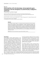
Figure 1 "Schematic representation of trans-epithelial loss of leu-
kocytes into airway lumen". Modified from references [61,16,112].
This scheme identifies some of the steps where future research is war-
ranted to delineate mechanisms involved in the trans-epithelial elimi-
nation of inflammatory cells from the airway wall. 1 After cell-to-cell
contact at the epithelial base paraepithelial crawling of the leukocyte
may begin by integrin binding to desmosomal junction adhesion mol-
ecules. 2 Several binding interactions and cellular signalling events in-
cluding cytosolic Ca++ fluxes may be involved as the leukocyte
continues to migrate between juxtapositioned epithelial cells. 3 Bind-
ing interactions involving junction adhesion-like proteins and recep-
tors such as the coxsackie and adenovirus receptor may be involved in
protein-tight passage of the leukocyte through the tight apical junc-
tion complex. 4 After its elimination from the airway wall the leukocyte
mixes with epithelial lining fluids and is finally eliminated by mucocili-
ary clearance.
1
4
Ca
2+
SIRP
2
3
Persson and Uller Respiratory Research 2010, 11:75
/>Page 8 of 12
lial exit of lymphocytes and other leukocytes. In accord
with this possibility, Caulfield et al [120] have suggested
that steroid-induced up-regulation of CXCR4 receptors
may move leukocytes away from inflamed airways in
asthma.
Clearance of cells from the airway lumen is essential
The potential role of trans-epithelial egression in resolu-
tion of inflammation underlines the importance of cell
clearance from the airway lumen. Clearing airway wall
leukocytes across the nasal [72] and bronchial epithelium
may be followed by swift and uneventful final elimination
from the lumen. Due to the lack of a mucociliary escala-
tor and lack of effect of coughing, clearance of leukocytes
across the alveolar epithelium may be more problematic.
Yet, in studies of lung inflammation in mice it appears
that egression of parenchymal leukocytes into the alveo-
lar air space is significantly beneficial; when this trans-
epithelial egression was prevented severe asphyxia
resulted [95,96]. We need to know to what extent apopto-
sis-related mechanisms can effect clearance of cellular
exudates from the bronchial as well as the alveolar lumen.
Interventions that can improve mucociliary clearance
[121-125] need increased attention. It is particularly
important that human peripheral airways can be freed
from leukocyte- and plasma protein-rich exudates that
otherwise will contribute to small airway closure
[14,126,127].
Use of sputum cell counts to adjust treatment
Analysis of induced sputum has advantages over determi-
nation of exhaled NO that recently was deemed to be of
little value as a guide to treatment interventions in
asthma [128]. Studies involving sequential sampling of
sputum in stable disease suggest that leukocyte counts in
induced sputum samples may exhibit acceptable repeat-
ability [129,130]. These data support the use of induced
sputum in monitoring disease severity and evaluating
anti-inflammatory treatments in stable asthma and
COPD. Of significant interest is the possibility that spu-
tum indices can predict disease exacerbations. This is an
area where sputum analysis has fared better than a clini-
cal strategy involving symptoms and spirometry [131-
135]. Since the first successful study of inhaled steroids
half a century ago sputum eosinophilia has shown its
value in predicting which patients will benefit from treat-
ment with these drugs. In a matter of days to weeks after
instituting steroid treatment both airway wall and lumen
eosinophils will be much reduced. Adjusting the steroid
dose to keep sputum eosinophil counts low successfully
reduces the exacerbation rate in asthma [131-133]. How-
ever, as stated by Jayaram et al [133] "the observation that
treatment to control sputum eosinophilia reduced
eosinophilic exacerbations may not be a surprise, since
treatment was designed to prevent these" (in this case by
keeping sputum eosinophils < 2%). It might also be
expected that the exacerbations that follow from tapering
steroid doses can be predicted by sputum eosino-
phils[134,135]. Interestingly, loss of control of asthma fol-
lowing rapid withdrawal of steroids was associated with
increased sputum neutrophils [136]. Reduced sputum
neutrophilia was also helpful as an index of therapeutic
effects of clarithromycin in refractory asthma [137]. It is
of note that sputum data, even better than bronchial
biopsy data, have identified individuals with regard to
risks for exacerbation [138]. This observation may reflect
the fact that sputum samples represent cumulative events
over a large surface area involving also more peripheral
airways than those available to biopsies. At growing
inflammation, the airway wall is increasingly infiltrated
with cells. A portion of these cells will migrate into the
lumen. In this situation the epithelial transmigration does
not reflect a resolving airway inflammation. However, a
'spill-over' of cells would be recorded in sputum samples
as a sign of an arriving exacerbation. Future studies spe-
cifically addressing the relationship between airway
lumen and airway wall eosinophils in developing exacer-
bations are warranted to further elucidate this possibility.
Leukocytes in the bronchial lumen in asthma and
COPD may differ between large and small bronchiolar-
alveolar airways. In accord there are differences as
regards the relative proportions of different leukocytes
occurring in sputum specimens and broncho-alveolar
lavage (BAL) fluids, respectively [139,140]. This is a con-
cern since much of the pathology of asthma and COPD
resides in the small airways. Tillie-Leblond and col-
leagues [141] further noted that only half of ten studies
on the subject could demonstrate a relationship between
eosinophils in induced sputum samples and symptoms of
asthma. Caution is also advised in interpretation of spu-
tum data since airway tissue and lumen may differ as to
which granulocyte, eosinophil or neutrophil [81,82], and
which T lymphocyte, especially Tc1 or Tc2 [116,142-144],
is predominant. Irrespective of such differences, it is
commonly assumed that numbers of leukocytes in spu-
tum samples reflect intensity of cell-mediated inflamma-
tory processes in diseased bronchial tissues. The present
hypothesis infers that the timing of obtaining samples in
relation to developing and resolving disease conditions is
crucial. Thus, during development of inflammation the
cell content of sputum samples may underestimate bron-
chial tissue cellularity. Reversely, during an active resolu-
tion phase when cells are being eliminated from the
airway wall the sputum samples could grossly overstate
the numbers of airway wall cells. Awareness of this con-
founding possibility may improve interpretation of spu-
tum data.
Persson and Uller Respiratory Research 2010, 11:75
/>Page 9 of 12
We have introduced a dual induction method [66]
whereby inhalation of histamine first induces a prompt
bronchial plasma exudation response. About an hour
later a second induction, this time of sputum, is
employed. The induced sputum then retrieves the exuded
plasma together with other mucosal interstitial proteins
that the travelling plasma may have picked up. This tech-
nique can improve the protein yield of induced sputum
and be employed to examine the pharmacology of plasma
exudation and the occurrence of exudative hyperrespon-
siveness. Although the laying down of exuded plasma
proteins (including fibronectin and fibrin) may pave the
way for cell traffic cells cannot be expected to migrate
into the airway lumen along with the bulk plasma. Per-
haps other inhalational challenges than histamine can be
developed that safely will bring cells into the airway
lumen to improve the cellular yield of a subsequent spu-
tum induction. As a bonus this work could lead to discov-
ery of interventions that will speed up resolution of
airway wall inflammation.
Conclusion
We have argued here that the occurrence of eosinophils,
neutrophils, lymphocytes, and mast cells in the bronchial
lumen can reflect their successful and non-injurious
elimination away from cell-mediated disease areas in the
airway wall. Evidence obtained in animal models together
with a large variety of clinical observations, previously
considered unexpected, support the importance of egres-
sion as a mode of eliminating pro-inflammatory leuko-
cytes from diseased airway tissues. These clinical reports
have been publicised during the last two decades. Simul-
taneously, the central role of leukocyte apoptosis in reso-
lution of airway diseases that we and others have been
seeking has not been confirmed. The possibility of reso-
lution through transepithelial exit of cells needs consider-
ation in studies of airway diseases and when assessing the
effects of drug interventions. Otherwise, data on airway
lumen leukocytes alone can lead to paradoxical conclu-
sions. Inhibiting pro-inflammatory, inciting processes in
the airways is important and so is rapid and complete
healing of epithelial injury [67]. However, it may not suf-
fice to reduce recruitment of inflammatory leukocytes to
the airway wall. We suggest that additional effects of pro-
moting transepithelial migration, together with a secured
clearance of cells from the airway lumen, are important
for accomplishing resolution of cell-mediated airways
diseases.
Authors' contributions
CP prepared the first draft. LU contributed several versions, made the figure,
and both approved the final manuscript.
Competing interests
The authors declare that they have no competing interests.
Acknowledgements
Our work on the present hypothesis is supported by the Swedish Medical
Research Council, the Swedish Lung and Heart Foundation, and Vinnova.
Author Details
1
Department of Clinical Pharmacology, Lund University Hospital, S-22185 Lund,
Sweden and
2
Department of Experimental Medical Science BMC D12, Lund
University, Lund, S-22184 Sweden
References
1. Serhan CN, Brain SD, Buckley CD, Gilroy DW, Haslett C, O'Neill LA, Perretti
M, Rossi AG, Wallace JL: Resolution of inflammation: state of the art,
definitions and terms. FASEB J 2007, 21(2):325-332.
2. Downey DG, Bell SC, Elborn JS: Neutrophils in cystic fibrosis. Thorax
2009, 64(1):81-88.
3. Porter JC: Epithelial Rho GTPases and the transepithelial migration of
lymphocytes. Methods Enzymol 2008, 439:205-217.
4. Persson CG: In vivo veritas: the continuing importance of discoveries in
complex biosystems. Thorax 1996, 51(4):441-443.
5. Persson C: Discoveries in complex biosystems. Nat Biotechnol 1997,
15(10):927.
6. Persson CG, Erjefalt JS, Uller L, Andersson M, Greiff L: Unbalanced
research. Trends Pharmacol Sci 2001, 22(10):538-541.
7. Persson CG, Erjefalt JS: Eosinophil lysis and free granules: an in vivo
paradigm for cell activation and drug development. Trends Pharmacol
Sci 1997, 18(4):117-123.
8. Uller L, Andersson M, Greiff L, Persson CG, Erjefalt JS: Occurrence of
apoptosis, secondary necrosis, and cytolysis in eosinophilic nasal
polyps. Am J Respir Crit Care Med 2004, 170(7):742-747.
9. Persson CG: Centennial notions of asthma as an eosinophilic,
desquamative, exudative, and steroid-sensitive disease. Lancet 1997,
350(9083):1021-1024.
10. Erjefalt JS, Persson CG: New aspects of degranulation and fates of airway
mucosal eosinophils. Am J Respir Crit Care Med 2000, 161(6):2074-2085.
11. Uller L, Persson CG, Kallstrom L, Erjefalt JS: Lung tissue eosinophils may
be cleared through luminal entry rather than apoptosis: effects of
steroid treatment. Am J Respir Crit Care Med 2001, 164(10 Pt
1):1948-1956.
12. Greiff L, Erjefalt JS, Andersson M, Svensson C, Persson CG: Generation of
clusters of free eosinophil granules (Cfegs) in seasonal allergic rhinitis.
Allergy 1998, 53(2):200-203.
13. Uller L, Persson CG, Erjefalt JS: Resolution of airway disease: removal of
inflammatory cells through apoptosis, egression or both? Trends
Pharmacol Sci 2006, 27(9):461-466.
14. Persson C, Uller L: Roles of plasma exudation in asthma and COPD. Clin
Exp Allergy 2009, 39(11):1626-1629.
15. Zemans RL, Colgan SP, Downey GP: Transepithelial migration of
neutrophils: mechanisms and implications for acute lung injury. Am J
Respir Cell Mol Biol 2009, 40(5):519-535.
16. Erjefalt JS, Uller L, Malm-Erjefalt M, Persson CG: Rapid and efficient
clearance of airway tissue granulocytes through transepithelial
migration. Thorax 2004, 59(2):136-143.
17. Porter JC, Falzon M, Hall A: Polarized localization of epithelial CXCL11 in
chronic obstructive pulmonary disease and mechanisms of T cell
egression. J Immunol 2008, 180(3):1866-1877.
18. Liu L, Mul FP, Lutter R, Roos D, Knol EF: Transmigration of human
neutrophils across airway epithelial cell monolayers is preferentially in
the physiologic basolateral-to-apical direction. Am J Respir Cell Mol Biol
1996, 15(6):771-780.
19. Kato Y, Fujisawa T, Shibano M, Saito T, Gatto W, Kamiya H, Hirai K, Sumida
M, Yoshie O: Airway epithelial cells promote transmigration of
eosinophils in a new three-dimensional chemotaxis model. Clin Exp
Allergy 2002, 32(6):889-897.
20. Lehmann C, Wilkening A, Leiber D, Markus A, Krug N, Pabst R, Tschernig T:
Lymphocytes in the bronchoalveolar space reenter the lung tissue by
means of the alveolar epithelium, migrate to regional lymph nodes,
and subsequently rejoin the systemic immune system. Anat Rec 2001,
264(3):229-236.
Received: 14 February 2010 Accepted: 11 June 2010
Published: 11 June 2010
This article is available from: 2010 Persson and Uller; licensee BioMed Central Ltd. This is an Open Access article distributed under the terms of the Creative Commons Attribution License ( which permits unrestricted use, distribution, and reproduction in any medium, provided the original work is properly cited.Respiratory Research 2010, 11:75
Persson and Uller Respiratory Research 2010, 11:75
/>Page 10 of 12
21. Erjefalt I, Luts A, Persson CG: Appearance of airway absorption and
exudation tracers in guinea pig tracheobronchial lymph nodes. J Appl
Physiol 1993, 74(2):817-824.
22. Korsgren M, Erjefalt JS, Korsgren O, Sundler F, Persson CG: Allergic
eosinophil-rich inflammation develops in lungs and airways of B cell-
deficient mice. J Exp Med 1997, 185(5):885-892.
23. Buckley CD, Ross EA, McGettrick HM, Osborne CE, Haworth O, Schmutz C,
Stone PC, Salmon M, Matharu NM, Vohra RK, Nash GB, Rainger GE:
Identification of a phenotypically and functionally distinct population
of long-lived neutrophils in a model of reverse endothelial migration. J
Leukoc Biol 2006, 79(2):303-311.
24. McGettrick HM, Hunter K, Moss PA, Buckley CD, Rainger GE, Nash GB:
Direct observations of the kinetics of migrating T cells suggest active
retention by endothelial cells with continual bidirectional migration. J
Leukoc Biol 2009, 85(1):98-107.
25. Haworth O, Buckley CD: Resolving the problem of persistence in the
switch from acute to chronic inflammation. Proc Natl Acad Sci USA 2007,
104(52):20647-20648.
26. Uller L, Lloyd CM, Rydell-Tormanen K, Persson CG, Erjefalt JS: Effects of
steroid treatment on lung CC chemokines, apoptosis and
transepithelial cell clearance during development and resolution of
allergic airway inflammation. Clin Exp Allergy 2006, 36(1):111-121.
27. Uller L, Ahlstrom Emanuelsson C, Andersson M, Erjefalt JS, Greiff L, Persson
CG: Early phase resolution of mucosal eosinophilic inflammation in
allergic rhinitis. Respir Res 2010, 11(1):54.
28. Spinozzi F, de Benedictis D, de Benedictis FM: Apoptosis, airway
inflammation and anti-asthma therapy: from immunobiology to
clinical application. Pediatr Allergy Immunol 2008, 19(4):287-295.
29. Wallen N, Kita H, Weiler D, Gleich GJ: Glucocorticoids inhibit cytokine-
mediated eosinophil survival. J Immunol 1991, 147(10):3490-3495.
30. Brunetti M, Martelli N, Colasante A, Piantelli M, Musiani P, Aiello FB:
Spontaneous and glucocorticoid-induced apoptosis in human mature
T lymphocytes. Blood 1995, 86(11):4199-4205.
31. Wissinger E, Goulding J, Hussell T: Immune homeostasis in the
respiratory tract and its impact on heterologous infection. Semin
Immunol 2009, 21(3):147-155.
32. Uller L, Bedke N, Sammut D, Green B, Howarth P, Holgate S, Davies D:
Double-stranded RNA induces disproportionate expression of thymic
stromal lymphopoietin versus interferon-beta in bronchial epithelial
cells from asthmatic donors. Thorax 2010.
33. Larche M: Regulatory T cells in allergy and asthma. Chest 2007,
132(3):1007-1014.
34. Murdoch JR, Lloyd CM: Chronic inflammation and asthma. Mutat Res
2009.
35. Dumas A, Pouliot M: [Neutrophil: foe or friend?]. Med Sci (Paris) 2009,
25(8-9):699-704.
36. Lawrence T, Fong C: The resolution of inflammation: Anti-inflammatory
roles for NF-kappaB. Int J Biochem Cell Biol 2009, 42(4):519-23.
37. Anderson P: Post-transcriptional regulons coordinate the initiation and
resolution of inflammation. Nat Rev Immunol 10(1):24-35.
38. Van Hove CL, Maes T, Joos GF, Tournoy KG: Chronic inflammation in
asthma: a contest of persistence vs resolution. Allergy 2008,
63(9):1095-1109.
39. Woolley KL, Gibson PG, Carty K, Wilson AJ, Twaddell SH, Woolley MJ:
Eosinophil apoptosis and the resolution of airway inflammation in
asthma. Am J Respir Crit Care Med 1996, 154(1):237-243.
40. Gibson PG, Saltos N, Fakes K: Acute anti-inflammatory effects of inhaled
budesonide in asthma: a randomized controlled trial. Am J Respir Crit
Care Med 2001, 163(1):32-36.
41. Walsh GM: Defective apoptotic cell clearance in asthma and COPD a
new drug target for statins? Trends Pharmacol Sci 2008, 29(1):6-11.
42. Rytila P, Plataki M, Bucchieri F, Uddin M, Nong G, Kinnula VL, Djukanovic R:
Airway neutrophilia in COPD is not associated with increased
neutrophil survival. Eur Respir J 2006, 28(6):1163-1169.
43. Watt AP, Courtney J, Moore J, Ennis M, Elborn JS: Neutrophil cell death,
activation and bacterial infection in cystic fibrosis. Thorax 2005,
60(8):659-664.
44. Watt AP, Brown V, Courtney J, Kelly M, Garske L, Elborn JS, Ennis M:
Neutrophil apoptosis, proinflammatory mediators and cell counts in
bronchiectasis. Thorax 2004, 59(3):231-236.
45. Matute-Bello G, Martin TR: Science review: apoptosis in acute lung
injury. Crit Care 2003, 7(5):355-358.
46. Kotecha S, Mildner RJ, Prince LR, Vyas JR, Currie AE, Lawson RA, Whyte MK:
The role of neutrophil apoptosis in the resolution of acute lung injury
in newborn infants. Thorax 2003, 58(11):961-967.
47. Druilhe A, Wallaert B, Tsicopoulos A, Lapa e Silva JR, Tillie-Leblond I, Tonnel
AB, Pretolani M: Apoptosis, proliferation, and expression of Bcl-2, Fas,
and Fas ligand in bronchial biopsies from asthmatics. Am J Respir Cell
Mol Biol 1998, 19(5):747-757.
48. O'Sullivan S, Cormican L, Burke CM, Poulter LW: Fluticasone induces T cell
apoptosis in the bronchial wall of mild to moderate asthmatics. Thorax
2004, 59(8):657-661.
49. Hodge S, Hodge G, Holmes M, Reynolds PN: Increased airway epithelial
and T-cell apoptosis in COPD remains despite smoking cessation. Eur
Respir J 2005, 25(3):447-454.
50. Cox G: Glucocorticoid treatment inhibits apoptosis in human
neutrophils. Separation of survival and activation outcomes. J
Immunol 1995, 154(9):4719-4725.
51. Gizycki MJ, Hattotuwa KL, Barnes N, Jeffery PK: Effects of fluticasone
propionate on inflammatory cells in COPD: an ultrastructural
examination of endobronchial biopsy tissue. Thorax 2002,
57(9):799-803.
52. Bourbeau J, Christodoulopoulos P, Maltais F, Yamauchi Y, Olivenstein R,
Hamid Q: Effect of salmeterol/fluticasone propionate on airway
inflammation in COPD: a randomised controlled trial. Thorax 2007,
62(11):938-943.
53. Reid DW, Wen Y, Johns DP, Williams TJ, Ward C, Walters EH:
Bronchodilator reversibility, airway eosinophilia and anti-inflammatory
effects of inhaled fluticasone in COPD are not related. Respirology 2008,
13(6):799-809.
54. Fukakusa M, Bergeron C, Tulic MK, Fiset PO, Al Dewachi O, Laviolette M,
Hamid Q, Chakir J: Oral corticosteroids decrease eosinophil and CC
chemokine expression but increase neutrophil, IL-8, and IFN-gamma-
inducible protein 10 expression in asthmatic airway mucosa. J Allergy
Clin Immunol 2005, 115(2):280-286.
55. Nguyen LT, Lim S, Oates T, Chung KF: Increase in airway neutrophils after
oral but not inhaled corticosteroid therapy in mild asthma. Respir Med
2005, 99(2):200-207.
56. Qiu Y, Zhu J, Bandi V, Guntupalli KK, Jeffery PK: Bronchial mucosal
inflammation and upregulation of CXC chemoattractants and
receptors in severe exacerbations of asthma. Thorax 2007,
62(6):475-482.
57. Ozol D, Aysan T, Solak ZA, Mogulkoc N, Veral A, Sebik F: The effect of
inhaled corticosteroids on bronchoalveolar lavage cells and IL-8 levels
in stable COPD patients. Respir Med 2005, 99(12):1494-1500.
58. Barnes NC, Qiu YS, Pavord ID, Parker D, Davis PA, Zhu J, Johnson M,
Thomson NC, Jeffery PK: Antiinflammatory effects of salmeterol/
fluticasone propionate in chronic obstructive lung disease. Am J Respir
Crit Care Med 2006, 173(7):736-743.
59. Vagaggini B, Cianchetti S, Bartoli M, Ricci M, Bacci E, Dente FL, Di Franco A,
Paggiaro P: Prednisone blunts airway neutrophilic inflammatory
response due to ozone exposure in asthmatic subjects. Respiration
2007, 74(1):61-68.
60. Alexis NE, Lay JC, Haczku A, Gong H, Linn W, Hazucha MJ, Harris B, Tal-
Singer R, Peden DB: Fluticasone propionate protects against ozone-
induced airway inflammation and modified immune cell activation
markers in healthy volunteers. Environ Health Perspect 2008,
116(6):799-805.
61. Chin AC, Parkos CA: Pathobiology of neutrophil transepithelial
migration: implications in mediating epithelial injury. Annu Rev Pathol
2007, 2:111-143.
62. Lukacs NW: Role of chemokines in the pathogenesis of asthma. Nat Rev
Immunol 2001, 1(2):108-116.
63. Teran LM: CCL chemokines and asthma. Immunol Today 2000,
21(5):235-242.
64. Moraes TJ, Rafii B, Niessen F, Suzuki T, Martin R, Vachon E, Vogel W, Ruf W,
O'Brodovich H, Downey GP: Protease-activated receptor (Par)1 alters
bioelectric properties of distal lung epithelia without compromising
barrier function. Exp Lung Res 2009, 35(2):136-154.
65. Martin TR: Neutrophils and lung injury: getting it right. J Clin Invest
2002, 110(11):1603-1605.
66. Persson CG, Erjefalt JS, Greiff L, Andersson M, Erjefalt I, Godfrey RW,
Korsgren M, Linden M, Sundler F, Svensson C: Plasma-derived proteins in
Persson and Uller Respiratory Research 2010, 11:75
/>Page 11 of 12
airway defence, disease and repair of epithelial injury. Eur Respir J 1998,
11(4):958-970.
67. Persson C, Andersson M, Uller L: Epithelial repair and function. In
Pulmonary Epithelium Edited by: Proud D. Chicester: Wiley; 2008:75-88.
68. Aalbers R, de Monchy JG, Kauffman HF, Smith M, Hoekstra Y, Vrugt B,
Timens W: Dynamics of eosinophil infiltration in the bronchial mucosa
before and after the late asthmatic reaction. Eur Respir J 1993,
6(6):840-847.
69. Frew AJ, St-Pierre J, Teran LM, Trefilieff A, Madden J, Peroni D, Bodey KM,
Walls AF, Howarth PH, Carroll MP, Holgate ST: Cellular and mediator
responses twenty-four hours after local endobronchial allergen
challenge of asthmatic airways. J Allergy Clin Immunol 1996,
98(1):133-143.
70. Brown JR, Kleimberg J, Marini M, Sun G, Bellini A, Mattoli S: Kinetics of
eotaxin expression and its relationship to eosinophil accumulation and
activation in bronchial biopsies and bronchoalveolar lavage (BAL) of
asthmatic patients after allergen inhalation. Clin Exp Immunol 1998,
114(2):137-146.
71. Juliusson S, Pipkorn U, Karlsson G, Enerback L: Mast cells and eosinophils
in the allergic mucosal response to allergen challenge: changes in
distribution and signs of activation in relation to symptoms. J Allergy
Clin Immunol 1992, 90(6 Pt 1):898-909.
72. Persson CG, Svensson C, Greiff L, Anderson M, Wollmer P, Alkner U, Erjefalt
I: The use of the nose to study the inflammatory response of the
respiratory tract. Thorax 1992, 47(12):993-1000.
73. Crimi E, Chiaramondia M, Milanese M, Rossi GA, Brusasco V: Increased
numbers of mast cells in bronchial mucosa after the late-phase
asthmatic response to allergen. Am Rev Respir Dis 1991,
144(6):1282-1286.
74. Gauvreau GM, Lee JM, Watson RM, Irani AM, Schwartz LB, O'Byrne PM:
Increased numbers of both airway basophils and mast cells in sputum
after allergen inhalation challenge of atopic asthmatics. Am J Respir Crit
Care Med 2000, 161(5):1473-1478.
75. Lommatzsch M, Julius P, Kuepper M, Garn H, Bratke K, Irmscher S,
Luttmann W, Renz H, Braun A, Virchow JC: The course of allergen-
induced leukocyte infiltration in human and experimental asthma. J
Allergy Clin Immunol 2006, 118(1):91-97.
76. Ichiyasu H, McCormack JM, McCarthy KM, Dombkowski D, Preffer FI,
Schneeberger EE: Matrix metalloproteinase-9-deficient dendritic cells
have impaired migration through tracheal epithelial tight junctions.
Am J Respir Cell Mol Biol 2004, 30(6):761-770.
77. Lambrecht BN, Carro-Muino I, Vermaelen K, Pauwels RA: Allergen-
induced changes in bone-marrow progenitor and airway dendritic
cells in sensitized rats. Am J Respir Cell Mol Biol 1999, 20(6):1165-1174.
78. Vermaelen K, Pauwels R: Accelerated airway dendritic cell maturation,
trafficking, and elimination in a mouse model of asthma. Am J Respir
Cell Mol Biol 2003, 29(3 Pt 1):405-409.
79. Bratke K, Lommatzsch M, Julius P, Kuepper M, Kleine HD, Luttmann W,
Christian Virchow J: Dendritic cell subsets in human bronchoalveolar
lavage fluid after segmental allergen challenge. Thorax 2007,
62(2):168-175.
80. Bratke K, Klug M, Bier A, Julius P, Kuepper M, Virchow JC, Lommatzsch M:
Function-associated surface molecules on airway dendritic cells in
cigarette smokers. Am J Respir Cell Mol Biol 2008, 38(6):655-660.
81. Wenzel SE: Asthma: defining of the persistent adult phenotypes. Lancet
2006, 368(9537):804-813.
82. O'Donnell R, Breen D, Wilson S, Djukanovic R: Inflammatory cells in the
airways in COPD. Thorax 2006, 61(5):448-454.
83. Ordonez CL, Shaughnessy TE, Matthay MA, Fahy JV: Increased neutrophil
numbers and IL-8 levels in airway secretions in acute severe asthma:
Clinical and biologic significance. Am J Respir Crit Care Med 2000, 161(4
Pt 1):1185-1190.
84. Louhelainen N, Rytila P, Haahtela T, Kinnula VL, Djukanovic R: Persistence
of oxidant and protease burden in the airways after smoking cessation.
BMC Pulm Med 2009, 9:25.
85. Willemse BW, ten Hacken NH, Rutgers B, Lesman-Leegte IG, Postma DS,
Timens W: Effect of 1-year smoking cessation on airway inflammation
in COPD and asymptomatic smokers. Eur Respir J 2005, 26(5):835-845.
86. Qiu Y, Zhu J, Bandi V, Atmar RL, Hattotuwa K, Guntupalli KK, Jeffery PK:
Biopsy neutrophilia, neutrophil chemokine and receptor gene
expression in severe exacerbations of chronic obstructive pulmonary
disease. Am J Respir Crit Care Med 2003, 168(8):968-975.
87. Yoshihara S, Yamada Y, Abe T, Linden A, Arisaka O: Association of
epithelial damage and signs of neutrophil mobilization in the airways
during acute exacerbations of paediatric asthma. Clin Exp Immunol
2006, 144(2):212-216.
88. Mosser AG, Brockman-Schneider R, Amineva S, Burchell L, Sedgwick JB,
Busse WW, Gern JE: Similar frequency of rhinovirus-infectible cells in
upper and lower airway epithelium. J Infect Dis 2002, 185(6):734-743.
89. Papi A, Bellettato CM, Braccioni F, Romagnoli M, Casolari P, Caramori G,
Fabbri LM, Johnston SL: Infections and airway inflammation in chronic
obstructive pulmonary disease severe exacerbations. Am J Respir Crit
Care Med 2006, 173(10):1114-1121.
90. Snoeck-Stroband JB, Lapperre TS, Gosman MM, Boezen HM, Timens W,
ten Hacken NH, Sont JK, Sterk PJ, Hiemstra PS: Chronic bronchitis sub-
phenotype within COPD: inflammation in sputum and biopsies. Eur
Respir J 2008, 31(1):70-77.
91. Andersson CK, Mori M, Bjermer L, Lofdahl CG, Erjefalt JS: Alterations in
lung mast cell populations in patients with chronic obstructive
pulmonary disease. Am J Respir Crit Care Med 2010, 181(3):206-217.
92. Tsuyuki S, Bertrand C, Erard F, Trifilieff A, Tsuyuki J, Wesp M, Anderson GP,
Coyle AJ: Activation of the Fas receptor on lung eosinophils leads to
apoptosis and the resolution of eosinophilic inflammation of the
airways. J Clin Invest 1995, 96(6):2924-2931.
93. Uller L, Rydell-Tormanen K, Persson CG, Erjefalt JS: Anti-Fas mAb-induced
apoptosis and cytolysis of airway tissue eosinophils aggravates rather
than resolves established inflammation. Respir Res 2005, 6:90.
94. Gerwin N, Gonzalo JA, Lloyd C, Coyle AJ, Reiss Y, Banu N, Wang B, Xu H,
Avraham H, Engelhardt B, et al.: Prolonged eosinophil accumulation in
allergic lung interstitium of ICAM-2 deficient mice results in extended
hyperresponsiveness. Immunity 1999, 10(1):9-19.
95. Corry DB, Kiss A, Song LZ, Song L, Xu J, Lee SH, Werb Z, Kheradmand F:
Overlapping and independent contributions of MMP2 and MMP9 to
lung allergic inflammatory cell egression through decreased CC
chemokines. FASEB J 2004, 18(9):995-997.
96. Corry DB, Rishi K, Kanellis J, Kiss A, Song Lz LZ, Xu J, Feng L, Werb Z,
Kheradmand F: Decreased allergic lung inflammatory cell egression
and increased susceptibility to asphyxiation in MMP2-deficiency. Nat
Immunol 2002, 3(4):347-353.
97. Sorkness RL, Herricks KM, Szakaly RJ, Lemanske RF Jr, Rosenthal LA:
Altered allergen-induced eosinophil trafficking and physiological
dysfunction in airways with preexisting virus-induced injury. Am J
Physiol Lung Cell Mol Physiol 2007, 292(1):L85-91.
98. Rohde G, Wiethege A, Borg I, Kauth M, Bauer TT, Gillissen A, Bufe A,
Schultze-Werninghaus G: Respiratory viruses in exacerbations of
chronic obstructive pulmonary disease requiring hospitalisation: a
case-control study. Thorax 2003, 58(1):37-42.
99. Johnston SL, Pattemore PK, Sanderson G, Smith S, Lampe F, Josephs L,
Symington P, O'Toole S, Myint SH, Tyrrell DA, Holgate ST: Community
study of role of viral infections in exacerbations of asthma in 9-11 year
old children. BMJ 1995, 310(6989):1225-1229.
100. Seemungal TA, Wedzicha JA: Viral infections in obstructive airway
diseases. Curr Opin Pulm Med 2003, 9(2):111-116.
101. Norman P: AZD-4818, a chemokine CCR1 antagonist: WO2008103126
and WO2009011653. Expert Opin Ther Pat 2009, 19(11):1629-1633.
102. Leckie MJ, ten Brinke A, Khan J, Diamant Z, O'Connor BJ, Walls CM, Mathur
AK, Cowley HC, Chung KF, Djukanovic R, Hansel TT, Holgate ST, Sterk PJ,
Barnes PJ: Effects of an interleukin-5 blocking monoclonal antibody on
eosinophils, airway hyper-responsiveness, and the late asthmatic
response. Lancet 2000, 356(9248):2144-2148.
103. Flood-Page P, Menzies-Gow A, Phipps S, Ying S, Wangoo A, Ludwig MS,
Barnes N, Robinson D, Kay AB: Anti-IL-5 treatment reduces deposition of
ECM proteins in the bronchial subepithelial basement membrane of
mild atopic asthmatics. J Clin Invest 2003, 112(7):1029-1036.
104. Haldar P, Brightling CE, Hargadon B, Gupta S, Monteiro W, Sousa A,
Marshall RP, Bradding P, Green RH, Wardlaw AJ, Pavord ID: Mepolizumab
and exacerbations of refractory eosinophilic asthma. N Engl J Med 2009,
360(10):973-984.
105. Nair P, Pizzichini MM, Kjarsgaard M, Inman MD, Efthimiadis A, Pizzichini E,
Hargreave FE, O'Byrne PM: Mepolizumab for prednisone-dependent
asthma with sputum eosinophilia. N Engl J Med 2009, 360(10):985-993.
106. Doerschuk CM: Leukocyte trafficking in alveoli and airway passages.
Respir Res 2000, 1(3):136-140.
Persson and Uller Respiratory Research 2010, 11:75
/>Page 12 of 12
107. Mitzner W, Lee W, Georgakopoulos D, Wagner E: Angiogenesis in the
mouse lung. Am J Pathol 2000, 157(1):93-101.
108. Persson CG, Erjefalt JS, Korsgren M, Sundler F: The mouse trap. Trends
Pharmacol Sci 1997, 18(12):465-467.
109. Muessel MJ, Scott KS, Friedl P, Bradding P, Wardlaw AJ: CCL11 and GM-
CSF differentially use the Rho GTPase pathway to regulate motility of
human eosinophils in a three-dimensional microenvironment. J
Immunol 2008, 180(12):8354-8360.
110. Rowe RG, Weiss SJ: Breaching the basement membrane: who, when
and how? Trends Cell Biol 2008, 18(11):560-574.
111. Burns AR, Smith CW, Walker DC: Unique structural features that
influence neutrophil emigration into the lung. Physiol Rev 2003,
83(2):309-336.
112. Chun J, Prince A: Ca2+ signaling in airway epithelial cells facilitates
leukocyte recruitment and transepithelial migration. J Leukoc Biol 2009,
86(5):1135-1144.
113. Chun J, Prince A: TLR2-induced calpain cleavage of epithelial junctional
proteins facilitates leukocyte transmigration. Cell Host Microbe 2009,
5(1):47-58.
114. Zen K, Parkos CA: Leukocyte-epithelial interactions. Curr Opin Cell Biol
2003, 15(5):557-564.
115. Erjefalt JS, Korsgren M, Malm-Erjefalt M, Conroy DM, Williams TJ, Persson
CG: Acute allergic responses induce a prompt luminal entry of airway
tissue eosinophils. Am J Respir Cell Mol Biol 2003, 29(4):439-448.
116. Saetta M, Mariani M, Panina-Bordignon P, Turato G, Buonsanti C, Baraldo S,
Bellettato CM, Papi A, Corbetta L, Zuin R, et al.: Increased expression of
the chemokine receptor CXCR3 and its ligand CXCL10 in peripheral
airways of smokers with chronic obstructive pulmonary disease. Am J
Respir Crit Care Med 2002, 165(10):1404-1409.
117. Taguchi M, Sampath D, Koga T, Castro M, Look DC, Nakajima S, Holtzman
MJ: Patterns for RANTES secretion and intercellular adhesion molecule
1 expression mediate transepithelial T cell traffic based on analyses in
vitro and in vivo. J Exp Med 1998, 187(12):1927-1940.
118. Cyster JG: Chemorepulsion and thymocyte emigration. J Clin Invest
2002, 109(8):1011-1012.
119. Vianello F, Kraft P, Mok YT, Hart WK, White N, Poznansky MC: A CXCR4-
dependent chemorepellent signal contributes to the emigration of
mature single-positive CD4 cells from the fetal thymus. J Immunol
2005, 175(8):5115-5125.
120. Caulfield J, Fernandez M, Snetkov V, Lee T, Hawrylowicz C: CXCR4
expression on monocytes is up-regulated by dexamethasone and is
modulated by autologous CD3+ T cells. Immunology 2002,
105(2):155-162.
121. Button B, Boucher RC: Role of mechanical stress in regulating airway
surface hydration and mucus clearance rates. Respir Physiol Neurobiol
2008, 163(1-3):189-201.
122. Daviskas E, Anderson SD, Eberl S, Chan HK, Young IH: The 24-h effect of
mannitol on the clearance of mucus in patients with bronchiectasis.
Chest 2001, 119(2):414-421.
123. Bhowmik A, Chahal K, Austin G, Chakravorty I: Improving mucociliary
clearance in chronic obstructive pulmonary disease. Respir Med 2009,
103(4):496-502.
124. Shah RV, Amin M, Sangwan S, Smaldone GC: Steroid effects on
mucociliary clearance in outpatient asthma. J Aerosol Med 2006,
19(2):208-220.
125. Bennett WD, Almond MA, Zeman KL, Johnson JG, Donohue JF: Effect of
salmeterol on mucociliary and cough clearance in chronic bronchitis.
Pulm Pharmacol Ther 2006, 19(2):96-100.
126. Sturton G, Persson C, Barnes PJ: Small airways: an important but
neglected target in the treatment of obstructive airway diseases.
Trends Pharmacol Sci 2008, 29(7):340-345.
127. Hogg JC: Pathophysiology of airflow limitation in chronic obstructive
pulmonary disease. Lancet 2004, 364(9435):709-721.
128. Szefler SJ, Mitchell H, Sorkness CA, Gergen PJ, O'Connor GT, Morgan WJ,
Kattan M, Pongracic JA, Teach SJ, Bloomberg GR, Bloomberg GR,
Eggleston PA, Gruchalla RS, Kercsmar CM, Liu AH, Wildfire JJ, Curry MD,
Busse WW: Management of asthma based on exhaled nitric oxide in
addition to guideline-based treatment for inner-city adolescents and
young adults: a randomised controlled trial. Lancet 2008,
372(9643):1065-1072.
129. Beeh KM, Beier J, Kornmann O, Mander A, Buhl R: Long-term repeatability
of induced sputum cells and inflammatory markers in stable,
moderately severe COPD. Chest 2003, 123(3):778-783.
130. Sapey E, Bayley D, Ahmad A, Newbold P, Snell N, Stockley RA: Inter-
relationships between inflammatory markers in patients with stable
COPD with bronchitis: intra-patient and inter-patient variability.
Thorax 2008, 63(6):493-499.
131. Green RH, Brightling CE, McKenna S, Hargadon B, Parker D, Bradding P,
Wardlaw AJ, Pavord ID: Asthma exacerbations and sputum eosinophil
counts: a randomised controlled trial. Lancet 2002,
360(9347):1715-1721.
132. Chlumsky J, Striz I, Terl M, Vondracek J: Strategy aimed at reduction of
sputum eosinophils decreases exacerbation rate in patients with
asthma. J Int Med Res 2006, 34(2):129-139.
133. Jayaram L, Pizzichini MM, Cook RJ, Boulet LP, Lemiere C, Pizzichini E,
Cartier A, Hussack P, Goldsmith CH, Laviolette M, Parameswaran K,
Hargreave FE: Determining asthma treatment by monitoring sputum
cell counts: effect on exacerbations. Eur Respir J 2006, 27(3):483-494.
134. Jatakanon A, Lim S, Barnes PJ: Changes in sputum eosinophils predict
loss of asthma control. Am J Respir Crit Care Med 2000, 161(1):64-72.
135. Leuppi JD, Salome CM, Jenkins CR, Anderson SD, Xuan W, Marks GB,
Koskela H, Brannan JD, Freed R, Andersson M, Chan HK, Woolcock AJ:
Predictive markers of asthma exacerbation during stepwise dose
reduction of inhaled corticosteroids. Am J Respir Crit Care Med 2001,
163(2):406-412.
136. Maneechotesuwan K, Essilfie-Quaye S, Kharitonov SA, Adcock IM, Barnes
PJ: Loss of control of asthma following inhaled corticosteroid
withdrawal is associated with increased sputum interleukin-8 and
neutrophils. Chest 2007, 132(1):98-105.
137. Simpson JL, Powell H, Boyle MJ, Scott RJ, Gibson PG: Clarithromycin
targets neutrophilic airway inflammation in refractory asthma. Am J
Respir Crit Care Med 2008, 177(2):148-155.
138. Lemiere C, Ernst P, Olivenstein R, Yamauchi Y, Govindaraju K, Ludwig MS,
Martin JG, Hamid Q: Airway inflammation assessed by invasive and
noninvasive means in severe asthma: eosinophilic and
noneosinophilic phenotypes. J Allergy Clin Immunol 2006,
118(5):1033-1039.
139. Maestrelli P, Saetta M, Di Stefano A, Calcagni PG, Turato G, Ruggieri MP,
Roggeri A, Mapp CE, Fabbri LM: Comparison of leukocyte counts in
sputum, bronchial biopsies, and bronchoalveolar lavage. Am J Respir
Crit Care Med 1995, 152(6 Pt 1):1926-1931.
140. Rutgers SR, Timens W, Kaufmann HF, van der Mark TW, Koeter GH, Postma
DS: Comparison of induced sputum with bronchial wash,
bronchoalveolar lavage and bronchial biopsies in COPD. Eur Respir J
2000, 15(1):109-115.
141. Tillie-Leblond I, Montani D, Crestani B, de Blic J, Humbert M, Tunon-de-
Lara M, Magnan A, Roche N, Ostinelli J, Chanez P: Relation between
inflammation and symptoms in asthma. Allergy 2009, 64(3):354-367.
142. Barcelo B, Pons J, Fuster A, Sauleda J, Noguera A, Ferrer JM, Agusti AG:
Intracellular cytokine profile of T lymphocytes in patients with chronic
obstructive pulmonary disease. Clin Exp Immunol 2006, 145(3):474-479.
143. Barczyk A, Pierzchala W, Kon OM, Cosio B, Adcock IM, Barnes PJ: Cytokine
production by bronchoalveolar lavage T lymphocytes in chronic
obstructive pulmonary disease. J Allergy Clin Immunol 2006,
117(6):1484-1492.
144. Freeman CM, Curtis JL, Chensue SW: CC chemokine receptor 5 and CXC
chemokine receptor 6 expression by lung CD8+ cells correlates with
chronic obstructive pulmonary disease severity. Am J Pathol 2007,
171(3):767-776.
doi: 10.1186/1465-9921-11-75
Cite this article as: Persson and Uller, Resolution of cell-mediated airways
diseases Respiratory Research 2010, 11:75