Báo cáo y học: "Pneumocystis cell wall β-glucan stimulates calcium-dependent signaling of IL-8 secretion by human airway epithelial cells" ppt
Bạn đang xem bản rút gọn của tài liệu. Xem và tải ngay bản đầy đủ của tài liệu tại đây (1.82 MB, 12 trang )
Carmona et al. Respiratory Research 2010, 11:95
/>Open Access
RESEARCH
© 2010 Carmona et al; licensee BioMed Central Ltd. This is an Open Access article distributed under the terms of the Creative Commons
Attribution License ( which permits unrestricted use, distribution, and reproduction in
any medium, provided the original work is properly cited.
Research
Pneumocystis
cell wall β-glucan stimulates
calcium-dependent signaling of IL-8 secretion by
human airway epithelial cells
Eva M Carmona, Jeffrey D Lamont, Ailing Xue, Mark Wylam and Andrew H Limper*
Abstract
Background: Respiratory failure secondary to alveolar inflammation during Pneumocystis pneumonia is a major cause
of death in immunocompromised patients. Neutrophil infiltration in the lung of patients with Pneumocystis infection
predicts severity of the infection and death. Several previous studies indicate that airway epithelial cells release the
neutrophil chemoattractant proteins, MIP-2 (rodents) and IL-8 (humans), in response to Pneumocystis and purified
Pneumocystis cell wall β-glucans (PCBG) through the NF-κB-dependent pathway. However, little is known about the
molecular mechanisms that are involved in the activation of airway epithelium cells by PCBG resulting in the secretion
of IL-8.
Method: To address this, we have studied the activation of different calcium-dependent mitogen-activated protein
kinases (MAPKs) in 1HAEo
-
cells, a human airway epithelial cell line.
Results: Our data provide evidence that PCBG induces phosphorylation of the MAPKs, ERK, and p38, the activation of
NF-κB and the subsequently secretion of IL-8 in a calcium-dependent manner. Further, we evaluated the role of
glycosphingolipids as possible receptors for β-glucans in human airway epithelial cells. Preincubation of the cells with
D-threo-1-phenyl-2-decanoylamino-3-morpholino-1-propanol (PDMP) a potent inhibitor of the glycosphingolipids
synthesis, prior to PCBG stimulation, significantly decreased IL-8 production.
Conclusion: These data indicate that PCBG activates calcium dependent MAPK signaling resulting in the release of IL-8
in a process that requires glycosphingolipid for optimal signaling.
Introduction
Pneumocystis pneumonia is an opportunistic infection,
caused by Pneumocystis jirovecii that predominantly
affects immunosuppressed patients, including those with
AIDS and malignancy. With the introduction of the
highly active retroviral therapy (HAART) the incidence of
Pneumocystis pneumonia among the HIV-infected
patients has decreased significantly, but still remains
among the most common severe opportunistic infection
in this group of patients [1]. In addition, in non-HIV
immunocompromised patients Pneumocystis infection is
associated with substantially greater morbidity and mor-
tality when compared with HIV-positive population
despite the available medication [2].
It has been postulated that one reason for the differen-
tial mortality rates between the two groups is based on
the differing abilities to mount inflammatory responses
in the face of infection; with non-HIV-infected patients
having a more robust inflammatory response against the
organism is elicited compared to HIV-infected individu-
als. Indeed, this exuberant inflammatory reaction
towards the organism has been shown to be more harm-
ful to the host than the organism burden itself [3-5]. Poly-
morphonuclear neutrophils (PMN) are one of the major
components of the lung inflammatory reaction seen in
patients affected with Pneumocystis pneumonia, though
CD8 cells and other cells are known to participate as well
[6-8]. Moreover, it has been documented that the degree
of neutrophil infiltration in the lung of these patients can
* Correspondence:
1
From the Thoracic Diseases Research Unit, Division of Pulmonary Critical Care
and Internal Medicine, Department of Medicine Mayo Clinic and Foundation,
Rochester, Minnesota, 55905, USA
Full list of author information is available at the end of the article
Carmona et al. Respiratory Research 2010, 11:95
/>Page 2 of 12
serve as a marker of the severity of respiratory failure and
death [3-5,9]. From theses observations, we have further
postulated that a balanced inflammatory response is nec-
essary to successfully control Pneumocystis infection.
Pneumocystis organisms are present within the alveolus
in at least two different developmental stages, namely the
trophic form and the cyst. The trophic form attaches
firmly to the alveolar epithelium, in a process that stimu-
lates organism proliferation [10]. The cyst form is charac-
terized by a thick β-glucan rich cell wall, which recent
studies have implicated as a major initiator of lung
inflammation during Pneumocystis infection [11,12].
However, the molecular mechanisms by which β-glucans
induce this exaggerated airway inflammatory response
have not yet been fully elucidated.
Airway epithelial cells actively participate in the
immune response during infection, not only by recogniz-
ing the microorganisms, but also by initiating appropriate
signal transduction pathways that will lead to the produc-
tion of a variety of cytokines and chemokines involved in
the recruitment of inflammatory cells to the site of infec-
tion. In the case of Pneumocystis, various studies have
demonstrated that Pneumocystis organisms closely asso-
ciate with airway epithelial cells; supporting the tenant
that binding of the organism to airway epithelial cells is
an integral component in the establishment of infection
[13,14]. While Pneumocystis trophic forms bind preferen-
tially to Type I alveolar cells, Pneumocystis cysts and
degraded components can be found in expectorated spu-
tum [15]. Thus, Pneumocystis components such as glucan
have ample opportunity to interact with epithelial cells in
the lower respiratory tract.
Our group has demonstrated that fungal β-glucans in
the wall of Pneumocystis induce NF-κB translocation and
TNF-α production in macrophages following contact
with the phagocyte [16]. In addition, we have also dem-
onstrated that Pneumocystis β-glucans (PCBG) stimulate
rat airway epithelial cells to secrete macrophage inflam-
matory protein-2 (MIP-2) through NF-κB dependent
mechanisms [17,18]. However, the events through which
PCBG initiate airway epithelial cells activation remain
unclear. Various bacterial pathogens such as Salmonella
and Pseudomonas species activate epithelial cells by
increasing intracellular calcium concentrations [19,20].
For instance, during pseudomonal infection, superficial
interactions of the microbe with airway epithelial cells are
sufficient to induce changes in calcium influx and subse-
quently stimulate NF-κB-dependent gene expression [19].
We, therefore, hypothesized that following binding of
PCBG to airway epithelial cells, the epithelial cells are
stimulated to express pro-inflammatory responses by
inducing changes in cytosolic calcium influx. These
changes in intracellular calcium subsequently activate
major signal transduction pathways that eventually lead
to cytokine secretion by airway epithelial cells.
Fungal adhesion to host tissues is an integral step for
colonization and subsequent infection [10,21,22]. Histo-
logical studies of Pneumocystis infected patients and ani-
mals demonstrate intimate association of Pneumocystis
organisms with alveolar epithelial cells [13]. Many recep-
tors have been proposed to bind Pneumocystis particles
including dectin-1, β2 integrin CD11b/CD18, and lacto-
sylceramide [16,17,23,24]. Airway epithelial cells specifi-
cally lack dectin-1 receptors, which are present in
macrophages. Based on our recent observations demon-
strating that lactosylceramide is responsible for MIP-2
production, we further evaluated the role of glycosphin-
golipids in cytokine signaling by airway epithelial cells
activated with PCBG [17,18].
Herein, we demonstrate that 1HAEo
-
human airway
epithelial cells simulated with PCBG induce the release of
the neutrophil chemokine IL-8, in a calcium-dependent
manner. We further demonstrate the participation of two
major MAPKs, ERK and p38, and that at least two major
transcription factors, NF-κB and AP-1, are necessary for
an adequate transcription of IL-8. Finally, we observed
that glycosphingolipids are necessary for the synthesis of
IL-8 by PCBG activated 1HAEo
-
cells.
Materials and methods
Reagents and antibodies
Endotoxin-free buffers and reagents were scrupulously
employed for all experiments. Saccharomyces cerevisiae
derived cell wall β-glucans, the calcineurin disrupting
agents TEMPO (2,2,6,6-Tetramethyl-1-piperidinyloxy,
free radical, 2,2,6,6-Tetramethylpiperidine 1-oxyl) and
cyclosporin B were purchased from Sigma Chemical Co,
(St. Louis, MO). The calcium chelator BAPTA/AM (1,2-
bis-(o-Aminophenoxy)-ethane-N,N,N',N'-tetraacetic
acid, tetraacetoxymethyl ester) was obtained from Alexis
Biochemical. The glucosylceramide synthase inhibitor
PDMP (D-threo-1-Phenyl-2-decanoylamino-3-mor-
pholino-1-propanol•HCl) was purchased from Matreya,
LLC (Pleasant Gap, PA), LPS from Escherichia coli
026:B6, EGTA, PD 98059, SB 202190, SB 202474, JNK
inhibitor II and other general reagents were from Calbio-
chem (Gibbstown, NJ), unless otherwise specified. Pneu-
mocystis carinii was derived originally from the
American Type Culture Collection stock (Manassas, VA)
and has been passaged though our immunosuppressed
rat colony [25]. All antibodies employed in these studies
were purchased from Cell Signaling Technologies (Dan-
vers, MA). The human airway epithelial cell line, 1HAEo
-
cells, were generously provided by Dr. Dieter Gruenert
(University of California, San Francisco) [26]. The cells
were routinely cultured in Modified Eagle's medium con-
Carmona et al. Respiratory Research 2010, 11:95
/>Page 3 of 12
taining 10% fetal bovine serum and 2 mM L-glutamine,
penicillin 10,000 units/liter, and streptomycin 1 mg/liter.
Plasmids
The NF-κB-dependent firefly luciferase reporter expres-
sion vector (κB-luc) was a kind gift of Dr. Carlos Paya
(Mayo Clinic, Rochester, MN)[27]. The IL-8, IL-8
mutated in AP-1, and NF-κB sites promoter-luciferase
reporter plasmids were gifts from Dr. Marc Hershenson
(University of Michigan)[28]. The pRL-TK expression
vector, which provides constitutive expression of Renilla
luciferase, was purchased from Promega (Madison, Wis-
consin).
Generation of Pneumocystis carinii β-Glucan-rich Cell Wall
Isolate
The Mayo Institutional Animal Care and Usage Commit-
tee approved all animal experimentation. A β-glucan-rich
cell wall fraction from P. car i nii was prepared as we previ-
ously described [11,18]. Pneumocystis pneumonia was
induced in dexamethasone-treated immunosuppressed
Lewis rats (Harlan, Inc., Indianapolis, IN) [25]. Pneumo-
cystis organisms were isolated from lungs of heavily
infected animals by homogenization and filtration
through 10-μm filters. The organisms were autoclaved
(120°C, 20 min) and disrupted by ultrasonication (200 W
for 3 min, six times), and the glucans were isolated by
NaOH digestion and lipid extraction as previously
detailed [11,18]. As we prior reported, the final product
contained predominantly carbohydrate (95.7%) and
released 82% of its content as D-glucose following hydro-
lysis [11]. Extensive measures were employed to ensure
that the fractions were free of endotoxin. Prior to use in
culture, the Pneumocystis cell wall fractions were washed
with 0.1% SDS and then vigorously washed with distilled
physiological saline to remove the detergent. The final
preparation was assayed for endotoxin with the Limulus
amebocyte lysate assay method and found to consistently
contain < 0.125 units of endotoxin [11].
IL-8 detection
IL-8 was measured in the supernatants of cultivated
1HAEo
-
cells by ELISA (BD OptEIA™, BD biosciences,
San Diego, CA). Cells were cultured to ~70% confluence
in a 96-well plates. Prior to activation with PCBG, the
cells were weaned from serum for 18 hours. For some
experiments, the cells were preincubated with various
calcium disrupting agents or MAPKs inhibitors for one
hour prior to stimulation. Supernatant was collected after
8 hour of stimulation with PCBG unless otherwise indi-
cated and stored at -70°C. All experiments were per-
formed in duplicate and repeated on a minimum of at
least three occasions.
Cellular Viability
Cell viability was confirmed using the XTT Cell Prolifera-
tion Kit II (Roche Molecular Biochemicals, Mannheim,
Germany). This assay measures the conversion of
sodium-3'-[1-(phenylaminocarbonyl)-3,4-tetrazolium]-
bis(4-methoxy-6-nitro) benzenesulfonic acid hydrate
(XTT) to a formazan dye through electron coupling in
metabolically active mitochondria using the coupling
reagent N-methyldibenzopyrazine methyl sulfate. Only
metabolically active cells are capable of mediating this
reaction, which is detected by absorbance of the dye at
450-500 nm. Greater than 80% survival was considered
acceptable cellular viability in all the experiments.
Intracellular calcium flux determination using digital video
fluorescence imaging
To me a sure intracellul ar Ca
2+
fluxes, cells were plated in 8
well borosilicate coverglass chambers and were incubated
with 5 μM Fura-2AM (acetoxy-methyl-2-[5-[bis[(ace-
toxymethoxy-oxomethyl)methyl]amino]-4-[2-[2-
[bis[(acetoxymethoxy-oxo methyl)methyl]amino]-5-
methyl-phenoxy]-ethoxy]benzofuran-2-yl]oxazole-5-car-
boxylate, a calcium imaging dye that binds to free Ca
2+
in
HBSS (Hanks balanced salt solution with 2.25 mM CaCl
2
,
0.8 mM MgSO
4
and 12 mM glucose; pH 7.4) for 60 min-
utes at room temperature. Cells were then washed twice
with fresh HBSS and subsequently maintained in HBSS.
Cells were continuously perfused during the acquisition
of Ca
2+
measurements. Fluorescence excitation, image
acquisition, and Ca
2+
data analyses were controlled using
a dedicated video fluorescence imaging system (Meta-
fluor; Universal Imaging Corporation). Cells were imaged
using an inverted Nikon Diaphot microscope equipped
with a Nikon Fluor X20 objective lens. Fura 2-loaded cells
were alternately excited at 340 and 380 nm using a
Lambda 10-2 filter changer (Sutter Instrument Com-
pany). Fluorescence emissions were collected separately
for each wavelength using a 510 nm barrier filter. Images
were acquired using a Micromax 12 bit camera system
(Princeton Instruments) approximately every 0.75 sec-
onds. Intracellular Ca
2+
concentrations were calculated
from the ratio of intensities at 340 nm and 380 nm, by
extrapolation from a calibration curve as previously
described [29]. For a positive control of intracellular cal-
cium release, cells were stimulated in parallel with PAR-2
Peptide (Anaspec, San Jose, Ca (Protease activated recep-
tor - 2)) at a final concentration of 100 μM.
Cell extraction and immunoblotting
To obtain total cellular proteins, cells were washed with
cold phosphate-buffered saline (PBS) twice and lysed in
RIPA buffer (50 mM Tris-HCl pH 7.4, 15 mM NaCl,
0.25% deoxycholic acid, 1% NP-40, 1 mM EDTA) freshly
Carmona et al. Respiratory Research 2010, 11:95
/>Page 4 of 12
supplemented with 2 μM phenylmethylsulfonyl fluoride
[PMSF], 10 μg/ml aprotinin, 1 μg/ml leupeptin, 1 μg/ml
pepstatin, 10 mM NaF and 300 μM Na orthovanadate.
Cell lysates were centrifuged at 12,000 × g for 1 min at
4°C. The resultant supernatant contained total cellular
protein. Protein concentrations in the clarified superna-
tants were determined using the Bio-Rad (Hercules,
Calif.) protein assay. For Western immunoblotting, equal
amounts of total cellular proteins were separated by 10%
SDS-PAGE and transferred to Immobilon-P membranes
(Millipore, Bedford, Mass.). Immunoblotting was per-
formed with specific antibodies and visualized using the
ECL enhanced chemiluminescence Western blotting
detection kit (Amersham, Buckinghamshire, England).
Densitometry analysis of the Immunoblots was per-
formed using the computer program ImageJ 1.42d,
National Institutes of Health, USA. The data was
expressed as fold increase of the ratio between the pro-
tein of interest and the loading control.
Gene transfection and reporter assays
Cells were seeded in 24-well plates. Lipofectamine Plus
(Invitrogen) was used to transfect DNA plasmids into the
1HAEo
-
cells according to the manufacturer's protocol.
Following trasfection, the 1HAEo-cells were cultured for
an additional 12 to 18 hours. Next, the cells were stimu-
lated for eight hours with PCBG (100 μg/ml). One hour
prior to stimulation, the cells were pretreated with
PD98059 (16 μM), SB202190 (30 μM), JNK inhibitor II
(10 μM) or BAPTA (1.2 μM). Following stimulation, the
cells were washed twice in cold PBS and lysed with 50-
100 μl of lysis buffer (Promega dual-luciferase reporter
assay system). Firefly and Renilla luciferase activities
from 10 μl of cell extracts were assayed with the Promega
dual-luciferase reporter assay system reagents and a
Berthold Lumat following the manufacturer's protocol.
The κB-luc and IL-8 luc activities were normalized for
Renilla expression. All transfection experiments were
performed in duplicate.
Effects of glycosphingolipids inhibitors on PCBG induced
IL-8 secretion and ERK phosphorylation by airway
epithelial cells
Cells were cultured as previously described, and incu-
bated with PDMP to reduce glycosphingolipid concentra-
tion, or media alone, for 72 hours prior to PCBG
stimulation. Phosphorylation of p44/42 was analyzed
from total cell lysates by immunoblotting and IL-8 was
measured by ELISA in the culture supernatant. To
exclude toxicity to the airway epithelial cells induced by
PDMP, XTT viability assays were performed under iden-
tical conditions. Greater than 80% viability was consid-
ered as acceptable cellular viability for all experimental
conditions.
Statistical and data analyses
All data are shown as the means ± SEM, unless otherwise
stated. Data were assessed for significance using the Stu-
dent t test or ANOVA with relevant posttests where
appropriate. Statistical differences were considered to be
significant if p was < 0.05. Statistical analysis was per-
formed using GraphPad Prism version 5 (GraphPad Soft-
ware, La Jolla, CA).
Results
PCBG induce IL-8 secretion from 1HAEo-cells
Since patients with severe Pneumocystis pneumonia
exhibit an intense neutrophil infiltration in their lungs,
we postulated that airway epithelial cells might partici-
pate in IL-8 secretion and subsequent recruitment of
inflammatory cells in response to infection [5,30,31]. Our
prior studies have been performed in rat primary alveolar
epithelial cells [17]. However, such primary cell cultures
are of rodent origin and, as primary cultures, have limited
ability to evaluate signaling pathways and promoter
mechanisms. Therefore, in this investigation we utilized
the 1HAEo-human airway epithelial cell line. Accord-
ingly, we first determined whether IL-8 was secreted by
1HAEo-airway epithelial cells challenged with either
PCBG or S. cerevisiae derived β-glucans. The 1HAEo
-
cells were exposed to the fungal β-glucan preparations, or
LPS, and IL-8 release was measured after 14 hours of
challenge. P. c ar inii and to a lesser degree Saccharomyces
derived β-glucans induced IL-8 secretion in a dose-
dependent manner compared with both unstimulated
and LPS challenged cells (Figure 1). Significantly, the
absence of response of these cells to LPS excluded the
possibility that endotoxin contamination of the β-glucan
preparation was responsible for the observed inflamma-
tory responses.
IL-8 secretion by airway epithelial cells stimulated with
PCBG is calcium-dependent
Since various microbial ligands are able to initiate intrac-
ellular calcium fluxes during cell stimulation, we next
investigated whether PCBG challenge of airway epithelial
cells triggered intracellular calcium release [31,32]. Con-
sistent with this, we observed that PCBG-treated cells
release intracellular calcium within a few seconds of stim-
ulation (Figure 2A). As a positive control, a potent PAR-2
agonist peptide was tested in parallel. The peak wave of
calcium release in PCBG treated cells appeared to be
somewhat slower and maybe more prolonged than in
PAR-2 treated cells. We believe that this is explained by
the differences in formulation between the two com-
pounds. While PAR-2 is a soluble reagent, and likely acts
quicker on the cells, PCBG is a particulate agonist with
slower action time.
Carmona et al. Respiratory Research 2010, 11:95
/>Page 5 of 12
Next, we sought to evaluate the importance of calcium
release in IL-8 secretion of PCBG stimulation of 1HAEo
-
epithelial cells. Accordingly, cells were pretreated with
various calcium-signaling disrupting agents prior to
PCBG stimulation and IL-8 release was determined in the
culture supernatants, after 8 hours of stimulation (Figure
2B and 2C). Cells pretreated with EGTA, an extracellular
calcium chelator [33], did not demonstrate any decrease
in IL-8 secretion. In contrast, epithelial cells preincubated
with the intracellular chelator BAPTA/AM [34], the cal-
cineurin disrupting agents TEMPO, or cyclosporin A [35]
each demonstrated significant decrease in IL-8 produc-
tion (Figure 2B and 2C). Together, these data indicate that
optimal secretion of IL-8 by airway epithelial cells stimu-
lated with PCBG requires intra-cellular, rather than
extra-cellular, calcium mobilization.
IL-8 secretion by airway epithelial cells is mediated by NF-
κB and AP-1
A variety of transcription factors including NF-κB and
AP-1 binding sites have been identified within the IL-8
promoter [36-42]. These transcription factors bind the
promoter as dimers, and various combinations of AP-1
and NF-κB have been shown to be important for optimal
activation of the IL-8 promoter, particularly in epithelial
cells [43]. Therefore, to further investigate the impor-
tance of NF-κB and AP-1, in IL-8 production induced by
β-glucans, we measured IL-8 activation in 1HAEo-cells
transiently transfected with the IL-8 luciferase reporter
construct or with an IL-8 luciferase reporter construct
that had targeted mutations in the NF-κB or AP-1 bind-
ing sites (Figure 3). PCBG failed to activate IL-8 tran-
scription in cells transfected with either the mutant NF-
κB or mutant AP-1 constructs, whereas IL-8 transcrip-
tion was activated normally in cells transfected with the
wild-type IL-8 promoter construct. From these observa-
tions, we can imply that both transcription factors are
necessary for optimal activation of IL-8 transcription by
airway epithelial cells following stimulation with PCBG.
IL-8 secretion by PCBG stimulated airway epithelial cells is
mediated by MAP Kinases
Since MAPKs has been implicated in IL-8 secretion by
airway epithelial cells, we next investigated whether
MAPK activation was necessary for β-glucan stimulation
of airway epithelial cells to release IL-8 [31,44,45]. To
accomplish this, 1HAEo
-
cells were preincubated with
PD98059, a specific pharmacological inhibitor of ERK,
prior to stimulation with PCBG. Cells pre-treated with
PD98059 exhibited a dose-dependent decrease in IL-8
production in response to the PCBG compared with
untreated cells (Figure 4A). To further understand the
kinetics of MAPK/ERK activation phosphorylation of
ERK was determined by western immunoblotting after
stimulation of the cells for different periods of time as
indicated in Figure 4B. Phosphorylation of ERK p44/42
was detected within five minutes of stimulation, and
remained slightly elevated as long as two hours after the
initial challenge (Figure 4B and 4C). In addition, the cal-
cineurin-disrupting agent TEMPO impaired ERK phos-
phorylation (Figure 4D and 4E).
Next, we evaluated whether p38, an independent major
MAPKs pathway, participated in β-glucan mediated IL-8
secretion from airway epithelial cells in response to
PCBG (Figure 5). The specific pharmacological inhibitor
of p38, SB202190, was administered prior to and
throughout PCBG stimulations of 1HAEo
-
cells. Notably,
SB202190 treated cells demonstrated significant reduc-
tion of IL-8 secretion in a dose-dependent manner, indi-
cating the participation of p38 in the release of IL-8
(Figure 5A). In addition, we further investigated the
kinetics of p38 activation following PCBG stimulation.
Phosphorylation of p38 was detected as early as 15 min-
utes following stimulation, and reached its peak after 30
minutes. Following one hour of PCBG stimulation, phos-
phorylation of p38 had returned to baseline levels (Figure
5B and 5C). These data verify differential kinetics of these
two MAPK signaling pathways, with the activation of p38
being substantially slower than the phosphorylation of
ERK p44/42.
Finally, we investigated whether another important
member of the MAPK signaling family, JNK, was also
involved in IL-8 secretion by airway epithelial cells fol-
lowing challenge with PCBG (Figure 6). The JNK inhibi-
tor II, a pharmacological antagonist of JNK was used
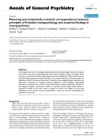
Figure 1 PCBG induces IL-8 release from 1HAEo
-
human airway
epithelial cells. Cells were incubated with LPS, Saccharomyces cerevi-
siae β-glucan and Pneumocystis β-glucan at the indicated doses for a
period of 14 hours. Release of IL-8 was measured by ELISA in the media
supernatant of the cells. Data were analyzed with one-way ANOVA and
posttest Dunnett's comparison test (*** denotes p < 0.001). The exper-
iment shown is representative of three independent experiments.
Carmona et al. Respiratory Research 2010, 11:95
/>Page 6 of 12
prior to and through stimulation of 1HAEo-cells over
PCBC for eight hours [46]. Interestingly, we did not
detect any inhibition of IL-8 secretion in PCBG stimu-
lated cells in the presence of the JNK-II inhibitor. To ver-
ify that the inhibitor was functionally active, we further
analyzed phosphorylation of JNK in PCBG stimulated
cells in the presence of JNK inhibitor II in comparison to
cells that were stimulated with PCBG in the absence of
the inhibitor, verifying that JNK phosphorylation was
indeed greatly reduced (data not shown). Nevertheless,
IL-8 secretion was not impacted by this inhibitor, indicat-
ing that the participation of ERK and p38 MAPK in air-
way epithelial cells stimulated with PCBG is specifically
restricted to those pathways, and that JNK does not par-
ticipate in this cytokine response.
MAPK activation in PCBG stimulated 1HAEo
-
cells
stimulates downstream NF-κB expression
We have previously shown that MIP-2 neutrophil
chemokine induced by PCBG in rodent primary lung epi-
thelial cells is mediated by NF-κB activation (10). We next
sought to determine whether MAPK activation following
β-glucan stimulation of human 1HAEo
-
cells resulted in
downstream NF-κB dependant activation (Figure 7). To
test this, we evaluated whether PCBG induced ERK and
p38 signaling resulted in NF-κB promoter dependent
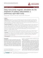
Figure 2 Intracellular calcium mobilization after PCBG stimulation. A. Airway epithelial cells (1HAEo-cells) were loaded with Fura-2AM and incu-
bated with either 100 ug/ml of PCBG or with PAR-2 Peptide control (100 μM) for the indicated times and transient intracellular calcium release mon-
itored by video fluorescence imaging. B. In additional experiments, airway epithelial cells were incubated with 100 ug/ml of PCBG. For one hour prior
to the addition of PCBG, the cells were preincubated with various calcium and calcineurin disrupting agents (EGTA, BAPTA, or TEMPO) at the concen-
tration indicated. IL-8 secretion was measured by ELISA in the supernatant of the cells after eight hours of incubation. C. Finally, airway epithelial cells
were incubated with 100 ug/ml of PCBG for eight hours in the presence of cyclosporine A at the indicated concentration and IL-8 secretion measured
by ELISA. Data were analyzed with one-way ANOVA and posttest Bonferroni comparison (*denotes p < 0.05; **denotes p < 0.01). The data shown are
representative of three independent experiments.
Carmona et al. Respiratory Research 2010, 11:95
/>Page 7 of 12
activation in 1HAEo
-
cells that were transiently trans-
fected with an NF-κB-dependent luciferase reporter plas-
mid. Prior to PCBG stimulation, the 1HAEo
-
cells were
incubated with either; the PD98059, SB202190, or the
JNK inhibitor II. Notably, pre-incubation of the cells with
either PD98059 or SB202190 significantly reduced NF-κB
dependent transcriptional activity in PCBG stimulated
cells. However, the addition of JNK inhibitor II again had
no effect on transcriptional activity related to NF-κB.
These data suggest that PCBG mediated MAPKs activa-
tion results in downstream NF-κB-dependent transcrip-
tional activation in target airway epithelial cells.
Inhibition of glycosphingolipids synthesis further impairs
IL-8 released by airway epithelial cells stimulated with
PCBG
Previous data from our laboratory indicate that PCBG
requires the glycosphingolipid lactosylceramide to induce
MIP-2 release in murine epithelial cells [17,47]. We,
therefore, sought to determine whether IL-8 secretion by
PCBG in these human airway cells was also dependent on
the presence of glycosphingolipids. To accomplish this,
we evaluated IL-8 secretion in PCBG stimulated cells in
the presence of PDMP, a potent glycosphingolipid syn-
thesis inhibitor. Serum free media cultivated cells were
treated with PDMP for 3 days prior to stimulation with
PCBG. IL-8 release from β-glucan stimulated airway epi-
thelial cells treated with the glycosphingolipid inhibitor
was significantly decreased compared to non-treated
cells (Figure 8A). We further investigated the effect of
PDMP on ERK phosphorylation. Cells were cultured with
media alone or in the presence of PDMP prior to activa-
tion with PCBG. Total cell lysates were analyzed for phos-
pho-p44/42 by immunoblotting (Figure 8B and 8C). The
phosphorylation of ERK p44/42 was reduced to baseline
in cells treated with PDMP compared with non-treated
cells. Taken together, these data strongly support our
findings that glycosphingolipids are important for PCBG
mediated ERK activation and subsequent IL-8 secretion
by airway epithelial cells in response to PCBG.
Discussion
Tissue inflammation is an essential component of host
defense against infection, however, exaggerated inflam-
matory response can be extremely deleterious to the host.
Considerable evidence reveals this to be particularly true
for Pneumocystis pneumonia. Early studies from our lab-
oratory, as well as from other investigators have docu-
mented that death and respiratory failure in patients with
Pneumocystis pneumonia is largely related to the intense
inflammatory reaction induced by the infection rather
than direct toxic effects of the fungus [3-5,9,30]. Many
patients with this infection present with intense neutro-
philic and CD8 lymphocytic infiltration in the lungs and
associated impaired oxygen exchange. What induces the
exaggerated recruitment of inflammatory cells in these
patients remains poorly understood. These studies were
undertaken to address the molecular mechanisms, which
regulates the potent neutrophil chemoattractant factor,
IL-8 in airway epithelial cells challenged with the potent
pro-inflammatory cell wall component of Pneumocystis
β-glucan.
Studies from our lab have documented the inflamma-
tory properties of PCBG, and have revealed that this car-
bohydrate-rich cell wall fraction is capable of inducing
specific chemokines and cytokines in cells such as mac-
rophages, dendritic cells (DC) and alveolar epithelial cells
[11,12,17,18]. Airway epithelial cells are the first cells to
come into contact with inhaled pulmonary pathogens.
Contrary to earlier beliefs that alveolar epithelial cells
were only involved in gas exchange, emerging evidence
has documented the importance of these cells as a rich
source of inflammatory mediators, particularly chemok-
ines. We have specifically demonstrated that rodent alve-
olar epithelial cells undergo NF-κB mediated MIP-2
release when challenged with Pneumocystis β-glucans. In
this regard, airway epithelial cells exhibit greater potency
than alveolar macrophages challenged with this cell wall
component (10, 19). In the present study, we further dem-
onstrate that human airway epithelial cells secrete signifi-
cant amounts of IL-8, the human homologue of MIP-2, in
response to Pneumocystis cell wall β-glucan. We have fur-
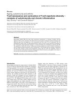
Figure 3 PCBG induced IL-8 expression requires NF-κB and AP-1
activation. 1HAEo
-
cells were transiently transfected with the IL-8 pro-
moter (WT), the IL-8 promoter mutated at the NF-κB site (mut kB) or the
IL-8 promoter mutated at the AP-1 site (mut AP-1). TK-renilla (10 ng)
was co-transfected as an internal control as indicated in material and
methods. Eighteen hours later, transfected 1HAEo
-
cells were chal-
lenged with 100 ug/ml of PCBG. After an additional eight hours of in-
cubation, the cells were harvested and luciferase activities were
measured. The IL-8 activity was normalized to Renilla luciferase activity
(relative lights units). Data were analyzed with one-way ANOVA and
posttest Bonferroni comparison (***denotes p < 0.01). The data shown
is the average of two independent experiments.
Carmona et al. Respiratory Research 2010, 11:95
/>Page 8 of 12
ther observed that airway epithelial cells mobilize intrac-
ellular calcium within seconds following β-glucan
stimulation. This intra-calcium flux initiates the activa-
tion of the two major MAPKs pathways, ERK and p38,
and subsequent activation of AP-1 and NF-κB, resulting
in the release of IL-8. Finally, we demonstrated that inhi-
bition of glycosphingolipids synthesis significantly
impairs the IL-8 response of these cells, suggesting an
important role for surface membrane glycosphingolipids
conferring inflammatory activation.
Glycosphingolipids, most notably lactosylceramide,
have been proposed as receptors for fungal β-glucans,
Figure 4 PCBG induces activation of ERK in 1HAEo
-
airway epithelial cells. A. 1HAEo
-
cells were challenged with 100 ug/ml of PCBG for eight
hours and IL-8 release assessed by ELISA in the culture supernatants. Cells were pretreated for 1 hour with the ERK inhibitor PD 98059 or vehicle solu-
tion as indicated prior to the addition of PCBG. Data were analyzed with one-way ANOVA and posttest Bonferroni comparison (**denotes p < 0.01;
***denotes p < 0.001). B. 1HAEo
-
cells were incubated with 100 ug/ml of PCBG for the indicated times, and phospho-p44/p42 and total p44/p42 were
detected by western blot in the total cell lysate. C. Densitometry analysis of phospho- p44/p42 to total-p44/p42 ratio. D. 1HAEo
-
cells were pre-incu-
bated for 1 hour with different concentrations of TEMPO prior to stimulation with 100 ug/ml of PCBG for 10 minutes, phospho-ERK p44/p42 was de-
tected by Western blot in the total cell lysate. Actin was shown as loading control. E. Densitometry analysis of phospho- p44/p42/Actin ratio. The data
shown is representative of at least two independent experiments.
Carmona et al. Respiratory Research 2010, 11:95
/>Page 9 of 12
and have been of particular interest in cellular activation
mediated by Pneumocystis (15, 16). In the present study,
we demonstrated that treatment of human airway epithe-
lial cells with PDMP, a glycosphingolipid synthesis inhibi-
tor, dramatically reduced the ability of Pneumocystis β-
glucans to stimulate IL-8 release, strongly indicating that
glycosphingolipids are important components initiating
Figure 5 Activation of p38 MAPK after PCBG stimulation of
1HAEo-cells. A. 1HAEo
-
cells were incubated with 100 μg/ml of PCBG
for a period of eight hours, and the media supernatants collected and
IL-8 measured by ELISA. Prior to the addition of PCBG the cells were
pretreated for 1 hour with the p38 inhibitor SB202190. Data were ana-
lyzed with one-way ANOVA and posttest Bonferroni comparison
(***denotes p < 0.001). B. 1HAEo
-
cells were challenged with 100 μg/
ml of PCBG for the times indicated and phospho-p38 and total p38 an-
alyzed by western blot in the total cell lysates. C. Densitometry analysis
of phospho-p38 to total p38 ratio. The data shown is representative of
three independent experiments.
Figure 6 IL-8 production by PCBG activated cells is not impaired
in the presence of a pharmacological inhibitor of JNK-II. 1HAEo
-
cells were incubated with 100 ug/ml of PCBG for a period of eight
hours. Prior to the addition of PCBG, the cells were preincubated for
one hour with JNK Inhibitor II at the concentration indicated. IL-8 se-
cretion was measured by ELISA in the media supernatant of the cells.
Data were analyzed with one-way ANOVA and posttest Bonferroni
comparison (not significantly different, p > 0.05). The data shown is
representative of two independent experiments.
Figure 7 NF-κB activation is impaired in the presence of MAPKs
inhibitors and an intra-calcium chelator, but not in the presence
of JNK inhibitor. 1HAEo
-
cells were transiently transfected with the
NF-κB reporter (50 ng) and TK-renilla (10 ng) as indicated in the Material
and Methods. Eighteen hours later, transfected 1HAEo
-
cells were chal-
lenged with 100 μg/ml of PCBG, prior stimulation the cells were prein-
cubated for 1 h with the different inhibitors. Eight hours later, the cells
were harvested and luciferase activities were measured. The NF-κB ac-
tivity was normalized to Renilla luciferase activity (relative lights units).
Data were analyzed with one-way ANOVA and posttest Bonferroni
comparison (*denotes p < 0.05; **denotes p < 0.01). The data shown is
representative of three independent experiments.
Carmona et al. Respiratory Research 2010, 11:95
/>Page 10 of 12
epithelial cell signaling. In the present study, we further
observed that intracellular calcium mobilization, as well
as activation of two major MAPK pathways (ERK and
p38), also participate in epithelial cells responses to
PCBG.
Intracellular calcium mobilization appears necessary
for IL-8 secretion, since PCBG does not activate airway
epithelial cells in the presence of the intracellular calcium
chelator BAPTA/AM or the calcineurin inhibitor
TEMPO. This early intracellular mobilization of calcium
acts through additional second messengers to induce
activation of the ERK and p38 MAPK pathways. Interest-
ingly, these two pathways are likely stimulated through
unique mechanisms, since their kinetics of activation
were significantly different. While ERK p42/44 was phos-
phorylated within five minutes of stimulation, p38 reach
its peak phosphorylation after 30 minutes. Ultimately,
ERK and p38 pathways were both found to impact down-
stream NF-κB activation at the transcriptional level.
In contrast, we did not observe any decrease in IL-8
levels nor NF-κB transcriptional activation in the pres-
ence of the specific pharmacological inhibitor of JNK,
suggesting that JNK does not participate in PCBG
induced cell stimulation. Recently, an interesting report
by Wang and coworkers demonstrated that whole Pneu-
mocystis induced the release of MCP-1 from alveolar epi-
thelial cells in a JNK-dependent fashion that did not
appear to require β-glucan [48]. The study of Wang and
colleagues utilized β-glucan derived from S. cerevisiae
[48]. While we observed some minimal activation of epi-
thelial cells by Saccharomyces β-glucan, PCBG was
shown to be far more potent in stimulating the epithelial
cells in a JNK independent manner in our hands.
The observations of our current study are comparable
to those of Slevogt and coworkers, who noted activation
of ERK and p38 but not participation of JNK in Moroxella
catarrhalis induced IL-8 production by epithelial cells
[49]. Interestingly, other studies have revealed differing
patterns of MAP activation in response to other microor-
ganisms. For instance, Lamont and coworkers has shown
that Porphyromonas gingivalis infection of epithelial cells
is associated with JNK activation, down regulation of
ERK and NF-κB activation, and decrease of IL-8 expres-
sion [50]. These studied support the notion that species-
specific stimuli result in specific, and often differing, cel-
lular IL-8 responses. In the case of Pneumocystis, two
predominant pathways appear to augment IL-8 responses
and neutrophilic recruitment in this pneumonia.
Regulation of IL-8 transcription is mediated by various
transcription factors including NF-κB, AP-1, and NF-IL-
6, which appear to be both stimuli and cell type specific
[51]. For instance, adequate induction of IL-8 by TNF-α
stimulated epithelial cells requires AP-1 and NF-κB bind-
Figure 8 Effect of glycosphingolipid synthesis inhibitors on
PCBG-mediated IL-8 secretion from 1HAEo-airway epithelial cells.
A. Cells were incubated with different concentrations of PDMP for 72
hours prior to stimulation with 100 ug/ml of PCBG, and the cells incu-
bated an additional 14 hours. Supernatants were assayed for IL-8 as de-
scribed. Data were analyzed with one-way ANOVA and posttest
Bonferroni comparison (***denotes p < 0.001). The data shown is rep-
resentative of three independent experiments. B. 1HAEo
-
cells were in-
cubated for 72 hours in the presence of PDMP at the concentrations
indicated, or media alone prior to stimulation with PCBG for 30 min.
Phospho-p44-42 was analyzed by western blot and actin was assessed
in parallel to verify equal loading. C. Densitometry analysis of phospho-
p44/p42 to Actin ratio.
Carmona et al. Respiratory Research 2010, 11:95
/>Page 11 of 12
ing activity to the IL-8 promoter, while AP-1 binding
activity does not appear to be necessary in TNF-α stimu-
lated endothelial cells [43]. This same group of investiga-
tors also demonstrated that AP-1 binding, and not the
NF-κB, is critical for IL-8 expression by H
2
O
2
stimulated
epithelial cells [43]. The current studies demonstrate that
IL-8 secretion and gene transcription induced by PCBG
in human airway epithelial cells requires the integrity of
NF-κB and AP-1 binding sites. This is noteworthy,
because distinct AP-1 dimers may selectively interact
with various NF-κB subunits and synergistically act to
augment IL-8 expression. Such interaction have been
demonstrated to occur in respiratory syncytial virus
(RSV) induced IL-8 expression [41]. Indeed, in RSV
infected cells, AP-1 cooperates preferentially with NF-κB,
while in TNF-α stimulated cells NF-IL-6 interacts with
NF-κB [41]. Based on these observations, we postulate
that alteration of the binding between these various tran-
scription factor subunits may help to initially promote IL-
8 secretion in Pneumocystis pneumonia and subsequently
to control neutrophil inflammation in this infection.
Conclusion
In summary, our investigations have demonstrated that
Pneumocystis cell wall β-glucans induce inflammatory
response in human airway epithelial cells. IL-8 secretion
by these cells involves membrane glycosphingolipid
receptors and the intracellular mobilization of calcium,
with subsequent phosphorylation of MAPKs pathways
including ERK and p38. These events lead to downstream
activation of the NF-κB and AP-1 transcription factors
and ultimately to IL-8 release. Abrogation of either one or
these MAPK pathways or these transcription factors
results in a blunted IL-8 response. Better knowledge of
the molecular mechanisms regulating chemokine genera-
tion will be essential to understand the recruitment of
inflammatory cells to the lung during Pneumocystis
pneumonia, and to design new treatment strategies for
the exuberant lung inflammation that accompanies this
infection.
Competing interests
The authors declare that they have no competing interests.
Authors' contributions
EMC performed the cytokine, signal transduction, and promoter assays and
participated in drafting the manuscript. JDL assisted with the signal transduc-
tion assays and cell culture work. AX participated in the calcium signaling stud-
ies. MW participated in its design and coordination of the calcium experiments.
AHL participated in the overall experimental design concept, review and inter-
pretation of data, preparation of the manuscript and secured all funding for
these studies. All authors read and approved the final manuscript.
Acknowledgements
These studies were funded by the Mayo Foundation and NIH grants R01-
HL62150 and R01-HL55934 to AHL. EMC was supported by funds from Mayo
Foundation. We thank Zvezdana Vuk-Pavlovic' and Joshua Burgess for many
helpful discussions. We further acknowledge the efforts of Deanne Hebrink
and Joseph Standing in the generation of the Pneumocystis carinii organisms
and the Pneumocystis β-glucan preparations used in these studies. Finally, we
appreciate the efforts of Ted Kottom for invaluable technical support for these
studies.
Author Details
From the Thoracic Diseases Research Unit, Division of Pulmonary Critical Care
and Internal Medicine, Department of Medicine Mayo Clinic and Foundation,
Rochester, Minnesota, 55905, USA
References
1. Kaplan JE, Hanson D, Dworkin MS, Frederick T, Bertolli J, Lindegren ML,
Holmberg S, Jones JL: Epidemiology of human immunodeficiency
virus-associated opportunistic infections in the United States in the era
of highly active antiretroviral therapy. Clin Infect Dis 2000, 30(Suppl
1):S5-14.
2. Russian DA, Levine SJ: Pneumocystis carinii pneumonia in patients
without HIV infection. Am J Med Sci 2001, 321(1):56-65.
3. Thomas CF Jr, Limper AH: Pneumocystis pneumonia. N Engl J Med 2004,
350(24):2487-2498.
4. Limper AH, Offord KP, Smith TF, Martin WJ: Pneumocystis carinii
pneumonia. Differences in lung parasite number and inflammation in
patients with and without AIDS. Am Rev Respir Dis 1989,
140(5):1204-1209.
5. Wright TW, Gigliotti F, Finkelstein JN, McBride JT, An CL, Harmsen AG:
Immune-mediated inflammation directly impairs pulmonary function
contributing to the pathogenesis of Pneumocystis carinii pneumonia.
J Clin Invest 1999, 104(9):1307-1317.
6. Wright TW, Gigliotti F, Finkelstein JN, McBride JT, An CL, Harmsen AG:
Immune-mediated inflammation directly impairs pulmonary function
contributing to the pathogenesis of Pneumocystis carinii pneumonia.
J Clin Invest 1999, 104(9):1307-1317.
7. Bhagwat SP, Gigliotti F, Xu H, Wright TW: Contribution of T cell subsets to
the pathophysiology of Pneumocystis-related immunorestitution
disease. Am J Physiol Lung Cell Mol Physiol 2006, 291(6):L1256-1266.
8. Gigliotti F, Crow EL, Bhagwat SP, Wright TW: Sensitized CD8+ T cells fail
to control organism burden but accelerate the onset of lung injury
during Pneumocystis carinii pneumonia. Infect Immun 2006,
74(11):6310-6316.
9. Mason GR, Hashimoto CH, Dickman PS, Foutty LF, Cobb CJ: Prognostic
implications of bronchoalveolar lavage neutrophilia in patients with
Pneumocystis carinii pneumonia and AIDS. Am Rev Respir Dis 1989,
139(6):1336-1342.
10. Limper AH, Thomas CF Jr, Anders RA, Leof EB: Interactions of parasite and
host epithelial cell cycle regulation during Pneumocystis carinii
pneumonia. J Lab Clin Med 1997, 130(2):132-138.
11. Vassallo R, Standing JE, Limper AH: Isolated Pneumocystis carinii cell wall
glucan provokes lower respiratory tract inflammatory responses. J
Immunol 2000, 164(7):3755-3763.
12. Lebron F, Vassallo R, Puri V, Limper AH: Pneumocystis carinii cell wall
beta-glucans initiate macrophage inflammatory responses through
NF-kappaB activation. J Biol Chem 2003, 278(27):25001-25008. Epub
22003 Apr 25025
13. Limper AH, Edens M, Anders RA, Leof EB: Pneumocystis carinii inhibits
cyclin-dependent kinase activity in lung epithelial cells. J Clin Invest
1998, 101(5):1148-1155.
14. Limper AH: Parasitic adherence and host responses in the development
of Pneumocystis carinii pneumonia. Semin Respir Infect 1991, 6(1):19-26.
15. Krajicek BJ, Thomas CF Jr, Limper AH: Pneumocystis pneumonia: current
concepts in pathogenesis diagnosis, and treatment. Clin Chest Med
2009, 30(2):265-278. vi
16. McCann F, Carmona E, Puri V, Pagano RE, Limper AH: Macrophage
internalization of fungal beta-glucans is not necessary for initiation of
related inflammatory responses. Infect Immun 2005, 73(10):6340-6349.
17. Evans SE, Hahn PY, McCann F, Kottom TJ, Pavlovic ZV, Limper AH:
Pneumocystis Cell Wall {beta}-Glucans Stimulate Alveolar Epithelial
Cell Chemokine Generation through Nuclear Factor-{kappa}B-
Dependent Mechanisms. Am J Respir Cell Mol Biol 2005, 32(6):490-497.
Received: 21 October 2009 Accepted: 13 July 2010
Published: 13 July 2010
This article is available from: 2010 Carmona et al; licensee BioMed Central Ltd. This is an Open Access article distributed under the terms of the Creative Commons Attribution License ( which permits unrestricted use, distribution, and reproduction in any medium, provided the original work is properly cited.Respiratory Research 2010, 11:95
Carmona et al. Respiratory Research 2010, 11:95
/>Page 12 of 12
18. Hahn PY, Evans SE, Kottom TJ, Standing JE, Pagano RE, Limper AH:
Pneumocystis carinii cell wall beta-glucan induces release of
macrophage inflammatory protein-2 from alveolar epithelial cells via a
lactosylceramide-mediated mechanism. J Biol Chem 2003,
278(3):2043-2050. Epub 2002 Nov 2044
19. Ratner AJ, Bryan R, Weber A, Nguyen S, Barnes D, Pitt A, Gelber S, Cheung
A, Prince A: Cystic fibrosis pathogens activate Ca2+-dependent
mitogen-activated protein kinase signaling pathways in airway
epithelial cells. J Biol Chem 2001, 276(22):19267-19275.
20. Gewirtz AT, Rao AS, Simon PO Jr, Merlin D, Carnes D, Madara JL, Neish AS:
Salmonella typhimurium induces epithelial IL-8 expression via Ca(2+)-
mediated activation of the NF-kappaB pathway. J Clin Invest 2000,
105(1):79-92.
21. Segal E: Pathogenesis of human mycoses: role of adhesion to host
surfaces. Microbiol Sci 1987, 4(11):344-347.
22. Douglas LJ: Adhesion of Candida species to epithelial surfaces. Crit Rev
Microbiol 1987, 15(1):27-43.
23. Brown GD, Gordon S: Immune recognition. A new receptor for beta-
glucans. Nature 2001, 413(6851):36-37.
24. Carmona EM, Vassallo R, Vuk-Pavlovic Z, Standing JE, Kottom TJ, Limper
AH: Pneumocystis cell wall beta-glucans induce dendritic cell
costimulatory molecule expression and inflammatory activation
through a Fas-Fas ligand mechanism. J Immunol 2006, 177(1):459-467.
25. Limper AH, Hoyte JS, Standing JE: The role of alveolar macrophages in
Pneumocystis carinii degradation and clearance from the lung. J Clin
Invest 1997, 99(9):2110-2117.
26. Cozens AL, Yezzi MJ, Chin L, Simon EM, Friend DS, Gruenert DC: Chloride
ion transport in transformed normal and cystic fibrosis epithelial cells.
Adv Exp Med Biol 1991, 290:187-194. discussion 194-186
27. Trushin SA, Pennington KN, Algeciras-Schimnich A, Paya CV: Protein
kinase C and calcineurin synergize to activate IkappaB kinase and NF-
kappaB in T lymphocytes. J Biol Chem 1999, 274(33):22923-22931.
28. Shimotake TK, Izhar FM, Rumilla K, Li J, Tan A, Page K, Brasier AR, Schreiber
MD, Hershenson MB: Interleukin (IL)-1 beta in tracheal aspirates from
premature infants induces airway epithelial cell IL-8 expression via an
NF-kappa B dependent pathway. Pediatr Res 2004, 56(6):907-913. Epub
2004 Oct 2020
29. White TA, Kannan MS, Walseth TF: Intracellular calcium signaling
through the cADPR pathway is agonist specific in porcine airway
smooth muscle. Faseb J 2003, 17(3):482-484.
30. Beck JM, Rosen MJ, Peavy HH: Pulmonary complications of HIV infection.
Report of the Fourth NHLBI Workshop. Am J Respir Crit Care Med 2001,
164(11):2120-2126.
31. Adamo R, Sokol S, Soong G, Gomez MI, Prince A: Pseudomonas
aeruginosa flagella activate airway epithelial cells through asialoGM1
and toll-like receptor 2 as well as toll-like receptor 5. Am J Respir Cell Mol
Biol 2004, 30(5):627-634. Epub 2003 Nov 2007
32. Mellstrom B, Naranjo JR: Mechanisms of Ca2+)-dependent
transcription. Curr Opin Neurobiol 2001, 11(3):312-319.
33. Lindenboim L, Haviv R, Stein R: Inhibition of drug-induced apoptosis by
survival factors in PC12 cells. J Neurochem 1995, 64(3):1054-1063.
34. Dieter P, Fitzke E, Duyster J: BAPTA induces a decrease of intracellular
free calcium and a translocation and inactivation of protein kinase C in
macrophages. Biol Chem Hoppe Seyler 1993, 374(3):171-174.
35. Nelson PA, Akselband Y, Kawamura A, Su M, Tung RD, Rich DH, Kishore V,
Rosborough SL, DeCenzo MT, Livingston DJ, Harding MW:
Immunosuppressive activity of [MeBm2t]1-, D-diaminobutyryl-8-, and
D-diaminopropyl-8-cyclosporin analogues correlates with inhibition of
calcineurin phosphatase activity. J Immunol 1993, 150(6):2139-2147.
36. Nakamura H, Yoshimura K, Jaffe HA, Crystal RG: Interleukin-8 gene
expression in human bronchial epithelial cells. J Biol Chem 1991,
266(29):19611-19617.
37. Brasier AR, Jamaluddin M, Casola A, Duan W, Shen Q, Garofalo RP: A
promoter recruitment mechanism for tumor necrosis factor-alpha-
induced interleukin-8 transcription in type II pulmonary epithelial
cells. Dependence on nuclear abundance of Rel A, NF-kappaB1, and c-
Rel transcription factors. J Biol Chem 1998, 273(6):3551-3561.
38. Mukaida N, Mahe Y, Matsushima K: Cooperative interaction of nuclear
factor-kappa B- and cis-regulatory enhancer binding protein-like
factor binding elements in activating the interleukin-8 gene by pro-
inflammatory cytokines. J Biol Chem 1990, 265(34):21128-21133.
39. Stein B, Baldwin AS Jr: Distinct mechanisms for regulation of the
interleukin-8 gene involve synergism and cooperativity between C/
EBP and NF-kappa B. Mol Cell Biol 1993, 13(11):7191-7198.
40. Matsusaka T, Fujikawa K, Nishio Y, Mukaida N, Matsushima K, Kishimoto T,
Akira S: Transcription factors NF-IL6 and NF-kappa B synergistically
activate transcription of the inflammatory cytokines, interleukin 6 and
interleukin 8. Proc Natl Acad Sci USA 1993, 90(21):10193-10197.
41. Kunsch C, Rosen CA: NF-kappa B subunit-specific regulation of the
interleukin-8 promoter. Mol Cell Biol 1993, 13(10):6137-6146.
42. Fiedler MA, Wernke-Dollries K, Stark JM: Mechanism of RSV-induced IL-8
gene expression in A549 cells before viral replication. Am J Physiol
1996, 271(6 Pt 1):L963-971.
43. Lakshminarayanan V, Drab-Weiss EA, Roebuck KA: H2O2 and tumor
necrosis factor-alpha induce differential binding of the redox-
responsive transcription factors AP-1 and NF-kappaB to the
interleukin-8 promoter in endothelial and epithelial cells. J Biol Chem
1998, 273(49):32670-32678.
44. Schmeck B, Zahlten J, Moog K, van Laak V, Huber S, Hocke AC, Opitz B,
Hoffmann E, Kracht M, Zerrahn J, Hammerschmidt S, Rosseau S, Suttorp N,
Hippenstiel S: Streptococcus pneumoniae-induced p38 MAPK-
dependent phosphorylation of RelA at the interleukin-8 promotor. J
Biol Chem 2004, 279(51):53241-53247. Epub 52004 Oct 53213
45. Griego SD, Weston CB, Adams JL, Tal-Singer R, Dillon SB: Role of p38
mitogen-activated protein kinase in rhinovirus-induced cytokine
production by bronchial epithelial cells. J Immunol 2000,
165(9):5211-5220.
46. Bennett BL, Sasaki DT, Murray BW, O'Leary EC, Sakata ST, Xu W, Leisten JC,
Motiwala A, Pierce S, Satoh Y, Bhagwat SS, Manning AM, Anderson DW:
SP600125, an anthrapyrazolone inhibitor of Jun N-terminal kinase.
Proc Natl Acad Sci USA 2001, 98(24):13681-13686.
47. Hahn PY, Limper AH: Pneumocystis carinii beta-glucan induces release
of macrophage inflammatory protein-2 from primary rat alveolar
epithelial cells via a receptor distinct from CD11b/CD18. J Eukaryot
Microbiol 2001:157S.
48. Wang J, Gigliotti F, Bhagwat SP, Maggirwar SB, Wright TW: Pneumocystis
stimulates MCP-1 production by alveolar epithelial cells through a JNK-
dependent mechanism. Am J Physiol Lung Cell Mol Physiol 2007,
292(6):L1495-1505.
49. Slevogt H, Schmeck B, Jonatat C, Zahlten J, Beermann W, van Laak V, Opitz
B, Dietel S, N'Guessan PD, Hippenstiel S, Suttorp N, Seybold J: Moraxella
catarrhalis induces inflammatory response of bronchial epithelial cells
via MAPK and NF-kappaB activation and histone deacetylase activity
reduction. Am J Physiol Lung Cell Mol Physiol 2006, 290(5):L818-826.
50. Watanabe K, Yilmaz O, Nakhjiri SF, Belton CM, Lamont RJ: Association of
mitogen-activated protein kinase pathways with gingival epithelial
cell responses to Porphyromonas gingivalis infection. Infect Immun
2001, 69(11):6731-6737.
51. Roebuck KA: Regulation of interleukin-8 gene expression. J Interferon
Cytokine Res 1999, 19(5):429-438.
doi: 10.1186/1465-9921-11-95
Cite this article as: Carmona et al., Pneumocystis cell wall ?-glucan stimulates
calcium-dependent signaling of IL-8 secretion by human airway epithelial
cells Respiratory Research 2010, 11:95