Báo cáo y học: " CTLA4-Ig interacts with cultured synovial macrophages from rheumatoid arthritis patients and downregulates cytokine production" ppt
Bạn đang xem bản rút gọn của tài liệu. Xem và tải ngay bản đầy đủ của tài liệu tại đây (1.26 MB, 10 trang )
Open Access
Available online />Page 1 of 10
(page number not for citation purposes)
Vol 11 No 6
Research article
CTLA4-Ig interacts with cultured synovial macrophages from
rheumatoid arthritis patients and downregulates cytokine
production
Maurizio Cutolo
1
, Stefano Soldano
1
, Paola Montagna
1
, Alberto Sulli
1
, Bruno Seriolo
1
,
Barbara Villaggio
2
, Pierfranco Triolo
3
, Paolo Clerico
3
, Lamberto Felli
4
and Renata Brizzolara
1
1
Research Laboratories and Academic Unit of Clinical Rheumatology, Department of Internal Medicine, University of Genova, Viale Benedetto XV,
16132 Genova, Italy
2
Clinical Academic Unit of Nephrology, Department of Internal Medicine, University of Genova, Viale Benedetto XV, 16132 Genova, Italy
3
Rheumatoid Arthritis Unit - Orthopedic Surgery Department, CTO Hospital, Via Zuretti 10126 Turin, Italy
4
Orthopedic Department, Largo Rosanna Benzi, University of Genova, 16132 Genova, Italy
Corresponding author: Maurizio Cutolo,
Received: 28 Mar 2009 Revisions requested: 27 Apr 2009 Revisions received: 4 Nov 2009 Accepted: 23 Nov 2009 Published: 23 Nov 2009
Arthritis Research & Therapy 2009, 11:R176 (doi:10.1186/ar2865)
This article is online at: />© 2009 Cutolo et al.; licensee BioMed Central Ltd.
This is an open access article distributed under the terms of the Creative Commons Attribution License ( />),
which permits unrestricted use, distribution, and reproduction in any medium, provided the original work is properly cited.
Abstract
Introduction Co-stimulatory signal B7(CD80/CD86):CD28 is
needed in order to activate T cells in immune response.
Cytotoxic T lymphocyte-associated antigen-4-immunoglobulin
(CTLA4-Ig) binding to the B7 molecules on antigen-presenting
cells downregulates this activation and represents a recent
biological treatment in rheumatoid arthritis (RA). Objectives of
the study were to investigate the presence of the B7.2 (CD86)
molecule and its masking by CTLA4-Ig on cultures of both RA
synovial macrophages (RA SM), and of macrophages
differentiated from THP-1 cells (M). In addition, the anti-
inflammatory effects of CTLA4-Ig on co-cultures of RA SM and
M with activated T cells were tested.
Methods All macrophages were co-cultured for 24 hours with
activated T cells, without or with CTLA4-Ig (10, 100, 500 μg/ml
for 1 hour, 3 hours and overnight, respectively).
Immunofluorescence (IF) staining for B7.2, and an analysis of
inflammatory cytokine expression (interleukin (IL) -6, tumor
necrosis factor (TNF) α, IL-1β, transforming growth factor (TGF)
β) by immunocytochemistry (ICC), western blot (WB) and
reverse transcriptase-polymerase chain reaction (RT-PCR) were
performed.
Results Macrophages showed intense B7.2 expression.
CTLA4-Ig/B7.2 masking was evident for all macrophages, even
after only 1 hour of cell culture (range from 10 to 100 μg/ml).
ICC of co-cultures showed a dose-dependent decrease in
inflammatory cytokines (P < 0.001 for IL-6, TNFα, IL-1β and
TGFβ). Data were confirmed by WB and RT-PCR analysis.
Conclusions Optimal concentrations of CTLA4-Ig for the
CTLA4-Ig/B7.2 masking on activated macrophages were
identified and were found to induce significant downregulation
in the cell production of IL-6, TNFα, IL1-β and TGFβ. In
conclusion, macrophages would appear to be a sensitive target
for CTLA4-Ig treatment in RA.
Introduction
Rheumatoid arthritis (RA) is a prototype of an immune-medi-
ated chronic inflammatory disease and is considered a model
for studying and validating new targeted biological therapies.
Migration of activated lymphocytes and monocytes into the
synovial tissue in RA is one of the first steps in synovial inflam-
mation, followed by subsequent damage of other joint compo-
nents [1-3]. In recent years, the role of T cells has regained
some importance in the immunopathology of RA, thus
APC: antigen-presenting cells; CTLA-4: cytotoxic T lymphocyte-associated antigen-4; DMARDs: disease-modifying antirheumatic drugs; DPBS: Dul-
becco's phosphate buffered saline; ELISA: enzyme-linked immunosorbent assay; IgG1: immunoglobulin G1; IL: interleukin; LPS: lipopolysaccharide;
MHC: major histocompatibility complex; PMA: phorbol myristate acetate; RA: rheumatoid arthritis; RT-PCR: reverse transcriptase-polymerase chain
reaction; SM: synovial macrophages; TCR: T cell receptor; TGF: transforming growth factor; TNF: tumor necrosis factor.
Arthritis Research & Therapy Vol 11 No 6 Cutolo et al.
Page 2 of 10
(page number not for citation purposes)
providing a rationale for the specific targeting of T cells with
biologic treatments [4-8].
T cell response is triggered by an initial signal delivered
through the T cell receptor (TCR), and it recognizes the anti-
genic peptide within the context of the major histocompatibility
complex (MHC) molecule on the antigen-presenting cells
(APC). In order to be fully activated, it needs to be followed by
another signal, which is provided by the signals of the co-stim-
ulatory molecules that are expressed on APC (such as den-
dritic cells, B-lymphocytes and macrophages) [9].
Among the known multiple co-stimulatory signals, one of the
best described is the CD80/CD86:CD28 pathway [10,11].
CD80 (B7.1) and CD86 (B7.2), which are expressed on APC,
bind the CD28 molecule on the T cells, thereby transducing
the co-stimulatory signal in the early phase of the immune
response. However, the activated T cells then express the
cytotoxic T lymphocyte-associated antigen-4 (CTLA-4) mole-
cule, which binds the B7 molecules on APC with a 10- to 20-
fold greater affinity compared with CD28, and downregulates
the T cell activation [12-14].
CTLA-4-Ig, a biological agent, is constructed by genetically
fusing the external domain of human CTLA-4 and a fragment
of the Fc domain of human immunoglobulin G1 (IgG1), which
has been modified to be non-complement fixing. Like the
native CTLA-4, the fusion protein (CTLA-4-Ig) binds more
avidly to CD80/CD86 (APC) than to CD28 (T cells), thus
interfering with CD28/B7 interaction [15,16].
Therefore, taking these mechanisms into consideration, sev-
eral randomized, double-blind, placebo-controlled clinical tri-
als have demonstrated that CTLA-4-Ig improves the signs and
symptoms of RA in patients with inadequate response to
methotrexate or/and anti-TNF agents [17-20].
However, because macrophages play a crucial role in various
steps of the synovial RA pathophysiology, the aim of this in
vitro study was firstly, to search for the presence of the B7.2
molecule on the surface of cultured synovial macrophages
(SM) obtained from active RA patients, and then to investigate
the modulatory effects of CTLA-4-Ig in a co-culture of RA SM
or macrophages together with an activated T cell line [21]. In
particular, the investigation focused on the effects of CTLA-4-
Ig on the production of peculiar mediators of inflammation pro-
duced by macrophages, such as cytokines (IL-6, TNFα, IL-1β)
and transforming growth factor beta (TGFβ).
Materials and methods
Rheumatoid arthritis synovial macrophages (RA SM)
cultures
RA SM were obtained from seven patients (five females and
two males, mean age: 47 ± 12 years, disease duration 4 ± 6
years, Disease Activity Score using 28 joint counts (DAS28)
>5.2) who fulfilled the 1987 revised criteria of the American
College of Rheumatology for adult RA and who underwent
therapeutic arthroscopic synoviectomy or knee replacement
surgery.
At the time of surgery, all patients had been taking non-steroi-
dal anti-inflammatory drugs for 20 days and low-dose gluco-
corticoids (prednisone range 5 to 7.5 mg/day) for three or four
months. No intra-articular treatments, systemic biological
drugs, antiproliferative drugs or other disease modifying anti-
rheumatic drugs (DMARDs) were being administered at the
time of knee surgery, nor had they been for at least four months
prior to it. The Ethics Committee of the University of Genova
approved the study and informed consent was obtained from
all patients.
After surgery, the synovial RA tissue samples were carefully
cut into small pieces (2 to 5 mm), washed in Dulbecco's phos-
phate buffered saline (DPBS; Sigma-Aldrich, Sigma Chemical
Division, Milan, Italy) and incubated in collagenase (0.75 mg/
ml; type IV from Clostridium histolyticum; Sigma-Aldrich,
Sigma Chemical Division, Milan, Italy) for one hour at 37°C.
The digest was passed through a pore size 58 Å ~150 mesh
wire to separate the synovial cells from the debris tissue. The
cells were then washed three times with DPBS, resuspended
in RPMI-1640 medium (Sigma-Aldrich, Sigma Chemical Divi-
sion, Milan, Italy) that had been supplemented with 10% fetal
bovine serum (containing < 0.5 EU/ml endotoxin), 2 mmol/l L-
glutamine, 100 μg/ml streptomycin, and 100 U/ml penicillin
(Sigma-Aldrich, Sigma Chemical Division, Milan, Italy). Viability
of the cells (90 to 95%) was tested by trypan blue exclusion.
The synovial cells were seeded into flexiperm chamber slides
(International, PBI S.p.a. Milan, Italy; 105 cells/well) and cul-
tured in 5% CO
2
air humidified atmosphere at 37°C. After one
hour, non-adherent cells were washed out, while adherent
cells (RA SM) were incubated in culture medium in the pres-
ence or in the absence of lipopolysaccharide (LPS) stimulus
(10 μg/ml) for one hour to investigate B7.2 expression [22].
B7.1 expression was not tested because it is expressed in
lower amounts on resting cells and is only upregulated with
prolonged T cell stimulation, while B7.2 is constitutively
expressed and rapidly upregulated on APC with an antigen-
specific signal, thus indicating that B7.2 is chiefly involved in
mediating initial T cell activation [10,23].
EBV+ transfected human B-lymphocytes were also incubated
in flexiperm chamber slides as a positive cell line control for
B7.2 expression.
THP-1 cell-line cultures
THP-1 (cell bank. Interlab Cell Line Collection. Clinical Pathol-
ogy Dept, IST, Genova, Italia) human monocytes were incu-
bated with phorbol myristate acetate (PMA; 0.5 μg/ml) for two
Available online />Page 3 of 10
(page number not for citation purposes)
hours to differentiate into macrophages, then cells were
seeded into flexiperm chamber slides (International, PBI S.p.a.
Milan, Italy; 10
5
cells/well) and cultured in 5% CO
2
air humid-
ified atmosphere at 37°C.
Macrophage and T cell co-cultures
Macrophage/Jurkat
The macrophages that were obtained from THP-1 as previ-
ously described, were co-cultured with human T cells for 24
hours (Jurkat T cells previously activated with concanavalin-A
(5 μg/ml) for 20 hours). They were then seeded into multiwell
flat bottom plates both in the absence and the presence of var-
ious concentrations of CTLA-4-Ig (10, 100, 500 μg/ml). The
macrophage/T cell ratio was 1:1.
At the end of the co-culture incubation period, the T cells were
removed with several washes in PBS. Macrophages were har-
vested, washed in PBS, and used for the various assays. Pre-
liminary immunocytostaining using a specific monoclonal
antibody to identify the macrophages (anti-human HAM56,
Dako, Carpinteria, CA, USA) was performed. All experiments
were performed in triplicate.
RA SM/Jurkat
RA SM that had been isolated from synovial tissue as previ-
ously described, were co-cultured into multiwell flat bottom
plates with T cells (Jurkat T cells previously activated with con-
canavalin-A (5 μg/ml) for 20 hours) for 24 hours, both in the
absence and presence of CTLA-4-Ig at various concentrations
(100, 500 μg/ml). The in vitro CTLA-4-Ig concentrations to be
tested were chosen on the basis of the available literature,
suggesting values that are very close to the therapeutic doses
used to treat RA [22,24,25]. The macrophage/T cell ratio was
1:1.
At the end of the co-culture incubation period, the T cells were
removed with several washes in PBS. The RA SM were har-
vested, washed in PBS, and used for the various test assays.
In order to identify macrophages, we performed preliminary
immunocytostaining with a specific monoclonal antibody (anti-
human HAM56, Dako, Carpinteria, CA, USA). All experiments
were performed in triplicate.
Immunofluorescence assay
Slides with cellular spots obtained from cultures of macro-
phages were seeded into flexiperm chamber slides, then fixed
in acetone for 30 seconds, air dried, and rehydratated in PBS.
The spots were then incubated with an fluorescein isothiocy-
anate anti-human CD86 (B7.2) mouse antibody (BD, Bio-
sciences, New York, NY, USA) at room temperature for 45
minutes, washed in PBS, and cover slipped with glycerol
medium. Evaluation of the fluorescence expression for CD86
(B7.2) was performed by fluorescence microscopy (Leica,
Cambridge, UK).
Immunocytochemistry assay
Macrophages from co-cultures that had been seeded into mul-
tiwell flat bottom plates were harvested mechanically and incu-
bated on glass slides for 45 minutes at 4°C, then the cellular
spots were fixed in acetone for 30 seconds, air dried, and
stored at -20°C until use.
After rehydration in PBS, the slides were incubated in a 3%
H
2
O
2
solution for 15 minutes at room temperature to prevent
non-specific reaction due to endogenous peroxidases.
The spots were then incubated with anti-cytokine (anti-IL6,
anti-TNFα, anti-IL-1β) and anti-TGFβ monoclonal antibodies
(all diluted 1:100 at room temperature for 45 minutes. Santa
Cruz Biotechnology, Santa Cruz, CA, USA).
Linked antibodies were detected by a biotinylated universal
(pan-specific) antibody and subsequently by horseradish per-
oxidase streptavidin (Vector Laboratories, Burlingame, CA,
USA). Each step was followed by two washes in PBS. The
staining reaction was developed by the diaminobenzidine sys-
tem (DakoCytomation, Dako North America, Inc., Carpinteria,
CA, USA). Finally, slides were counter-stained with hematoxy-
lin, fixed with ethanol, and cover-slipped with Eukitt mounting
medium for microscope preparations (O. Kindler GmbH,
Friburg, Germany). Negative controls were treated the same
way, except that the primary antibodies were omitted.
Image analysis of immunocytochemistry slides was performed
using the Leica Q-Win image analysis system (Leica, Cam-
bridge, UK). Approximately 100 cells were analysed for each
sample, and pixels/mm
2
(positive area) were quantified by the
Leica Q-Win software (Leica, Cambridge, UK). The individual
cells were randomly selected by the operators using the cur-
sor and were then automatically measured as positive areas.
Immune enzymatic assay (ELISA)
After 24 hours of treatment, the culture medium was harvested
and stored at -20°C until analysis. The enzymatic immu-
noassay for quantitative determination of IL-6, TNFα and IL-1β
was carried out with a microplate kit system (Diaclone,
Besançon, France). The results were obtained with a multiwell
plate automatic processor (Techno Genetics, Milan, Italy).
Western blot analysis
After the various treatments, macrophage pellets were lysed in
a buffer containing 20 mmol/l Tris-HCl pH 8, 150 mmol/l NaCl,
1 mmol/l phenylmethylsulphonyl fluoride, 5 mg/ml aprotinin,
and 0.5% Nonidet P-40 (Promega, Milan, Italy) for one hour at
4°C. The lysates were then centrifuged for 10 minutes at
13,000 rpm. Protein samples (20 mg) were diluted with sam-
ple buffer and separated by 10% SDS-PAGE. The proteins
were transferred to a Hibond-C nitrocellulose membrane
(GeHealthCare Europe, Friburg, Germany), after which the
Arthritis Research & Therapy Vol 11 No 6 Cutolo et al.
Page 4 of 10
(page number not for citation purposes)
membrane was blocked for one hour at room temperature in
PBS containing 5% non-fat powdered milk.
To carry out immunoblot analysis, the membrane was incu-
bated with rabbit anti-IL-6 (diluted 1:500), goat anti-TNFα
(1:200) and goat anti-TGFβ (1:200) antibodies (Santa Cruz
Biotechnology, Santa Cruz, CA, USA) overnight at 4°C. Then
membranes were washed in 1% PBS + 0.05% Tween 20, pH
7.4 and incubated for one hour at room temperature with a
secondary anti-rabbit antibody for IL-6 (1:5,000; Santa Cruz
Biotechnology, Santa Cruz, CA, USA) and a secondary anti-
goat antibody for TNFα and TGFβ (1:65,000; Sigma, Milan,
Italy). After three further washes with PBS/Tween, a bound
secondary antibody was detected through emitting chemilumi-
nescent reaction (Immobilon, Millipore, CA, USA).
Western blot analysis was not performed on RA SM because
it would have been difficult to obtain enough cells from the pri-
mary culture to allow full analysis of the cytokines being
investigated.
Densitometry analysis of the western blot bands were per-
formed by Delta Sistemi Analysis Software (version 3.0.02,
Latina, Italy).
Reverse transcriptase-polymerase chain reaction
mRNA extraction from the various experimental conditions of
macrophage/Jurkat co-cultures was performed with the Rne-
asy Mini Kit (Qiagen, Valencia, CA, USA).
The samples were analysed by RT-PCR, using both IL-6-,
TNFα-, TGFβ-specific primers (Invitrogen S.R.L., Milan, Italy)
and beta-actin-specific primers (Promega, Milan, Italy) as the
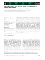
Figure 1
B7.2 expression on macrophages by immunofluorescence analysisB7.2 expression on macrophages by immunofluorescence analysis. B7.2 expression by immunofluorescence analysis (a) on primary cultures of
rheumatoid arthritis synovial macrophages (RA SM) untreated, (b) on macrophages differentiated from THP-1 untreated, (c) on SM pre-treated with
lipopolysaccharide and (d) on EBV+ B lymphocytes (positive control cell line).
Available online />Page 5 of 10
(page number not for citation purposes)
internal positive control. Statistical analysis was carried out by
the non-parametric Wilcoxon T test.
Statistical analysis was performed to compare the paired treat-
ments. P < 0.05 was considered statistically significant.
Results
B7.2 (CD86) positivity on cultured macrophages
Macrophages showed intense positivity for B7.2 and a diffuse
distribution pattern on the cell surface both in RA SM cultures
and in macrophage cultures. No differences were found
between LPS-treated or untreated cells, and no differences
were found between cultured macrophages and the positive
control cell line either (EBV+ transfected human B-lym-
phocytes; Figure 1).
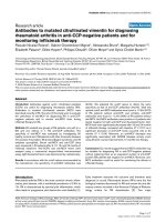
In vitro CTLA-4-Ig/B7.2 masking on macrophages
Analysis by both fluorescence and optic microscopy in light
field showed a reduction of B7.2 positivity on macrophages
treated with CTLA-4-Ig, certainly due to CTLA4-Ig binding to
B7.2 and subsequent B7.2 expression masking.
The positive staining of B7.2 showed a gradual decrease from
CTLA-4-Ig-untreated macrophages (controls) to CTLA-4-Ig-
treated macrophages (from 10 to 500 μg/ml; Figure 2). B7.2
masking on macrophages was still evident within the CTLA-4-
Ig range from 10 to 100 μg/ml after one hour.
Effects of CTLA-4-Ig on cytokine expression in
macrophage and T cell co-cultures
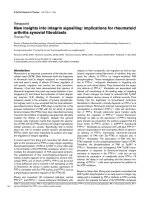
Macrophage/Jurkat
Macrophages that had been co-cultured with T cells following
the addition of CTLA-4-Ig (range from 100 to 500 μg/ml),
showed a decrease in pro-inflammatory cytokine content, as
evaluated by immunocytochemistry (Figures 3a to 3d). The
cytokines we evaluated were IL-6, TNFα, IL-1β and TGFβ, as
growth factor.
The changes that were observed in IL-6, TNFα and TGFβ
expression included a significant, dose-dependent decrease
(P < 0.001), following treatment with CTLA-4-Ig at both 100
and 500 μg/ml. IL-1beta expression only showed a significant
decrease for CTLA-4-Ig (500 μg/ml) treatment (P < 0.001).
Interestingly, as compared with untreated macrophages (con-
trols), we found a significant decrease (P < 0.05) which was
limited to TNFα expression, even after administering only 10
μg/ml of CTLA-4-Ig (Figures 3e to 3h).
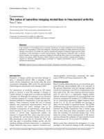
ELISA for IL-6 and IL-1β confirmed a slight, dose-dependent
reduction in cytokine production in the media of co-cultures
after treatment with CTLA-4-Ig as compared with untreated
cells (Figure 4). Interestingly, we were unable to dose TNFα
concentrations in culture medium because they were beyond
the standard curve, even following a 1:10 diluition of the cul-
ture medium.
Figure 2
B7.2 expression in macrophages after CTLA4-IG treatment by immunofluorescence analysisB7.2 expression in macrophages after CTLA4-IG treatment by immunofluorescence analysis. B7.2 expression by immunofluorescence analysis on
primary cultures of rheumatoid arthritis synovial macrophages (RA SM) (a) untreated, (b) treated with CTLA4-Ig 10 μg/ml, (c) treated with CTLA4-Ig
100 μg/ml and (d) treated with CTLA4-Ig 500 μg/ml; (e) on macrophages differentiated from THP-1 untreated, (f) treated with CTLA4-Ig 10 μg/ml,
(g) CTLA4-Ig 100 μg/ml and (h) CTLA4-Ig 500 μg/ml.
Arthritis Research & Therapy Vol 11 No 6 Cutolo et al.
Page 6 of 10
(page number not for citation purposes)
Analysis of mRNA expression in macrophages treated with
CTLA-4-Ig versus untreated macrophages (controls) showed
a downregulation of cytokine production for IL-6, TNFα and
TGFβ (Figures 5a to 5d). Western blot results by densitometry
analysis showed a slight, overall decrease in IL-6 and TNFα
protein expression (Figures 5e and 5f) in CTLA-4-Ig-treated
cells versus untreated cells, while TGFβ levels were not
detectable.
RA SM/Jurkat
RA SM that were co-cultured with T cells following the addi-
tion of CTLA-4-Ig (range from 100 to 500 μg/ml) induced a
decrease in the pro-inflammatory cytokine expression (Figures
6a and 6b). In particular, IL-6 and TNFα showed a significant
downregulation following the addition of CTLA-4-Ig at 500 μg/
ml (P < 0.05 and P < 0.001, respectively). Of note, in RA SM
and T cell co-cultures, adding CTLA-4-Ig at a lower concentra-
tion (100 μg/ml) induced a significant modulation in TNFα
expression alone (P < 0.001; Figures 6c and 6d).
TGFβ did not show any change in expression following the
addition of CTLA-4-Ig (range from 100 to 500 μg/ml) com-
pared with untreated RA SM (controls; data not shown).
Discussion
The current report is the first to describe the positivity for the
B7.2 co-stimulatory molecule in RA SM and its interaction with
the CTLA-4 competitor (CTLA-4-Ig).
Figure 3
Immunocytochemistry for IL-6, TNFalpha, IL-1beta and TGFbeta expression in co-cultures of macrophage/JurkatImmunocytochemistry for IL-6, TNFα, IL-1β and TGFβ expression in co-cultures of macrophage/Jurkat. Immunocytochemistry in co-cultures of mac-
rophage/Jurkat, (a) untreated (controls) and (a1) treated with CTLA4-Ig 500 μg/ml for IL-6, (b) untreated (controls) and (b1) treated with TNFalpha,
(c) untreated (controls) and (c1) treated with IL-1β and (d) untreated (controls) and (d1) treated with TGFβ. Quantification (mean value ± standard
deviation) of immunocytochemistry (ICC-QWin) for (e) IL-6, (f) TNFα, (g) IL-1β and (h) TGFβ expression in co-cultures of macrophage/Jurkat,
untreated (controls) and treated with CTLA4-Ig (10, 100, 500 μg/ml). * P < 0.05; ** P < 0.01; *** P < 0.001.
Available online />Page 7 of 10
(page number not for citation purposes)
Earlier studies had already detected the B7.2 molecule within
the RA synovial tissue (while B7.1 was found to be almost alto-
gether absent), and particularly so in areas enriched with
CD68-positive macrophages, such as the lining layer and
close to CD3-positive T cells [23]. However, the real implica-
tions of this observation were not determined at that time.
Researchers merely believed that it suggested the presence of
important therapeutic targets, such as co-stimulatory mole-
cules (B7 complex), in the synovial tissue, at least as far as the
early intervention on the immune response in RA was
concerned.
The present study shows that the CTLA-4-Ig fusion protein is
able to downregulate SM activation (co-cultured with T cells)
by interacting with the B7.2 molecule expressed on their sur-
face and by decreasing their production of inflammatory RA
cytokines (IL-6, TNFα, IL-1β).
Furthermore, for the first time, the present study shows that
CTLA4-Ig treatment reduces the expression of TGFbeta in
human macrophages.
Recent reports suggest that TGFβ-induced effects have an
autocrine pathogenetic importance in RA; for example, in
inducing matrix metalloproteinase-mediated matrix degrada-
tion/remodeling. It has also been suggested that in the context
of an inflammatory cytokine milieu, TGFβ supports de novo dif-
ferentiation of IL-17-producing T cells. Therefore, these obser-
vations may be strengthened by the present results [26,27].
Finally, we must now consider SM as possible direct targets of
CTLA-4-Ig-mediated downregulation. As a matter of fact, cur-
rent concepts suggest that inhibiting T cell co-stimulation is
the key mode of CTLA-4 action.
However, additional mechanisms of action may be related to
the fact that CTLA-4 directly binds to APC (such as macro-
phages) and might directly modulate their function. Thus,
CTLA-4 might not only indirectly affect T cell interaction with
APC, but CTLA-4 may also directly affect its primary cellular
target, which is the cells of the monocytic lineage (i.e. macro-
phages) that strongly express the B7-2 molecule (CD86) in
RA synovial tissue.
Figure 4
Evaluation of cytokine production by ELISA assayEvaluation of cytokine production by ELISA assay. Evaluation by ELISA assay of IL-6 and IL-1β production in supernatants of co-cultured macro-
phage/Jurkat untreated (controls (cnt)) and treated with CTLA4-Ig (10, 100, 500 μg/ml). Results are expressed as mean value ± standard deviation
from three experiments. * P < 0.05; ** P < 0.01; *** P < 0.001.
Arthritis Research & Therapy Vol 11 No 6 Cutolo et al.
Page 8 of 10
(page number not for citation purposes)
As is well known, macrophages are not only one of the main
inflammatory cytokine-producing cells in the RA synovial tis-
sue, but they also differentiate into bone resorbing osteo-
clasts, which are implicated in RA inflammatory bone erosions
and joint damage [28,29]. Recently, it was found that CTLA-4-
Ig directly inhibits the Receptor Activator for Nuclear Factor k
B Ligand (RANKL) - as well as the TNFα-mediated osteoclas-
togenesis in vitro in a dose-dependent manner (which is
clearly evident and significant at 100 μg/ml), without the need
for the concomitant presence of T cells [30].
Furthermore, CTLA-4-Ig was effective at inhibiting the TNF-
induced osteoclast formation in a non-T cell-dependent TNF-
induced model of arthritis, as well as at inhibiting the formation
of inflammatory bone erosions in vivo [30].
Therefore, even if CTLA-4-Ig typically works by inhibiting T cell
co-stimulation and T cell activation, we must now consider that
a further primary cellular target of CTLA-4-Ig might also be
mononuclear APC, such as SM that express the B.7 complex.
Although we co-cultured macrophages with T cells, the results
clearly suggest direct downregulation of the CTLA-4-Ig fusion
protein on the RA SM pro-inflammatory function. The next step
is to test these effects on SM monocultures.
Interestingly, recent concepts, such as reverse signalling, have
defined that the cell-cell interactions can be rather bidirec-
tional, which means that the 'ligand'-expressing cell (i.e. mac-
rophage) undergoes a functional change upon engaging with
the receptor [31]. For example, the binding of CTLA-4 to its lig-
Figure 5
Reverse transcriptase-polymerase chain reaction and western blot analysis for IL-6, TNFalpha and TGFbeta expression in co-cultures of macrophage/JurkatReverse transcriptase-polymerase chain reaction and western blot analysis for IL-6, TNFα and TGFβ expression in co-cultures of macrophage/Jur-
kat. Reverse transcriptase-polymerase chain reaction assay in co-cultures of macrophage/Jurkat, untreated (line 1: control) and treated with CTLA4-
Ig 10 μg/ml (line 2), CTLA4-Ig 100 μg/ml (line 3), CTLA4-Ig 500 μg/ml (line 4), for (a) β-actin (internal positive control), (b) IL-6, (c) TNFα and (d)
TGFβ. Line 5: molecular weight. Densitometry analysis of western blot in co-cultures of macrophage/Jurkat, untreated (line 2: control) and treated
with CTLA4-Ig 10 μg/ml (line 3), CTLA4-Ig 100 μg/ml (line 4), CTLA4-Ig 500 μg/ml (line 5) for (e) IL-6 and (f) TNFα protein expression in co-cul-
tures of macrophage/Jurkat. Line 1: molecular weight.
Available online />Page 9 of 10
(page number not for citation purposes)
and B7 might lead to a functional change in APC, as already
observed [32,33].
A different approach was previously used to study the role of
the CTLA-4 molecule in the inflammatory reaction of RA joints.
It involved blocking CTLA-4 with an anti-CTLA-4 antibody and
then assessing its effects on TNFα and IL-1 production in syn-
ovial fluid mononuclear cell cultures [34]. As expected, adding
the anti-CTLA-4 antibody enhanced TNFα and IL-1 production
in a dose-dependent manner.
Conclusions
In conclusion, the CTLA-4-Ig interaction we observed with SM
and their subsequent downregulation further support the key
role they play in various steps of RA, and may explain the ben-
eficial effects of CTLA-4-Ig fusion protein treatment in control-
ling the signs and symptoms of RA, even in advanced phases
of the disease.
Competing interests
The authors declare that they have no competing interests.
Authors' contributions
MC defined the design and coordination of the study, partici-
pated in interpretation of data, drafted the manuscript and pro-
vided general supervision and final approval of the version to
be published. SS participated in the design and coordination
of the study and carried out western blot analysis. PM partici-
pated in the design and coordination of the study, performed
the statistical analysis, cultured the cells and carried out RT-
PCR analysis. AS participated in patient selection and clinical
evaluation of data. BS participated in patient selection and
clinical evaluation of data. BV participated in acquisition and
data collection. PT provided surgical tissue samples (synovial
tissues) in order to culture the RA SM. PC provided surgical
tissue samples (synovial tissues) in order to culture the RA
SM. LF provided surgical tissue samples (synovial tissues) in
order to culture the RA SM. RB participated in the design and
coordination of the study, cultured the cells, carried out immu-
nocytochemistry analysis and helped draft the manuscript.
Acknowledgements
The study was partially supported by a research grant from Bristol-Myers
Squibb Company (In Vitro Study IM 101-157/11.07.2007), who also
provided the CTLA-4-Ig molecule.
Figure 6
Immunocytochemistry for IL-6 and TNFalpha expression in co-cultures of RA SM/JurkatImmunocytochemistry for IL-6 and TNFα expression in co-cultures of RA SM/Jurkat. Immunocytochemistry in co-cultures of rheumatoid arthritis syn-
ovial macrophages (RA SM)/Jurkat, (a) untreated (controls) and (a1) treated with CTLA4-Ig 500 μg/ml for IL-6 and (b) untreated (controls) and (b1)
treated with CTLA4-Ig 500 μg/ml for TNFα. Quantification (mean value ± standard deviation) of immunocytochemistry (ICC-QWin) for (c) IL-6 and
(d) TNFα. * P < 0.05; ** P < 0.01; *** P < 0.001.
Arthritis Research & Therapy Vol 11 No 6 Cutolo et al.
Page 10 of 10
(page number not for citation purposes)
References
1. Lundy SK, Sarkar S, Tesmer LA, Fox DA: Cells of the synovium
in rheumatoid arthritis. T lymphocytes. Arthritis Res Ther 2007,
9:202.
2. Brennan FM, McInnes JIB: Evidence that cytokines play a role in
rheumatoid arthritis. J Clin Invest 2008, 118:3537-3545.
3. Cutolo M, Straub RH, Bijlsma JW: Neuroendocrine-immune
interactions in synovitis. Nat Clin Pract Rheumatol 2007,
3:627-634.
4. Cope AP, Schulze-Koops H, Aringer M: The central role of T cells
in rheumatoid arthritis. Clin Exp Rheumatol 2007, 25:S4-11.
5. Miossec P: Dynamic interactions between T cells and dendritic
cells and their derived cytokines/chemokines in the rheuma-
toid synovium. Arthritis Res Ther 2008, 10:S2.
6. Olsen NJ, Stein MC: New drugs for rheumatoid arthritis. N Engl
J Med 2004, 350:2167-2179.
7. Abbott JD, Moreland LW: Rheumatoid arthritis: Developing
pharmacological therapies. Expert Opin Investig Drugs 2004,
13:1007-1018.
8. Mariette X: Emerging biological therapies in rheumatoid
arthritis. Joint Bone Spine 2004, 71:470-479.
9. Goronzy JJ, Weyand CM: T-cell co-stimulatory pathways in
autoimmunity. Arthritis Res Ther 2008, 10:S3.
10. Trikudanathan S, Sayegh MH: The evolution of the immunobiol-
ogy of co-stimulatory pathways: clinical implications. Clin Exp
Rheumatol 2007, 25:S12-21.
11. Ueda H, Howson JM, Esposito L, Heward J, Snook H, Chamberlain
G, Rainbow DB, Hunter KM, Smith AN, Di Genova G, Herr MH,
Dahlman I, Payne F, Smyth D, Lowe C, Twells RC, Howlett S,
Healy B, Nutland S, Rance HE, Everettt V, Smink LJ, Lam AC,
Cordell HJ, Walzer NM, Bordin C, Hulme J, Motzo C, Cucca F,
Hess JF, et al.: Association of the T-cell regulatory gene CTLA-
4 with susceptibility to autoimmune disease. Nature 2003,
423:506-511.
12. Walunas TL, Lenschow DJ, Bakker CY, Linsley PS, Freeman GJ,
Green JM, Thompson CB, Bluestone JA: CTLA-4 can function as
a negative regulator of T cell activation. Immunity 1994,
1:405-413.
13. Alegre ML, Frauwirth KA, Thompson CB: T-cell regulation by
CD28 and CTLA-4. Nat Rev Immunol 2001, 1:220-228.
14. Leibson PJ: The regulation of lymphocyte activation by inhibi-
tory receptors. Curr Opin Immunol 2004, 16:328-336.
15. Alten R, Bosch F Van den, Appelboom T, Leon M, Emery P, Cohen
S, Luggen M, Shergy W, Nuamah I, Becker JC: Co-stimulatory
blockade in patients with rheumatoid arthritis: A pilot, dose-
finding, double-blind, placebo-controlled clinical trial evaluat-
ing CTLA-4-Ig and LEA29Y eighty-five days after the first
infusion. Arthritis Rheum 2002, 46:1470-1479.
16. Kremer JM, Westhovens R, Leon M, Di Giorgio E, Alten R, Stein-
feld S, Russell A, Dougados M, Emery P, Nuamah IF, Williams GR,
Becker JC, Hagerty DT, Moreland LW: Treatment of rheumatoid
arthritis by selective inhibition of T-cell activation with fusion
protein CTLA-4Ig. N Engl J Med 2003, 349:1907-1915.
17. Malstrom V, Trollmo C, Klareskog L: Modulating co-stimulation:
A rational strategy in the treatment of rheumatoid arthritis?
Arthritis Res Ther 2005, 7:S15-20.
18. Buch MH, Vital EM, Emery P: Abatacept in the treatment of
rheumatoid arthritis. Arthritis Res Ther 2008, 10:S5.
19. Rozelle AL, Genovese MC: Efficacy results from pivotal clinical
trials with abatacept. Clin Exp Rheumatol 2007, 25:S30-34.
20. Michaud K, Bombardier C, Emery P: Quality of life in patients
with rheumatoid arthritis: Does abatacept make a difference?
Clin Exp Rheumatol 2007, 25:S35-45.
21. Cutolo M, Capellino S, Montagna P, Sulli A, Seriolo B, Villaggio B:
Anti-inflammatory effects of Leflunomide in combination with
methotrexate on co-culture of T lymphocytes and synovial
macrophages from rheumatoid arthritis patients. Ann Rheum
Dis 2006, 65:728-735.
22. Goodier MR, Londei M: Low concentrations of lipopolysaccha-
ride synergize with peptides to augment human T-cell prolif-
eration and can prevent the induction of non-responsiveness
by CTLA-4-Ig. Immunology 2001, 102:15-23.
23. Balsa A, Dixey J, Sansom DM, Maddison PJ, Hall ND: Differential
expression of the co-stimulatory molecules B7.1 (CD80) and
B7.2 (CD86) in rheumatoid synovial tissue. Br J Rheumatol
1996, 35:33-37.
24. Thomas R, Quinn C: Functional differentiation of dendritic cells
in rheumatoid arthritis. Role of the CD86 in the synovium. J
Immunol 1996, 156:3074-3086.
25. Gao YH, Wang P, Takagi K, Shimozato O, Yagita H, Okigaki T,
Matasumura M: Expression of a soluble form of CTLA-4 on
macrophage and its biological activity. Cell Research 1999,
9:189-199.
26. Pohlers D, Beyer A, Koczan D, Wilhelm D, Thiesen HJ, Kinne RW:
Constitutive upregulation of the transforming growth factor-β
pathway in rheumatoid arthritis synovial fibroblasts. Arthritis
Res Ther 2007, 9:R59.
27. Mangan PR, Harrington LE, Quinn DB, Helms WS, Bullard DC,
Elson CO, Hatton RD, Wahl SM, Schoeb TR, Weaver CT: Trans-
forming growth factor-beta induces development of the
T(H)17 lineage. Nature 2006, 441:231-234.
28. Teitelbaum SL: Bone resorption by osteoclasts. Science 2000,
289:1504-1508.
29. McInnes IB, Schett G: Cytokines in rheumatoid arthritis. Nat
Rev Immunol 2007, 7:429-442.
30. Axmann R, Herman S, Zaiss M, Franz S, Polzer K, Zwerina J, Her-
rmann M, Smolen J, Schett G: CTLA-4 directly inhibits osteo-
clast formation. Ann Rheum Dis 2008, 67:1603-1609.
31. Alegre ML, Fallarino F: Mechanisms of CTLA-4-Ig in tolerance
induction. Curr Pharm Des 2006, 12:149-160.
32. Fallarono F, Grohmann U, Hwang KW, Orabona C, Vacca C,
Bianchi R, Belladonna ML, Fioretti MC, Alegre ML, Puccetti P:
Modulation of tryptophan catabolism by regulatory T cells. Nat
Immunol 2003, 4:1206-1212.
33. Grohmann U, Orabona C, Fallarono F, Vacca C, Calcinaro F, Fal-
orni A, Candeloro P, Belladonna ML, Bianchi R, Fioretti MC, Puc-
cetti P: CTLA-4-Ig regulates tryptophan catabolism in vivo. Nat
Immunol 2002, 3:1097-1101.
34. Liu MF, Yang CY, Li JS, Lai KA, Chao SC, Lei HY: Increased
expression of down-regulatory CTLA-4 molecule on T lym-
phocytes from rheumatoid synovial compartment. Scand J
Immunol 1999, 50:68-72.