Báo cáo y học: "Serum levels of autoantibodies against C-reactive protein correlate with renal disease activity and response to therapy in lupus nephritis" pps
Bạn đang xem bản rút gọn của tài liệu. Xem và tải ngay bản đầy đủ của tài liệu tại đây (500.54 KB, 9 trang )
Open Access
Available online />Page 1 of 9
(page number not for citation purposes)
Vol 11 No 6
Research article
Serum levels of autoantibodies against C-reactive protein
correlate with renal disease activity and response to therapy in
lupus nephritis
Christopher Sjöwall
1
, Agneta Zickert
2
, Thomas Skogh
1
, Jonas Wetterö
1
and Iva Gunnarsson
2
1
Rheumatology/AIR, Clinical and Experimental Medicine, Linköping University, SE-581 85 Linköping, Sweden
2
Department of Medicine, Rheumatology Unit, Karolinska University Hospital, Solna, SE-171 76 Stockholm, Sweden
Corresponding author: Christopher Sjöwall,
Received: 2 Aug 2009 Revisions requested: 26 Aug 2009 Revisions received: 3 Dec 2009 Accepted: 11 Dec 2009 Published: 11 Dec 2009
Arthritis Research & Therapy 2009, 11:R188 (doi:10.1186/ar2880)
This article is online at: />© 2009 Sjöwall et al.; licensee BioMed Central Ltd.
This is an open access article distributed under the terms of the Creative Commons Attribution License ( />),
which permits unrestricted use, distribution, and reproduction in any medium, provided the original work is properly cited.
Abstract
Introduction Serum levels of C-reactive protein (CRP) seldom
reflect disease activity in systemic lupus erythematosus (SLE).
We have previously shown that autoantibodies against neo-
epitopes of CRP often occur in SLE, but that this does not
explain the modest CRP response seen in flares. However, we
have repeatedly found that anti-CRP levels parallel lupus
disease activity, with highest levels in patients with renal
involvement; thus, we aimed to study anti-CRP in a material of
well-characterized lupus nephritis patients.
Methods Thirty-eight patients with lupus nephritis were
included. Treatment with corticosteroids combined with
cyclophosphamide, mycophenolate mofetil or rituximab was
started after baseline kidney biopsy. A second biopsy was taken
after ≥ 6 months. Serum creatinine, cystatin C, complement,
anti-dsDNA, anti-CRP and urinalysis were done on both
occasions. Biopsies were evaluated regarding World Health
Organisation (WHO) class and indices of activity and chronicity.
Renal disease activity was estimated using the British Isles
Lupus Assessment Group (BILAG) index.
Results At baseline, 34/38 patients had renal BILAG-A; 4/38
had BILAG-B. Baseline biopsies showed WHO class III (n = 8),
IV (n = 19), III to IV/V (n = 3) or V (n = 8) nephritis. Seventeen
out of 38 patients were anti-CRP-positive at baseline, and six at
follow-up. Overall, anti-CRP levels had dropped at follow-up (P
< 0.0001) and anti-CRP levels correlated with renal BILAG (r =
0.29, P = 0.012). A positive anti-CRP test at baseline was
superior to anti-dsDNA and C1q in predicting poor response to
therapy as judged by renal BILAG. Baseline anti-CRP levels
correlated with renal biopsy activity (r = 0.33, P = 0.045), but
not with chronicity index. Anti-CRP levels were positively
correlated with anti-dsDNA (fluorescence-enhanced
immunoassay: r = 0.63, P = 0.0003; Crithidia luciliae
immunofluorescence microscopy test: r = 0.44, P < 0.0001),
and inversely with C3 (r = 0.35, P = 0.007) and C4 (r = 0.29, P
= 0.02), but not with C1q (r = 0.14, P = 0.24). No associations
with urinary components, creatinine, cystatin C or the glomerular
filtration rate were found.
Conclusions In the present study, we demonstrate a statistically
significant correlation between anti-CRP levels and
histopathological activity in lupus nephritis, whereas a baseline
positive anti-CRP test predicted poor response to therapy. Our
data also confirm previous findings of associations between
anti-CRP and disease activity. This indicates that anti-CRP
could be helpful to assess disease activity and response to
therapy in SLE nephritis, and highlights the hypothesis of a
pathogenetic role for anti-CRP antibodies in lupus nephritis.
Introduction
Systemic lupus erythematosus (SLE) is characterized by mul-
tiple organ involvement, by production of a wide range of anti-
nuclear antibodies and by the presence of immune complexes
in the inflamed organs [1]. Impaired clearance of cellular
debris by the reticuloendothelial system is considered a key
event in the initiation and maintenance of SLE. Autoantigens
escaping physiological clearance may thus become exces-
sively presented to the adaptive immune system, resulting in
loss of peripheral tolerance and occurrence of a multitude of
BILAG: British Isles Lupus Assessment Group; BSA: bovine serum albumin; CLIFT: Crithidia luciliae immunofluorescence microscopy test; CRP: C-
reactive protein; dsDNA: double-stranded DNA; ELISA: enzyme-linked immunosorbent assay; IFN: interferon; IL: interleukin; mCRP: monomeric C-
reactive protein; SLE: systemic lupus erythematosus; TNF: tumour necrosis factor; WHO: World Health Organisation.
Arthritis Research & Therapy Vol 11 No 6 Sjöwall et al.
Page 2 of 9
(page number not for citation purposes)
autoantibodies - the waste disposal theory [2]. Antibodies
against dsDNA are frequently found both in serum and inflam-
matory lesions in glomerulonephritis [3]. The circulating levels
of anti-dsDNA often correlate with disease activity, and these
autoantibodies are presumed to be of pathogenetic impor-
tance in lupus nephritis [4-6].
The pentraxins constitute an evolutionarily conserved group of
proteins, which are expressed during infection, systemic
inflammation or tissue damage and participate in the acute
phase response in many species [7]. The pentraxin family
includes long pentraxins (such as pentraxin 3, produced by
mononuclear cells in response to lipopolysaccharides, IL-1β
and TNF) and the liver-derived short pentraxins C-reactive pro-
tein (CRP) and serum amyloid P component mainly generated
by stimulation with IL-6 [7]. Despite raised levels of IL-6 and
extensive systemic inflammation, serum CRP concentrations
typically remain low in lupus flares [8], although differences
between certain disease manifestations [9] and conflicting
data have been reported [10]. The novel in vitro finding that
IFNα mediates suppression of IL-6-induced CRP expression
in human hepatocytes, however, could possibly explain the
weak CRP response in SLE flares [11].
CRP has several biological functions that are related to affinity
for molecules exposed on bacteria and apoptotic cells/cell
debris, such as phosphorylcholine, nucleosomes, and ribonu-
cleoproteins (snRNPs), thereby resembling a primitive form of
a natural antibody [12]. In addition, like IgG class antibodies,
CRP interacts with cellular Fcγ receptors, thereby facilitating
the phagocytic clearance of circulating opsonized material.
Activation of the classical complement pathway is considered
one of the main physiological functions of CRP. In contrast to
IgG-mediated classical activation, however, CRP-mediated
activation appears to be essentially limited to the initial stages
involving C1 to C4, with less formation of the membrane attack
complex [13]. Furthermore, at sufficient concentrations, solu-
ble native CRP may prevent activation of the classical comple-
ment pathway on biological surfaces due to consumption of
soluble C1q without binding C2/C4 [14].
In line with its role as a scavenger of autoantigens from dead
or dying cells, single nucleotide polymorphisms of the CRP
gene have been found to associate with low baseline levels of
CRP, with production of antinuclear antibodies, and with
increased susceptibility to SLE [8]. Furthermore, in two murine
lupus models, subcutaneous CRP injections delayed the dis-
ease onset, reversed nephritis, and prolonged the survival of
the animals - indicating a preventive and disease-modifying
role for CRP in SLE [8,13]. Very recently, however, this finding
was contradicted by others [15].
The presence of autoantibodies against CRP in lupus was
originally described by Frank A Robey and coworkers in 1985
[16]. Later, Bell and colleagues reported a high frequency of
autoantibodies against a certain dissociated and tissue-bound
form of CRP, recognized as monomeric CRP (mCRP) in SLE,
and at lower prevalence rates in subacute cutaneous lupus
erythematosus and primary biliary cirrhosis [17]. Since then
several groups have confirmed the finding of IgG class autoan-
tibodies against monomeric CRP (anti-CRP) in SLE and in
some other rheumatic conditions [18-22]. In addition, the
presence of autoantibodies against pentraxin 3 was recently
shown in SLE patients [23,24].
In a series of papers, we have demonstrated the strong corre-
lations between anti-CRP antibody level and disease activity
as reflected by the SLE disease activity index, anti-dsDNA anti-
body levels, and complement levels [12]. The anti-CRP assay
has been shown to be antigen-specific [17], without false pos-
itive results due to the presence of immune complexes [19,25]
or antibodies to DNA or nucleosomes [26]. We have also con-
sistently found that most patients with raised anti-CRP anti-
body levels appear to have a disease phenotype with renal
involvement. The latter was recently confirmed by Tan and col-
leagues, who found elevated anti-CRP in SLE patients - where
the antibody levels paralleled disease activity, particularly in
individuals with lupus nephritis [27]. The authors also studied
renal histopathology and found that anti-CRP antibody levels
correlated with tubulointerstitial lesions and the chronicity
index, but not with the activity index [27]. The aim of the
present study was therefore to compare anti-CRP antibody
levels in well-characterized lupus nephritis patients and to
seek potential associations with histopathology, renal activity
and response to therapy.
Materials and methods
Patients
Thirty-eight patients meeting the 1982 American College of
Rheumatology classification criteria for SLE [28] were
included in the study. All patients had biopsy-proven active
lupus nephritis (during the period 1995 to 2006) and partici-
pated in a prospective control programme at the rheumatology
clinic of Karolinska University Hospital (Stockholm, Sweden).
Thirty-four of the 38 patients (89%) were women (mean age,
33.0 years; range, 19 to 61 years) and four patients were men
(mean age, 34.8 years; range, 18 to 50 years). Thirty-three out
of 38 patients (87%) were Caucasian. The mean duration of
SLE was 6.9 years (range, 0 to 34 years). At baseline, 27
patients displayed proliferative nephritis (World Health Organ-
isation (WHO) class III or IV), eight patients showed membra-
nous pattern (WHO class V), and biopsies in three patients
were classified as both proliferative and membranous.
The patients were treated in accordance with clinical routine
for lupus nephritis [29], including corticosteroids combined
with cyclophosphamide intravenously (n = 27) or orally (n = 1),
rituximab (n = 6), and mycophenolate mofetil (n = 3). One
patient was initially treated with mycophenolate mofetil, but
switched to intravenous cyclophosphamide after 3 months. At
Available online />Page 3 of 9
(page number not for citation purposes)
the timepoint for the first renal biopsy, clinical data, blood sam-
ples and urinary samples were collected. Serum samples were
kept frozen at -70°C for future analyses. After induction ther-
apy (mean time, 8 months; range, 6 to 15 months), the patients
underwent a second renal biopsy and further clinical and lab-
oratory data were collected. Additional data are presented in
Table 1.
Renal histopathology
Renal biopsies were performed by percutaneous ultrasonog-
raphy-guided puncture in accordance with a standard proto-
col. The renal tissue obtained was staged according to the
WHO classification for lupus nephritis [30]. All biopsies were
evaluated by light microscopy, immunofluorescence and elec-
tron microscopy. The biopsies were graded according to a
standardized semiquantitative histological scoring protocol for
activity and chronicity indices [31].
Renal disease activity and response to therapy
Renal disease activity was estimated using the classical Brit-
ish Isles Lupus Assessment Group (BILAG) index [32]. An
improvement of at least two grades in the renal domain of
BILAG (that is, from A to C or from B to D) at follow-up was
required for the patient to be regarded as a responder. The
BILAG index was translated into numerical data for correlation
analyses (A = 9, B = 3, C = 1 and D = 0) as suggested by Dr
David A Isenberg, London (personal communication).
Laboratory and serological measures
Renal function was monitored by urinalysis (dip-slide proce-
dure), urinary sediment assessment, 24-hour urine albumin
excretion, serum creatinine, the glomerular filtration rate
assessed by urinary clearance of iohexol according to clinical
routine, and cystatin C (turbidimetry).
Analyses of complement component C1q were performed by
rocket electrophoresis using polyclonal rabbit anti-C1q
(DAKO, Glostrup, Denmark). Levels of C1q were expressed
as the percentage of the levels of healthy blood donors (nor-
mal range, 76 to 136%). C3 (normal range, 0.70 to 1.3 g/l)
and C4 (normal range, 0.13 to 0.32 g/l) were determined by
nephelometry.
Assessments of serum IgG anti-dsDNA antibodies were made
by the ImmunoCAP fluorescence-enhanced immunoassay
(Pharmacia, Uppsala, Sweden), normal range <15 IU/ml, and
by the Crithidia luciliae immunofluorescence microscopy test
(CLIFT) with cut-off titre 1:10.
Anti-C-reactive protein antibody assay
IgG anti-CRP antibodies were measured with an ELISA as
described previously [26]. To avoid systematic errors, samples
from patients and controls were always randomly mixed on the
microtitre plates and analysed on the same occasion. Anti-
CRP antibody levels were expressed as the percentage of a
positive reference sample from a SLE patient at flare repre-
senting 100 arbitrary units. No differences were apparent con-
sidering the groups of men and women in the control material.
To exclude the possibility of non-specific binding, each serum
was also tested in the same way on uncoated plates.
Statistics
Figures were prepared in GraphPad Prism (version 4.0;
GraphPad Software Inc., San Diego, CA, USA). Correlation
analyses were performed using Spearman's rank correlation
(SPSS for Windows version 15.0.0; SPSS Inc. (IBM), Chi-
cago, IL, USA), and differences between groups were calcu-
lated with the Wilcoxon signed rank test or the Mann-Whitney
U test (GraphPad). Response to therapy was compared by
chi-square analysis using StatCalc (Epi Info version 3.5.1;
Centers for Disease Control and Prevention, Atlanta, GA,
USA). Two-tailed P < 0.05 was considered significant.
Ethics
Informed consent was obtained from all subjects. The
research protocol was approved by the regional ethics com-
mittee in Stockholm.
Results
Anti-CRP antibody levels were determined at baseline and fol-
low-up in each of the 38 patients. The cut-off value for positive
reaction was set at 8 units, calculated from the 95th percentile
in 100 healthy blood donors (controls). As indicated in Table
1, 17 out of 38 patients (45%) were judged anti-CRP anti-
body-positive at baseline and six patients (16%) were positive
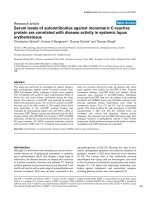
at follow-up. Overall, the anti-CRP antibody levels were signif-
icantly reduced at follow-up (P < 0.0001) (Figure 1).
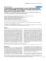
Anti-CRP antibody levels were more efficiently reduced in
patients who responded to therapy as judged by renal histopa-
thology (data not shown), and baseline anti-CRP levels were
significantly higher (P = 0.0097) in patients who did not reach
a renal BILAG improvement of at least two grades (Figure 2).
Neither a baseline lowered C1q (relative risk = 1.58, 95%
confidence interval = 0.87 to 2.84) nor a positive anti-dsDNA
antibody test as judged by the CLIFT (relative risk = 1.18, 95%
confidence interval = 0.42 to 3.26) predicted response to
therapy, whereas a positive anti-CRP test did (relative risk =
2.16, 95% confidence interval = 1.20 to 3.89). Response to
therapy in relation to anti-CRP, adjusted for C1q and anti-
dsDNA antibody status, respectively, is illustrated in Tables 2
and 3. A positive anti-CRP test was thus associated with a
poor response to therapy, particularly in patients with normal
C1q levels and a positive anti-dsDNA antibody test (CLIFT).
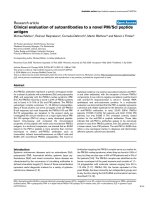
Patients with proliferative nephritis (WHO class III or IV) had a
greater reduction of anti-CRP antibody levels compared with
patients with membranous (WHO class V) nephritis (Figure 3),
although this was not statistically significant (P = 0.08).
Arthritis Research & Therapy Vol 11 No 6 Sjöwall et al.
Page 4 of 9
(page number not for citation purposes)
Table 1
Clinical characteristics and laboratory data for patients
Baseline Follow-up
Anti-CRP-positive Anti-CRP-negative Mann-Whitney Anti-CRP-positive Anti-CRP-negative Mann-Whitney
Age (years) 30.5 (13.1) 35.0 (11.6) NS 27.0 (8.2) 35.1 (12.7) NS
Gender
Female 16 18 5 29
Male13 13
Ethnicity
Caucasian 16 17 5 28
Iranian Caucasian 0 3 0 3
Iraqi Caucasian 0 1 0 1
Asian 1 0 1 0
Creatinine (μmol/l) 95.3 (43.3) 97.0 (51.0) NS 78.3 (9.6) 83 (46.2) NS
Albuminuria (g/day) 1.7 (1.4) 2.7 (2.3) NS 1.6 (1.3) 0.8 (0.9) NS
C3 (g/l) 0.47 (0.26) 0.57 (0.24) NS 0.85 (0.28) 0.80 (0.27) NS
C4 (g/l) 0.09 (0.06) 0.10 (0.05) NS 0.14 (0.03) 0.14 (0.07) NS
C1q (% of normal
reference)
57.9 (37.3) 79.0 (36.7) NS 73.0 (32.4) 83.0 (28.1) NS
Anti-dsDNA-positive 16 18 3 21
CLIFT titre, median 200 25 P = 0.04 25 10 NS
Renal histopathology
Class I 0 0 0 1
Class II00 113
Class III 1 7 1 2
Class IV 12 7 2 4
Class III to IV/V 1 2 0 2
Class V 3 5 2 10
Activity index 7.0 (3.1) 5.5 (3.5) NS 3.5 (2.0) 2.7 (3.2) NS
Chronicity index 2.3 (1.9) 1.4 (2.0) NS 2.7 (1.0) 2.7 (2.4) NS
BILAG index
A1420 15
B31 414
C00 08
D00 15
Treatment
Prednisolone, mean
daily dose (mg)
10.1 (10.5) 12.9 (17.8) NS 14.5 (7.2) 10.6 (5.0) NS
Mycophenolate
mofetil
12
Cyclophosphamide 15 13
Rituximab 0 6
Mycophenolate
mofetil/
cyclophosphamide
10
Data presented as mean (standard deviation) or n. BILAG, British Isles Lupus Assessment Group; CLIFT, Crithidia luciliae immunofluorescence
microscopy test; CRP, C-reactive protein; NS, not significant.
Available online />Page 5 of 9
(page number not for citation purposes)
Of the 17 anti-CRP antibody-positive patients at baseline, 16
patients had received cyclophosphamide and two patients
received mycophenolate mofetil (one in combination with
cyclophosphamide). One individual increased dramatically in
anti-CRP, from 8 units at baseline to 214 units at follow-up.
This patient with WHO class IVc nephritis received cyclophos-
phamide intravenously, but did not respond to therapy and
was regarded as an outlier (not included in all analyses). All six
anti-CRP antibody-positive patients at follow-up were positive
also at baseline, and all had received cyclophosphamide intra-
venously as induction therapy. Five of these six patients did not
respond to therapy as judged by renal histopathology; one
patient switched from WHO class IVb to a IIa pattern, but the
remaining five patients had a similar or worsened histopatho-
logic picture with increment in the chronicity index. Using the
BILAG index, the same five individuals with persistently raised
anti-CRP levels did not respond to therapy (renal BILAG
improvement ≥ 2 grades). In the whole material, no obvious
relation between anti-CRP antibody reduction and the type of
induction therapy was found. All six patients receiving rituxi-
mab were consistently anti-CRP-negative. As illustrated in Fig-
ure 4, anti-CRP antibody levels at baseline showed a
moderate but statistically significant positive correlation with
the renal biopsy activity index (r = 0.33, P = 0.045), whereas
the association at follow-up did not reach statistical signifi-
cance (r = 0.30, P = 0.061). Accumulated anti-CRP data
yielded an even stronger correlation with histopathological
activity (r = 0.37, P = 0.0017). No associations between anti-
CRP levels and the chronicity index were found (not shown).
Figure 5 shows the correlation between anti-CRP antibody
levels and the BILAG index (r = 0.29, P = 0.012); when solely
anti-CRP-positive samples were included, the coefficient was
slightly decreased (r = 0.20). Using only anti-CRP-positive/
anti-dsDNA-negative samples (as judged by the CLIFT), anti-
CRP levels were barely associated with renal BILAG, but the
number of observations here were small (r = 0.10). The corre-
lation between BILAG and anti-dsDNA antibodies was weaker
still, however, measured by the fluorescence-enhanced immu-
noassay (r = 0.04) as well as by the CLIFT (r = 0.14); when
anti-DNA-negative samples (CLIFT) were excluded, the coeffi-
cient was further decreased (r = 0.08). Looking at anti-CRP-
negative/anti-dsDNA-positive samples (CLIFT), anti-DNA was
not significantly associated with renal BILAG. On the other
hand, anti-CRP levels correlated with anti-dsDNA antibodies
as measured with fluorescence-enhanced immunoassay (r =
0.63, P = 0.0003) and the CLIFT (r = 0.44, P < 0.0001).
Inverse correlations between anti-CRP and C4 (r = 0.29, P =
0.02) as well as between anti-CRP and C3 (r = 0.35, P =
0.007) were observed, while the tendency to an inverse rela-
tion between anti-CRP and C1q was not significant (r = 0.14,
p = 0.24). No associations between anti-CRP and urinary
components, creatinine, cystatin C or the glomerular filtration
rate were found.
Discussion
In the present study, we demonstrate a moderate but statisti-
cally significant correlation between anti-CRP antibody levels
and renal biopsy activity index in lupus nephritis. Elevated
baseline levels of anti-CRP were also found to be predictive of
the therapeutic response as judged by renal BILAG at follow-
Figure 1
Anti-C-reactive protein antibody levels in systemic lupus erythematosus patients before and after induction therapyAnti-C-reactive protein antibody levels in systemic lupus erythematosus
patients before and after induction therapy. Anti-C-reactive protein
(anti-CRP) antibody levels at baseline and follow-up in 37 systemic
lupus erythematosus patients (outlier excluded). Paired data were ana-
lysed with the Wilcoxon signed rank test.
Figure 2
Baseline anti-C-reactive protein antibody levels in responders and non-responders to given therapyBaseline anti-C-reactive protein antibody levels in responders and non-
responders to given therapy. At baseline, there was a highly significant
difference in anti-C-reactive protein (anti-CRP) antibody levels between
patients that would respond (n = 16) and would not respond (n = 22)
to therapy (analysed with Mann-Whitney U test). Response to therapy
was defined as a renal British Isles Lupus Assessment Group improve-
ment ≥ 2 grades. The limitations extend down from the lowest value
and up to the highest. Median value for responders was 4 units, and
was 9 units for nonresponders. Boxes show the 25th to 75th percen-
tile, with median values marked inside.
Arthritis Research & Therapy Vol 11 No 6 Sjöwall et al.
Page 6 of 9
(page number not for citation purposes)
up. Finally, the present study also confirms previous findings of
associations between anti-CRP and lupus disease activity as
assessed by common clinical and laboratory disease activity
measures, such as complement and anti-dsDNA levels.
Although the results of this descriptive study do not allow con-
clusions regarding nephritogenic properties of anti-CRP anti-
bodies, our data imply that anti-CRP antibody testing is a
useful tool to support the clinician's evaluation of disease
activity and response to therapy in lupus nephritis.
Distinction of disease activity from organ damage in SLE
remains a challenge. Indirect assessment of disease activity
such as the BILAG index has proven reliable and sensitive to
change [33], but it is time consuming and requires a deft cer-
tified clinician. Microscopic examination of a kidney biopsy,
with classification and estimation of indices of renal disease
activity and chronicity, today offers the best possibility to esti-
mate renal disease and its response to therapy. Biopsy is
costly and is associated with considerable risks for the patient,
however, and can therefore not be done ad infinitum. Labora-
tory variables such as circulating complement components
and anti-dsDNA antibodies can be helpful, but there is an
urgent need for additional reliable biomarkers of disease activ-
ity in SLE.
We have previously demonstrated that the occurrence of
autoantibodies against the tissue-based/monomeric CRP is a
common finding in SLE, particularly in patients with nephritis
[12], and that anti-CRP antibody levels correlate with SLE dis-
ease activity index, anti-dsDNA and complement components
[26]. These findings have been confirmed by several groups
[18,20-22], although not in a recent study by Kessel and col-
leagues [34]. According to our experience, the prevalence of
positive anti-CRP tests in SLE is 30 to 50% depending on dis-
ease activity and disease phenotype [19,25,26]. Since the
present study was limited to patients with lupus nephritis, the
prevalence of 45% positive anti-CRP antibody tests was
slightly below expectation. A higher prevalence rate was
reported by Bell and colleagues [17], whereas an appreciably
lower frequency was found by Shoenfeld and colleagues [22].
Apart from differences in patient selection and disease pheno-
type (that is, renal involvement), the diverging results in these
studies [18,20-22,27] may be due to differences in methodo-
logical details regarding anti-CRP antibody analysis. Contrast-
ing to most other studies, we refrain from the use of BSA to
block nonspecific IgG-binding in our anti-CRP antibody assay,
since serum antibodies to BSA (and other dietary proteins) are
common and may thus affect the results, regardless of
whether or not BSA is also included in the dilution buffer [35].
In a recent study by Tan and colleagues, positive correlations
were reported regarding anti-CRP antibody levels and chronic
Figure 3
Anti-C-reactive protein antibody levels in patients with proliferative ver-sus membranous lupus nephritisAnti-C-reactive protein antibody levels in patients with proliferative ver-
sus membranous lupus nephritis. The levels of anti-C-reactive protein
(anti-CRP) antibodies in the 26 patients with proliferative lupus nephri-
tis (World Health Organisation (WHO) class III/IV) were reduced to a
greater extent than the eight patients with membranous lupus nephritis
(WHO class V), although not statistically significant by the Mann-Whit-
ney U test (mean difference -8.3 vs. -1.1 units; P = 0.08). The vertical
bars denote 95% confidence intervals. The outlier as well as the three
patients with class III to IV/V nephritis were not included in this analysis.
20
18
16
14
12
10
8
6
4
2
0
-2
-4
-6
WHO III/IV
WHO V
Baseline
Follow-up
Anti-CRP antibody level (arbitrary units)
Table 2
Renal BILAG response and the C1q/anti-CRP status demonstrated
BILAG response <2 BILAG response ≥ 2
C1q low
Anti-CRP-positive 8 2
Anti-CRP-negative 5 5
Relative risk 1.60 (0.80 to 3.20)
C1q normal
Anti-CRP-positive 6 1
Anti-CRP-negative 3 8
Relative risk 3.14 (1.14 to 8.64)
Relative risks presented as chi-square (95% confidence interval). BILAG, British Isles Lupus Assessment Group; CRP, C-reactive protein.
Available online />Page 7 of 9
(page number not for citation purposes)
renal histology features such as tubular atrophy, interstitial
fibrosis and the chronicity index score, as well as regarding
disease activity assessments (that is, interstitial inflammation
and the SLE disease activity index) [27]. In contrast to the cur-
rent study, however, anti-CRP levels were not associated with
the renal activity index [27]. Our previous findings [25,26], as
well as the results from the present study, support the notion
that anti-CRP primarily reflects disease activity rather than
chronicity, severity or organ damage. The connection with
renal involvement is clear cut, but seems to be stronger in
WHO class III or IV than in membranous nephritis (Figure 3).
In this context, very interestingly, the presence of surface-
bound CRP has been demonstrated in the renal mesangium
and in glomerular capillary walls in specimens from patients
with lupus nephritis [36]. Since CRP was found to co-localize
with IgG, it is probable that this actually represent immune
complexes consisting of mCRP-anti-CRP. Further studies on
this interesting matter are underway.
During induction therapy, anti-CRP appears to behave simi-
larly to anti-dsDNA antibodies [26,37], but differently from
autoantibodies to SS-A/Ro and SS-B/La [25] and cardiolipin
(unpublished data). It has been hypothesized that anti-CRP
antibodies could play a role in lupus-related atherosclerosis
[38]. Although Figueredo and colleagues reported a weak
association between anti-CRP and anti-phospholipid antibod-
ies in SLE and non-SLE patients, however, they found no asso-
Table 3
Renal BILAG response and the anti-dsDNA/anti-CRP status demonstrated
BILAG response <2 BILAG response ≥ 2
Anti-dsDNA-positive
Anti-CRP-positive 13 3
Anti-CRP-negative 7 11
Relative risk 2.09 (1.12 to 3.90)
Anti-dsDNA-negative
Anti-CRP-positive 1 0
Anti-CRP-negative 1 2
Relative risk 3.00 (0.61 to 14.86)
Relative risks presented as chi-square (95% confidence interval). BILAG, British Isles Lupus Assessment Group; CRP, C-reactive protein.
Figure 4
Correlation between anti-C-reactive protein antibody levels and the renal histopathology activity indexCorrelation between anti-C-reactive protein antibody levels and the
renal histopathology activity index. Anti-C-reactive protein (anti-CRP)
antibody levels at baseline correlated with the renal histopathology
activity index according to the description by Austin and colleagues
[31] (r = 0.33, P = 0.045), whereas the association at follow-up did not
reach statistical significance (r = 0.30, P = 0.061). Correlations were
analysed with Spearman's rank correlation. Anti-CRP levels in relation
to the renal activity index are shown for baseline and follow-up samples,
respectively, in 37 patients (outlier excluded).
Figure 5
Anti-C-reactive protein antibody level correlation with the renal British Isles Lupus Assessment Group indexAnti-C-reactive protein antibody level correlation with the renal British
Isles Lupus Assessment Group index. Anti-C-reactive protein (anti-
CRP) antibody levels correlated significantly with renal disease activity,
as assessed by the classical British Isles Lupus Assessment Group
(BILAG) index [32] using Spearman's rank correlation. Analysis from
baseline and follow-up observations in 37 patients (outlier excluded).
Arthritis Research & Therapy Vol 11 No 6 Sjöwall et al.
Page 8 of 9
(page number not for citation purposes)
ciation with vascular events or foetal loss [21]. Neither did we
find, in our study of non-lupus patients with acute coronary
syndrome, any raised anti-CRP levels compared with the age-
matched controls that were anamnestically healthy and with-
out medication [39].
The growing interest for mCRP has highlighted its importance
in several disease states [40-42] and revealed bioactivities in
vitro and in vivo regarding elimination of immune complexes
[43], interaction with the complement system [14,44,45],
affinity for different Fcγ receptors [46], and proinflammatory
effects on platelets [47] and blood lipids [48]. Given all of
these mCRP-mediated biological effects, anti-CRP antibodies
may participate in the pathogenesis of lupus nephritis and sev-
eral mechanisms could be hypothesized. One possibility is
that native CRP dissociates into mCRP as it binds to nuclear
structures planted on the renal glomerular basement mem-
brane [6,49] due to impaired waste disposal [2]. Similar to
anti-dsDNA antibodies [50], anti-CRP antibodies may possibly
form in situ renal immune complexes, which initiate or amplify
the tissue inflammation [36]. Further, if the tissue microenvi-
ronment becomes acidic due to inflammation, CRP dissoci-
ates to mCRP, which may enhance binding of circulating
soluble immune complexes to phagocytic Fcγ receptors
[12,43] and constitute a vicious circle.
Conclusions
We have demonstrated a statistically significant correlation
between anti-CRP antibody levels and renal biopsy activity
index in patients with lupus nephritis. Anti-CRP antibody levels
have previously been found to correlate with disease activity,
but the present study is the first to show an association with
renal disease activity assessed with the BILAG index. In addi-
tion, the study suggests that a positive anti-CRP antibody test
is superior to anti-dsDNA antibodies and C1q in predicting
poor response to therapy in lupus nephritis as judged by renal
BILAG.
Competing interests
The authors declare that they have no competing interests.
Authors' contributions
CS contributed to the original idea, laboratory work, interpre-
tation of data and manuscript writing. AZ contributed to
patient characterization, acquisition of data and statistics. TS
contributed to the original idea, interpretation of data and man-
uscript writing. JW contributed to interpretation of data, statis-
tics and manuscript writing. IG contributed to patient
characterization, acquisition of data and manuscript writing.
Acknowledgements
The study was financed by grants from the Swedish Society Against
Rheumatism, the Swedish Research Council (Project K2009-52X-
14594-07-03), the Swedish Society of Medicine, King Gustaf V 80-
Year Foundation, and the Siv Olsson, the Karin Svensson, the Gunnar
Trosell, the Österlund's, the Greta and Johan Kock, the Nanna Svartz,
the Magn. Bergvall, the Ingrid Asp, the Lars Hierta and the Golje
research foundations.
References
1. Rahman A, Isenberg DA: Systemic lupus erythematosus. N
Engl J Med 2008, 358:929-939.
2. Janko C, Schorn C, Grossmayer GE, Frey B, Herrmann M, Gaipl
US, Munoz LE: Inflammatory clearance of apoptotic remnants
in systemic lupus erythematosus (SLE). Autoimmun Rev 2008,
8:9-12.
3. Vlahakos DV, Foster MH, Adams S, Katz M, Ucci AA, Barrett KJ,
Datta SK, Madaio MP: Anti-DNA antibodies form immune
deposits at distinct glomerular and vascular sites. Kidney Int
1992, 41:1690-1700.
4. ter Borg EJ, Horst G, Hummel EJ, Limburg PC, Kallenberg CG:
Measurement of increases in anti-double-stranded DNA anti-
body levels as a predictor of disease exacerbation in systemic
lupus erythematosus. A long-term, prospective study. Arthritis
Rheum 1990, 33:634-643.
5. Bootsma H, Spronk PE, ter Borg EJ, Hummel EJ, de Boer G, Lim-
burg PC, Kallenberg CG: The predictive value of fluctuations in
IgM and IgG class anti-dsDNA antibodies for relapses in sys-
temic lupus erythematosus. A prospective long-term
observation. Ann Rheum Dis 1997, 56:661-666.
6. Mortensen ES, Rekvig OP: Nephritogenic potential of anti-DNA
antibodies against necrotic nucleosomes. J Am Soc Nephrol
2009, 20:696-704.
7. Manfredi AA, Rovere-Querini P, Bottazzi B, Garlanda C, Mantovani
A: Pentraxins, humoral innate immunity and tissue injury. Curr
Opin Immunol 2008, 20:538-544.
8. Gaitonde S, Samols D, Kushner I: C-reactive protein and sys-
temic lupus erythematosus. Arthritis Rheum 2008,
59:1814-1820.
9. Sturfelt G, Sjöholm AG: Complement components, comple-
ment activation, and acute phase response in systemic lupus
erythematosus. Int Arch Allergy Appl Immun 1984, 75:75-83.
10. Williams RC Jr, Harmon ME, Burlingame R, Du Clos TW: Studies
of serum C-reactive protein in systemic lupus erythematosus.
J Rheumatol 2005, 32:454-461.
11. Enocsson H, Sjöwall C, Skogh T, Eloranta ML, Rönnblom L, Wet-
terö J: Interferon-α mediates suppression of C-reactive pro-
tein: Explanation for muted C-reactive protein response in
lupus flares? Arthritis Rheum
2009, 60:3755-3760.
12. Sjöwall C, Wetterö J: Pathogenic implications for autoantibod-
ies against C-reactive protein and other acute phase proteins.
Clin Chim Acta 2007, 378:13-23.
13. Marnell L, Mold C, Du Clos TW: C-reactive protein: ligands,
receptors and role in inflammation. Clin Immunol 2005,
117:104-111.
14. Sjöwall C, Wetterö J, Bengtsson T, Askendal A, Almroth G, Skogh
T, Tengvall P: Solid-phase classical complement activation by
C-reactive protein (CRP) is inhibited by fluid-phase CRP-C1q
interaction. Biochem Biophys Res Commun 2007,
352:251-258.
15. Carlucci F, Terence Cook H, Garg A, Pepys MB, Botto M: Lack of
effect of a single injection of human C-reactive protein on
murine lupus or nephrotoxic nephritis. Arthritis Rheum 2009,
62:245-249.
16. Robey FA, Jones KD, Steinberg AD: C-reactive protein mediates
the solubilization of nuclear DNA by complement in vitro. J Exp
Med 1985, 161:1344-1356.
17. Bell SA, Faust H, Schmid A, Meurer M: Autoantibodies to C-
reactive protein (CRP) and other acute-phase proteins in sys-
temic autoimmune diseases. Clin Exp Immunol 1998,
113:327-332.
18. Minatani M, Aotsuka S, Satoh T: Autoantibodies against C-reac-
tive protein (CRP) in sera of patients with systemic rheumatic
diseases. Mod Rheumatol 2001, 11:127-131.
19. Sjöwall C, Eriksson P, Almer S, Skogh T: Autoantibodies to C-
reactive protein is a common finding in SLE, but not in primary
Sjögren's syndrome, rheumatoid arthritis or inflammatory
bowel disease. J Autoimmun 2002, 19:155-160.
20. Rosenau BJ, Schur PH: Antibodies to C-reactive protein. Ann
Rheum Dis 2006, 65:674-676.
Available online />Page 9 of 9
(page number not for citation purposes)
21. Figueredo MA, Rodriguez A, Ruiz-Yagüe M, Romero M, Fernandez-
Cruz A, Gomez-de la Concha E, Patiño R: Autoantibodies
against C-reactive protein: clinical associations in systemic
lupus erythematosus and primary antiphospholipid syndrome.
J Rheumatol 2006, 33:1980-1986.
22. Shoenfeld Y, Szyper-Kravitz M, Witte T, Doria A, Tsutsumi A, Tat-
suya A, Dayer JM, Roux-Lombard P, Fontao L, Kallenberg CG, Bijl
M, Matthias T, Fraser A, Zandman-Goddard G, Blank M, Gilburd B,
Meroni PL: Autoantibodies against protective molecules - C1q,
C-reactive protein, serum amyloid P, mannose-binding lectin,
and apolipoprotein A1: prevalence in systemic lupus
erythematosus. Ann N Y Acad Sci 2007, 1108:227-239.
23. Augusto JF, Onno C, Blanchard S, Dubuquoi S, Mantovani A,
Chevailler A, Jeannin P, Subra JF: Detection of anti-PTX3 autoan-
tibodies in systemic lupus erythematosus. Rheumatology
(Oxford) 2009, 48:442-444.
24. Bassi N, Zampieri S, Ghirardello A, Tonon M, Zen M, Cozzi F, Doria
A: Pentraxins, anti-pentraxin antibodies, and atherosclerosis.
Clin Rev Allergy Immunol 2009, 37:36-43.
25. Mathsson L, Åhlin E, Sjöwall C, Skogh T, Rönnelid J: Cytokine
induction by circulating immune complexes and signs of in-
vivo complement activation in systemic lupus erythematosus
are associated with the occurrence of anti-Sjögren's syn-
drome A antibodies. Clin Exp Immunol 2007, 147:513-520.
26. Sjöwall C, Bengtsson AA, Sturfelt G, Skogh T: Serum levels of
autoantibodies against monomeric C-reactive protein are cor-
related with disease activity in systemic lupus erythematosus.
Arthritis Res Ther 2004, 6:R87-R94.
27. Tan Y, Yu F, Yang H, Chen M, Fang Q, Zhao MH: Autoantibodies
against monomeric C-reactive protein in sera from patients
with lupus nephritis are associated with disease activity and
renal tubulointerstitial lesions. Hum Immunol 2008,
69:840-844.
28. Tan EM, Cohen AS, Fries JF, Masi AT, McShane DJ, Rothfield NF,
Schaller JG, Talal N, Winchester RJ: The 1982 revised criteria for
the classification of systemic lupus erythematosus. Arthritis
Rheum 1982, 25:1271-1277.
29. Bertsias G, Boumpas DT: Update on the management of lupus
nephritis: let the treatment fit the patient. Nat Clin Pract
Rheumatol 2008, 4:464-472.
30. Churg J, Bernstein J, Glassock RJ: Renal Disease: Classification
and Atlas of Glomerular Diseases
2nd edition. New York:Igaku-
Shoin; 1995.
31. Austin HA, Muenz LR, Joyce KM, Antonovych TA, Kullick ME, Klip-
pel JH, Decker JL, Balow JE: Prognostic factors in lupus nephri-
tis: contribution of renal histologic data. Am J Med 1983,
75:382-391.
32. Hay EM, Bacon PA, Gordon C, Isenberg DA, Maddison P, Snaith
ML, Symmons DP, Viner N, Zoma A: The BILAG index: a reliable
and valid instrument for measuring clinical disease activity in
systemic lupus erythematosus. Q J Med 1993, 86:447-458.
33. Gordon C, Sutcliffe N, Skan J, Stoll T, Isenberg DA: Definition and
treatment of lupus flares measured by the BILAG index. Rheu-
matology (Oxford) 2003, 42:1372-1379.
34. Kessel A, Rosner I, Halasz K, Grushko G, Shoenfeld Y, Paran D,
Toubi E: Antibody clustering helps refine lupus prognosis.
Semin Arthritis Rheum 2009, 39:66-70.
35. Mogues T, Li J, Coburn J, Kuter DJ: IgG antibodies against
bovine serum albumin in humans - their prevalence and
response to exposure to bovine serum albumin. J Immunol
Methods 2005, 300:1-11.
36. Zuniga R, Markowitz GS, Arkachaisri T, Imperatore EA, D'Agati VD,
Salmon JE: Identification of IgG subclasses and C-reactive pro-
tein in lupus nephritis: the relationship between the composi-
tion of immune depositis and Fcγ receptor type IIA alleles.
Arthritis Rheum 2003, 48:460-470.
37. Vallerskog T, Gunnarsson I, Widhe M, Risselada A, Klareskog L,
van Vollenhoven R, Malmström V, Trollmo C: Treatment with
rituximab affects both the cellular and the humoral arm of the
immune system in patients with SLE. Clin Immunol 2007,
122:62-74.
38. O'Neill SG, Isenberg DA, Rahman A: Could antibodies to C-reac-
tive protein link inflammation and cardiovascular disease in
patients with systemic lupus erythematosus? Ann Rheum Dis
2007, 66:989-991.
39. Wetterö J, Nilsson L, Jonasson L, Sjöwall C: Reduced serum lev-
els of autoantibodies against monomeric C-reactive protein
(CRP) in patients with acute coronary syndrome. Clin Chim
Acta 2009, 400:128-131.
40. Verma S, Szmitko PE, Yeh ET: C-reactive protein: structure
affects function. Circulation 2004, 109:
1914-1917.
41. Schwedler SB, Guderian F, Dämmrich J, Potempa LA, Wanner C:
Tubular staining of modified C-reactive protein in diabetic
chronic kidney disease. Nephrol Dial Transplant 2003,
18:2300-2307.
42. Slevin M, Krupinski J: A role for monomeric C-reactive protein in
regulation of angiogenesis, endothelial cell inflammation and
thrombus formation in cardiovascular/cerebrovascular
disease? Histol Histopathol 2009, 24:1473-1478.
43. Motie M, Brockmeier S, Potempa LA: Binding of model soluble
immune complexes to modified C-reactive protein. J Immunol
1996, 156:4435-4441.
44. Li SH, Szmitko PE, Weisel RD, Wang CH, Fedak PW, Li RK,
Mickle DA, Verma S: C-reactive protein upregulates comple-
ment-inhibitory factors in endothelial cells. Circulation 2004,
109:833-836.
45. Mihlan M, Stippa S, Józsi M, Zipfel PF: Monomeric CRP contrib-
utes to complement control in fluid phase and on cellular sur-
faces and increases phagocytosis by recruiting factor H. Cell
Death Differ 2009, 16:1630-1640.
46. Heuertz RM, Schneider GP, Potempa LA, Webster RO: Native
and modified C-reactive protein bind different receptors on
human neutrophils. Int J Biochem Cell Biol 2005, 37:320-335.
47. Eisenhardt SU, Habersberger J, Murphy A, Chen YC, Woollard KJ,
Bassler N, Qian H, von Zur Muhlen C, Hagemeyer CE, Ahrens I,
Chin-Dusting J, Bobik A, Peter K: Dissociation of pentameric to
monomeric C-reactive protein on activated platelets localizes
inflammation to atherosclerotic plaques. Circ Res 2009,
105:128-137.
48. Schwedler SB, Hansen-Hagge T, Reichert M, Schmiedeke D, Sch-
neider R, Galle J, Potempa LA, Wanner C, Filep JG: Monomeric
C-reactive protein decreases acetylated LDL uptake in human
endothelial cells. Clin Chem 2009, 55:1728-1731.
49. Mjelle JE, Rekvig OP, Fenton KA: Nucleosomes possess a high
affinity for glomerular laminin and collagen IV and bind nephri-
togenic antibodies in murine lupus-like nephritis. Ann Rheum
Dis 2007, 66:1661-1668.
50. Mjelle JE, Kalaaji M, Rekvig OP: Exposure of chromatin and not
high affinity for dsDNA determines the nephritogenic impact
of anti-dsDNA antibodies in (NZBxNZW)F
1
mice. Autoimmunity
2009, 42:104-111.