Báo cáo y học: " Expression and function of human hemokinin-1 in human and guinea pig airways" pdf
Bạn đang xem bản rút gọn của tài liệu. Xem và tải ngay bản đầy đủ của tài liệu tại đây (586.42 KB, 12 trang )
RESEARC H Open Access
Expression and function of human hemokinin-1
in human and guinea pig airways
Stanislas Grassin-Delyle
1*
, Emmanuel Naline
1
, Amparo Buenestado
1
, Paul-André Risse
1,2
, Edouard Sage
3
,
Charles Advenier
1
, Philippe Devillier
1
Abstract
Background: Human hemokinin-1 (hHK-1) and endokinins are peptides of the tachykinin family encoded by the
TAC4 gene. TAC4 and hHK-1 expression as well as effects of hHK-1 in the lung and airways remain however
unknown and were explored in this study.
Methods: RT-PCR analysis was performed on human bronchi to assess expression of tachykinin and tachykinin
receptors genes. Enzyme immunoassay was used to quantify hHK-1, and effects of hHK-1 and endokinins on
contraction of human and guinea pig airways were then evaluated, as well as the role of hHK-1 on cytokines
production by human lung parenchyma or bronchi explants and by lung macrophages.
Results: In human bronchi, expression of the genes that encode for hHK-1, tachykinin NK
1
-and NK
2
-receptors was
demonstrated. hHK-1 protein was found in supernatants from explants of human bronchi, lung parenchyma and
lung macrophages. Exogenous hHK-1 caused a contractile response in human bronchi mainly through the
activation of NK
2
-receptors, which blockade unmasked a NK
1
-receptor involvement, subject to a rapid
desensitization. In the guinea pig trachea, hHK-1 caused a concentration-dependant contraction mainly mediated
through the activation of NK
1
-receptors. Endokinin A/B exerted similar effects to hHK-1 on both human bronchi
and guinea pig trachea, whereas endokinins C and D were inactive. hHK-1 had no impact on the production of
cytokines by explants of human bronchi or lung parenchyma, or by human lung macrophages.
Conclusions: We demonstrate endogenous expression of TAC4 in human bronchi, the encoded peptide hHK-1
being expressed and involved in contraction of human and guinea pig airways.
Background
The mammalian tachykinins are a family of structurally
related peptides which are derived from three distinct
genes. TAC1 encodes for substance P (SP) and neurokinin
A (NKA) through alternative splicing, while TAC3 encodes
for neurokinin B (NKB) [1,2]. TAC4 was identified recently
in lymphoid B haematopoietic cells of the mouse bone
marrow and encodes for hemoki nin-1 (HK-1) [3 ]. The
same peptide is encoded by the rat TAC4 [4] and is conse-
quently named rat/mouse hemokinin-1 (r/mHK-1). In
human, TAC4 encodes for hemokinin-1 (hHK-1), but its
sequence is different from its murine counterpart. A more
detailed analysis of the TAC4 gene in humans showed that
it is spliced into four alternative transcripts (a,ß,g and δ)
that give rise to four different peptides which have been
named endokinins, endokinin A (EKA), B (EKB), C (EKC)
and D (EKD). Extensive TAC4 expression has been shown
in a number of murine tissues including brain, spleen, sto-
mach, skin, breast, bone marrow, thymus, prostate, uterus,
skeletal muscle, lymph node, eyes, as well as in lung [5]. In
human, TAC4 expression has been observed in several tis-
sues including brain, cerebellum, thymus, prostate, testis,
uterus, adrenal gland, fetal liver and spleen for aTAC4;
heart, liver adrenal gland, bone marrow, prostate and testis
for bTAC4,whereasg-and δTAC4 where ub iquitously
expressed, with the most prolific expression in the adrenal
gland and placenta [4-6]. TAC4 expression in human lung
was reported in multi-tissue cDNA expression panels, but
without distinction of the different anatomical entities
(bronchi, parenchyma ) [4,6].
* Correspondence:
1
Laboratory of pulmonary pharmacology UPRES EA220, Foch Hospital,
University Versailles-Saint Quentin en Yvelines, 11 rue Guillaume Lenoir,
92150 Suresnes, France
Full list of author information is available at the end of the article
Grassin-Delyle et al. Respiratory Research 2010, 11:139
/>© 2010 Grassin-Delyle et al; licensee BioMed Central Ltd. This is an Open Access article distributed under the terms of the Creative
Commons Attribution License (http://creativ ecommons.org/licenses/by/2.0), which permits unrestrict ed use, distribution, and
reproduction in any medium, provided the original wor k is properly cited.
The biological action of tachykinins are mediated by at
least three different transmembrane G-protein coupled
receptors, namely NK
1
,NK
2
and NK
3
receptors which
are stimulated preferentiall y, but not exclusively by SP,
NKA and NKB, respectively [ 7-9]. r/mHK-1 has similar
affinity to SP at the human NK
1
receptor [4,6,10-12],
while hHK-1 binds to the human NK
1
receptor with a
14-fold lower affinity than SP [4]. Human HK-1 also
binds to the human NK
2
and NK
3
receptors, with an
affinity about 200-250-fold lower than for NK
1
receptors
[4,10,13].
HK-1 is involved in a variety of biological effects.
Many studies have focused on its actions on immunolo-
gical regulation and inflammation. Indeed, r/mHK-1 was
initially found to be an important growth and survival
factor for mouse early B-cells [3,14-16] and can play a
role in murine T-cell development [15]. With respect to
smooth muscle preparations, r/mHK-1 was found to
cause a relaxation of the porcine coronary arteries [17]
but to induce a contraction of the isolated rat urinary
bladder [10], mouse and human uterus [18,19]. hHK-1
was also able to induce coronary vasodilatation followed
with coronary vasorelaxation in the isolated guinea pig
heart [20]. Numerous reports have focused on the invol-
vement of the nonadrenergic noncholinergic system in
the regul ation of airway tone, demonst rating contractile
properties for SP and NKA in human bronchi [21-25]
and guinea pig airways [23,26]. Tachykinins released
from the sensory unmyelinated C-fibers can cause the
contraction of airway smooth muscle, an increase in vas-
cular permeability, glandular secretion, a nd in choliner-
gic neurotransmission [27]. Tachykinins have been also
involved in the recruitment and the activation of inflam-
matory cells such as mast cells [28 ], eosinophils [29],
neutrophils [30,31], lymphocytes [32], monocytes and
macrophages [33]. Tachykinins are also produc ed by
immune and inflammatory cells airway smooth muscle
cells, endothelial and epithelial cells, and fibroblasts
[3,14,34-36]. This non neuronal production may be
involved in the pulmonary effects of t achykinins. These
peptides can induce bronchoconstriction in man, asth-
matics being more se nsitive than normal subjects, in
agreement with the in vitro enhanced sensitivity and
maximal response to ta chykinins of human bronchi pre-
treated with serum from patients with atopic asthma
[37]. However, in contrast to the charact erizati on of SP-
or NKA-mediated effects, little is known about the
expression of hHK-1 and the contractile and i nflamma-
tory effects o f this peptide in human airways. Thus, the
aims of the present study were to determine the pre-
sence of tachykinins, tachykinin receptors and tachykinins
degra ding enzyme neutral endopeptidase (NEP) mRNAs,
and hHK-1 protein in human bronchial tissues, and to
characterize the effects of hHK-1, EKA/B (common
C-terminal decapeptide of EKA and EKB [6,38]), EKC
and EKD in human and guinea-pig isolated airways.
Finally, effects of hHK-1 on the production of cytokines
by explants of human bronchi or lung parenchyma and
by human lung macrophages were assessed in compari-
son to those of SP. We report for the first time the endo-
genous expression of TAC4 and hHK-1 in human
bronchi, together with a role of hHK-1 and endokinins in
the contraction of human and guinea pig airways.
Methods
Human bronchi and guinea pig airways preparations
Human bronchial tissues were removed from 47 patients
undergoi ng surgical resection at Foch Hospit al (Sur-
esnes, France) or Val d’or Clinic (Saint Cloud, France)
for lung c ancer (31 men and 16 women; age = 64 ± 9
years). Just after resection, segments of human bronchi
with an inner diameter (ID) of 1 to 3 mm were taken as
far as possible from the malignant lesion. Male Hartley
guinea pigs (Charles River , L’Arbresle, France) weighing
300 to 350 g were sacrificed by cervical dislocation, and
tracheas and proximal bronchi were removed. After the
removal of adhering lung parenchyma and connective tis-
sues, rings from human bronchi (5-7 mm long, 0.5-1 mm
ID) and guinea pig trachea (3 mm long, 3 mm ID) or
proximal airways (3 mm long, 1 mm ID) were prepared.
8to24segmentsofhumanbronchiwereobtainedfrom
each patient, whereas 8 trac hea segments and 2 to 3
main bronc hi segments were obtained from each guinea
pig. For RT-PCR analysis, human bronchi were isolated
within 1 hour after resection, immediately disrupted and
homogenized in TRIzol reagent (Invitrogen) with a Potter
Elv ehjem homogenizer, and homogenates were kept fro-
zen at -80°C until mRNA extractio n. Experiments with
human lung tissues were approved by the Regional Ethics
Committee for Biomedical Research and animals were
used as recommended by animal care guidelines.
Reverse Transcriptase-Polymerase Chain Reaction (RT-
PCR)
Total RNA was extracted from human bronchi (n =4)
using TRIzol reagent. After a DNase step (DNase I, Invi-
trogen) , total RNA (1 μg) was reverse-transcribed using a
High Capacity RNA-to-cDNA Synthesis Kit (Applied
Biosystems, Les Ulis, France). The resulting product
(cDNA) was used as template in endpoint or real-time
PCR. Amplification was performed from 20 ng cDNA
with Power SYBR Green PCR Master Mix (Applied Bio-
systems) in a MiniOpticon Real-Time PCR Detection
System (Bio-Rad, Marnes-la-Coquette, France). Thermal
cycling conditions were designed as follows: initial dena-
turation at 95°C for 10 min, followed by 40 cycles at 95°C
for 15 sec and 60°C for 1 min. Total reaction volume was
25 μL with 300 nM of each reverse and forward primer.
Grassin-Delyle et al. Respiratory Research 2010, 11:139
/>Page 2 of 12
The primers used for tachykinins and their receptors
were designed against sequences common to all
described isoforms and were synthesized by Eurogentec
(Angers, France). The primer pairs used for PCR were
as follows: 5’ -AAAGGGCTCCGGCAGTTC-3’ and
5’-TGCAGAAGAAATAGGAGCCAATG-3’ for TAC1;
5’ -GAAGTCATGCAT GTCACGTTTCTC-3’ and 5’-
GACTCTTCAAAAGCCACTCATCTCT-3’ for TAC3;
5’ -TACGGCGAAGCTGTGCATT-3’ and 5’ -TCACA-
CAAGGCCCACACTG A-3’ for TAC4;5’ -GTAGGG-
CAGGAGGAAGAAGATGT-3’ and 5’ -CAAGGTGGT
CAAAATGATGATTGT-3’ for TACR1;5’ -GAGGCC-
GATGACGCTGTAG-3’ and 5’ -CAAGACGCTCCTC
CTGTACCA-3’ for TACR2;5’ -ATATACCT GTC-
CACCGCAATGG-3’ and 5’-CGCTTCCAGAACTT CTT
TCCTATC-3’ for TACR3. Expected amplicon sizes were
91, 110, 90, 85, 84 and 80 bp respectively. For NEP,pri-
merpairwas5’ -GGAGCTGGTCTCGGGAATG-3’ and
5’ -AGCCTCTCGGTCCTTGTCCT-3’ [39] (amplicon
expected size: 219 bp). To control for the recovery of
intact cellular RNA and for the uniform efficiency of
each reverse transcription reaction, a hypoxanthine phos-
phoribosyltransferase (HPRT) fragment was amplified by
real-time RT-PCR (primer pair: 5’-TAATCCAGCAGGT-
CAGCAAAG-3’ and 5’ -CTGAGGATTTGGAAAGG
GTGT-3’ ; expected size: 157 bp) on the same plate as
that with tachykinins or tachykinins receptors cDNAs.
The absence of secondary, non-specific amplification
products in our experiment s was assessed by analyzing
melting curves and by separating PCR reaction products
on agarose gel. The identity of each PCR product was
established by DNA sequence analysis. With each sam-
ple, control samples without the RT step or with water
instead of cDNA template wer e amplified to ensure there
was no genomic DNA contamination and that all
reagents were free of target sequence contamination. For
each tachykinin and tachykinin receptor gene, a positive
control sample of human fetal brain total mRNA
(Ozyme, Saint Quentin en Yvelines, France) was also
included in each run.
In vitro bronchomotor responses
Human bronchial rings and guinea pig tracheal and
bronchial rings were suspended on hooks in 5 mL organ
bath containing a modified Krebs-Henseleit solution
(NaCl 119, KCl 4.7, CaCl
2
2.5, KH
2
PO
4
1.2, NaHCO
3
25
and glucose 11.7 mM), maintained at 37°C and oxyge-
nated with 95% O
2
and 5% CO
2
. An initial tension of 2 g
was applied to tissues, according to previously
described protocols [21,26] . Changes of tension were
measured isometr ically with Gould strain gauges (UF1;
Piodem, Canterburry, Kent, UK); and were recorded
and post-processed with IOX and Datanalyst softwares
(Emka Technologies France, Paris). During the initial
stabilization period (30 min), tissues were washed
every 10 minutes with Krebs-Henseleit solution. Phos-
phoramidon was used to inhibit enzymatic degradation
of tachykinins by NEP [21,40]. Phosphoramidon (10
-6
M) was added in organ bath with or without NK
1
-,
NK
2
-orNK
3
-receptor antagonists (SR 140333, SR
48968 and SR 142801, 10
-7
M) after the first stabiliza-
tion period. Antagonist concentrations we re chosen
based on their reported affinities for human tachykinin
receptors [41-43] and on their ability to antagonize
HK-1-induced responses at similar concentrations in
other models [10,13,44]. Tissues were then equilibrated
1 hour and concentration-response curves to tachyki-
nins and related peptides were established by applying
cumulative concentrations of peptides at 5 to 10 min
intervals in semi-logarithmic increments, or by apply-
ing a single conc entration of peptide. Only one con-
centration-response curve to tachykinins was recorded
in each strip, and each experiment was performed in
duplicate. Maximal response was determined by a final
addition of acetylcholine hydrochloride (ACh, 3 mM).
Contractile responses to tachykinins and related com-
pounds were expressed as percentage of that induced
by ACh. The pD
2
(defined as the negative log of the
molar drug concentration that caused 50% of maximal
effect) were calculated from the log concentration-
effect curves. When the pD
2
value was not assessable
(maximal effect (E
max
) not reached), it was replaced by
the -log EC
20
(defined as the negative log of the drug
concen tration that caused 20% of maximal contraction
with ACh). All values in the text and in the figures
are expressed as arithmetic mean ± standard error of
the mean (s.e.m) of duplicate experiments on tissues
from the given (n) number of individuals or animals.
Short-term culture of human bronchi and lung
parenchyma explants and of lung macrophages
Explants of lung parenchyma and bronchi were pre-
pared according to Mitsuta et al. [45].Briefly,small
bronchi (1 mm ID) removed from 4 patients and lung
parenchyma from 6 patients were cut under sterile
conditions into small fragments and rinsed once in
RPMI 1640 su pplemented with antibiotics (100 μg/mL
streptomycin and 100 U/mL penicillin) and 2 mM
L-glutamine. Explants were then conserved overnight
at +4°C in RPMI supplemented medium. Fragments
( ≈ 50 mg) were pre-incubated in 12-well (bronchi) or
6-well (parenchyma) culture plates for 1 hour (37°C,
5% CO
2
) in the presence of phosphoramidon (10
-6
M)
in 2.5 mL (bronchi) or 5 mL (parenchyma) of RPMI
supplemented medium, before hHK-1 or SP (both 10
-9
to 10
-5
M) was applied.
Lung macrophages from 6 patients were isolated and
cultured as previously described[46]andexposedto
Grassin-Delyle et al. Respiratory Research 2010, 11:139
/>Page 3 of 12
either hHK-1 or SP (both 10
-9
to 10
-5
M) after a 1-hour
pre-incubation with phospho ramidon (10
-6
M). After a
24 hour incubation of bronchi and parenchyma explants
or lung macrophages, supernatants were collected,
centrifuged and frozen at -80°C until subsequent cyto-
kine quantification.
Cytokines and hHK-1 assays
Cytokines production (TNF-a,IL-6,IL-8,MIP-1a,
MCP-1, ENA-78, GRO-a,MIG,andMIF)wasassessed
by m easuring their concentrations in the culture super-
natants with enzyme-linked immunosorbent assays
(ELISA, Duoset Development System), according to the
manufacturer’s instructions (R&D Systems Europe, Lille,
France). hHK-1 concentrations were determined with
enzyme immunoassay (EIA) according to the manufac-
turer’s instructions (Bachem, Weil am Rhein, Germany).
Specifications of this EIA indicate absence of cross-
reactivity with SP, NKA or NKB, and appropriate negative
(RPMI alone) and positive (RPMI spiked with hHK-1)
controls were included in the assay. Supernatants were
diluted as appropriate and the optical density was deter-
mined at 450 nm with an MRX II microplate reader from
Dynex Technologies (Saint-Cloud, France). Concentra-
tions were expressed as pg per 100 mg tissue (bronchi and
parenchyma explants) or pg per million cells (lung macro-
phages). The detection limits of these assays were 8 pg/ml
for M IP-1a,9pg/mlforIL-6,16pg/mlforTNF-a,
MCP-1 and ENA-78, 32 pg/ml for IL-8, GRO-a and MIF,
and 62 pg/ml for MIG.
Sources of chemicals and reagents
Substance P (RPKPQQFFGLM-NH
2
), [Sar
9
,Met(O
2
)
11
]
substance P (selective for NK
1
receptors), neurokinin A
(HKTDSFVGLM-NH
2
), [b-Ala
8
]-NKA (4-10) (selective
for NK
2
receptors), neurokinin B (DMHDFFVGLM-
NH
2
) were provided from Bachem and human hemoki-
nin-1 (TGKASQFFGLM-NH
2
) from NeoMPS (Stras-
bourg, France). Custom synthesized endokinin A/B,
endokinin C and endokinin D were supplied from Phoe-
nix Pha rma (Belmont, California, USA), SR 1 40333 ((S)
1-(2-[3-(3,4-dichlorophenyl)-1-(3-isopropoxyphenylace-
tyl)piperidin-3-yl] ethyl)-4-phenyl-1-azoniabicyclo
[2.2.2]octane chloride), SR 48968 ((S)-N-methyl-N-
[4-acetylamino-4-phenylpiperidino-2-(3,4-dichlorophenyl)
butyl]benzamide) and SR 142801 ((S)-(N)-(1-(3-(1-ben-
zoyl-3-(3,4-dichlorophenyl)piperidin-3-yl)propyl)-4-
phenylpiperidin-4-yl)-N-methylacetamide) were kindly
provided by Dr Emonds-A lt (Sanofi Research Center,
Montpellier, France) and dissolved in ethanol. Phosphora-
midon (N-(a-L-rhamnopyranosyloxyhydroxyphosphinyl)-
L-leucyl-L-tryptophan), penicillin/streptomycin stabilized
solution, L-glutamine and acetylcholine hydrochloride
were obtained from Sigma (Saint Louis, M O, United
States); RPMI 1640 medium from Eurobio Biotechnology
(Les Ulis, France). All tachykinins except NKB were dis-
solved in sterile distilled water and kept in aliquots at -20°
C until used. Solutions of NKB were prepared in 20%
dimethylsulfoxide and then diluted in distilled water. Max-
imal final concentrations of dimethylsulfoxide achieved in
organ baths were found to have no effect on resting bron-
chial tone and on acetylcholine-induced responses.
Statistical analysis of results
GraphPad Prism software (version 5.01 for Windows,
GraphPad Software®, San Diego California, United
States) was used to determine pD
2
and E
max
and to per-
form a statistical analysis of the results, using ANOVA
followed with Bonferroni post-tests. A p value lower
than 0.05 (p < 0.05) was considered to be significant.
Results
Tachykinins, tachykinin receptors and neutral
endopeptidase expression
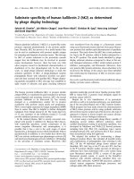
In human bronchi, TAC4, TACR1 and TACR2 mRNAs
were found in all samples whereas TAC1 and TACR3
mRNAs were not detected (fig. 1). A low TAC3 mRNA
expression was found for one patient only, and NEP
mRNA was expressed in high amounts in three of
the four samples. All of these mRNAs were highly
expressed in fetal brain positive control samples,
except TACR2 mRNA which was not found in this
tissue.
In addition to TAC4 mRNA expression, hHK-1 p ro-
tein was found in the supernatants of bronchial explants
(1.40 ± 0.31 pg/100 mg (n = 11)), parenchyma explants
(1.15 ± 0.29 pg/ 100 mg (n = 11)) and lung macrophages
(1.85 ± 0.89 pg/10
6
cells (n = 6)) cultured for 24 hours
in the presence of phosphoramidon.
Characterization of hHK-1- and endokinins-induced
responses in human airways
Contractile effects of hHK-1 and endokinins in isolated
human bronchi
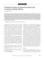
On human isolated bronchi and in the presence of
phosphoramidon, hHK-1 produced concentration-
dependent contractions reaching 80 ± 2% of the con-
traction induced by acetylcholine with a pD
2
of 5.6 ±
0.2 (n = 12) (fig. 2A). In comparison, E
max
and pD
2
values for the contractions induced by the NK
2
receptor
agonist NKA were 87 ± 1% and 8.5 ± 0 .1 (curves not
shown). EKA/B caused concentration-dependent con-
traction on human isolated bronchi and was equipotent
to hHK-1 (respective -log EC
20
of7.2±0.3(n =3)and
7.0 ± 0.5 (n = 3)), whereas EKC and EKD were devoid
of any contractile activity (fig. 2B).
Grassin-Delyle et al. Respiratory Research 2010, 11:139
/>Page 4 of 12
Effects of tachykinin receptor antagonists on cumulative
additions of hHK-1 to human bronchi
The NK
2
receptor antagonist SR 48968 (10
-7
M), com-
pletely abolished the contractile e ffects of cumulative
additions of hHK-1 on human isolated bronchi, whereas
the NK
1
receptor antagonist SR 140333 (10
-7
M) only
exerted a small but not statistically significant reduction
of hHK-1-induc ed contraction at the lowest concentra-
tions (10
-8
M-10
-7
M) (fig. 2A). Finally, the NK
3
recep-
tor antagonist SR 142801 (10
-7
M) did not alter the
concentration-response curve to hHK-1.
Desensitization of the human tachykinin NK
1
receptor
SincearapidNK
1
receptor desensitization has been
reported in human isolated bronchi [22], and in order
to clarify the role of the NK
1
receptor in the responses
to hHK-1, we compare d the effects of single or cumula-
tive additions of hHK-1 and of the specific NK
1
receptor
agonist [Sar
9
,Met(O
2
)
11
] SP. Experiments were per-
formedinthepresenceoftheNK
2
receptor antagonist
SR 48968 (10
-7
M) to block the NK
2
receptor-mediated
component. Cumulative additions of both peptides
induced small contractions of human isolated bronchi
(E
max
= 9 ± 3% and 13 ± 3%, respectively), characterized
by inverted U-shaped concentration-response curves
(fig. 3A and 3B). On the other hand, single a dditions of
hHK-1 or [Sar
9
,Met(O
2
)
11
]SPdidnotleadtoan
inverted U-shaped curve but to a sigmoid response
curve, and maximal contractions reached 43 ± 5% and
26 ± 7% respectively, with pD
2
values of 6.6 ± 0.3 (n =
5-7) and 8.0 ± 0.4 (n = 10). In contrast, concentration-
response curves for NKA and hHK-1 in the presence of
the NK
1
receptor antagonist SR 140333 (10
-7
M) were
similar whatever the protocol used (fig. 3C and 3D).
Effects of tachykinin receptor antagonists on single addition
of hHK-1 to human bronchi
SR 140333 and SR 48968 reduced weakly but not signifi-
cantly the response of human bronchi to a single addition
of 10
-6
M hHK-1 (31 ± 5% and 31 ± 4% respectively, ver-
sus control 42 ± 4% (n = 6-12)) (fig. 4A). However, the
association of both SR 140333 and SR 48968 was synergic
and abolished the smooth muscle contraction. In con-
trast, the response to [Sar
9
,Met(O
2
)
11
]SP(10
-6
M), was
specifically abolished by SR 140333 but unmodified by
SR 48968 (fig. 4B).
TAC4
TACR1 TACR2
TAC1 TAC3 TACR3 MME HPRT
Human bronchi
Positive control
Figure 1 Expression of tachykinin, tachykinin receptor and NEP mRNAs in human bronchi. RT-PCR product o f the housekeeping gene
HPRT used as normalization standard is also represented. Equal aliquots of each cDNA sample (human bronchi or human fetal brain positive
control) were amplified for 40 PCR cycles with their respective specific primer pairs. Since TACR2 was not expressed in human fetal brain, another
bronchi sample was used as positive control for this gene.
67891011
0
20
40
60
80
100
Endokinin A/B
Endokinin C
Endokinin D
Hemokinin-1
- log [Agonist]
Contraction (% ACh 3 mM)
456789
0
20
40
60
80
100
Control
SR 140333
SR 48968
SR 142801
- log [HK-1]
Contraction (% ACh 3 mM)
AB
(M)
(M)
Figure 2 (A) Cumulative concentration-r esponse curves of hHK-1 on human bronchi (n = 5-12) in the abs ence (control) and presence
of NK
1
,NK
2
or NK
3
receptor antagonists SR 140333, SR 48968 or SR 142801 (10
-7
M). (B) Cumulative concentration-response curves of
hHK-1, EKA/B, EKC and EKD on human bronchi (n = 3). Experiments were performed in the presence of phosphoramidon (10
-6
M). Values are
expressed in percentage (mean ± s.e.m.) of maximal contraction obtained with ACh 3 mM.
Grassin-Delyle et al. Respiratory Research 2010, 11:139
/>Page 5 of 12
Cross-desensitization of tachykinin NK
1
receptor between
hHK-1 and [Sar
9
,Met(O
2
)
11
]SP
Since [Sar
9
,Met(O
2
)
11
] SP and hHK-1 are both able to
induce a desensitization of NK
1
receptors, we performed
cross-desensitization experiments with the two com-
pounds in order to assess if tissues desensitized with
one peptide were still responsive to a subsequent addi-
tion of the other peptide. Fig. 5 shows that after an
initial contraction induced by a single addition of [Sar
9
,
Met(O
2
)
11
]SP(10
-7
M), the response to a seco nd addi-
tion of this peptide was abolished (33 ± 7% for the first
addition, 4 ± 1% for the second, n =5,p < 0.01),
whereas under similar conditions, after an i nitial addi-
tion of [Sar
9
,Met(O
2
)
11
]SP,theresponsetohHK-1
(3.10
-7
M) wa s maintaine d (32 ± 7% an d 34 ± 5%
respectively, n = 5). When hHK-1 was added in a first
step to the bath, the response to [Sar
9
,Met(O
2
)
11
]SP
was abolished, whereas the response to a second a ddi-
tion of hHK-1 itself was partially reduced (48 ± 8% and
30 ± 2% respectively, n = 5), suggesting a cross-desensi-
tization between hHK-1 and [Sar
9
,Met(O
2
)
11
]SPforthe
NK
1
receptor.
Characterization of hHK-1- and endokinins-induced
responses in guinea pig airways
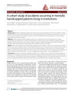
Contractile effects of hHK-1 and endokinins in isolated
guinea pig airways
Human hemokinin-1 induced concentration-dependent
contractions of the guinea-pig trachea (fig. 6A). This
effect was reproducible and independent of the protocol
5678910
0
20
40
60
80
100
- log [[Sar
9
Met(O
2
)
11
] SP]
Contraction (% ACh 3 mM)
5678910
0
20
40
60
80
100
- log [HK-1]
Contraction (% ACh 3 mM)
5678910
0
20
40
60
80
100
- log [NKA]
Contraction (% ACh 3 mM)
5678910
0
20
40
60
80
100
- log [HK-1]
Contraction (% ACh 3 mM)
Cumulative additions
Single addition
AB
CD
Tachykinin NK
1
receptor
Tachykinin NK
2
receptor
(M)
(M)
(M)
(M)
Figure 3 Desensitization of tachykinin NK
1
(A and B) and NK
2
(C and D) receptors. Human bronchi were pre-treated with SR 48968 (10
-7
M)
(A and B) or SR 140333 (10
-7
M) (C and D) before cumulative or non cumulative additions of hHK-1 (B and D, n = 5), [Sar
9
,Met(O
2
)
11
] SP (A, n = 10)
or NKA (C, n = 5). Experiments were performed in the presence of phosphoramidon (10
-6
M). Values are expressed in percentage (mean ± s.e.m.) of
maximal contraction obtained with ACh 3 mM.
Grassin-Delyle et al. Respiratory Research 2010, 11:139
/>Page 6 of 12
***
0
20
40
60
80
100
[Sar
9
Met(O
2
)
11
]SP 10
-6
M
Contraction (% ACh 3 mM)
0
20
40
60
80
100
HK-1 10
-6
M
Contraction (% ACh 3 mM)
Control
SR 140333
SR 48968
SR 140333 + SR 48968
**
***
AB
Figure 4 Contraction induced with single additions of 10
-6
MhHK-1(leftgraph,n = 6-12) or 10
-6
MspecificNK
1
receptor agonist
[Sar
9
,Met(O
2
)
11
] SP (right graph, n = 6) on human bronchi in the absence (control) and presence of NK
1
or NK
2
receptor antagonists
SR 140333 and SR 48968 (10
-7
M). Experiments were performed in the presence of phosphoramidon (10
-6
M). Values are expressed in
percentage (mean ± s.e.m.) of maximal contraction obtained with ACh 3 mM. Statistical analysis was performed with one-way ANOVA followed
with Bonferroni post-test. ** p < 0.01 and *** p < 0.001 versus paired control.
0
20
40
60
80
100
First agonist
Second agonist
Contraction (% ACh 3 mM)
**
***
HK-1 HK-1 HK-1 HK-1 Sar
9
Sar
9
Sar
9
Sar
9
NS
Figure 5 Cross-desensitization of NK
1
receptors after consecutive applications of hHK-1 (3.10
-7
M) and [Sar
9
,Met(O
2
)
11
] SP (10
-7
M) on
human bronchi (n =5). All combinations of hHK-1 and [Sar
9
,Met(O
2
)
11
] SP were assessed. Experiments were performed in the presence of
phosphoramidon (10
-6
M). Values are expressed in percentage (mean ± s.e.m.) of maximal contraction obtained with ACh 3 mM. Statistical
analysis was performed with two-way ANOVA for repeated measures followed with Bonferroni post-test. ** p < 0.01 and *** p < 0.001 for
contraction obtained after the second application versus the first application. (Sar
9
= [Sar
9
,Met(O
2
)
11
] SP).
Grassin-Delyle et al. Respiratory Research 2010, 11:139
/>Page 7 of 12
used for the addition of hHK-1 (cumulative or noncu-
mulative). Table 1 shows that hHK-1 potency was simi-
lar to that of SP, but was 11-fold lower than that of
[Sar
9
,Met(O
2
)
11
] SP and 49- and 72-fold lower than that
of the NK
2
-receptor agonists, NKA and [b-Ala
8
]-NKA
(4-10), respectively. EKA/B (10
-8
M-10
-6
M) exerted
similar effects to hHK-1, whereas EKC and EKD were
without effect (fig. 6A). In guinea-pig isolated bronchi
(fig. 6B), hHK-1 and EKA/B exerted similar effects but
were less potent than SP and [Sar
9
,Met(O
2
)
11
] SP.
Effects of tachykinin receptor antagonists on cumulative
additions of hHK-1 to guinea pig airways
Contractions induced by hHK-1 on the isolated guinea-
pig trachea (fig. 7A and 7B) and main bronchi (fig. 7C)
were abolished by the NK
1
receptor antagonist SR
140333 (10
-7
M), and were altered to a lesser extent by
the NK
2
receptor antagonist SR 48968 (10
-7
M). In addi-
tion, fig. 7A shows that SR 140333 reduced maximal
contractions induced by hHK-1 in the guinea-pig tra-
chea, suggesting a non competitive antagonism in line
with previous data on the rabbit pulmonary artery and
on the guinea pig ileum [42].
Effects of hHK-1 and SP on cytokine production by
human bronchi or lung parenchyma explants and by lung
macrophages
hHK-1 and SP up to 10
-5
M had no impact on TNF-a,IL-
8andMIP-1a production by bronchial explants (n =4).
Similarly, both peptides did not alter TNF-a,IL-6,MIP-
1a, MCP-1, ENA-78, GRO-a, MIG, and MIF production
by lung parenchyma (n = 6 differe nt preparations) and
TNF-a, IL-6, MIF, MIG and MIP-1a production by lung
macrophages (n = 3 to 6 different preparations) (data not
shown). LPS caused a clear-cut increase of these cytokines
in all preparations.
Discussion
Inthepresentstudywehavedemonstratedtheexpres-
sion of TAC4 transcript and protein in human bronchi
and shown tha t hHK-1 and EKA/B exert a contractile
effect in human and guinea pig airways. In human iso-
lated bronchi, the response is mediated mainly through
NK
2
receptor stimulation, the NK
1
receptor-mediated
effect being unmasked in t he presence of SR 48968 and
subject to rapid desensitization. In guinea pig trachea
and main bronchi, the response is mediated mainly
through NK
1
receptor stimulation and to a minor extent
4567891011
0
20
40
60
80
100
Hemokinin-1
Endokinin A/B
Endokinin C
[Sar
9
,Met(O
2
)
11
] SP
Substance P
- log [Agonist]
Contraction (% ACh 3 mM)
4567891011
0
20
40
60
80
100
Hemokinin-1
Endokinin A/B
Endokinin C
Endokinin D
Substance P
Neurokinin A
[Sar
9
,Met(O
2
)
11
] SP
[ -Ala
8
]-NKA (4-10)
- log [Agonist]
Contraction (% ACh 3 mM)
A. Trachea B. Main bronchi
(M)
(M)
Figure 6 Concentration- respo nse curves to (A) cumulativ e additions of hHK-1, EKA/B, EKC, EKD, SP, NKA and specific NK
1
([Sar
9
,Met(O
2
)
11
]SP)
and NK
2
([b-Ala
8
]-NKA (4-10)) receptor agonists on guinea pig trachea (n = 6-12); (B) cumulative additions of hHK -1, EKA/B, EKC, SP and
[Sar
9
,Met(O
2
)
11
]SP on guinea pig main bronchi (n =6-7). E xperiments were performed in the presence of phosphoramidon (10
-6
M). Values are
expressed in percentage (mean ± s.e.m.) of maximal contraction obtained with ACh 3 mM.
Table 1 Functional potencies and maximal effects of
human hemokinin-1 and various tachykinin peptides on
guinea-pig trachea
Agonist N pD
2
E
max
(% of Ach 3 mM)
hHK-1 12 6.4 ± 0.03 73 ± 5
EKA/B 6 ND ND
SP 12 6.7 ± 0.1 84 ± 2
[Sar
9
,Met(O
2
)
11
] SP 12 7.5 ± 0.1 76 ± 2
NKA 6 8.1 ± 0.1 95 ± 2
[b-Ala
8
]-NKA (4-10) 6 8.3 ± 0.6 80 ± 5
hHK-1: human hemokinin-1, EKA/B: endokinin A/B, SP: substance P, NKA:
neurokinin A.
ND: not determined because an asymptote was not reached with the highest
concentration (1 μM) applied.
Values are presented as pD
2
and percentage of maximal contraction obtained
with Ach 3 mM (mean ± s.e.m.) for n determinations.
Grassin-Delyle et al. Respiratory Research 2010, 11:139
/>Page 8 of 12
through NK
2
receptors. The N-terminally extended form
of human hHK-1, EKA/B, exerts similar effects to hHK-
1 on both human br onchi and guinea pig airways,
whereas EKC and EKD, p eptides a lso derive d from
TAC4, did not induce functional responses. Finally, we
have shown that hHK-1 did not alter cytokine produc-
tion by human bronchi or parenchyma explants, or by
human lung macrophages.
Our study shows that the TAC4 gene encoding for
hHK-1 is constitutively present and expressed in human
airways. Only a few numbers of studies have been
devoted to the presence of TAC4 in human tissues, parti-
cularly in lung, and none of them has previously reported
expression of hHK-1 protein. Indeed, TAC4 expression
was not found in the mouse lung by northern blot analy-
sis [3], but was demonstrated by semi-quantitative PCR
in murine lung (mouse, gerbil) [4,5]. In human, a wide
expression of TAC4 has been reported with a strong
expression in tissues such as heart, skeletal muscle, skin,
thyroid, spinal cord, placenta, adrenal gland, spermatozoa
and blood circulating cells and a weaker expressi on in
whole lung, kidney, testis and liver [4,6,39,47]. In contrast
to TAC4, we have shown that TAC1, which encodes for
SP and NKA, was not detected under our experimental
conditions and that TAC3 was observed in only one of
four samples. In a previous study, Pinto et al. showed in
a human total mRNA master panel (BD Biosciences
Clontech) that TAC1 and TAC3 mRNAs were undetect-
able in the lung, but they observed a low expression of
these transcripts in samples of hum an bronchi obtai ned
from patients who had undergone lobectomy or pneu-
mectomy for lung carcinoma, a high expression being
observed in pulmonary arteries [48]. Concerning the
genes that encode for tachykinin receptors, we have iden-
tified the mRNA expression of TACR1 (NK
1
receptor)
and TACR2 (NK
2
receptor), in agreement with Pinto et al.
and in agreement with previous immunohistochemical
evidences of NK
1
and NK
2
receptor expressions in human
bronchial smooth muscle, bronchial glands and bronchial
vessels [48,49]. We did not find TACR3 (NK
3
receptor)
expression whereas Pinto et al. identified this transcript in
all assayed tissues [48]. These discrepancies in the results
of tachykinin transcript expression could be related to dif-
ferences either within human samples or to differences in
expression patterns of tachykinin genes along the respira-
tory tract since we used smaller bronchi than in the work
of Pinto et al. and since differences in the response to
tachykinins have been reported according to the size of
human bronchi [22]. It should be noted that we found
TAC4 transcript and hHK-1 protein expressions in human
bronchi similar in size (1 to 3 mm) to the bronchi used for
the functional studies, substantiating a role for hHK-1 in
the regulation of airway tone. Finally, our results showing
NEP mRNA expression are also consistent with previous
studies reporting a strong expression of NEP in human
bronchi [50].
In human isolated bronchi pre-treated with phosphor-
amidon, hHK-1 exerts a contra ctile effect which was
abolished by the NK
2
receptor antagonist SR 48968,
while the NK
1
receptor antagonist SR 140333 only
weakly reduced the effects of hHK-1 at low concentra-
tions. In human bronchi, hHK-1 appears 800-fold less
potent than NKA. This result is in agreement with pre-
vious d ata obtained on NK
2
receptors eithe r with CHO
cells [4] or rabbit pulmonary artery [13].
A rapid functional desensitization of NK
1
receptors
has been reported with SP and specific NK
1
receptor
agonists in different tissues [51,52] including airways
[22]. In addition, HK-1 has been reported to induce a
desensitization of NK
1
receptors in human embryonic
5678910
0
20
40
60
80
100
Control
SR 140333 3.10
-9
M
SR 140333 10
-8
M
SR 140333 10
-9
M
SR 140333 10
-7
M
- log [HK-1]
Contraction (% ACh 3 mM)
5678910
0
20
40
60
80
100
SR 48968 10
-7
M
SR 140333 10
-7
M
Control
- log [HK-1]
Contraction (% ACh 3 mM)
ABC
5678910
0
20
40
60
80
100
Control
SR 48968 10
-7
M
- log [HK-1]
Contraction (% ACh 3 mM)
**
**
*
***
***
*
***
***
***
***
***
***
(M)
(M)
(M)
Trachea Trachea
Main bronchi
Figure 7 Cumulative concentration-response curves to hHK-1 on guinea pig trachea (A and B) and main bronchi (C) pre-treated with
NK
1
or NK
2
receptor antagonists. (A) Cumulative additions of hHK-1 on guinea pig trachea in the absence (control) and presence of various
concentrations of NK
1
receptor antagonist SR 140333 (10
-9
to 10
-7
M) (n = 4-11). (B) Cumulative additions of hHK-1 on guinea pig trachea in the
absence (control) and presence of NK
2
receptor antagonist SR 48968 (10
-7
M) (n = 6). (C) Cumulative additions of hHK-1 on guinea pig main
bronchi in the absence (control) and presence of NK
1
or NK
2
receptor antagonists SR 140333 and SR 48968 (10
-7
M) (n = 5-7). Experiments were
performed in the presence of phosphoramidon (10
-6
M). Values are expressed in percentage (mean ± s.e.m.) of maximal contraction obtained
with ACh 3 mM. Statistical analysis was performed with two-way ANOVA for repeated measures followed with Bonferroni post-test. * p < 0.05,
** p < 0.01 and *** p < 0.001 versus paired control.
Grassin-Delyle et al. Respiratory Research 2010, 11:139
/>Page 9 of 12
kidney cells [11], rabbit jugular veins [13], U251 MG
astrocytoma cells [53] and scratc hing behavior in rats
[54]. We also observed desensitization of N K
1
receptors
in huma n bronchi, since the magnitude of the c ontrac-
tile response caused by the second a pplication of the
NK
1
-receptor specific agon ist was lower than after the
first addition, even with a 10-fold higher concentration.
Such a desensitization can b e due to receptor internali-
zation, which is a common phenomenon for NK
1
recep-
tor signaling [55] and has already been described with
hHK-1 on astrocytoma cells[53].Wehavedemon-
strated a cross-desensitization between hHK-1 and
[Sar
9
,Met(O
2
)
11
] SP substantiating NK
1
-receptor acti-
vation and desensitization by hHK-1. In addition, since
a first exposure to [Sar
9
,Met(O
2
)
11
] SP was able to
desensitize the NK
1
receptor, preventing a second
response to this specific NK
1
-receptor agonist, but was
unable to prevent the response to hHK-1, these cross-
desensitization experiments further substantiate the
NK
2
-receptor mediated component of the contractile
response to hHK-1. As expected in the single addition
protocol, the contractile effect of [Sar
9
,Met(O
2
)
11
]SP
was abolished by the NK
1
receptor antag onist SR
140333 and unmod ified by the NK
2
receptor antagonist
SR 48968. In contrast, the effect of hHK-1 was not
inhibited by SR 140333 or SR 48968 when used alone,
but was abolished by concomitant addition of the two
antagonists, demonstrating that hHK-1 cont racts
bronchi through NK
1
-andNK
2
receptors. However, it
can also be suggested that [Sar
9
,Met(O
2
)
11
]SPandSP
on the one hand, and hHK-1 on the other hand, may
bind to different sites of the NK
1
receptor and interact
in a different manner with receptor antagonists [5,56].
In contrast with the results in huma n bronchi, SP and
the specific NK
1
-receptor agonist produced maximal
responses similar to those of NKA and hHK-1 in guinea
pig airways providing evide nce of t he higher involve-
ment of NK
1
receptors in this animal species than in
humans as already reported [23,40] . In support of this
notion, hHK-1 exerted a contractile effect mainly
through NK
1
receptor stimulation since this effect was
abolished by the NK
1
receptor antagonist SR 140333,
but was only weakly reduced in th e presence of the NK
2
receptor antagonist SR 48968. Howev er, the NK
2
recep-
tors play a predominant role in guinea pig airways con-
traction since NKA is approximately 10-fold more
potent than SP [23,40]. T he weak effect of SR 48968
against hHK-1 induced bronchoconstriction in the gui-
nea pig airways is likely explained by the higher affinity
of hHK-1 for NK
1
- than for NK
2
receptors in a prepara-
tion fully responsive to NK
1
-mediated response
[4,10,13]. It is noteworthy that the potency of hHK-1 in
the guinea pig airways w as lower than that reported for
r/mHK-1 in specific NK
1
-receptor animal tissues such
as rabbit jugular vein [13], rat urinary bladder [10] and
pig coronary artery [17].
In our study of cytokines production, we were not
able to reproduce the weak TNF-a production that was
observed in SP-stimulated human alveolar macrophages
from healthy subjects [57]. This result may be related to
the underlying disease or the smoking status of the
patients that were all ex-smokers in o ur study since SP-
induced TNF-a releaseismorepronouncedinsmokers
[57]. SP-induced release of inflammatory mediators by
human monocytes/macrophages still remains controver-
sial and may have been related to the presence of endo-
toxin at low levels [58-61]. In addition to the lung
macrophages, explants of lung parenchyma and bronchi
did not produce pro-inflammatory cytokines in response
to hHK-1 or SP, suggesting that hHK-1 may not be
involved in lung inflammatory pathways through the
release of these cytokines. H owever, hHK-1 may exert
other inflammatory effects as already described for SP
or NKA (reviewed in [36]).
SP expression has been reported in the human
respiratory tract [62] and is increased in airways [63],
bronchoalveolar or n asal lavages [64] , sputum [65] or
plasma [66] from asthmatics. It has been demonstrated
that the antibodies used in such studies were directed
against the C-terminal portion of SP, which is shared by
hHK-1, leading to cross-reactivity with hHK-1 [5].
Immunoreactivity attributed to SP expression in human
lungs may therefore be also related to hHK-1 expres-
sion. The use of specific assays for hHK-1 is required to
evaluate the respective expression of SP and hHK-1 in
the respiratory tracts of healthy subjects and in patients
with asthma.
Conclusion
In conclusio n, our results provide evidence for a consti-
tutive expression of TAC4 and hHK-1 in human
bronchi. Our findings indicate that hHK-1 could induce
contraction of human bronchi and guinea pig airways.
This hHK-1-induced contraction could be mainly attrib-
uted to NK
2
rec ept ors in humans and to NK
1
receptors
in guinea pig. The absence cytokine release from lung
explants and macrophages suggests that hHK-1 does not
participate in airways inflammation by inducing the
release of the patt ern of cytokines measured in the pre-
sent study. hHK-1 is therefore involved in the tachyki-
nin-driven contractile response of human airways, but
further studies are n eeded for a better understanding of
hHK-1 involvement in airway diseases such as asthma.
Author details
1
Laboratory of pulmonary pharmacology UPRES EA220, Foch Hospital,
University Versailles-Saint Quentin en Yvelines, 11 rue Guillaume Lenoir,
92150 Suresnes, France.
2
Meakins-Christie Laboratories, Department of
Grassin-Delyle et al. Respiratory Research 2010, 11:139
/>Page 10 of 12
Medicine, McGill University, Montreal, QC, Canada.
3
Department of thoracic
surgery, Foch Hospital, University Versailles-Saint Quentin en Yvelines, 40 rue
worth, 92150 Suresnes, France.
Authors’ contributions
SGD carried out the molecular genetic studies, the contractile function
studies, the cultures of lung explants, the immunoassays, participated to the
interpretation of data, performed the statistical analysis and drafted the
manuscript. EN participated to the contractile function studies and to the
analysis and interpretation of data. AB and PAR participated to the cultures
of lung explants ant to the immunoassays. ES provided human tissues and
critically revised the manuscript. CA and PD conceived the study,
participated in its design and coordination and drafted the manuscript. All
authors read and approved the final manuscript.
Competing interests
The authors declare that they have no competing interests.
Received: 16 April 2010 Accepted: 7 October 2010
Published: 7 October 2010
References
1. Kotani H, Hoshimaru M, Nawa H, Nakanishi S: Structure and gene
organization of bovine neuromedin K precursor. Proc Natl Acad Sci USA
1986, 83(18):7074-7078.
2. Page NM, Woods RJ, Lowry PJ: A regulatory role for neurokinin B in
placental physiology and pre-eclampsia. Regul Pept 2001, 98(3):97-104.
3. Zhang Y, Lu L, Furlonger C, Wu GE, Paige CJ: Hemokinin is a
hematopoietic-specific tachykinin that regulates B lymphopoiesis. Nat
Immunol 2000, 1(5):392-397.
4. Kurtz MM, Wang R, Clements MK, Cascieri MA, Austin CP, Cunningham BR,
Chicchi GG, Liu Q: Identification, localization and receptor
characterization of novel mammalian substance P-like peptides. Gene
2002, 296(1-2):205-212.
5. Page NM: Hemokinins and endokinins. Cell Mol Life Sci 2004,
61(13):1652-1663.
6. Page NM, Bell NJ, Gardiner SM, Manyonda IT, Brayley KJ, Strange PG,
Lowry PJ: Characterization of the endokinins: human tachykinins with
cardiovascular activity. Proc Natl Acad Sci USA 2003, 100(10):6245-6250.
7. Almeida TA, Rojo J, Nieto PM, Pinto FM, Hernandez M, Martin JD,
Candenas ML: Tachykinins and tachykinin receptors: structure and
activity relationships. Curr Med Chem 2004, 11(15):2045-2081.
8. Maggi CA: The mammalian tachykinin receptors. Gen Pharmacol 1995,
26(5):911-944.
9. Regoli D, Boudon A, Fauchere JL: Receptors and antagonists for
substance P and related peptides. Pharmacol Rev 1994, 46(4):551-599.
10. Bellucci F, Carini F, Catalani C, Cucchi P, Lecci A, Meini S, Patacchini R,
Quartara L, Ricci R, Tramontana M, et al: Pharmacological profile of the
novel mammalian tachykinin, hemokinin 1. Br J Pharmacol 2002,
135(1):266-274.
11. Morteau O, Lu B, Gerard C, Gerard NP: Hemokinin 1 is a full agonist at the
substance P receptor. Nat Immunol 2001, 2(12):1088.
12. Duffy RA, Hedrick JA, Randolph G, Morgan CA, Cohen-Williams ME,
Vassileva G, Lachowicz JE, Laverty M, Maguire M, Shan LS, et al: Centrally
administered hemokinin-1 (HK-1), a neurokinin NK1 receptor agonist,
produces substance P-like behavioral effects in mice and gerbils.
Neuropharmacology 2003, 45(2):242-250.
13. Camarda V, Rizzi A, Calo G, Guerrini R, Salvadori S, Regoli D:
Pharmacological profile of hemokinin 1: a novel member of the
tachykinin family. Life Sci 2002, 71(4):363-370.
14. Metwali A, Blum AM, Elliott DE, Setiawan T, Weinstock JV: Cutting edge:
hemokinin has substance P-like function and expression in
inflammation. J Immunol 2004, 172(11):6528-6532.
15. Zhang Y, Paige CJ: T-cell developmental blockage by tachykinin
antagonists and the role of hemokinin 1 in T lymphopoiesis. Blood 2003,
102(6):2165-2172.
16. Weinstock JV: The role of substance P, hemokinin and their receptor in
governing mucosal inflammation and granulomatous responses. Front
Biosci 2004, 9:1936-1943.
17. Long Y, Fu CY, Tian XZ, Chen J, Han M, Wang R: Mechanisms of relaxing
response induced by rat/mouse hemokinin-1 in porcine coronary
arteries: roles of potassium ion and nitric oxide. Eur J Pharmacol 2007,
569(1-2):119-125.
18. Patak E, Pennefather JN, Gozali M, Candenas L, Kerr K, Exintaris B, Ziccone S,
Potteck H, Chetty N, Page NM, et al: Functional characterisation of
hemokinin-1 in mouse uterus. Eur J Pharmacol 2008, 601(1-3):148-53.
19. Pennefather JN, Patak E, Ziccone S, Lilley A, Pinto FM, Page NM, Story ME,
Grover S, Candenas ML: Regulation of the Stimulant Actions of
Neurokinin A and Human Hemokinin-1 on Human Uterus: a Comparison
with Histamine. Biol Reprod 2006, 75(3):334-41.
20. Kong ZQ, Yang WL, Tao Y, Shi XM, Fu CY, Zhao RF, Wang R: Effects of rat/
mouse hemokinin-1, human hemokinin-1 and human hemokinin-1(4-
11), mammalian tachykinin peptides, on rate and perfusion pressure in
the isolated guinea pig heart. Neuropeptides 2010, 44(5):437-44.
21. Naline E, Devillier P, Drapeau G, Toty L, Bakdach H, Regoli D, Advenier C:
Characterization of neurokinin effects and receptor selectivity in human
isolated bronchi. Am Rev Respir Dis 1989, 140(3):679-686.
22. Naline E, Molimard M, Regoli D, Emonds-Alt X, Bellamy JF, Advenier C:
Evidence for functional tachykinin NK1 receptors on human isolated
small bronchi. Am J Physiol 1996, 271(5 Pt 1):L763-767.
23. Advenier C, Naline E, Drapeau G, Regoli D: Relative potencies of
neurokinins in guinea pig trachea and human bronchus. Eur J Pharmacol
1987, 139(2):133-137.
24. Lundberg JM, Martling CR, Saria A: Substance P and capsaicin-induced
contraction of human bronchi. Acta Physiol Scand 1983, 119(1):49-53.
25. Frossard N, Barnes J: Effect of tachykinins in small human airways.
Neuropeptides 1991, 19(3):157-161.
26. Girard V, Feletou M, Advenier C, Canet E: Effects of tachykinins and
capsaicin on the mechanical and electrical activity of the guinea-pig
isolated trachea. Br J Pharmacol 1997, 122(5):841-848.
27. Groneberg DA, Quarcoo D, Frossard N, Fischer A: Neurogenic mechanisms
in bronchial inflammatory diseases. Allergy 2004, 59(11)
:1139-1152.
28. Heaney LG, Cross LJ, Stanford CF, Ennis M: Substance P induces histamine
release from human pulmonary mast cells. Clin Exp Allergy 1995,
25(2):179-186.
29. Wiedermann FJ, Kahler CM, Reinisch N, Wiedermann CJ: Induction of
normal human eosinophil migration in vitro by substance P. Acta
Haematol 1993, 89(4):213-215.
30. Carolan EJ, Casale TB: Effects of neuropeptides on neutrophil migration
through noncellular and endothelial barriers. J Allergy Clin Immunol 1993,
92(4):589-598.
31. Iwamoto I, Nakagawa N, Yamazaki H, Kimura A, Tomioka H, Yoshida S:
Mechanism for substance P-induced activation of human neutrophils
and eosinophils. Regul Pept 1993, 46(1-2):228-230.
32. Schratzberger P, Reinisch N, Prodinger WM, Kahler CM, Sitte BA, Bellmann R,
Fischer-Colbrie R, Winkler H, Wiedermann CJ: Differential chemotactic
activities of sensory neuropeptides for human peripheral blood
mononuclear cells. J Immunol 1997, 158(8):3895-3901.
33. Boichot E, Lagente V, Paubert-Braquet M, Frossard N: Inhaled substance P
induces activation of alveolar macrophages and increases airway
responses in the guinea-pig. Neuropeptides 1993, 25(5):307-313.
34. Nelson DA, Bost KL: Non-neuronal mammalian tachykinin expression.
Front Biosci 2004, 9:2166-2176.
35. Pennefather JN, Lecci A, Candenas ML, Patak E, Pinto FM, Maggi CA:
Tachykinins and tachykinin receptors: a growing family. Life Sci 2004,
74(12):1445-1463.
36. Maggi CA: The effects of tachykinins on inflammatory and immune cells.
Regul Pept 1997, 70(2-3):75-90.
37. Ben-Jebria A, Marthan R, Rossetti M, Savineau JP: Effect of passive
sensitization on the mechanical activity of human isolated bronchial
smooth muscle induced by substance P, neurokinin A and VIP. Br J
Pharmacol 1993, 109(1):131-136.
38. Yoshioka D, Takebuchi F, Nishimori T, Naono R, Ikeda T, Nakayama T:
Intrathecal administration of the common carboxyl-terminal
decapeptide in endokinin A and endokinin B evokes scratching
behavior and thermal hyperalgesia in the rat. Neurosci Lett 2006,
410(3):193-197.
39. Ravina CG, Seda M, Pinto FM, Orea A, Fernandez-Sanchez M, Pintado CO,
Candenas ML: A role for tachykinins in the regulation of human sperm
motility. Hum Reprod 2007, 22(6):1617-1625.
40. Devillier P, Advenier C, Drapeau G, Marsac J, Regoli D: Comparison of the
effects of epithelium removal and of an enkephalinase inhibitor on the
Grassin-Delyle et al. Respiratory Research 2010, 11:139
/>Page 11 of 12
neurokinin-induced contractions of guinea-pig isolated trachea. Br J
Pharmacol 1988, 94(3):675-684.
41. Emonds-Alt X, Bichon D, Ducoux JP, Heaulme M, Miloux B, Poncelet M,
Proietto V, Van Broeck D, Vilain P, Neliat G, et al: SR 142801, the first
potent non-peptide antagonist of the tachykinin NK3 receptor. Life Sci
1995, 56(1):PL27-32.
42. Emonds-Alt X, Doutremepuich JD, Heaulme M, Neliat G, Santucci V,
Steinberg R, Vilain P, Bichon D, Ducoux JP, Proietto V, et al: In vitro and in
vivo biological activities of SR140333, a novel potent non-peptide
tachykinin NK1 receptor antagonist. Eur J Pharmacol 1993, 250(3):403-413.
43. Emonds-Alt X, Vilain P, Goulaouic P, Proietto V, Van Broeck D, Advenier C,
Naline E, Neliat G, Le Fur G, Breliere JC: A potent and selective non-
peptide antagonist of the neurokinin A (NK2) receptor. Life Sci 1992,
50(15):PL101-106.
44. Berger A, Tran AH, Paige CJ: Co-regulated decrease of Neurokinin-1
receptor and Hemokinin-1 gene expression in monocytes and
macrophages after activation with pro-inflammatory cytokines. J
Neuroimmunol 2007, 187(1-2):83-93.
45. Mitsuta K, Shimoda T, Fukushima C, Obase Y, Ayabe H, Matsuse H, Kohno S:
Preoperative steroid therapy inhibits cytokine production in the lung
parenchyma in asthmatic patients. Chest 2001, 120(4):1175-1183.
46. Buenestado A, Grassin Delyle S, Arnould I, Besnard F, Naline E, Blouquit-
Laye S, Chapelier A, Bellamy JF, Devillier P: The role of adenosine
receptors in regulating production of tumour necrosis factor-alpha and
chemokines by human lung macrophages. Br J Pharmacol 2010,
159(6):1304-1311.
47. Klassert TE, Pinto F, Hernandez M, Candenas ML, Hernandez MC, Abreu J,
Almeida TA: Differential expression of neurokinin B and hemokinin-1 in
human immune cells. J Neuroimmunol 2008, 196(1-2):27-34.
48. Pinto FM, Almeida TA, Hernandez M, Devillier P, Advenier C, Candenas ML:
mRNA expression of tachykinins and tachykinin receptors in different
human tissues. Eur J Pharmacol 2004, 494(2-3):233-239.
49. Mapp CE, Miotto D, Braccioni F, Saetta M, Turato G, Maestrelli P, Krause JE,
Karpitskiy V, Boyd N, Geppetti P, et al: The distribution of neurokinin-1
and neurokinin-2 receptors in human central airways. Am J Respir Crit
Care Med 2000, 161(1):207-215.
50. Baraniuk JN, Ohkubo K, Kwon OJ, Mak J, Ali M, Davies R, Twort C, Kaliner M,
Letarte M, Barnes PJ: Localization of neutral endopeptidase (NEP) mRNA
in human bronchi. Eur Respir J 1995, 8(9):1458-1464.
51. Culman J, Tschope C, Jost N, Itoi K, Unger T: Substance P and neurokinin
A induced desensitization to cardiovascular and behavioral effects:
evidence for the involvement of different tachykinin receptors. Brain Res
1993, 625(1):75-83.
52. Nakanishi S: Mammalian tachykinin receptors. Annu Rev Neurosci
1991,
14:123-136.
53. Berger A, Paige CJ: Hemokinin-1 has Substance P-like function in U-251
MG astrocytoma cells: a pharmacological and functional study. J
Neuroimmunol 2005, 164(1-2):48-56.
54. Naono R, Yoshioka D, Ikeda T, Nakayama T, Nishimori T: The common
carboxyl-terminal region of novel tachykinin peptides contributes to
induce desensitization in scratching behavior of rats. Brain Res Bull 2007,
71(5):461-465.
55. Hokfelt T, Pernow B, Wahren J: Substance P: a pioneer amongst
neuropeptides. J Intern Med 2001, 249(1):27-40.
56. Kong ZQ, Fu CY, Chen Q, Wang R: Cardiovascular responses to
intravenous administration of human hemokinin-1 and its truncated
form hemokinin-1(4-11) in anesthetized rats. Eur J Pharmacol 2008, 590(1-
3):310-316.
57. Bardelli C, Gunella G, Varsaldi F, Balbo P, Del Boca E, Bernardone IS,
Amoruso A, Brunelleschi S: Expression of functional NK1 receptors in
human alveolar macrophages: superoxide anion production, cytokine
release and involvement of NF-kappaB pathway. Br J Pharmacol 2005,
145(3):385-396.
58. Lieb K, Fiebich BL, Busse-Grawitz M, Hull M, Berger M, Bauer J: Effects of
substance P and selected other neuropeptides on the synthesis of
interleukin-1 beta and interleukin-6 in human monocytes: a re-
examination. J Neuroimmunol 1996, 67(2):77-81.
59. Brunelleschi S, Guidotto S, Viano I, Fantozzi R, Pozzi E, Ghio P, Albera C:
Tachykinin activation of human alveolar macrophages in tobacco smoke
and sarcoidosis: a phenotypical and functional study. Neuropeptides 1996,
30(5):456-464.
60. Pujol JL, Bousquet J, Grenier J, Michel F, Godard P, Chanez P, de Vos C,
Crastes de Paulet A, Michel FB: Substance P activation of bronchoalveolar
macrophages from asthmatic patients and normal subjects. Clin Exp
Allergy 1989, 19(6):625-628.
61. Derocq JM, Segui M, Blazy C, Emonds-Alt X, Le Fur G, Brelire JC, Casellas P:
Effect of substance P on cytokine production by human astrocytic cells
and blood mononuclear cells: characterization of novel tachykinin
receptor antagonists. FEBS Lett 1996, 399(3):321-325.
62. Lundberg JM, Hokfelt T, Martling CR, Saria A, Cuello C: Substance P-
immunoreactive sensory nerves in the lower respiratory tract of various
mammals including man. Cell Tissue Res 1984, 235(2):251-261.
63. Ollerenshaw SL, Jarvis D, Sullivan CE, Woolcock AJ: Substance P
immunoreactive nerves in airways from asthmatics and nonasthmatics.
Eur Respir J 1991, 4(6):673-682.
64. Nieber K, Baumgarten CR, Rathsack R, Furkert J, Oehme P, Kunkel G:
Substance P and beta-endorphin-like immunoreactivity in lavage fluids
of subjects with and without allergic asthma. J Allergy Clin Immunol 1992,
90(4 Pt 1):646-652.
65. Tomaki M, Ichinose M, Miura M, Hirayama Y, Yamauchi H, Nakajima N,
Shirato K: Elevated substance P content in induced sputum from
patients with asthma and patients with chronic bronchitis. Am J Respir
Crit Care Med 1995, 151(3 Pt 1):613-617.
66. Cardell LO, Uddman R, Edvinsson L: Low plasma concentrations of VIP
and elevated levels of other neuropeptides during exacerbations of
asthma. Eur Respir J 1994, 7(12):2169-2173.
doi:10.1186/1465-9921-11-139
Cite this article as: Grassin-Delyle et al.: Expression and function of
human hemokinin-1 in human and guinea pig airways. Respiratory
Research 2010 11:139.
Submit your next manuscript to BioMed Central
and take full advantage of:
• Convenient online submission
• Thorough peer review
• No space constraints or color figure charges
• Immediate publication on acceptance
• Inclusion in PubMed, CAS, Scopus and Google Scholar
• Research which is freely available for redistribution
Submit your manuscript at
www.biomedcentral.com/submit
Grassin-Delyle et al. Respiratory Research 2010, 11:139
/>Page 12 of 12