Báo cáo y học: " Phosphodiesterase 6 subunits are expressed and altered in idiopathic pulmonary fibrosis" pdf
Bạn đang xem bản rút gọn của tài liệu. Xem và tải ngay bản đầy đủ của tài liệu tại đây (5.24 MB, 14 trang )
RESEA R C H Open Access
Phosphodiesterase 6 subunits are expressed and
altered in idiopathic pulmonary fibrosis
Sevdalina Nikolova
1
, Andreas Guenther
1,2
, Rajkumar Savai
1
, Norbert Weissmann
1
, Hossein A Ghofrani
1
,
Melanie Konigshoff
3
, Oliver Eickelberg
3
, Walter Klepetko
4
, Robert Voswinckel
1,5
, Werner Seeger
1,5
,
Friedrich Grimminger
1
, Ralph T Schermuly
1,5
, Soni S Pullamsetti
1,5*
Abstract
Background: Idiopathic Pulmonary Fibrosis (IPF) is an unresolved clinical issue. Phosphodiesterases (PDEs) are
known therapeutic targets for various proliferative lung diseases. Lung PDE6 expression and function has received
little or no attention. The present study aimed to characterize (i) PDE6 subunits expression in human lung, (ii) PDE6
subunits expression and alteration in IPF and (iii) functionality of the specific PDE6D subunit in alveolar epithelial
cells (AECs).
Methodology/Principal Findings: PDE6 subunits expression in transplant donor (n = 6) and IPF (n = 6) lungs was
demonstrated by real-time quantitative (q)RT-PCR and immunoblotting analysis. PDE6D mRNA and protein levels
and PDE6G/H protein levels were significantly down-regulated in the IPF lungs. Immunohistochemical analysis
showed alveolar epithelial localization of the PDE6 subunits. This was confirmed by qRT-PCR from human primary
alveolar type (AT)II cells, demonstrating the down-regulation pattern of PDE6D in IPF-derived ATII cells. In vitro,
PDE6D pro tein depletion was provoked by transforming growth factor (TGF)-b1 in A549 AECs. PDE6D siRNA-
mediated knockdown and an ectopic expression of PDE6D modified the proliferation rate of A549 AECs. These
effects were mediated by increased intracellular cGMP levels and decreased ERK phosphorylation.
Conclusions/Significance: Collectively, we report previously unrecognized PDE6 expression in human lungs,
significant alterations of the PDE6D and PDE6G/H subunits in IPF lungs and characterize the functional role of
PDE6D in AEC proliferation.
Introduction
IPF is a progressive interstitial lung disease of unknown
etiology associated with high morbidity and mortality
[1], and further characterized by abnormal a lveolar
epithelial and fibro-proliferative responses, excessive
extra-cellular matrix deposition, patchy inflammatory
infiltrations and progressive loss of normal lung struc-
ture [2]. At present there is no effective t herapy for
blocking or reversing the progression of the disease [3].
This situation demands a better understanding of the
molecular and cellular mechanisms involved in the
pathogenesis of IPF.
PDEs comprise a family of related p roteins which can
be subdivided into 11 families based on their amino acid
sequences, sensitiv ity to different activators and inhibi-
tors and their ability to preferentially hydrolyze either
cAMP or cGMP, or both [4]. Of these, PDE6 is a
cGMP-specific PDE family and presents multi-compo-
nent enzyme complexes [5]. The rod PDE6 enzyme is
comprised of two catalytic subunits, PDE6a and PDE6b,
encoded by the PDE6A and PDE6B genes respectively,
two identical inhibitory subunits PDE6g, encoded by
PDE6G [6,7], and one regulatory subunit PDE6δ,
encoded by the PDE6D gene [8]. The cone PDE6
enzyme represents two identical catalytic subunits of
PDE6a’ and two identical inhibitory subunits PDE6g’ ,
encoded by the PDE6C and PDE6H genes, respectively
[9]. Primarily localized in the rod and cone photorecep-
tive cells of the mammalian retina, PDE6 has been
widely studied in the context of visual dysfunctions
[10,11]. Until now, the expression and characterization
of PDE6 in other organs outside of the retina has
* Correspondence:
1
University of Giessen Lung Centre (UGLC), Giessen, Germany
Full list of author information is available at the end of the article
Nikolova et al. Respiratory Research 2010, 11:146
/>© 2010 Nikolova et al; licensee BioMed Central Ltd. This is an Open Access article distributed under the terms of the Creative
Commons Attribution License ( which permits unrestricted use, distribution, and
reproduction in any medium, provided the original work is properly cite d.
received little attention. However, recent reports suggest
functionality of PDE6 apart from the classical photo-
transduction cascade [12-14]. PDE6 activity has been
coupled to non canonical Wnt5a-Frizzl ed-2 signaling in
non-retinal tissue [12-14]. Recently, a significant
increase of Wnt signaling in ATII cells derived from IPF
patients and its involvement in epithelial cell injury and
hyperplasia has been documented [15].
More interestingly, the specific PDE6D subunit has
been reported to regulate the membrane association of
Ras and Rap GTPases [16]. The striking similarity
between PDE6D and Rho guanine nucleotide dissocia-
tion inhibitor (GDI) reasons involvement of PDE6D in
cytoskeleto n reorgani zation, membrane trafficking, tran-
scriptional regulation and cell growth control [17]. The
study of Cook TA et al. demonstrates that PDE6D can
modify cGMP hydro lytic activity in preparations of bro-
ken rod outer segments [ 18]. cGMP plays a role in con-
trolling key epithelial cell functions such as ciliary
motility, cytokine production and proliferation [19-21].
We therefore hypothesized that i) the PDE6 subunits
potentiality can be expressed in the lung, ii) the subunits
are differentially regulated in IPF and iii) the specific
subunit of PDE6, PDE6D, modulates the proliferation
rate of AECs. To this end, we achieved our aim to eluci-
date previously unrecognized PDE6 expression in nor-
mal human lungs, significant alterations of the PDE6D
andPDE6G/HsubunitsinIPF-derivedlungsandchar-
acterize the functional role of PDE6D in AEC
proliferation.
Materials and methods
Ethics Statement
The study protocol for tissue donation was approved by
the Ethics Committee of the Justus-Liebig-University
School of Medicine (AZ 31/93). Informe d consent was
obtained from each individual patient or the patient’ s
next of kin.
Human Tissues
Explanted lung tissues from IPF subjects (n = 6) or
donor (n = 6) were obtained during lung transplantation
at the Department of Cardiothora cic Surgery, University
of Vienna, Austria. Diagnosis was established on the
basis of a proof of a usu al interstitial pneumonitis (UIP)
pattern in the explanted lungs from lung transplant reci-
pients [(n = 6; 4 males, 2 females; mean age = 63.33 ±
1.71 yr, mean forced vital capacity (FVC) = 39.00 ± 2.58
(% of standard); mean forced expiratory volume (FEV) =
44.67 ± 6.39 (% of standard); mean carbon monoxide
lung diffusion capacity (DL
co
) = 30.5 ± 1.5 (% of pre-
dicted)]. Apart from IPF subjects, tissue was also
obtained from 6 donor lungs, which could not be uti-
lized due to size limitations bet ween donor and putative
recipient (mostly single lobes) or due to incompatibility
between donor and recipient.
Isolation of human ATII cells
Primary human ATII cells were isolated, as previously
described [22]. Briefly, the lung was digested and
minced. The cell-rich fraction was filtered, layered onto
a Percoll density gradient, and centrifuged. The cells
were then incubated with anti-CD14 antibody-coated
magnetic beads. The remaining cell s uspension was
incubated in human IgG-coated tissue culture dishes at
37°C in a 5% CO
2
,95%O
2
atmosphere. The purity of
isolated human ATII cells was examined by Papanico-
laou staining. The purity and viability of ATII cell pre-
parations was consistently between 90 and 95%.
Cell culture
The A549 human AEC li ne (American Type Culture
Collection, Manassas, VA, USA) was maintained in
Dulbecco’s modified Eagle’s (DMEM) F12 medium (Invi-
trogen, Carlsbad, CA, USA) supplemented with 10%
heat-in activated fetal bovine seru m (FBS) (PAA Labora-
tories GmbH, Pasching, Austria), 100 units/ml penicillin,
0.1 mg/ml streptomycin, and 2 mM L-glutamine at 37°C
in a 5% CO
2
, 95% O
2
atmosphere. For cytokine stimula-
tion A549 cells were cultured in the absence or presence
of TGF-b1 (R&D System s, Minnea polis, USA, final con-
centration: 2 ng/ml and 5 ng/ml) for 12 h and 24 h. For
studies w ith inhibitors A549 cells were cultured in the
absence or presence of ERK inhibitor, U 0126 (Cell Sig-
naling Technology, Beverly, USA, final concentration: 10
μMand20μM, solvent: dimethylsulfoxide (DMSO)) or
p38a/b inhibitor, SB 203580 (Axon Medchem, Gronin-
gen, The Netherlands, final concentration: 10 μMand
20 μM, solvent: DMSO) [23], details are specified in
Measurement of Cell proliferation section from Materi-
als and Methods.
RNA isolation, cDNA synthesis and mRNA quantification
by qRT-PCR or semi-quantitative RT-PCR
Total RNA was isolated from frozen human lung tissues
and cell pellets using Trizol reagent (Invitrogen, Carls-
bad, CA, USA). cDNA synthesis was carried out with an
ImProm-II-™ reverse transcription system (Promega
Corporation, Madison, WI, USA) by incubating 5 μgof
RNA, following the manufacturer’ s protocol.
qRT-PCR was performed with 2 μlcDNAsetupwith
the Platinum SYBRGreen qPCR SuperMix UDG (Invi-
trogen, Carlsbad, CA, USA), final volume: 25 μl, using
the Mx3000P Real-Time PCR System (Stratagene, La
Jolla, CA, USA). Porphobilinogen deaminase (PBGD)
and pro-surfactant protein C (SPC), ubiquitously as well
as consistently expressed genes were used as reference
in total lung homogenates and ATII cells qRT-PCR
Nikolova et al. Respiratory Research 2010, 11:146
/>Page 2 of 14
reactions, resp ectively. The oligonu cleotide primer pairs
(human origin): PBGD FP: 5’ -T GT CTG GTA ACG
GCA ATG CG-3’ ;RP:5’-CCCACGCGA ATCACTCT -
CAT-3’ ,pro-SPCFP:5’-TGA AAC GCC TTC TTA
TCG TG-3’;RP:5’-CTA GTG AGA GCC TCA AGA
CTG G-3’,PDE6AFP:5’ -TGG CAA AGA GGA CAT
CAA AGT-3’ ;RP:5’-TAA TCA TCC ATC CAG ACT
CAT CC-3’ ,PDE6BFP:5’-GCA GA A CAA TAG GAA
AGA GTG GA-3’ ;RP:5’-CAG GAT A CA GCA G GT
TGA AGA CT-3’,PDE6CFP:5’-AAG AAT GT T TTG
TCC CTG CCT A-3’ ;RP:5’ -AAG AGT GGC TTT
GGT TTG GTT-3’ ,PDE6DFP:5’ -AAT GGT TCT
TCG AGT TTG GC-3’ ;RP:5’-AAA GTC TCA CTC
TGG ATG TGC T-3’ ,PDE6GFP:5’-TTT AAG CAG
CGA CAG ACC AG-3’ ;RP:5’-ATA TTG GGC CAG
CTC GTG-3’ , PDE6H FP: 5’ -TGA GTG ACA ACA
CTA CTC TGC CT-3’;RP:5’-ATG CAA TTC CAG
GTG GCT-3’, (final concentration of 200 nM). Relative
changes in transcr ipt abundance were expressed as ΔC
T
values (ΔC
T
=DC
T
reference
-DC
T
target
), where higher
ΔC
T
values indicate higher transcript abundances [24].
For semi-quantitative RT-PCR 1 μg cD NA was ampli-
fied in 50 μl reaction mixture using 0.5 U GoTaq DNA
polymerase (Promega, Madison, WI, USA) and 0.5 μM
of the following oligonucleotide primer pairs: PDE6A
FP: 5’-TGG CAA AGA GGA CAT CAA AGT-3’;RP5’-
TAA TCA TCC ATC CAG ACT CAT CC-3’ ,PDE6B
FP: 5’-GCA GAA CAA TAG GAA AGA GTG GA-3’ ;
5’ -CAG GAT ACA GCA GGT TGA AGA CT-3’,
PDE6C FP: 5’-AAG AAT GTT TTG TCC CTG CCT
A-3’;RF:5’-AAG AGT GGC TTT GGT TTG G TT-3’,
PDE6D FP: 5’-GGA TGC TGA GAC AGG GAA GAT
A-3’;RP:5’-GCC AGG TAT TTG TGG AGT T AG G-
3’ ,PDE6GFP:5’ -GAC AGA CCA GGC AGT TCA
AGA G-3’ ;RP:5’-TGA GCA GGG TTT AGA GCA
CAG T-3’,PDE6HFP:5’-GAC AAC ACT ACT CTG
CCT GCT C-3 ’;RP5’-GTC ATC TCC AAA TCC TTT
CAC AC-3’, glyceraldehyde-3-phosphate dehydrogenase
(GAPDH) FP: 5’-CAC CGT CAA GGC TGA GAA C-3’;
RP: 5’ -CAG TAG AGG CAG GGA T GA TGT T-3’ .
The PCR products were sequence analyzed.
Immunoblotting
Total protein extracts were isolated from frozen human
lung tissues, pig retina a nd cell pellets homogenized in a
lysis buffer containing 150 mM NaCl, 1% Nonidet P-40,
0.1% SDS, 20 mM Tris-HCl pH 7.6, 5 mM EDTA, 1 mM
EGTA, 1 mM PMSF and 1× complete mini protease inhi-
bitor cocktail (Roche Diagnostics GmbH, Mannheim,
Germany) by centrifugation at 13000 rpm for 2 0 min at
4
°
C. The protein lysates (25-50 μg) were subjected to SDS-
PAGE and immunoblotting for anti-PDE6A, anti-PDE6B,
anti-PDE6D, anti-PDE6G/H (FabGennix, Shreveport, LA,
USA; Santa Cruz Biotechnology Inc., Heidelberg,
Germany, 1:1,000 dilution), anti-His-horseradish peroxi-
dase (HRP) c onjugated (C lontech, Heidelberg, Germany,
1: 2,000 dilution), phospho-specific and total anti-ERK
(Santa Cruz Biotechnology Inc., Heidelb erg, Germany,
1:1,000 dilution), phospho-specific and total anti-p38 a/b
(Abcam, Cambridge, UK and Cell Signaling Technologies,
Danvers, USA, respectively, 1:500 dilution) and anti-
GAPDH (Novus Biologicals, Hiddenhausen, Germany,
1:4,000 dilution) antibodies. The signals were visual ized
using appropriate HRP- conjugated secondary antibodies
and developed with an enhanced chemiluminescence
(ECL) kit (GE Healthcare UK limited, Buckinghamshire,
UK) [25].
Blocking with immunizing peptides
Anti-PDE6A and -PDE6B antibodies specificity was vali-
dated with PDE6A blocking p eptide (M(1)GEVTAEE-
VEKFLDSN(16)C, Abcam, Cambridge, UK) and PDE6B
blocking peptide (H(20)QYFG(K/R)KLSPENVAGAC
(36), Abcam, Cambridge, UK), respectively. The s ignals
were developed with an ECL kit as described above. The
signal that disappeared when using the blocking peptid e
(BP) was considered specific to the antibody. GAPDH
was used as a control for equal loading.
Immunohistochemistry
Serial sections of paraffin embedded lung tissue slides (3
μm) were co-stained with anti-PDE6A, anti-PDE6B,
anti-PDE6D, anti-PDE6G/H antibodies (Abcam, Cam-
bridge, UK; Proteintech Group Inc., Manchester, UK;
Santa Cruz Biotechnology Inc., Heidelberg, Germany,
1:200 dilution) and anti-pro-SPC antibody (Chemicon
International Inc., Temecula, CA, USA, 1:1000 dilution).
Staining was developed using a rabbit primary amino-
ethylcarbazole (AEC) kit (Zymed Laboratories Inc., San
Francisco, CA, USA), following the manufacturer’ s
instructions [25].
Overexpression
For overexpression, the PDE6D gene was PCR amplified
from total human lung homogenates by use of platinium
high fidelity Taq DNA polymerase (Invitrogen, Carlsbad,
CA, USA) and oligonucleotide primer pair: FP: 5’-ACC
AGA GTG AGA AAG CCG-3’ and RP: 5’-CAG TTT
CCT CCT CCC TCC AA-3’, cloned into the pGEM-T
easy v ector system (Promega, Madison, W I, USA) and
thereafter subcloned into pcDNA3.1/V5-His TOPO
eukaryotic expression vector system (Invitrogen, Carls-
bad, CA, USA), oligonucleo tide primer pair: FP: 5’-CAC
CAT GTC AGC CAA GGA C-3; RP: 5’ -AAC ATA
GAA AAG TCT CAC TCT GGA-3’. Plasmid DNAs for
transfection experiments were purified with an endofree
plasmid maxi kit (Qiagen, Hilden, Germany).
Nikolova et al. Respiratory Research 2010, 11:146
/>Page 3 of 14
siRNA
Endogeneous PDE6D expression in A549 cells was
knockdown with PDE6D siRNA target sequence (sense
5’-GGC AGU GUC UCG AGA ACU U-3’;antisense5’-
AAG UUC UCG AGA CAC UGC C-3’ ; Eurogen tec,
Seraing, Belgium, 100 nM). Negative control siRNA
sequence (Eurogentec, Seraing, Belgium, 100 nM) was
used as a specificity control.
Transient transfection assays
A549 cells were used at 80% confluence. The transient
transfection was c arried out with Lipofectamine™ 2000
transfection reagent (Invitrogen, Carlsbad, CA, USA) as
per the manufacturer ’s protocol. The transfection efficiency
was assessed with anti-PDE6D (FabGennix, Shreveport,
LA, U SA) and where appropriate with anti-His-HRP c onju-
gated (Clontech, Heidelberg, Germany) an tibodies [ 26].
Measurement of cell proliferation
A549 cells were transfected under starvation conditions
for 6 h, rendered quiescence for 24 h in 0.1% FBS
DMEM F12 medium and then subjected to serum sti-
mulation (10% FBS) for 24 h. The effects on cell growth
were measured by 3-(4,5-dimethylthiazol-2-yl)-2,5-
diphenyltetrazolium bromide (MTT) and [
3
H]-Thymi-
dine uptake assay. For studies with inhibitors, A549 cells
were rendered quiescence for 24 h in 0.1% FBS DMEM
F12 me dium and pretreated with U 0126 or SB 2 03580
for 30 min prior to serum stimulation for 12 h and 24
h.Theeffectsoncellgrowthweremeasuredby[
3
H]-
Thymidine uptake assay.
[
3
H]-Thymidine uptake assay
[
3
H] Thymidine (GE Hea lthcare UK limited, Buckin-
ghamsh ire, UK) was used at a concent ration 0.1 μCi per
well. The [
3
H]-Thymi dine content of the cell lysates was
determined by a scintillation counter (Canberra-Packard,
TRI-CARB 2000, Meriden, USA) and the values were
expressed as counts per minute (cpm)/number of cells
[25]. In addition, cell number was analyzed using the
Casy-1 System (Schaerfe, Reutlingen, Germany), based
on the Coulter Counter principle.
PDE activity assay
The A549 cell protein was extracted with RIPA buffer
(Santa Cruz, Heidelberg, Germany) and equalized to the
same concentration for use. The reactions were per-
formed with 10 μg protein in 100 μl HEPES buffer
(40mM)atpH7.6consistingofMgCl
2
(5 mM), BSA
(1 mg/ml) and [
3
H]-cGMP (1 μCi/ml, Amersham Bios-
ciences, Munich, Germany) at 37°C for 15 min. The
samples were boiled for 3 min, subsequently cooled for
5minandincubatedwith25μlCrotalusatroxsnake
venom (20 mg/ml, Sigma-Aldrich, Munich, Germany)
for 15 min at 37°C. After being chilled on ice, the sam-
ples were applied to QAE Sephadex A-25 (Amersham
Biosciences, Munich, Germany) mini-chromatography
columns and eluted with 1 ml ammonium formate
(30 mM, pH 7.5). The elutes were collec ted in 2 ml
scintillation solution (Rotiszint®eco plus, Roth, Germany)
and counted by a beta-counter with CPM (counts per
minute) values. Each assay was performed in triplicate
and repeated twice independently . Data are expressed as
picomoles of cGMP per minute per milligram of pro-
tein. (pmol cGMP/minute/mg protein).
cGMP enzyme immunoassay (EIA)
At the end of culture, cells were washed with PBS twice
and lysed in 0.1 M HCl at room temperature for 10
min. After centrifugation the supernatants were equal-
ized to the same protein concentration for use. 50 μl
protein samples which were pre-diluted to 0.3 μg/ml
and standard solutions were incubated with 50 μl tracer
and 50 μl antibody in darkness at 4°C overnight. After
washing 5 times, plates were incubated with Ellman’ s
solution for 90-120 min at room temperature with gen-
tle shaking. The plates were read at a wavelength of 405
nm and the concentration was calculated by the ready-
made Cayman EIA Double workbook. The standard
curve was made as a plot of the %B/B0 value (%Bound/
Maximum Bound) vs concentration of a series of known
standards using a linear (y ) and log (x) axis. Using the
4-parameter logistic equation obtained from the stan-
dard curve, the cGMP concentration of samples was
determined and is given as nmol/mg protein. Each sam-
ple was determined in duplicate and repeated twice.
Statistical analysis
Alldatawereexpressedasthemeans±S.E.Datawere
compa red using a two-tailed Student’s t-test, or a 1-way
ANOVA with the Bonferroni’s post hoc test for studies
with more than 2 groups. Statistical significance was
assumed when P < 0.05.
Results
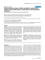
mRNA detection of the PDE6 enzyme subunits
The mRNA expression of each P DE6 subunit in lung tis-
sue homogenates of donors and IPF patients was analyzed
by qRT-PCR technique. As illustrated in Figure 1A,
PDE6A, PDE6B, PDE6C, PDE6D, PDE6G and PDE6H
mRNAs were expressed in the human lung. PDE6A,
PDE6B, PDE6C and PDE6G showed no significant altera-
tions in the IPF lungs as compared to donor lungs. In con-
trast, PDE6D subunit was significantly down-regulated in
the IPF lungs as compared to the donor lungs ( relative
mRNA expression: 2.44 ± 0.28 and 0.30 ± 0.56, respec-
tively) and PDE6H showed a tendency of down-regulation
in the IPF lungs as compared to the donor lungs (relative
mRNA expression: -7.22 ± 0.34 and -8.98 ± 0.66,
Nikolova et al. Respiratory Research 2010, 11:146
/>Page 4 of 14
respectively). In addition, the resultant PCR products were
validated by direct sequencing, followed by BLAST analy-
sis that confirmed the similar sequence alignment for each
subunit (Figure 1B).
Protein expression of the PDE6 enzyme subunits
The protein content of the PDE6 subunits in whole lung
tissue homogenates of donors and IPF patients was quan-
tified by immunoblotting. As illustrated in F igure 2A,
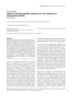
Figure 1 PDE6 mRNA detection in lung tissues from donors and IPF patients. (A) qRT-PCR analysis was used to assess PDE6 subunits
expression in whole lung tissue homogenates from donors (n = 6) and IPF patients (n = 6), white square donor and black square IPF lungs.
Each reaction was performed in quadriplicates. Data were present as mean ± S.E, *P < 0.001 versus donor for PDE6D mRNA expression. (B)
Sequence alignment of the PDE6 subunits.
Nikolova et al. Respiratory Research 2010, 11:146
/>Page 5 of 14
immunoreactivity was detected for PDE6A (~105 kDa),
PDE6B (~105 kDa), PDE6D (~17 kDa) and PDE6G/H
(~11 kDa) subunits. PDE6A and PDE6B blocking peptide
studies were c arried out to reconfirm the specificity of
PDE6A and PDE6B immunoreactivity (Figure 2C and
2D). Additionally, pig retinal lysate served as a positive
control f or immunoreactivity and proper protein size
(Figu re 2E). Nota bly, the PDE6D and PDE6G/H subuni ts
were significantly down-regulated in the IPF lungs as
compared to donor lungs, whereas PDE6A and PDE6B
showed no significant alterations between donor and
IPF-derived lung tissues (Figure 2B).
Cellular localization of the PDE6 enzyme subunits
The cellular localization of the PDE6 subunits was
assessed by serial immunohistochemical stainings on
tissue sectio ns from donor and IPF lungs. As shown in
Figure 3A, PDE6A, PDE6B, PDE6D and PDE6G/H were
co-stained with pro-SPC, suggesting the presence of
PDE6 subunits in ATII cells. PDE6A immunoreactivity
was recognized in th e cytoplasm and membrane of ATII
cells, PDE6B immunoreactivity was recognized in the
nuclei, PDE6D immunoreactivity in the cytoplasm and
PDE6G/H immunoreactivity in the membrane of ATII
cells.
PDE6 enzyme subunits expression in human AECs
To confirm the AEC localization pattern, the PDE6 sub-
units were qRT-PCR amplified from primary human
donor and IP F-derived ATII cells. All PDE6 subunits
(except for PDE6C, no amplicons were detected by
qRT-PCR) were found to be expressed by these cells
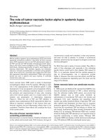
Figure 2 PDE6 immunoreactivity in lung tissues from donors and IPF patients. (A) Immunoblotting was used to assess PDE6A (~105 kDa),
PDE6B (~105 kDa), PDE6D (~17 kDa) and PDE6G/H (~11 kDa) expression in lung tissue homogenates from donors (n = 6) and IPF patients (n =
6). GAPDH (~37 kDa) served as a loading control. (B) Corresponding densitometric analysis normalized to GAPDH expression, white square donor
and black square IPF lungs. Data were present as mean ± S.E, *P < 0.01 versus donor for PDE6D protein expression and *P < 0.001 versus donor
for PDE6G/H protein expression. (C) Demonstration of PDE6A and PDE6B antibodies specificity by an antigen (peptide/protein) blocking
technique. GAPDH served as a loading control. (D) Corresponding densitometric analysis normalized to GAPDH expression, BP (blocking peptide),
white square without BP and black square with BP. (E) Immunoblotting showing PDE6A and PDE6B immunoreactivity in pig retina and human
lung.
Nikolova et al. Respiratory Research 2010, 11:146
/>Page 6 of 14
(Figure 3B). Notably, P DE6D mRNA levels were signifi-
cantly decreased in IPF-derived ATII cells as compared
to donor ATII cells (relative mRNA expression: 1.56 ±
1.05 and -3.80 ± 1.40, respectively). In contrast, PDE6A,
PDE6B, PDE6G and PDE6H were not differentially regu-
lated in AECIIs from IPF versus control lungs.
TGF-b1 down-regulates PDE6D in A549 cells
A549 cells were used as an in vi tro AEC model. Firstly,
the cells were characterized for the expression of PDE6
subunits. mRNAs of all PDE6 subunits (except for
PDE6C and PDE6H) and the complete set of PDE6 pro-
teins w ere found to be expressed by these cells (Figure
4A and 4B). Next, to explore whether TGF-b1 promotes
PDE6D down-regulation in AECs, A549 cells were trea-
ted with two different concentrations of TGF-b1(2ng/
ml and 5 ng/ml) for 12 and 24 h. Decrease in PDE6D
protein e xpression was cle arly evident at concentration
as low as 2 ng/ml (Figure 4C and 4D), with no further
decrease at higher concentration (5 ng/ml) (Figure 4E
and 4F ). PDE6D down-regulation occurred within 12 h
of TGF-b1 stimulation and was sustained up to 24 h
(Figure 4C-F).
Effects of PDE6D modulations on A549 cells proliferation
Further, we studied the functional impact of PDE6D mod-
ulations on A549 cells proliferation. siRNA silencing of
PDE6D resulted in a significant loss of PDE6D protein
expression 24 and 48 h post tr ansfection. Transfection
with non-targeting siRNA caused no change in PDE6D
protein expression (Figure 5A). The loss of PDE6D expres-
sion was coupled to a significantly decreased cell number
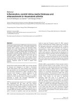
Figure 3 Cellular and sub-cellular localization of the PDE6 subunits. (A) Immunohistochemical stainings were perfor med on serial tissue
sections of donor (upper row) and IPF (bottom row) lungs. PDE6A, PDE6B, PDE6D and PDE6G/H were co-stained with pro-SPC, a marker specific
for ATII cells. PDE6A immunoreactivity was recognized in the cytoplasm and membrane of ATII cells, PDE6B immunoreactivity was recognized in
the nuclei, PDE6D immunoreactivity in the cytoplasm and PDE6G/H immunoreactivity in the membrane of ATII cells. The red and dark brown color
is indicative of immunoreactivity. Tissue slides were counterstained with hematoxylin (blue color). Isotype control stands for rabbit serum react ion
and null control stands for no antibody reaction, magnification 630×. Arrows indicate stained cells. (B) PDE6 mRNA expression in human primary
donor and IPF-derived ATII cells. Primary human ATII cells were isolated from whole lung tissue of donor and IPF patients as described in Material
and Methods. The mRNA levels of PDE6A, PDE6B, PDE6D, PDE6G and PDE6H were analyzed by qRT-PCR. Results are derived from 3 different donor
and IPF patients. Each reaction was performed in quadriplicates. Data were present as mean ± S.E, *P < 0.01 versus donor ATII cells.
Nikolova et al. Respiratory Research 2010, 11:146
/>Page 7 of 14
(Figure 5B) and [
3
H]-Thymidine uptake (Figure 5C) as
compared to control siRNA and no siRNA transfected
cells 24 h post serum stimulation. Complementary, transi-
ent overexpression of PDE6D in A549 cells resu lted in a
significantly enhanced PDE6D expression and detection of
PDE6D His-tagged protein 24 and 48 h post transfection.
Empty vector transfection caused no change in PDE6D
protein expression (Figure 6A). The gain of PD E6D
expression was coupled to a significantly increased cell
number (Figure 6B) and [
3
H]-Thymidine uptake (Figure
6C) as compared to empty vector expressing cells and no
DNA transfected cells 24 h post serum stimulation.
Figure 4 TGF-b1-induced PDE6D down-regulation in A549 AECs. (A) mRNAexpressionprofileofPDE6subunitsinA549AECs.(B) Protein
expression profile of PDE6A (~105 kDa), PDE6B (~105 kDa), PDE6D (~17 kDa) and PDE6G/H (~11 kDa) subunits in A549 AECs. (C) TGF-b1 effects o n
PDE6D expression in A549 cells. A549 cells were rendered quiescence for 24 h in 0.1% FBS DMEM F12 medium, stimulated with TGF-b1(2ng/ml)for
12 and 24 h and PDE6D (~17 kDa) expression was measured by immunoblotting. GAPDH (~37 kDa) served as a loading control. (D) Corresponding
densitometric analysis, normalized to GAPDH expression. Data were present as mean ± S.E, *P < 0.001 versus unstimulated cells. (E) TGF-b1 effects o n
PDE6D expression in A549 cells. A549 cells were rendered quiescence for 24 h in 0.1% FBS DMEM F12 medium, stimulated with TGF-b1(5ng/ml)for
12 and 24 h and PDE6D (~17 kDa) expression was measured by immunoblotting. GAPDH (~37 kDa) served as a loading control. (F) Corresponding
densitometric analysis, normalized to GAPDH expression. Data were present as mean ± S.E, *P < 0.01 versus unstimulated cells.
Nikolova et al. Respiratory Research 2010, 11:146
/>Page 8 of 14
PDE6D knockdown regulates cGMP levels and ERK
phosphorylation
We then opted to explore signaling pathways related to
PDE6D-mediated proliferative responses. In particular, we
studied the effects of PDE6D down-regulation on (i)
cGMP hydrolyzing PDE acti vity, (ii) intracellular cGMP
levels and (iii) serum induced phosphorylation of ERK
protein in A549 cells. cGMP hydrolyzing PDE activity was
decreased in PDE6D siRNA as compared to non-targeting
siRNA and mock transfectio n 24 h post serum stimula-
tion. In corroboration, intracellular cGMP determined by
EIA assay was increased 1.6 fold by PDE6D down-
regulation (Figure 7A and 7B). ERK phosphorylation was
increased 1 h, 12 h and 24 h post serum stimulation as
compared to unstimulated cells (0.1% FBS). siRNA
mediated loss of PDE6D protein expression was detectable
12 h and 24 h post serum stimulation and this was related
to a decrease in ERK phosphorylation as compared to con-
trol siRNA treated cells (Figure 7C-E). However, no appar-
ent changes in the phospho-p38a/b levels were o bserved
by PDE6D down-regulation, suggesting the specificity of
PDE6D for ERK signaling (Figure 7C-E).
Figure 5 Knockdown of endogenous PDE6D expression decelerates the proliferation rate of A549 AECs. (A) Demonstration of PDE6D
knockdown in A549 cells: upper panel: decreased PDE6D (~17 kDa) immunoreactive protein 0-48 h post transfection with 100 nM PDE6D siRNA.
The negative control siRNA (csiRNA, 100 nM) caused no change in PDE6D protein expression. The bottom panel represents GAPDH (~37 kDa)
used as a loading control. (B) Bar graph presentation of cell counts from PDE6D siRNA transfected cells 24 h post serum stimulation. Data were
expressed as % of control. Serum stimulation was significant
#
P < 0.001 versus 0.1% FBS stimulated cells. Cell number from PDE6D knockdown
cells was significantly decreased as compared to csiRNA transfected and no siRNA transfected (only lipofectamine (Lf)) cells (*P < 0.001 versus
csiRNA 100 nM transfected cells,
†
P<0.01 versus Lf treated cells). (C) Bar graph presentation of [
3
H]-Thymidine uptake in PDE6D knockdown cells
24 h post serum stimulation. Data were expressed as cpm/×10
5
cells. Serum stimulation was significant
#
P < 0.001 versus 0.1% FBS stimulated
cells. [
3
H]-Thymidine uptake of PDE6D knockdown cells was significantly decreased as compared to csiRNA transfected and Lf treated cells (*P <
0.001 versus csiRNA 100 nM transfected cells,
†
P<0.001 versus Lf treated cells). Lf concentration was kept constant throughout the experimental
settings and had no effect on cell viability (P = 0.2699).
Nikolova et al. Respiratory Research 2010, 11:146
/>Page 9 of 14
ERK inhibition inhibits A549 cells proliferation
Supplementary, employing ERK (U 0126) and p38a /b
(SB 203580) ph armacological inhibitors, we showed that
ERK1/2 inhibitor (U 0126) signific antly inhibits [
3
H]-
Thymidine uptake 12 h and 24 h post serum stimulation
as compared to control (no DMSO) and DMSO treated
A549 cells. The effects of U 0126 were dose dependent.
Additionally, we used the p38a/b inhibitor (SB 203580)
as a control. SB 203580 had no effect on [
3
H]-Thymi-
dine uptake by A549 cells (Figure 7F and 7G).
Discussion
In the present study, we report previously unrecognized
PDE6 expression in the human lung. The members of
the PDE family, PDE1, PDE2, PDE3, PDE4 and PDE5
are highly expressed in the lung and have been shown
to potentially contribute to the pathogenesis of various
lung diseases [27,28]. Nevertheless, to our knowledge
this is the first report that has described the expression
and characterization of P DE6 subunits in both the phy-
siology and pathophysiology of the lung. Among these,
PDE6D (mRNA and protein levels) and PDE6G/H subu-
nit (protein levels) were found significantly down-
regulated in the IPF lungs as compared to the donor
lungs. All PDE 6 subunits wer e detected in ATII cells,
with PDE6D significantly down-re gulated in IPF-derived
ATII cells. PDE6D down-regulation was induced in vitro
by TGF-b 1 in A549 cells, suggesting a link between the
Figure 6 Overexpression of PDE6D accelerates the proliferation rate of A54 9 AECs. (A) Demonstration of PDE6D overexpression in A549
cells: upper panel: increased PDE6D (~17 kDa) immunoreactive protein 0-48 h post transfection with pcDNA3.1/His-PDE6D vector (PDE6D). These
expressional changes were not observed in pcDNA3.1/His-lacZ empty vector (EV) or no DNA transfected (only lipofectamine (Lf)) cells. Middle
panel: The membrane was probed with anti-His-HRP conjugated antibody. A band of ~23 kDa was detected in the PDE6D transfected cells but
not in EV transfected or Lf treated cells. The bottom panel represents GAPDH (~37 kDa) used as a loading control. (B) Bar graph presentation of
cell counts from PDE6D overexpressing cells 24 h post serum stimulation. Data were expressed as % of control. Serum stimulation was
significant
#
P < 0.05 versus 0.1% FBS stimulated cells. Cell number from PDE6D overexpressing cells was significantly increased as compared to
EV transfected and Lf treated cells (*P < 0.01 versus EV transfected cells,
†
P<0.01 versus Lf treated cells). (C) Bar graph presentation of [
3
H]-
Thymidine uptake in PDE6D overexpressing cells 24 h post serum stimulation. Data were expressed as cpm/×10
5
cells. Serum stimulation was
significant
#
P < 0.001 versus 0.1% FBS stimulated cells. [
3
H]-Thymidine uptake of PDE6D overexpressing cells was significantly increased as
compared to EV transfected and Lf treated cells (*P < 0.01 versus EV transfected cells,
†
P<0.01 versus Lf treated cells). Lf concentration was kept
constant throughout the experimental settings and had no effect on cell viability (P = 0.3552).
Nikolova et al. Respiratory Research 2010, 11:146
/>Page 10 of 14
Figure 7 PDE6D siRNA knockdown inhibits cGMP hydrolyzing PDE activity, increases cGMP levels and inhibits serum stimulated ERK
phosphoprylation in A549 AECs. (A) cGMP hydrolyzing PDE activity in PDE6D siRNA transfected cells 24 h post serum stimulation. cGMP PDE
activity in PDE6D knockdown cells was significantly decreased as compared to control siRNA (csiRNA) and no siRNA transfected (only
lipofectamine (Lf)) cells (*P < 0.05 versus csiRNA 100 nM transfected cells,
†
P<0.01 versus Lf treated cells). (B) Intracellular cGMP levels in PDE6D
siRNA transfected cells 24 h post serum stimulation. Intracellular cGMP levels were significantly increased as compared to csiRNA transfected and
Lf treated cells (*P < 0.01 versus csiRNA 100 nM transfected cells,
†
P<0.01 versus Lf treated cells). (C, D, E) Immunoblotting of ERK (~42/44 kDa)
and p38a/b (~38/41 kDa) phosphorylation in PDE6D siRNA transfected cells 1 h, 12 h and 24 h post serum stimulation, respectively. GAPDH
(~37 kDa) was used as a loading control for PDE6D expression and total ERK (~42/44 kDa) for ERK phosphorylation. (F, G) Bar graph presentation
of [
3
H]-Thymidine uptake in U 0126 (10 μM, 20 μM) and SB 203580 (10 μM, 20 μM) treated cells 12 h and 24 h post serum stimulation,
respectively. Serum stimulation was significant
#
P < 0.001 versus 0.1% FBS stimulated cells. [
3
H]-Thymidine uptake of U 0126 (10 μM, 20 μM)
treated cells was significantly decreased as compared to control (no DMSO) and DMSO treated cells, data were present as mean ± S.E from two
independent experiments, *P < 0.001 versus DMSO treated 10% FBS stimulated cells. SB 203580 (10 μM, 20 μM) exerted no effect.
Nikolova et al. Respiratory Research 2010, 11:146
/>Page 11 of 14
observed PDE6D down-regulation in IPF specimens and
the pathogenesis of the disease [29]. Furthermore, using
A549 ce lls as an in vitro AECs model, we were able to
show that PDE6D modulates the proliferation rate of
these cells (siRNA and ectopic expression studies). More
interestingly, we sho wed that mecha nisms accounting
for PDE6D effects o n AEC proliferation is related to
PDE6D increasing the intracellular cGMP levels and
suppressing the phosphorylation of ERK.
This finding is further supported by the reports that
have demonstrated solitary PDE6 subunit expression in
a variety of non-retinal tissues. The rod catalytic PDE6A
and PDE6B subunits were found to be weakly expressed
in brain [30,31]. Piriev et al. demonstrated that the cata-
lytic core of the rod PDE6 enzyme can be synthesized in
human ki dney cells with conseque nt expression of enzy-
maticactivity[32].Theregulatoryandtheinhibitory
PDE6D and PDE6G subunits, respectively, have been
reported to be expressed in a variety of heterogeneous
tissues, including the lung [8,33]. Identifying the expres-
sion of PDE isoforms in organs and cells that had not
been reported previously is a subject gaining interest.
For example, PDE5, known to express in lung, recently
reported to be also expressed i n vascular, ganglion and
bipolar cell layers of retinal tissue. It was c laimed to
play a physiological role in the retina and might contri-
bute to PDE5 inhibitor-associated ocular side effects [34].
Although at present the physiological roles of the
PDE6 subunits in the lung are unknown and the func-
tionality of the PDE6 enzyme in IPF needs to be
explored, the study of Wang et al. [14] does provide evi-
dence for the presence of functional PDE6 enzyme in
non-retinal tissues. Based on our findings, all the PDE6
subunits appear to be expressed and localize in human
lung alveolar epithelium. Among those, PDE 6D and
PDE6G/H subunit protein levels were found significantly
down-regulated in the IPF lungs as compared to the
donor lungs, suggesting a plausi ble contribution of these
PDE6 subunits to the pathogenesis of IPF. Thus, we
believe that PDE6 alterations may play a crucial role in
epithelial apoptosis, proliferation, surfactant synthesis
and reactive oxygen species (ROS) generation abnormal-
ities associated with IPF [35].
In fact, based on the data obtained from human donor
and IPF lungs, it is not possible at present to determine
whether PDE6 functions as a compl ex or each PDE6
subunit has a solitary function. However, considering
the requirement for multiple subunits assembly to pro-
duce functional rod and cone PDE6 enzymes and the
difficulties in expressing functionally active rod and
cone PDE6 enzymes in various systems [36], we herein
explored independent functionality of the specific
PDE6D subunit in AEC proliferation. In addition, we
assessed the contribution of PDE6D to PDE6 as well as
tothepresenceorabsenceofcGMP.Inourstudies,
gain of function (overexpression) or loss of function
(targeting siRNA) of PDE6D affected AEC proliferation,
with increased PDE6D resulting in increased AEC prolif-
eration. The anti-proliferative effects encountered in
response to PDE6D knockdown were largely due to a
decrease in cGMP hydrolyzing PDE activity that may
subsequently stimulate the intracellular levels of cGMP.
Ofnote,wewereabletomeasureonlytotalcGMP
hydrolyzing activity (Figure 7B), but not PDE6 specific
cGMP hydrolyzing activity due to less selectivity of
PDE6 inhibitors. Several classes of PDE inhibitors inhibit
PDE6 equally as well as the PDE family to which they
are targeted [37]. Similarly, further studies are need ed to
explore the role of PDE6 inhibitor y subunits (PDE6G
andPDE6H)thatwerefounddownregulatedatthe
protein level in IPF lungs. Several lines of evidenc e
reported that the inhibitory PDE6G/H subunits of the
PDE6 are expressed in non-retinal tissues [33] and are
involved in the stimulation of the p 42/p44 mitogen-
activated protein kinase (MAPK) pathway by growth
factors and G-protein-coupled receptor agonists in
human embryonic kidney 293 cells [38].
Impaired AECs proliferation is a significant finding in
IPF [39]. Multiple studies have reported rapid prolifera-
tion of ATII cells following injury [40,41] or reduced
proliferative capacity of ATII cells and inability to differ-
entiate into ATI ce lls in both experimental lung fibrosis
[42] and IPF [39]. Herein, we report modulatory effects
of the specific PDE6D subunit on AECs proliferation, as
deduce d from PDE6D siRNA-m ediated knockdo wn and
over-expression studies in A549 cells. This functional
property of PDE6D is significant, considering its c-Myc/
E2F 4 cont rolled expression (http ://www.unleashedinfor-
matics.com). In line with these studies, PDE6D (-/-)
mice are consistently smaller in size, indicating a plausi-
ble involvement of PDE6D in growth arrest [43]. Thus,
it can be imagined that the proliferative phenotype of
IPF-derived ATII cells is associated with the observed
PDE6D down regulation in IPF lungs.
ERK activation has been shown to be o f critical
importance for ATII cell proliferation [44]. ERK signal-
ing has also been documented to regulate differentiation
of fetal ATII cells [45]. In agreement, our study indi-
cates that ERK is a key mediator of A549 AECs prolif-
eration and that PDE6D mediated proliferative
responses are related to ERK signaling. siRNA mediated
inhibition of PDE6D decreased the serum induced phos-
phorylation of ERK in a time response fashion. Thus, we
propose PDE6D as a critical regulator of ERK mediated
ATII cells proliferation.
In conclusion, these data demonstrate previously unrec-
ognized PDE6 e xpression in human lung, significant
alterations of the PDE6D and PDE6G/H subunits in
Nikolova et al. Respiratory Research 2010, 11:146
/>Page 12 of 14
IPF-deri ved lungs and characterize the functional role of
PDE6D in AEC proliferation. For a further consolidation
of the proposed pathomechanistic link between PDE6D
content and type II cell proliferation on an in vivo level,
transgenic mice with epithelial cell-specific PDE6D knock-
out would have to be generated. Hence, we can, right now,
only postulate that decre ased PDE6D expression in IPF
might be involved in attenuation of type II cell hyperplasia.
Further, it is tempting t o speculate that therapeutic pre-
vention of PDE6D down-regulation and/or PDE6D over-
expression in animal models of pulmonary fibrosis may be
beneficial to boost up alveolar re-epithelization and may
represent a therapeutic option in IPF.
Acknowledgements
The authors would like to thank Eva Dony and Michael Seimetz for their
valuable assistance.
Author details
1
University of Giessen Lung Centre (UGLC), Giessen, Germany.
2
Lung Clinic
Waldhof Elgershausen, Greifenstein, Germany.
3
Comprehensive Pneumology
Center, University Hospital Grosshadern, Ludwig-Maximilians-University, and
Helmholtz Zentrum München, Munich, Germany.
4
Department of
Cardiothoracic Surgery, University of Vienna, Vienna, Austria.
5
Max-Planck-
Institute for Heart and Lung Research, Bad Nauheim, Germany.
Authors’ contributions
Conceived and designed the experiments: SN, NW, HAG, RTS, SSP.
Performed the experiments: SN, RS, SSP. Analyzed the experiments: AG, WS,
FG. Contributed reagents/Materials: MK, OE, WK, RV. Wrote the paper: SN,
RTS, SSP. All authors read and approved the manuscript.
Competing interests
The authors declare that they have no competing interests.
Received: 11 July 2010 Accepted: 27 October 2010
Published: 27 October 2010
References
1. American Thoracic Society: Idiopathic pulmonary fibrosis: diagnosis and
treatment. International consensus statement. American Thoracic Society
(ATS), and the European Respiratory Society (ERS). Am J Respir Crit Care
Med 2000, 161(2 Pt 1):646-664.
2. Selman M, King TE, Pardo A: Idiopathic pulmonary fibrosis: prevailing and
evolving hypotheses about its pathogenesis and implications for
therapy. Ann Intern Med 2001, 134(2):136-151.
3. Thannickal VJ, Flaherty KR, Hyzy RC, Lynch JP: Emerging drugs for
idiopathic pulmonary fibrosis. Expert Opin Emerg Drugs 2005,
10(4):707-727.
4. Beavo JA: Cyclic nucleotide phosphodiesterases: functional implications
of multiple isoforms. Physiol Rev 1995, 75(4):725-748.
5. Cote RH: Characteristics of photoreceptor PDE (PDE6): similarities and
differences to PDE5. Int J Impot Res 2004, 16(Suppl 1):S28-33.
6. Baehr W, Devlin MJ, Applebury ML: Isolation and characterization of cGMP
phosphodiesterase from bovine rod outer segments. J Biol Chem 1979,
254(22):11669-11677.
7. Deterre P, Bigay J, Forquet F, Robert M, Chabre M: cGMP
phosphodiesterase of retinal rods is regulated by two inhibitory
subunits. Proc Natl Acad Sci USA 1988, 85(8):2424-2428.
8. Lorenz B, Migliaccio C, Lichtner P, Meyer C, Strom TM, D’Urso M, Becker J,
Ciccodicola A, Meitinger T: Cloning and gene structure of the rod cGMP
phosphodiesterase delta subunit gene (PDED) in man and mouse. Eur J
Hum Genet 1998, 6(3):283-290.
9. Gillespie PG, Beavo JA: Characterization of a bovine cone photoreceptor
phosphodiesterase purified by cyclic GMP-sepharose chromatography. J
Biol Chem 1988, 263(17):8133-8141.
10. Muradov KG, Granovsky AE, Artemyev NO: Mutation in rod PDE6 linked to
congenital stationary night blindness impairs the enzyme inhibition by
its gamma-subunit. Biochemistry 2003, 42(11):3305-3310.
11. Stockman A, Sharpe LT, Tufail A, Kell PD, Ripamonti C, Jeffery G: The effect
of sildenafil citrate (Viagra) on visual sensitivity. JVis2007, 7(8):4.
12. Ahumada A, Slusarski DC, Liu X, Moon RT, Malbon CC, Wang HY: Signaling
of rat Frizzled-2 through phosphodiesterase and cyclic GMP. Science
2002, 298(5600):2006-2010.
13. Ma L, Wang HY: Mitogen-activated protein kinase p38 regulates the Wnt/
cyclic GMP/Ca2+ non-canonical pathway. J Biol Chem 2007,
282(39):28980-28990.
14. Wang H, Lee Y, Malbon CC: PDE6 is an effector for the Wnt/Ca2+/cGMP-
signalling pathway in development.
Biochem Soc Trans 2004, 32(Pt
5):792-796.
15. Konigshoff M, Balsara N, Pfaff EM, Kramer M, Chrobak I, Seeger W,
Eickelberg O: Functional Wnt signaling is increased in idiopathic
pulmonary fibrosis. PLoS ONE 2008, 3(5):e2142.
16. Nancy V, Callebaut I, El Marjou A, de Gunzburg J: The delta subunit of
retinal rod cGMP phosphodiesterase regulates the membrane
association of Ras and Rap GTPases. J Biol Chem 2002,
277(17):15076-15084.
17. Van Aelst L, D’Souza-Schorey C: Rho GTPases and signaling networks.
Genes Dev 1997, 11(18):2295-2322.
18. Cook TA, Ghomashchi F, Gelb MH, Florio SK, Beavo JA: The delta subunit
of type 6 phosphodiesterase reduces light-induced cGMP hydrolysis in
rod outer segments. J Biol Chem 2001, 276(7):5248-5255.
19. Friedman DL: Role of cyclic nucleotides in cell growth and
differentiation. Physiol Rev 1976, 56(4):652-708.
20. Geary CA, Davis CW, Paradiso AM, Boucher RC: Role of CNP in human
airways: cGMP-mediated stimulation of ciliary beat frequency. Am J
Physiol 1995, 268(6 Pt 1):L1021-1028.
21. Stadnyk AW: Cytokine production by epithelial cells. FASEB J 1994,
8(13):1041-1047.
22. Fang X, Song Y, Hirsch J, Galietta LJ, Pedemonte N, Zemans RL,
Dolganov G, Verkman AS, Matthay MA: Contribution of CFTR to apical-
basolateral fluid transport in cultured human alveolar epithelial type II
cells. Am J Physiol Lung Cell Mol Physiol 2006, 290(2):L242-249.
23. Ogura H, Tsukumo Y, Sugimoto H, Igarashi M, Nagai K, Kataoka T: ERK and
p38 MAP kinase are involved in downregulation of cell surface TNF
receptor 1 induced by acetoxycycloheximide. Int Immunopharmacol 2008,
8(6):922-926.
24. Pullamsetti S, Kiss L, Ghofrani HA, Voswinckel R, Haredza P, Klepetko W,
Aigner C, Fink L, Muyal JP, Weissmann N, et al: Increased levels and
reduced catabolism of asymmetric and symmetric dimethylarginines in
pulmonary hypertension. FASEB J 2005, 19(9):1175-1177.
25. Schermuly RT, Dony E, Ghofrani HA, Pullamsetti S, Savai R, Roth M,
Sydykov A, Lai YJ, Weissmann N, Seeger W, et al: Reversal of experimental
pulmonary hypertension by PDGF inhibition. J Clin Invest 2005,
115(10):2811-2821.
26. Hanze J, Eul BG, Savai R, Krick S, Goyal P, Grimminger F, Seeger W, Rose F:
RNA interference for HIF-1alpha inhibits its downstream signalling and
affects cellular proliferation. Biochem Biophys Res Commun 2003,
312(3):571-577.
27. Schermuly RT, Pullamsetti SS, Kwapiszewska G, Dumitrascu R, Tian X,
Weissmann N, Ghofrani HA, Kaulen C, Dunkern T, Schudt C, et al:
Phosphodiesterase 1 upregulation in pulmonary arterial hypertension:
target for reverse-remodeling therapy. Circulation 2007,
115(17):2331-2339.
28. Galie N, Ghofrani HA, Torbicki A, Barst RJ, Rubin LJ, Badesch D, Fleming T,
Parpia T, Burgess G, Branzi A, et al: Sildenafil citrate therapy for pulmonary
arterial hypertension. N Engl J Med 2005, 353(20):2148-2157.
29. Bergeron A, Soler P, Kambouchner M, Loiseau P, Milleron B, Valeyre D,
Hance AJ, Tazi A: Cytokine profiles in idiopathic pulmonary fibrosis
suggest an important role for TGF-beta and IL-10. Eur Respir J 2003,
22(1):69-76.
Nikolova et al. Respiratory Research 2010, 11:146
/>Page 13 of 14
30. Kuenzi F, Rosahl TW, Morton RA, Fitzjohn SM, Collingridge GL, Seabrook GR:
Hippocampal synaptic plasticity in mice carrying the rd mutation in the
gene encoding cGMP phosphodiesterase type 6 (PDE6). Brain Res 2003,
967(1-2):144-151.
31. Taylor RE, Shows KH, Zhao Y, Pittler SJ: A PDE6A promoter fragment
directs transcription predominantly in the photoreceptor. Biochem
Biophys Res Commun 2001, 282(2):543-547.
32. Piriev NI, Yamashita C, Samuel G, Farber DB: Rod photoreceptor cGMP-
phosphodiesterase: analysis of alpha and beta subunits expressed in
human kidney cells. Proc Natl Acad Sci USA 1993, 90(20):9340-9344.
33. Tate RJ, Arshavsky VY, Pyne NJ: The identification of the inhibitory
gamma-subunits of the type 6 retinal cyclic guanosine monophosphate
phosphodiesterase in non-retinal tissues: differential processing of
mRNA transcripts. Genomics 2002, 79(4):582-586.
34. Foresta C, Caretta N, Zuccarello D, Poletti A, Biagioli A, Caretti L, Galan A:
Expression of the PDE5 enzyme on human retinal tissue: new aspects of
PDE5 inhibitors ocular side effects. Eye 2008, 22(1):144-149.
35. Horowitz JC, Thannickal VJ: Idiopathic pulmonary fibrosis: new concepts
in pathogenesis and implications for drug therapy. Treat Respir Med 2006,
5(5):325-342.
36. Ionita MA, Pittler SJ: Focus on molecules: rod cGMP phosphodiesterase
type 6. Exp Eye Res 2007, 84(1):1-2.
37. Zhang X, Feng Q, Cote RH: Efficacy and selectivity of phosphodiesterase-
targeted drugs in inhibiting photoreceptor phosphodiesterase (PDE6) in
retinal photoreceptors. Invest Ophthalmol Vis Sci 2005, 46(9):3060-3066.
38. Wan KF, Sambi BS, Frame M, Tate R, Pyne NJ: The inhibitory gamma
subunit of the type 6 retinal cyclic guanosine monophosphate
phosphodiesterase is a novel intermediate regulating p42/p44 mitogen-
activated protein kinase signaling in human embryonic kidney 293 cells.
J Biol Chem 2001, 276(41):37802-37808.
39. Kasper M, Haroske G: Alterations in the alveolar epithelium after injury
leading to pulmonary fibrosis. Histol Histopathol 1996, 11(2):463-483.
40. Stephens RJ, Sloan MF, Evans MJ, Freeman G: Early response of lung to
low levels of ozone. Am J Pathol 1974, 74(1):31-58.
41. Evans MJ, Cabral LJ, Stephens RJ, Freeman G: Renewal of alveolar
epithelium in the rat following exposure to NO2. Am J Pathol 1973,
70(2):175-198.
42. Adamson IY, Young L, Bowden DH: Relationship of alveolar epithelial
injury and repair to the induction of pulmonary fibrosis. Am J Pathol
1988, 130(2):377-383.
43. Zhang H, Li S, Doan T, Rieke F, Detwiler PB, Frederick JM, Baehr W: Deletion
of PrBP/delta impedes transport of GRK1 and PDE6 catalytic subunits to
photoreceptor outer segments. Proc Natl Acad Sci USA 2007,
104(21):8857-8862.
44. Thrane EV, Schwarze PE, Thoresen GH, Lag M, Refsnes M: Persistent versus
transient map kinase (ERK) activation in the proliferation of lung
epithelial type 2 cells. Exp Lung Res 2001, 27(4):387-400.
45. Sanchez-Esteban J, Wang Y, Gruppuso PA, Rubin LP: Mechanical stretch
induces fetal type II cell differentiation via an epidermal growth factor
receptor-extracellular-regulated protein kinase signaling pathway. Am J
Respir Cell Mol Biol 2004, 30(1):76-83.
doi:10.1186/1465-9921-11-146
Cite this article as: Nikolova et al.: Phosphodiesterase 6 subunits are
expressed and altered in idiopathic pulmonary fibrosis. Respiratory
Research 2010 11:146.
Submit your next manuscript to BioMed Central
and take full advantage of:
• Convenient online submission
• Thorough peer review
• No space constraints or color figure charges
• Immediate publication on acceptance
• Inclusion in PubMed, CAS, Scopus and Google Scholar
• Research which is freely available for redistribution
Submit your manuscript at
www.biomedcentral.com/submit
Nikolova et al. Respiratory Research 2010, 11:146
/>Page 14 of 14