Báo cáo y học: "Anabolic and catabolic responses of human articular chondrocytes to varying oxygen percentages" ppt
Bạn đang xem bản rút gọn của tài liệu. Xem và tải ngay bản đầy đủ của tài liệu tại đây (1.22 MB, 15 trang )
RESEA R C H ARTIC L E Open Access
Anabolic and catabolic responses of human
articular chondrocytes to varying oxygen
percentages
Simon Ströbel
1
, Marko Loparic
1,2
, David Wendt
1
, Andreas D Schenk
2
, Christian Candrian
1,3
, Raija LP Lindberg
4
,
Florina Moldovan
5
, Andrea Barbero
1*
, Ivan Martin
1
Abstract
Introduction: Oxygen is a critical parameter proposed to modulate the functions of chondrocytes ex-vivo as well
as in damaged joints. This article investigates the effect of low (more physiological) oxygen percentage on the
biosynthetic and catabolic activity of human articular chon drocytes (HAC) at different phases of in vitro culture.
Methods: HAC expanded in monolayer were cultured in pellets for two weeks (Phase I) or up to an additional two
weeks (Phase II). In each Phase, cells were exposed to 19% or 5% oxygen. Resulting tissues and culture media were
assessed to determine amounts of produced/released proteoglycans and collagens, me talloproteinases (MMPs),
collagen degradation products and collagen fibril organization using biochemical, (immuno)-histochemical, gene
expression and scanning electron microscopy analyses. In specific experiments, the hypoxia-inducible factor-1a
(HIF-1a) inhibitor cadmium chloride was supplemented in the culture medium to assess the involvement of this
pathway.
Results: Independent from the oxygen percentage during expansion, HAC cultured at 5% O
2
(vs 19% O
2
) durin g
Phase I accumulated higher amounts of glycosaminoglycans and type II collagen and expressed reduced levels of
MMP-1 and MMP-13 mRNA and protein. Switching to 19% oxygen during Phase II resulted in reduced synthesis of
proteoglycan and collagen, increased release of MMPs, accumulation of type II collagen fragments and higher
branching of collagen fibrils. In contrast, reducing O
2
during Phase II resulted in increased proteoglycan and type II
collagen synthesis and reduced expression and release of MMP-13 mRNA and protein. Supplementation of
cadmium chloride during differentiation culture at 5% O
2
drastically reduced the up-regulation of type II collagen
and the down-regulation of MMP-1 mRNA.
Conclusions: The application of more physiologic oxygen percentage during specific phases of differentiation
culture enhanced the biosynthetic activity and reduced the activity of catabolic enzymes implicated in cartilage
breakdown. Modulation of the oxygen percentage during HAC culture may be used to study pathophysiological
events occurring in osteoarthritis and to enhance properties of in vitro engineered cartilaginous tissues.
Introduction
Homeostasis of normal cartilage in adults represents a
delicate balance between the synthesis and the degrada-
tion of extra cellular matrix components to maint ain the
functional integrity of the joint. In elderly individuals,
together with changes in proliferation activity, energy
metabolism and response to growth factors [1],
chondrocytes become less resistant to extrinsic stress.
This in turn causes a disturbance of tissue homeostasis
and thus the risk of degenerative pathologies of osteoa r-
thritic nature [2]. In particular the oxidative stress is
proposed to play a key role in cartilage degeneration.
Oxygen is a critical parameter proposed to modulate
chondrocyte metabolic activity [3]. Indeed, articular car-
tilage is generally exposed to a finely regulated gradient
of relatively low oxygen percentages (from about 10% at
the surface to about 1% in the deepest layers) [4], whic h
* Correspondence:
1
Departments of Surgery and of Biomedicine, University Hospital Basel,
Hebelstrasse 20, Basel, 4031, Switzerland
Ströbel et al. Arthritis Research & Therapy 2010, 12:R34
/>© 2010 Ströbel et al.; l icensee BioMed Central Ltd. This is an open access article distributed und er the terms of the Creative Co mmons
Attribution License (http://c reativecommons.or g/license s/by/2.0), which permits unrestricted use, distribu tion, and reproduction in
any medium, prov ided the original work is properly cited.
is essential for maintenance of specialized tissue func-
tion [5]. During the onset of cartilage degeneration, pos-
sibly due to surface fibrillation and/or microfractures of
the subchondral bone, such gradients have been pro-
posed to break down [6], thus contributing to the pro-
gression of the disease.
The influence of various oxygen percentages on chon-
drocyte function has been investigated in a broad variety
of models, differing with respect to (i) the cell source
used (species: bovine, chicken, rodents, human, and ana-
tomical locations of cell harvesting: knee, hip, interpha-
langeal joint, nose), (ii ) the c haracteristic of the don or
(age, stage of cartilage degeneration), (iii) the oxygen
percentage applied (from less then 1% to more than
60%), (iv) the hy drodynamic culture conditions (static
culture or mixing within bioreactors), and ( v) the stage
of cell differentiation (cells in native tissue, de-differen-
tiated cells, re-differentiating expanded cells in pellets,
alginate gels, or different types of porous scaffolds). It is
thus not surprising that the data reported in literature
on the influence of oxygen percentage on chondrocyte
behavior are rather controversial [3]. For instance, as
compared to culture under normoxic conditions (18 to
21% oxygen), culture at more physiological, low oxygen
percentages (1 to 8%) has been reported to increase
[7-10], decrease [11,12] or have no effect on the chon-
drocyte proliferation rate [6,13-15]. Moreover, the
expression of cartilage specific genes and/or the extent
of matrix protein synthesis/deposition was reported to
be up-regulated [6-9,12,15-22], down-regulated
[10,23-26] or not modulated at all [6,9] by culture under
more physiological oxygen percentages.
Importantly, in addition to the still controversial find-
ings, in the above mentioned studies the effect of oxy-
gen percentage on chondrocytes has mainly been
investigated with regard to the cell biosynthetic activity,
without considering and exploring chondrocyte catabolic
processes. We thus aimed our study at investigating the
effect of a low (more physiological) oxygen percentage
both on the cartilage tissue forming capacity of human
articular chondrocytes (HAC), and on their pro-cata-
bolic, matrix degradative activity. In particular, we
hypothesized that culture at a more physiological oxygen
percentage has a dual role in the chondrocyte metabo-
lism, by enhancing their biosynt hetic activity and at the
same time reducing the expression of matrix degradative
enzymes. To test these hypotheses, HAC were exposed
to normoxic conditions (19%) or to a low oxygen per-
centage (5%) during culture in two simple and widely
used model systems ( that is, monolayer expansion or
differentiation in micromass pellets), as well as at differ-
ent phases of t issue development (that is., during de-
novo tissue formation or in pre-formed tissues). We
further investigated whether the applied oxygen
percentage influences the structural organization of the
collagen fibrils produced by HAC and whether those
features have a patho physiological coun terpart in
healthy and osteoarthritic cartilage tissue. Finally, in
order to address whether the metabolic effects of HAC
culture at low oxygen percentage involve signaling
through the hypo xia-induci ble factor-1a (HIF-1a) path-
way, some cultures were supplemented with the specific
inhibitor cadmium chloride.
Materials and methods
Cartilage samples collection
Macroscopically normal human articular cartilage sam-
ples (Mankin Score: 2 to 3) were obtained post mortem
(within 24 hours after death) from the knee joints of a
total of six donors with no clinical history of joint disor-
ders(meanage:56years,range:43to65years),after
informed consent by relatives and in accordance with
the local ethics committee (University Hospital Basel,
Switzerland). Cells from different donors were used for
independent experimental runs. Osteoarthritic cartilage
tissues (Mankin Score: 6 to 7) harvested from three
patients undergoing total or partial knee replacement
(female:male = 2:1, mean age: 67 years, range 6 5 to 71
years) were used as controls for degenerated structural
organization of collagen fibrils.
Chondrocyte isolation and expansion
Cartilage tissues were minced in small pieces and
digested with 0.15% ty pe II collagenase (10 ml solution/g
tissue) for 22 hours. The isolated human articular chon-
drocytes (HAC) were expanded for two passages with
Dulbecco’sEagle’s Medium (DMEM) containing 4.5 mg/
ml D-glucose, 0.1 m M nonessential amino acids, 1 mM
sodium pyruvate, 100 mM HEPES buffer, 100 U/ml peni-
cillin, 100 μg/ml streptom ycin and 0.29 mg/ml
L-glutamate supplemented with 10% of foetal bovine
serum (complete medium) and 1 ng/ml of Transforming
Growth Factor b1(TGFb-1), 5 ng/ml of Fibroblast
Growth Factor 2, and 10 ng/mL of Platelet-Derived
Growth Factor-BB (all from R&D Systems, Minneapolis,
MN, USA) (expansion medium) [27] i n a humidified
incubator (37°C/5% CO
2
) at e ither normoxic condition
(19% O
2
)orlow, more physiological oxygen tension (5%
O
2
). Expansion medium was equilibrated under 5% and
19% O
2
for at least six hours before each media change.
Expanded cells were subsequently cultivated in pellets as
described below.
3D pellet cultures
The chondrogenic capacity of expanded HAC was inves-
tigated in pellet cultures under the two oxygen condi-
tions (19% O
2
and 5% O
2
) used for the expansion.
Chondrocytes were re-suspended in complete medium
Ströbel et al. Arthritis Research & Therapy 2010, 12:R34
/>Page 2 of 15
supplemented with 10 μg/ml insulin (ACTRAPID HM),
0.1 mM ascorbic acid 2-phosphate (SIGMA, San Gallen,
Switzerland), 10 ng/mL Transforming Growth Factor-b3
(Novartis, Basel, Switzerland) (chondrogenic medium)
[27]. Chondrogenic medium was equilibrated under 5%
and 19% O
2
for at least six hours before each media
change.
Pellets generated by cells from two donors after two
weeks of culture under the two oxygen perc entages
(19% O
2
or 5% O
2
) (Phase I) were further cultured for
up to two weeks ( Phase II) in chondrogenic med ium at
the same or at interchanged oxygen percentages (that is,
from 5% to 19% O
2
or from 19% to 5% O
2
)(Figure1).
For the HIF-1a inhibition experiments, pellets generated
by cells from three donors after two weeks of cultur e at
19% O
2
were subsequently exposed to 5% O
2
and cul-
tured for six hours or three days in chondrogenic med-
ium supplemented with 5 μM cadmium chloride (CdCl
2
,
SIGMA) [28].
Resulting tissues were analyzed histologically, immu-
nohistochemically, biochemically and v ia scanning elec-
tronic microscopy to d etermine the quality of generated
tissue, anabolic and catabolic cell functions and collagen
fibril organization.
Pellet characterization
Biochemical analyses
For the determination of the glycosaminoglycan (GAG)
and DNA contents, pellets were digested with protease
K (0.5 ml of 1 mg/ml protease K in 50 mM Tris with 1
mM EDTA, 1 mM iodoacetamide, and 10 μg/ml pepsta-
tin-A for 15 hours at 56°C) as previously described [29].
GAG contents of p ellets were measured spectrophoto-
metrically using the dimethylmethylene blue (DMMB)
assay [30]. The DNA amount was measured spe ctro-
fluorometrically using the CyQUANT® Kit (Molecular
Probes,Eugene,OR,USA)followingthekit’ sinstruc-
tion. GAG contents were reported as μg GAG/μg DNA.
19%O
2
5%O
2
19%O
2
19%O
2
5%O
2
5%O
2
5%O
2
19%O
2
19%O
2
5%O
2
Differentiation
Phase I
(2 weeks)
Expansion
(2 - 3 weeks)
Differentiation
Phase II
(4 days - 2 weeks)
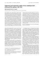
Figure 1 Experimental design. Human articular cartilage were cultured in monolayer (Expansion) under 5% and 19% oxygen percentages. Cells
were then cultured for two weeks again under the two oxygen percentages (Differentiation Phase I). Pellets generated at 5% and 19% oxygen
were further cultured at the same conditions or at interchanged oxygen percentages (Differentiation Phase II).
Ströbel et al. Arthritis Research & Therapy 2010, 12:R34
/>Page 3 of 15
Measurement of [
35
S]SO
4
and [
3
H]proline incorporation
The proteoglycan and collagen synthesis of pellets were
measured by assessing the incorporation of (
35
S)SO
4
and (
3
H)proline for a p eriod of 24 h as described pre-
viously [31]. Briefl y, pellets were incubated in the pre-
sence of both (
35
S)SO
4
(1 μCi/culture) to label
proteoglycans and (
3
H)proline (1.5 μCi/culture) to label
collagen. For the assessment of the released ECM frac-
tion, radiolabeled proteoglycan and collagen were preci-
pitated overnight at 4°C using respectively 100% ethanol
and 70% ammonium sulphate and subsequently, resus-
pended in 4 M guanidine hydro chloride or 10% sodium
dodecyl sulphate in Tris buffer (0.1 M, pH 7.0) respec-
tively for proteoglycan and collagen. For the assessment
of the incorporated ECM fraction, tissue pellets were
digested with protease K as previously described. The
incorporation of (
35
S)SO
4
and (
3
H)proline in culture
pellet and in conditioned medium was measured in a
Packard b-liquid scintillation counter with scintillation
fluid (Ultima Gold , Perkin Elmer, Schwer zenbach, Swit-
zerland). The amount of synthesised molecules was nor-
malized to the DNA content of the tissue.
Histological and immunohistochemical analyses
Pellets were fixed in 4% formalin, embedded in paraffin
and cross-sectioned (5 μm thick sections). The sections
were stained with Safranin O for sulfated GAG and pro-
cessed for immunohistochemistry to visualize type II
collagen (II-II6B3, Hybridoma Bank, University of Iowa,
Iowa City, IA, USA), as described in Grogan et al. [32]
and type II collagen fragments according to Roy-Beau-
dry et al. [33].
Electronic microscopy (SEM)
Images obtained from both scanning electron microscopy
(SEM) and transmission elect ron microscopy (TEM)
were used for t he structural analysis of collagen fibrils.
Pel let samples were glued onto a Teflon d isc with a five-
minute curing epoxy glue (Devcon Epoxy, ITW Brands,
Wood Dale, IL, USA). After which, the mounted speci-
mens were placed in a vibratory microtome (VT 1000 E,
Leica, He idelberg, Germany) to trim off the outermost,
approxim ately 150 μm thick cartilage layer parallel to the
support surface to minimize inhomogenities across the
surface among samples. The surface layer of the adult
healthy and OA cartilage was examined w ithout any
modification. The samples were then prepared for SEM
and TEM analysis as previous ly described [34]. For TEM
analysis, the samples were further homogenised into
small pieces in order to isolate single collagen fibrils.
Image analysis
Quantitative data on the collagen fibril organization
were obtained using the Image Processing Library &
Toolbox (IPLT) image analysis software package ( Basel,
Switzerland) [35]. A Canny edge detection algorithm
[36], followed by a skeletonization algorithm [37] was
applied to identify the collagen fibrils. The skeletonized
data were subjected to an algorithm identifying the end
points and intersections of the skeleton. Using this
information, the individual line segments were identified
and analyzed. Finally, the following parameters were
determined from each pellet condition: (i) the bending
ratio, calculated as the mean-squared end-to-end dis-
tance divided by the mean-squared contour length and
(ii) the persistence length, calculated using a previously
described model [38]. Both these parameters were
required to correlate the linearity of the fibrils and
length before branching of each i ndividual fibril to its
mechanical properties, respectively [39].
Total RNA extraction and cDNA synthesis
Total RNA of pellets was extracted using Trizol (Life
Technologies, Basel, Switzerland) and the standard sin-
gle-step acid-phenol guanidinium method. RNA was
treated with DNAseI using the DNA-free
™
Kit (Ambion,
Austin, Texas) and quantified spectrometrically. cDNA
was generated from 3 μgofRNAbyusing500μg/ml
random hexamers (Promega AG Dübendorf, Switzer-
land) and 1 μlof50U/mlStratascript
™
reverse tran-
scriptase (Stratagene, Amsterdam, NL), in the presence
of dNTPs. Real-time RT-PCR reactions wer e per formed
and monitored using the ABI Prism 7700 Sequence
Detection System (Perkin- Elmer/Applied Biosystems,
Rotkreuz, Switzerl and). Cycle te mperatures and times as
well as primers and probes used for the reference gene
(18-S rRNA) and the genes of interest (collagen type II
and aggrecan) were as previously described [40]. Assays
on-Demand(AppliedBiosystem)wereusedtomeasure
the expression of MMP-1 (Hs00233958_m1), MMP-2
(Hs00234422_m1), MMP-9 (Hs00234579_m1) and
MMP-13 (Hs00233992_m1). For ea ch cDNA sample,
the threshold cycle (Ct) value of each target sequence
was subtracted to the Ct value of 18-S rRNA, to derive
ΔCt. The level of gene expression was calculated as
2
ΔCt
. Each sample was assessed at least in d uplicate for
each gene of interest.
Quantification of released matrix metalloproteinases
Matrix metalloproteinases (MMP) were quantified in
media coll ected from cultured pellets by using the Mul-
tiAnalyte Profiling MMP base Kit (Fluorokine
®
MAP:
LMP000) complemented with the specific MMPs
(MMP-1: LMP901; MMP-3: LMP513; MMP-9: LMP911;
MMP-13: LMP511, R&D Systems, Minneapolis, MN,
USA). The assay was performed on a Luminex 100
™
analyzer (Austin, Texas, USA) following the manufac-
turer’s instructions. The amount of released MMPs was
normalized to the DNA content of the tissue.
Statistical analysis
For each a nalysis, triplicate pellets for each condition
and donor were assessed. Statistical evaluation was
Ströbel et al. Arthritis Research & Therapy 2010, 12:R34
/>Page 4 of 15
performed using SPSS software version 7.5 software
(SPSS, Sigma Stat, Erkrath, Germany). Values are pre-
sented as mean ± standard deviation (SD). Differences
between groups were assessed by Mann Whitney tests.
Differences in the persistence length and bending ratio
of collagen fibrils from different conditions were
assessed by one-way analysis of variance (ANOVA) with
Bonferroni post hoc test. Values of P <0.05werecon-
sidered statistically significant.
Results
Chondrogenic differentiation of HAC cultured under
different oxygen percentages
HAC were initially cultured in monolayer with expansion
medium at 5% or 19% O
2
and subsequently re-differen-
tiated in t hree-dimen sional p ellets at the two different
oxygen percentages (Phase I) (See Figure 1 for the experi-
mental design). HAC proliferated at comparable rates
(less than 5% variation in the number of doublings/day;
data not shown) at the two oxygen conditions. Cells
expanded at either oxygen percentage and subsequently
differentiated at 19% O
2
produced tissues faintly stained
forGAGandtypeIIcollagen(Figures2A,Iand2IIand
2B, I and 2II). Instead, reducing oxygen percentage dur-
ing differentiation enhance d the amount of car tilaginous
matrix accumulation, as evidenced by a qualitative
increased size of the generated tissues (Figure 2A, low
magnification), an increased intensity of Safranin O and
type II collagen stain (Figure 2A, B) and a statistically sig-
nificant higher amount of GAG (3.4- and 3.1-fold for
HAC e xpanded at 19% or 5% O
2
respectively) (Figure
2C). Due to the fact that expansion at 5% O
2
did not
influence the extent of HAC differentiation, further
assessments were only performed with cells expanded at
19% O
2
. In agreement with the histological and biochem-
ical results, the RT-P CR ana lysis confirmed statistically
significant higher expression of the cartilage specific
genes type II collagen (86.6-fold) and aggrecan (8.5-fold)
at 5% O
2
than at 19% O
2
aft er the Phase I differentiation
culture (Figure 2D, E).
Expression of catabolic mediators
We then investigated the possible role of oxygen
percentage in modulating the expression of catabolic
mediators. Analysis of specific matrix metalloproteinases
(that is, MMP-1, MMP-2, MMP-9 and MMP-13)by
RT-PCR indicated that low oxygen percentage applied
during the Phase I differentiation culture selectively
down-regulated MMP-1 and MMP-13 mRNA expres-
sion (7.7- and 3.5-fold, respectively). MMP-2 mRNA
was h ighly expressed and not modulated by the oxygen
percentag e. The expression of MMP-9 mRNA remained
unaffected and was at the limit of detection at both
oxygen percentages (Figure 3A).
The protein levels of MMP-1, -2, -9, -13 were assessed
in the supernatant of pellet cultures at the end of Phase
I. Consistent with the mRNA results, the amounts of
MMP-1 and -13 released were reduced in the pellets
cultured at 5% O
2
as compared to those cultured at 19%
O
2
(8.2- and 11.3-fold respectively). The protein expres-
sion levels of MMP-2 and -9 remained similar at the dif-
ferent oxygen percentages (Figure 3B).
Effect of oxygen percentage on HAC anabolic and
catabolic activity in pre-formed cartilaginous tissues
We next investigated the influence of oxygen in anabolic
(synthesis and accumulation of cartilaginous matrix pro-
teins) and catabolic (MMPs expression, activity and
degradation products) processes of pre-formed tissues.
Pellets generated after two weeks of culture at 19% O
2
or 5% O
2
(Phase I) were subsequen tly cultured up to an
additional two weeks (Phase II) at the same or at inter-
changed oxygen percentages (Figure 1).
Accumulation and synthesis of cartilaginous matrix proteins
In agreement w ith the above described results, pellets
cultured for four weeks (two weeks of Phase I and two
weeks of Phase II) at 5% O
2
were more strongly stained
for Safranin O and type II collagen, and accumulated
larger amounts of GAG (4.0-fold) as compared to those
cultured for the same time at 19% O
2
(Figu re 4A, B, C).
Reducing oxygen percentage during Phase II for pellets
cultured at 19% during Phase I resulted i n an improved
quality of the cartilaginous tissues, as assessed by an
increased accumulation of cartilaginous matrix positive
for GAG and type II collagen (Figure 4A, B) and by a
higher GAG content (3.3-fold) (Figure 4C). Conversely,
increasing oxygen percentage during Phase II for pellets
cultured at 5% during Phase I resulted in a reduced
accumulation of cartilaginous matrix (Figu re 4A, B) and
GAG content (1.9-fold) (Figure 4C).
Results from the radiolabelling experiments indicated
that similar amounts of total collagen and proteoglycan
(that is, released + accumulated) were synthesized by
pellets cultured for 18 days (two weeks of Phase I and
four days of Phase II) at the two oxygen percentages.
However, as compared to 19% oxygen (Phase I and
Phase II), the released fractions of these newly synthe-
sized macromolecules by pellets cultured at 5% O
2
(Phase I and Phase II) were markedly and statistically
significantly lower (2.0- and 2.9-fold respectively for col-
lagen and proteoglycan), while the accumulated fractions
were higher (2.1- and 6.6-fold respectively for collagen
and proteoglycan). Consistent with the biochemical
results, the culture at 5% O
2
during Phase II of tissues
pre-formed at 19% O
2
during Phase I resulted in an
augmented synthesis of collagen and proteoglycan
(respectively by 2.7- and 1.4-fold). In particular, the
increased synthesis of the newly synthesized
Ströbel et al. Arthritis Research & Therapy 2010, 12:R34
/>Page 5 of 15
ED
Type II collagen mRNA Aggrecan mRNA
Differentiation
Safranin-O
19%O
2
Type II collagen
III
VIIII
BA
5%O
2
noisnapxEnoisnapxE
19%O
2
5%O
2
19%O
2
5%O
2
VIIII
III
1.0E-06
1.0E-05
1.0E-04
1.0E-03
Diff 20% Diff 5%
*
Fold differences
from 18S
Diff 19% Diff 5%
1.0E-06
1.0E-05
1.0E-04
1.0E-03
Diff 20% Diff 5%
*
Fold differences
from 18S
Diff 19% Diff 5%
GAG/DNA ( g/
g)
C
0
2
4
6
8
10
20% Diff 5% Diff 20% Diff 5% Diff
20% expansion 5% expansion
GAG accumulation
*
*
Diff 19% Diff 5% Diff 19% Diff 5%
Expansion 19% Expansion 5%
Figure 2 Anabolic response of HAC to different oxygen percentages during the expansion and differentiation Phase I. (A - B) Safranin
O and type II collagen immunohistochemical stainings of representative tissues generated by human articular chondrocytes (HAC) expanded at
19% (I and III)or5%(II and IV) oxygen and further cultured in pellets at 19% (I and II)or5%(III and IV) oxygen. Bar = 100 μm. (C)
Quantification of glycosaminoglycans (GAG) accumulated normalized to the amount of DNA. (D - E) Real time reverse transcription-polymerase
chain reaction analysis of the expression of type II collagen and aggrecan mRNA by HAC cultured in pellets at 19% and 5% O
2
. Levels are
expressed as fold of difference from ribosomal 18S. For the gene expression analysis only expansion at 19% O
2
was considered. Values are mean
± SD of measurements obtained from three independent experiments. * = significantly different from the 19% O
2
.
Ströbel et al. Arthritis Research & Therapy 2010, 12:R34
/>Page 6 of 15
MMPs mRNA expression
A
B
Fold differences from 18S
1.0E-06
1.0E-05
1.0E-04
1.0E-03
1.0E-02
1.0E-01
Diff 20% Diff 5% Diff 20% Diff 5% Diff 20% Diff 5% Diff 20% Diff 5%
MMP-1 MMP-2 MMP-9 MMP-13
*
*
Diff 19% Diff 5% Diff 19% Diff 5% Diff 19% Diff 5% Diff 19% Diff 5%
MMP-1 MMP-2 MMP-9 MMP-13
0
5
10
15
20
25
30
Diff 20% Diff 5% Diff 20% Diff 5% Diff 20% Diff 5% Diff 20% Diff 5%
MMP-1 MMP-2 MMP-9 MMP-13
*
*
Protein/DNA (ng/
g)
MMPs protein release
Diff 19% Diff 5% Diff 19% Diff 5% Diff 19% Diff 5% Diff 19% Diff 5%
MMP-1 MMP-2 MMP-9 MMP-13
Figure 3 Quantification of MMPs produced by HAC cultured at different oxygen percentages during the Phase I.(A) Real ti me reverse
transcription-polymerase chain reaction analysis of the expression of MMP-1, -2, -9, -13 mRNA by human articular chondrocytes (HAC) cultured
in pellets at 19% and 5% O
2
. Levels are expressed as fold of difference from ribosomal 18S. (B) Quantification of MMP-1, -2, -9, -13 released in
the culture medium. Levels are normalized to the amount of DNA measured in relative pellets. Values are mean ± SD of measurements obtained
from three independent experiments. * = significantly different from the 19% O
2
.
Ströbel et al. Arthritis Research & Therapy 2010, 12:R34
/>Page 7 of 15
B
Type II collagen
II
IV
Safranin-O
I
III
A
II
IV
I
III
D
E
35
S-PG/DNA (cpm/
g)
Phase II: 19%O
2
Phase I: 19%O
2
Phase I: 19%O
2
Phase II: 5%O
2
Phase II: 19%O
2
Phase II: 5%O
2
Phase II: 5%O
2
Phase I: 5%O
2
Phase I: 5%O
2
Phase II: 19%O
2
Phase II: 5%O
2
Phase II: 19%O
2
C
3
H-proline/DNA (cpm/
g)
0
10000
20000
30000
40000
Phase II:
20%
Phase II:
5%
Phase II:
5%
Phase II:
20%
Phase I: 20% Phase I: 5%
Phase II:
19%
Phase II:
5%
Phase II:
5%
Phase II:
19%
Phase I: 19% Phase I: 5%
0
10000
20000
30000
40000
Phase II:
20%
Phase II:
5%
Phase II:
5%
Phase II:
20%
Phase I: 20% Phase I: 5%
Phase II:
19%
Phase II:
5%
Phase II:
5%
Phase II:
19%
Phase I: 19% Phase I: 5%
released
accumulated
Collagen synthesis
*
*
°
a
a, r
a, r
Proteoglycan synthesis
released
accumulated
*
*
°
aa, r
a, r
0
2
4
6
8
10
Phase II: 19% Phase II: 5% Phase II: 5% Phase II: 19%
Phase I: 19% Phase I: 5%
GAG/DNA (
g/
g)
*
GAG accumulation
°
*
Figure 4 Anabolic response of HAC to different oxygen percentages during differentiation Phase I and II.(A - B) Safranin O and type II
collagen stainings of representative tissues generated by human articular chondrocytes (HAC) cultured in pellets for two weeks (Phase I) at 19%
(I and II)or5%(III and IV) oxygen and further cultured for two additionally weeks (Phase II) at 19% (I and III)or5%(II and IV) oxygen. Bar =
100 μm. (C) Quantification of glycosaminoglycans (GAG) accumulated in pellets cultured as described in (A - B) normalized to the amount of
DNA. (D - E) Amounts of newly synthesized collagen (D) and proteoglycan (E) measured in pellets cultured for 18 days (two weeks of Phase I
and four days of Phase II). The upper and lower parts of the columns represent the released and accumulated fractions respectively. Values are
mean ± SD of measurements obtained from two independent experiments. * = significantly different from the group cultured with the same
oxygen percentage in Phase I but with different oxygen tension in Phase II; ° = significantly different from the group cultured entirely at 19% O
2
;
a = accumulated, r = released.
Ströbel et al. Arthritis Research & Therapy 2010, 12:R34
/>Page 8 of 15
macromolecules was mainly reflected by an augmented
accumulation (up to 5.9-fold). Instead, the culture at
19% O
2
during Phase II of tissues pre-formed at 5% O
2
during Phase I differently modulated the synthesis of
the two extracellular matrix molecules: while a
decreased accumulation ( 2.3-fold) and an increased
released (2.6-fold) was measured for collagen, only a
reduction of the accumulated fraction was demonstrated
for proteoglycan (8.6-fold) (Figure 4D, E).
MMPs production and activity
Pellets cultured for four weeks (two weeks of Phase I
and two weeks of Phase II) at 5% O
2
released lower
amounts o f MMP-1 and -13 (6.1- and 10.1-fold respec-
tively) as compared to those cultured for the same time
at 19% O
2
.Cultureat5%O
2
during Phase II of tissues
pre-formed at 19% O
2
during Phase I resulted in
reduced pro duction of both MMPs, though only MMP-
13 by statistically significant levels (by 1.8-fold). Instead,
culture at 19% O
2
during Phase II of pellets pre-formed
at 5% O
2
during Phase I resulted in increased release of
both MMP-1 and MMP-13 (4. 0-and6.2-foldrespec-
tively) (Figure 5A, B).
In order to assess whether the observed increased pro-
duction of MMPs corresponded to an increased protei-
nase activity, pellets cultured for a total of four weeks at
the different oxygen percentages were assessed immuno-
histochemically t o detect the presence of type II colla gen
C-telopeptides, derived by MMP-1 and -13 collagenolytic
activity [33]. Analyses indicated that only the pellets
formed at 5% O
2
during Phase I and subsequently cul-
turedat19%O
2
during Phase II were intensely stained
for the type II collagen fragments (Figure 5C).
Collagen fibril organization
To determine whether increasing oxygen percentage
during cultivation Phase II of tissues pre-formed at 5%
O
2
would change the structure and arrangement of the
collagen fibril network, pellets were qualitatively and
quantitatively assessed via EM. Images indicated that the
collagen fibrils of pellets cultured at 5% O
2
during
Phase I and then for two weeks at 19% O
2
during Phase
II were less linear than those of pellets cultured for four
weeks at 5% O
2
. Interestingly, a similar trend was also
observed in the OA cartilage as compared to healthy
cartilage samples (Figure 6A, B). In pellets, the collagen
network was comprised of single fibrils with diameters
ranging from 20 to 30 nm. In healthy adult cartilage,
the network contained bundled and twisted collagen
fibrils three- to four-fold larger in diameter. Quantitative
image analysis indicated that increasing the oxygen per-
centage during Phase II resulted in a significant reduc-
tion of persistence length as well as bending ratio
(47.9% and 10.5% respectively). Intere stingly, both para-
meters were higher in healthy as compared to OA tis-
sues (30.0% and 6.6% respectively for persistence length
and bending ratio). Considerable decrease in persistence
length and bending ratio w ould indicate softening and
gradual deterioration of cartilage physiological function
[39].
Response to low oxygen under CdCl
2
-treatment
Todeterminewhethertheobservedpro-anabolicand
anti-catabolic effects of low oxygen percentage are
mediated by HIF-1a, HAC from three donors were pre-
cultured in pellets during Phase I at 19% O
2
. During the
subsequent culture Phase II, the pre-cultured pellets
were maintained at 19% O
2
or exposed to 5% O
2
,with
or without treatment with CdCl
2
forsixhoursorthree
days (Figure 7A). Following culture at low oxygen per-
centage, type II collagen m RNA was up-regulated to a
higher extent after six hours (up to 33.0-fold; Figure 7B)
than after three days (data not shown), while MMP-1
mRNA was down-regulated to a higher extent after
three days (up to 65.5-fold; Figure 7C) than after s ix
hour s (data not shown). Supplementation of CdCl
2
dur-
ing this culture phase almost abrogated the aforemen-
tioned low O
2
-mediated effects, so that the expression
of type II collagen and MMP-1 mRNA reached levels
comparable to those of cells cultured at 19% O
2
for the
corresponding times (Figure 7B, C).
Discussion
In this study we found that culture at low, more physio-
logical (5%) oxygen percentage has a dual role in HAC
metabolism, namely to enhance the proteoglycan and
collagen synthesis and at the same time to reduce the
activity of two key catabolic enzymes involved in carti-
lage breakdown (that is, MMP-1 and MMP-13). As a
consequence, HAC exposure to 19% oxygen reduced the
de novo formation of cartilage tissue and induced degra-
dation of pre-deposited collagen fibrils, leading to struc-
tural features similar to those found in osteoarthritic
tissue. Interestingly, HAC appeared to be highly sensi-
tive to the oxygen percentage applied during differentia-
tion culture in pellets, but not during expansion in
monolayers. The anti-anabolic and pro-catabolic effects
mediated by low oxygen percentage were HIF1a- depen-
dent, as assessed by specific inhibition of this factor by
CdCl
2
treatment.
The application of 5% oxygen percentage during the
HAC mono layer expansion did not influence the prolif -
eration rate and chondrogenic capacity o f HAC. This is
in contrast with results reported by Egli et al. [7], indi-
cating that bovine articular chondrocytes expanded
under hypoxic conditions generated tissues with higher
amounts of cartilaginous matrix as compared to those
expanded under normoxic conditions. The discrepancy
between our results and those generated by Egli et al.
[7]canberelatedtothedifferenttypeofcellsused
(human vs bovine), the stage of cell de-differentiation
Ströbel et al. Arthritis Research & Therapy 2010, 12:R34
/>Page 9 of 15
Protein/DNA (ng/
g)
Phase II: 19%O
2
Phase II: 5%O
2
III
III IV
MMP-1 protein release MMP-13 protein release
BA
C
Type II collagen fragments
Phase I: 19%O
2
Phase II: 5%O
2
Phase II: 19%O
2
Phase I: 5%O
2
Protein/DNA (ng/
g)
0
5
10
15
20
25
30
Phase II:
20%
Phase II:
5%
Phase II:
5%
Phase II:
20%
Phase I: 20% Phase I: 5%
0
5
10
15
20
Phase II:
20%
Phase II:
5%
Phase II:
5%
Phase II:
20%
Phase I: 20% Phase I: 5%
*
°
*
*
°
19% 19% 19% 19%
Phase I: 19% Phase I: 5% Phase I: 19% Phase I: 5%
Figure 5 Catabolic response of HAC to different oxygen percentages during differentiation Phase I and II.(A - B) Quantification of MMP-
1(A) and MMP-13 (B) released in the medium by human articular chondrocytes (HAC) cultured in pellets for four weeks (two weeks of Phase I
and two weeks of Phase II). Levels are normalized to the amount of DNA measured in relative pellets. Values are mean ± SD of measurements
obtained from two independent experiments. * = significantly different from the group cultured with the same oxygen percentage in Phase I
but with different oxygen tension in Phase II; ° = significantly different from the group cultured entirely at 19% O
2
(Phase I and Phase II). (C)
Immunohistochemical detection of type II collagen fragments of pellets cultured under conditions described in (A - B). Bar = 100 μm.
Ströbel et al. Arthritis Research & Therapy 2010, 12:R34
/>Page 10 of 15
(second passaged vs first passaged cells) and/or the spe-
cific oxygen percentage tested ( 5% vs 1.5%). Indeed,
HAC culture at lower than 5% oxygen during expansion
may lead to a benefit in their redifferentiation capacity,
and remains to be investigated.
The influence of oxygen percentage during the de-
novo tissue formation was evaluated by culturing HAC
in micromass pellets, a model commonly used to inves-
tigate in vitro cartilage development. Our results indi-
cate that the application of 5% as compared to 19%
oxygen percentage critically enhanced the chondrogenic
capacity of HAC, as assessed by a greater accumulation
of GAG and type II collagen. Similar responses to
reduced oxygen percentage have been reported [9] using
human nasal chondrocytes statically cultured in pellets
for three days and subsequently transferred to a
dynamic bioreacto r system. We also investigated
whether culture of chondrocytes at low oxygen percen-
tage modulated the production of specific metalloprotei-
nases involved in the degradation of extracellular matrix
proteins. We observed that the expression of MMP-1
and MMP-13, both at mRNA and protein levels, was
reduced in cells cultured at 5% as compared to 19% oxy-
gen. Interestingly, MMP-1 (or collagenase-1) and/or
MMP-13 (or collagenase-3) are among the enzymes
expressed b y human chondrocytes in degenerative
C
Collagen structure
D
Persistence length Bending ratio
nm
0
200
400
600
800
Phase II:
5%
Phase II:
20%
Healthy OA
Phase I: 5% Native tissues
nm/nm
*
°
Phase II:
5%
Phase II:
19%
Healthy OA
Phase I: 5% Native tissues
0.70
0.75
0.80
0.85
0.90
Phase II:
5%
Phase II:
20%
Healthy OA
Phase I: 5% Native tissues
*
°
5% 19%
BA
Healthy OsteoarthriticPhase II: 5%O
2
Phase I: 5%O
2
III
Phase II: 19%O
2
Pellets Native tissues
SEM images
I
II
II
Figure 6 SEM images and structural analysi s of extracell ular coll agen -fibrils from engineered, healthy and osteoarthritic cartilage
samples. Representative scanning electron microscopy (SEM) images of (A) tissues generated by culturing human articular chondrocytes in
pellets for four weeks (two weeks of Phase I and two weeks of Phase II) or (B) native human tissue biopsies from healthy or osteoarthritic (OA)
cartilage. (C) Persistence length and (D) bending ratio assessment of the extracellular fibril network of engineered and native tissues. * =
significant different from 19% O
2
; ° = significant different from OA tissues.
Ströbel et al. Arthritis Research & Therapy 2010, 12:R34
/>Page 11 of 15
patholo gies of cartilage, namely osteoarthritis and rheu-
matoid arthritis [41] and are thus thought to play a cri-
tical role in cartilage destruction. In particular, it has
been shown that both MMPs are involved in the initial
phase of type II collagen breakd own [42,43], and MMP-
13 is the collagenase with highest affinity for type II col-
lagen [44]. However, the expression of other MMPs or
degradative enzymes (for example, aggrecanases) not
included in our study might also be regulated by culture
at low oxygen tension.
Our results prompted us to hypothesize that different
oxygen percentages could regulate not only cartilage
generation, but also its further maturation and stability.
We thus exposed tissues form ed at the different oxygen
percentages for two weeks (Phase I) to interchanged
oxygen percentages in a subsequent culture phase
(Phase II). Results obtained from the radiolabelling
experiments indicated that the exposure of tissues to 5%
oxygen during Phase II induced higher synthesis and
accumulation of c ollagen and proteoglycan. It remains
to be assessed whether low oxygen perc entages also
enhance expression of molecules involved in stabiliza-
tion of the newly synthesized extracellular matrix com-
ponents (for exa mple, decorin, fibromodulin, l ink
protein, type IX collagen) [45]. Importantly, the pre-
sence of type II collagen cleavage products, indicative of
MMP activity, was immunohistochemically detected [33]
only in the pellets pre-formed at 5% oxygen (Phase I)
and subsequently cultured for additional two weeks at
19% oxygen (Phase II). These results, together with the
observed enhanced expression of MMP-1 and -13 at
19% oxygen, strongly indicate a direct involvement of
Fold differences from 18S
MMP-1 mRNA expression
A
Fold differences from 18S
1.0E-06
1.0E-05
1.0E-04
1.0E-03
1.0E-02
1.0E-01
Donor 1 Donor 2 Donor 3
1.0E-06
1.0E-05
1.0E-04
1.0E-03
Donor 1 Donor 2 Donor 3
Type II collagen mRNA expression
19%, ChM
5%, ChM
5%, ChM + CdCl
2
19%O
2
19%O
2
Phase I
(2 weeks)
Phase II
(6 hours – 3 days)
19%O
2
ChM
5%O
2
5%O
2
ChM
ChM + CdCl
2
CB
Figure 7 Effects inhibition of HIF-1a on anabolic and catabolic gene regulation at low oxygen percentage.(A) Experimental desig n:
human articular chondrocytes from three donors were cultured as pellets in chondrogenic medium (ChM) at 19% O
2
(Phase I) and subsequently
maintained at the same oxygen percentage or exposed to 5% O
2
in the absence or presence of 5 μM CdCl
2
for six hours or three days (Phase
II). Real time reverse transcription-polymerase chain reaction analysis of type II collagen mRNA expression after six hours (B) and of MMP-1 mRNA
expression after three days (C). Levels are expressed as fold of difference from ribosomal 18S. Values for each donor are mean ± SD of
measurements obtained from three independent pellets.
Ströbel et al. Arthritis Research & Therapy 2010, 12:R34
/>Page 12 of 15
oxygen in regulating the MMP-mediated breakdow n of
cartilaginous tissues. The result that pellets entirely cul-
turedat19%O
2
negatively stained for type II collagen
fragments could be explained by the insufficient accu-
mulation of the MMP substrate (that is, type II collagen)
during the initial cultivation Phase I.
The presence of type II collagen fragments correlated
well with the branched/tangled collagen fibril organiza-
tion and decreased values of bending ratio and persis-
tence length in pellets exposed to 19% oxygen. This
couldpossiblyresultfroman increased enzymatic clea-
vage of the extracellular matrix molecules by specific
MMPs. Conclusively, increased activity of catabolic
enzymes is affecting the collagen fibril network that
exhibits lower value s of bending ratio and persistence
length. Based on this correlation, both parameters could
potentially represent valuable markers for determining
the degree of collagen deterioration. Exposure of carti-
lage tissues formed at physio logical oxygen pe rcentages
to higher oxygen levels resembled degradation events
occurring during the progression of OA, where, follow-
ing initial pathologic events, the normal oxygen gradi-
ents break down [6]. Therefore, our tissue engineering
model would be instrumental to investigation of the
evolution of cartilage damage following alteration of the
oxygen levels and to assess the effect of possible thera-
peutic targets.
The observed pro-anabolic and anti-catabolic effects of
low oxygen culture were mediated by the hypoxia indu-
cible signaling pathway, since reduction of the oxygen
percentage did not regulate type II colla gen and MMP-1
mRNA expression in the presence of the HIF-1a inhibi-
tor cadmium chlo ride (CdCl
2
) [28]. While the impor-
tance of HIF-1a in modulating the expression/synthesis
of cartilage-specific genes was recently addressed
[28-46], the involvement of this factor in the oxygen-
dependent modulation of catabolic genes, recently
reported for porcine pulmonary artery endothelial and
smooth muscle cells [47], has not been previously postu-
lated for HAC.
Conclusions
The present study demonstrates that low oxygen percen-
tage applied during the differentiation phases of human
articular chondrocyte culture enhances cell biosynthetic
activity as well as reduces the activity of catabolic
enzymes known to play key roles in the breakdown of
cartilage matrix during degenerative pathologies. These
findings indicate that regulation o f oxygen percentages
during in vitro culture could be used to improve the
generation of functional cartilage substitutes, and thus
prompt the development of tools enabl ing accurate con-
trol of oxyg en levels for tissues of clinically relevant size
[48]. Moreover, modulation of oxygen tension in
cultured HAC may be used as a tool to model and
study in vitro pathophysiological events occ urring in
osteoarthritis. Finally, following such investigations, the
identification of innovative strategies to maintain local
in vivo oxygen percentages to defined levels could repre-
sent a powerful tool for preventing the progression of
degenerative cartilage diseases.
Abbreviations
ANOVA: analysis of variance; cDNA: complementary deoxyribonucleic acid;
CO
2
:carbondioxide;Ct:thresholdcycle;DMEM:Dulbecco’s modified
Eagle’ s medium; DMMB: dimethyl methylene blue; dNTP:
deoxyribonucleotide; ECM: extra cellul ar matrix; EDTA:
ethylenediaminetetraacetic acid; EM: electronic microscopy; GAG:
glycosaminoglycans; HAC: h uman articular chondrocytes; HEPES: 4-(2-
hydroxyethyl)-1-piperazineethanesulfonic acid; HIF-1a: hypoxia-inducible
factor-1alpha; IPLT: Image Processing Library & Toolbox; MMP:
metalloproteinase; mRNA: messenger ribonucleic acid; O
2
:oxygen;OA:
osteoarthritis; PBS: phosphate buffered saline; RNA: ribonucleic acid; rRNA:
ribosomal ribonucleic acid; RT- PCR: reverse-transcriptas e polymerase chain
reaction; SD: standard deviation; SEM: scanning electron microscopy; TEM:
transmission electron microscopy; TGFb1 : transforming growth factor
beta-1.
Acknowledgements
We would like to acknowledge the European Union for financial support
(STEPS; FP6-#NMP3-CT-2005-500465) and the National Competence Center in
Research (NCCR) program Nanoscale Science, awarded by the Swiss National
Science Foundation, for support to Mr. M. Loparic. We are grateful to Mrs F.
Wolf and Mrs D. Thuillard for their assistance with immunohistochemical
processing, to Dr. Riccardo Gottardi from Department for Biophysical
Engineering (Genova, Italy) for his assistance with EM analysis and Dr. M.
Duggelin and Ms. Melanie Burkhardt for the imaging analysis. We thank Dr.
Christgau from Nordic Immunology (Tilburg, NL) for the generous supply of
the antibodies against type II collagen fragments.
Author details
1
Departments of Surgery and of Biomedicine, University Hospital Basel,
Hebelstrasse 20, Basel, 4031, Switzerland.
2
M.E. Müller Institute for Structural
Biology, Biozentrum University of Basel, Klingelbergstrasse 50/70, Basel, 4056,
Switzerland.
3
Department of Orthopaedic Surgery and Traumatology,
Ospedale Regionale di Lugano, Via Tesserete 46, Lugano, 6900, Switzerland.
4
Departments of Biomedicine and Neurology, University Hospital Basel,
Hebelstrasse 20, Basel, 4031, Switzerland.
5
Faculty of Dentistry and CHU
Sainte-Justine, University of Montreal, 3175 Côte Sainte-Catherine, Montreal,
H3T1C5, Canada.
Authors’ contributions
SS participated in study conception and design, acquisition of data
(biochemistry, histology, immunohistochemistry for type II collagen, RT-PCR
analysis and cell culture), in the study design, in the interpretation of data
and drafting the manuscript. ML participated in acquisition of the data
(scanning electronic microscopy and image analysis) and in the
interpretation of data. DW participated in study conception in the study
design and revised the manuscript. ADS participated in analysis (image
analysis). CC participated in study conception and provided the patient
biopsies and their clinical data. RLPL participated in the development of the
Luminex assays. FM participated in the acquisition of data
(immunohistochemistry for type II collagen fragments) and revised the
manuscript. AB and IM were responsib le for study design, supervision of the
experiments, interpretation of data and participated in writing the
manuscript. All authors read and approved the final manuscript.
Competing interests
The authors declare that they have no competing interests.
Received: 28 September 2009 Revised: 9 February 2010
Accepted: 2 March 2010 Published: 2 March 2010
Ströbel et al. Arthritis Research & Therapy 2010, 12:R34
/>Page 13 of 15
References
1. Martin JA, Brown T, Heiner A, Buckwalter JA: Post-traumatic osteoarthritis:
the role of accelerated chondrocyte senescence. Biorheology 2004,
41:479-491.
2. Martin JA, Buckwalter JA: Roles of articular cartilage aging and
chondrocyte senescence in the pathogenesis of osteoarthritis. Iowa
Orthop J 2001, 21:1-7.
3. Malda J, Martens DE, Tramper J, van Blitterswijk CA, Riesle J: Cartilage
tissue engineering: controversy in the effect of oxygen. Crit Rev
Biotechnol 2003, 23:175-194.
4. Silver IA: Measurement of pH and ionic composition of pericellular sites.
Philos Trans R Soc Lond B Biol Sci 1975, 271:261-272.
5. Gonsalves M, Barker AL, Macpherson JV, Unwin PR, O’Hare D, Winlove CP:
Scanning electrochemical microscopy as a local probe of oxygen
permeability in cartilage. Biophys J 2000, 78:1578-1588.
6. Grimshaw MJ, Mason RM: Bovine articular chondrocyte function in vitro
depends upon oxygen tension. Osteoarthritis Cartilage 2000, 8:386-392.
7. Egli RJ, Bastian JD, Ganz R, Hofstetter W, Leunig M: Hypoxic expansion
promotes the chondrogenic potential of articular chondrocytes. J Orthop
Res 2008, 26:977-985.
8. Hansen U, Schunke M, Domm C, Ioannidis N, Hassenpflug J, Gehrke T,
Kurz B: Combination of reduced oxygen tension and intermittent
hydrostatic pressure: a useful tool in articular cartilage tissue
engineering. J Biomech 2001, 34:941-949.
9. Malda J, van Blitterswijk CA, van Geffen M, Martens DE, Tramper J, Riesle J:
Low oxygen tension stimulates the redifferentiation of dedifferentiated
adult human nasal chondrocytes. Osteoarthritis Cartilage 2004, 12:306-313.
10. Nevo Z, Beit-Or A, Eilam Y: Slowing down aging of cultured embryonal
chick chondrocytes by maintenance under lowered oxygen tension.
Mech Ageing Dev 1988, 45:157-165.
11. Lane JM, Brighton CT, Menkowitz BJ: Anaerobic and aerobic metabolism
in articular cartilage. J Rheumatol 1977, 4:334-342.
12. Murphy CL, Sambanis A: Effect of oxygen tension and alginate
encapsulation on restoration of the differentiated phenotype of
passaged chondrocytes. Tissue Eng 2001, 7:791-803.
13. Malda J, Brink van den P, Meeuwse P, Grojec M, Martens DE, Tramper J,
Riesle J, van Blitterswijk CA: Effect of oxygen tension on adult articular
chondrocytes in microcarrier bioreactor culture. Tissue Eng 2004,
10:987-994.
14. Marcus RE: The effect of low oxygen concentration on growth, glycolysis,
and sulfate incorporation by articular chondrocytes in monolayer
culture. Arthritis Rheum 1973, 16:646-656.
15. Saini S, Wick TM: Effect of low oxygen tension on tissue-engineered
cartilage construct development in the concentric cylinder bioreactor.
Tissue Eng 2004, 10:825-832.
16. Domm C, Schunke M, Christesen K, Kurz B: Redifferentiation of
dedifferentiated bovine articular chondrocytes in alginate culture under
low oxygen tension. Osteoarthritis Cartilage 2002, 10:13-22.
17. Kurz B, Domm C, Jin M, Sellckau R, Schunke M: Tissue engineering of
articular cartilage under the influence of collagen I/III membranes and
low oxygen tension. Tissue Eng 2004, 10:1277-1286.
18. Martinez I, Elvenes J, Olsen R, Bertheussen K, Johansen O: Redifferentiation
of in vitro expanded adult articular chondrocytes by combining the
hanging-drop cultivation method with hypoxic environment. Cell
Transplant 2008, 17:987-996.
19. Murphy CL, Sambanis A: Effect of oxygen tension on chondrocyte
extracellular matrix accumulation. Connect Tissue Res 2001, 42:87-96.
20. Murphy CL, Polak JM: Control of human articular chondrocyte
differentiation by reduced oxygen tension. J Cell Physiol 2004,
199:451-459.
21. Nevo Z, Horwitz AL, Dorfmann A: Synthesis of chondromucoprotein by
chondrocytes in suspension culture. Dev Biol 1972, 28:219-228.
22. Scherer K, Schunke M, Sellckau R, Hassenpflug J, Kurz B: The influence of
oxygen and hydrostatic pressure on articular chondrocytes and
adherent bone marrow cells in vitro. Biorheology 2004, 41:323-333.
23. Brighton CT, Lane JM, Koh JK: In vitro rabbit articular cartilage organ
model. II. 35S incorporation in various oxygen tensions. Arthritis Rheum
1974, 17:245-252.
24. Clark CC, Tolin BS, Brighton CT: The effect of oxygen tension on
proteoglycan synthesis and aggregation in mammalian growth plate
chondrocytes. J Orthop Res 1991, 9:477-484.
25. Obradovic B, Carrier RL, Vunjak-Novakovic G, Freed LE: Gas exchange is
essential for bioreactor cultivation of tissue engineered cartilage.
Biotechnol Bioeng 1999, 63:197-205.
26. Ysart GE, Mason RM: Responses of articular cartilage explant cultures to
different oxygen tensions. Biochim Biophys Acta 1994, 1221:15-20.
27. Barbero A, Grogan S, Schafer D, Heberer M, Mainil-Varlet P, Martin I: Age
related changes in human articular chondrocyte yield, proliferation and
post-expansion chondrogenic capacity. Osteoarthritis Cartilage 2004,
12:476-484.
28. Duval E, Leclercq S, Elissalde JM, Demoor M, Galéra P: Hypoxia-inducible
factor-a inhibits the fibroblast-like markers type I and type III collagen
during hypoxia-induced chondrocyte redifferentiation: Hypoxia not only
induces type II collagen and aggrecan, but it also inhibits type I and
type III collagen in the hypoxia-inducible factor 1a-dependent
redifferentiation of chondrocytes. Arthritis Rheum 2009,
60:3038-3048.
29. Hollander AP, Heathfield TF, Webber C, Iwata Y, Bourne R, Rorabeck C,
Poole AR: Increased damage to type II collagen in osteoarthritic articular
cartilage detected by a new immunoassay. J Clin Invest 1994,
93:1722-1732.
30. Farndale RW, Buttle DJ, Barrett AJ: Improved quantitation and
discrimination of sulphated glycosaminoglycans by use of
dimethylmethylene blue. Biochim Biophys Acta 1986, 883:173-177.
31. Waldman SD, Couto DC, Grynpas MD, Pilliar RM, Kandel RA: A single
application of cyclic loading can accelerate matrix deposition and
enhance the properties of tissue-engineered cartilage. Osteoarthritis
Cartilage 2006, 14:323-330.
32. Grogan SP, Rieser F, Winkelmann V, Berardi S, Mainil-Varlet P: A static,
closed and scaffold-free bioreactor system that permits chondrogenesis
in vitro. Osteoarthritis Cartilage 2003, 11:403-411.
33. Roy-Beaudry M, Martel-Pelletier J, Pelletier JP, M’Barek KN, Christgau S,
Shipkolye F, Moldovan F: Endothelin 1 promotes osteoarthritic cartilage
degradation via matrix metalloprotease 1 and matrix metalloprotease 13
induction. Arthritis Rheum 2003, 48:2855-2864.
34. Stolz M, Gottardi R, Raiteri R, Miot S, Martin I, Imer R, Staufer U, Raducanu A,
Duggelin M, Baschong W, Daniels AU, Friederich NF, Aszodi A, Aebi U: Early
detection of aging cartilage and osteoarthritis in mice and patient
samples using atomic force microscopy. Nat Nanotechnol 2009, 4:186-192.
35. Philippsen A, Schenk AD, Signorelli GA, Mariani V, Berneche S, Engel A:
Collaborative EM image processing with the IPLT image processing
library and toolbox. J Struct Biol 2007, 157:28-37.
36. Canny JF: A computational approach to edge detection. IEEE Trans Pattern
Anal Mach Intell 1986, 8:679-698.
37. Gonzalez RC, Woods RE: Digital Image Processing Prentice Hall, Inc., USA, 3 2007.
38. Hagerman PJ: Flexibility of DNA. Annu Rev Biophys Biophys Chem 1988,
17:265-286.
39. Raub CB, Unruh J, Suresh V, Krasieva T, Lindmo T, Gratton E, Tromberg BJ,
George SC: Image correlation spectroscopy of multiphoton images
correlates with collagen mechanical properties. Biophys J 2008,
94:2361-2373.
40. Jakob M, Demarteau O, Schafer D, Hintermann B, Dick W, Heberer M,
Martin I: Specific growth factors during the expansion and
redifferentiation of adult human articular chondrocytes enhance
chondrogenesis and cartilaginous tissue formation in vitro. J Cell Biochem
2001, 81:368-377.
41. Burrage PS, Mix KS, Brinckerhoff CE: Matrix metalloproteinases: role in
arthritis. Front Biosci 2006, 11:529-543.
42. Dahlberg L, Billinghurst RC, Manner P, Nelson F, Webb G, Ionescu M,
Reiner A, Tanzer M, Zukor D, Chen J, van Wart HE, Poole AR: Selective
enhancement of collagenase-mediated cleavage of resident type II
collagen in cultured osteoarthritic cartilage and arrest with a synthetic
inhibitor that spares collagenase 1 (matrix metalloproteinase 1). Arthritis
Rheum 2000, 43:673-682.
43. Wu W, Billinghurst RC, Pidoux I, Antoniou J, Zukor D, Tanzer M, Poole AR:
Sites of collagenase cleavage and denaturation of type II collagen in
aging and osteoarthritic articular cartilage and their relationship to the
distribution of matrix metalloproteinase 1 and matrix metalloproteinase
13. Arthritis Rheum 2002, 46:2087-2094.
44. Reboul P, Pelletier JP, Tardif G, Cloutier JM, Martel-Pelletier J: The new
collagenase, collagenase-3, is expressed and synthesized by human
chondrocytes but not by synoviocytes. A role in osteoarthritis. J Clin
Invest 1996, 97:2011-2019.
Ströbel et al. Arthritis Research & Therapy 2010, 12:R34
/>Page 14 of 15
45. Poole AR, Kojima T, Yasuda T, Mwale F, Kobayashi M, Laverty S:
Composition and structure of articular cartilage: a template for tissue
repair. Clin Orthop Relat Res 2001, 391:26-33.
46. Pfander D, Cramer T, Schipani E, Johnson RS: HIF-1alpha controls
extracellular matrix synthesis by epiphyseal chondrocytes. J Cell Sci 2003,
116:1819-1826.
47. Ye H, Zheng Y, Ma W, Ke D, Jin X, Liu , Wang D: Hypoxia down-regulates
secretion of MMP-2, MMP-9 in porcine pulmonary artery endothelial and
smooth muscle cells and the role of HIF-1. J Huazhong Univ Sci Technolog
Med Sci 2005, 25:382-384.
48. Wendt D, Stroebel S, Jakob M, John GT, Martin I: Uniform tissues
engineered by seeding and culturing cells in 3D scaffolds under
perfusion at defined oxygen tensions. Biorheology 2006, 43:481-488.
doi:10.1186/ar2942
Cite this article as: Ströbel et al.: Anabolic and catabolic responses of
human articular chondrocytes to varying oxygen percentages. Arthritis
Research & Therapy 2010 12:R34.
Submit your next manuscript to BioMed Central
and take full advantage of:
• Convenient online submission
• Thorough peer review
• No space constraints or color figure charges
• Immediate publication on acceptance
• Inclusion in PubMed, CAS, Scopus and Google Scholar
• Research which is freely available for redistribution
Submit your manuscript at
www.biomedcentral.com/submit
Ströbel et al. Arthritis Research & Therapy 2010, 12:R34
/>Page 15 of 15