Báo cáo y học: "Calcium deposition in osteoarthritic meniscus and meniscal cell culture" pps
Bạn đang xem bản rút gọn của tài liệu. Xem và tải ngay bản đầy đủ của tài liệu tại đây (1.23 MB, 9 trang )
Sun et al. Arthritis Research & Therapy 2010, 12:R56
/>Open Access
RESEARCH ARTICLE
BioMed Central
© 2010 Sun et al.; licensee BioMed Central Ltd. This is an open access article distributed under the terms of the Creative Commons At-
tribution License ( which permits unrestricted use, distribution, and reproduction in any
medium, provided the original work is properly cited.
Research article
Calcium deposition in osteoarthritic meniscus and
meniscal cell culture
Yubo Sun*
1
, David R Mauerhan
1
, Patrick R Honeycutt
1
, Jeffrey S Kneisl
1
, H James Norton
2
, Natalia Zinchenko
1
,
Edward N Hanley Jr
1
and Helen E Gruber
1
Abstract
Introduction: Calcium crystals exist in the knee joint fluid of up to 65% of osteoarthritis (OA) patients and the presence
of these calcium crystals correlates with the radiographic evidence of hyaline cartilaginous degeneration. This study
sought to examine calcium deposition in OA meniscus and to investigate OA meniscal cell-mediated calcium
deposition. The hypothesis was that OA meniscal cells may play a role in pathological meniscal calcification.
Methods: Studies were approved by our human subjects Institutional Review Board. Menisci were collected during
joint replacement surgeries for OA patients and during limb amputation surgeries for osteosarcoma patients. Calcium
deposits in menisci were examined by alizarin red staining. Expression of genes involved in biomineralization in OA
meniscal cells was examined by microarray and real-time RT-PCR. Cell-mediated calcium deposition in monolayer
culture of meniscal cells was examined using an ATP-induced
45
calcium deposition assay.
Results: Calcium depositions were detected in OA menisci but not in normal menisci. The expression of several genes
involved in biomineralization including ENPP1 and ANKH was upregulated in OA meniscal cells. Consistently, ATP-
induced calcium deposition in the monolayer culture of OA meniscal cells was much higher than that in the monolayer
culture of control meniscal cells.
Conclusions: Calcium deposition is common in OA menisci. OA meniscal cells calcify more readily than normal
meniscal cells. Pathological meniscal calcification, which may alter the biomechanical properties of the knee meniscus,
is potentially an important contributory factor to OA.
Introduction
Osteoarthritis (OA) is a disease characterized by the
breakdown of hyaline articular cartilage and the forma-
tion of osteophytes. A gradual realization, however, is
that OA is not merely a cartilage disease, but a disease of
the whole joint [1,2]. The OA synovial membrane and
subchondral bone have drawn considerable attention
recently. Aberrant gene expression in the OA synovium,
OA fibroblast-like synoviocytes and OA subchondral
bone has been detected [3-5]. The knee menisci are spe-
cialized tissues that play a vital role in load transmission,
shock absorption and joint stability. Increasing evidence
suggests that the knee meniscus may not be a passive
bystander in the disease process of OA.
A previous study examined the incidence of horizontal
cleavage lesions of the knee menisci in 100 random
necropsy specimens and found that the coincidence of
horizontal cleavage lesions and OA was frequent [6].
Another study found among persons with radiographic
evidence of OA and knee pain or stiffness that the preva-
lence of meniscal tears was 63%, but the corresponding
prevalence among persons without radiographic evi-
dence of OA and knee pain or stiffness was only 23% [7].
Several studies have demonstrated that meniscal degen-
eration is a general feature of OA knee joints as revealed
by magnetic resonance imaging [8-10] and that meniscal
degeneration contributes to joint space narrowing [11].
These findings and observations together suggest that
pathological changes have occurred in OA menisci.
Calcium crystals are found in the knee joint fluid of up
to 65% of OA patients [12-14]. Calcium crystals are also
found in hyaline articular cartilage of OA patients [15-
* Correspondence:
1
Department of Orthopaedic Surgery, Carolinas Medical Center, PO Box 32861,
Charlotte, NC 28232, USA
Full list of author information is available at the end of the article
Sun et al. Arthritis Research & Therapy 2010, 12:R56
/>Page 2 of 9
17]. There is compelling evidence indicating that these
crystals may worsen joint degeneration. Injection of crys-
tals into the knee joint of dogs induced a severe inflam-
matory response [18]. In cell culture, crystals stimulated
mitogenesis [19,20] and the production of matrix metal-
loproteinases [21,22] and inflammatory cytokines [23,24].
Several proteins, including ectonucleotide pyrophos-
phatase/phosphodiesterase 1 (ENPP1), progressive anky-
losis homolog (ANKH), tissue nonspecific alkaline
phosphatase and transglutaminase-2, have been impli-
cated in pathological calcification in OA hyaline articular
cartilage [25-28].
Meniscal calcification is common in calcium pyrophos-
phate dihydrate crystal deposition disease [29-31]. Stud-
ies found that 86% of patients with calcium
pyrophosphate dihydrate deposition disease had calcified
meniscus [29] and that meniscal calcification increased
with age and correlated with cartilage lesions both in
patients with no history of arthritis and in cadavers
[32,33]. Studies investigating calcification in human OA
menisci and OA meniscal cell culture, however, are lack-
ing.
In the present study, we examined calcium deposition
in OA menisci and investigated the expression of several
genes implicated in the biomineralization biological pro-
cess, including ENPP1, ANKH and matrix Gla protein.
We also examined calcium deposition in the monolayer
culture of OA meniscal cells and normal meniscal cells.
The main purpose of this study was to test the hypothesis
that OA meniscal cells may play a role in pathological
meniscal calcification.
Materials and methods
Dulbecco's modified Eagle's medium (DMEM), fetal
bovine serum and stock antibiotic/antimycotic mixture
were products of Invitrogen (Carlsbad, CA, USA). Cal-
cium phosphocitrate (CaPC) was prepared according to
the methods described [34,35].
45
Calcium was obtained
from Perkin-Elmer (Boston, MA, USA). All other chemi-
cals are purchased from Sigma (St Louis, MO, USA).
Meniscal specimens
Meniscal specimens were collected, with the approval of
the authors' Institutional Review Board, from eight con-
secutive unselected OA patients who underwent total
joint replacement surgery and from three osteosarcoma
patients who underwent lower limb amputation surgery
at our medical center. Hyaline articular cartilage speci-
mens were also collected. The need for informed consent
was waived since these tissues were surgical waste of rou-
tine joint replacement surgery and lower limb amputa-
tion surgery, and since there was no patient private
information being collected.
Alizarin red staining analysis
Medial menisci were processed to remove fatty and syn-
ovial tissues, and were divided from the middle into two
portions. The anterior portion was processed to prepare
meniscal cells. The posterior portion was processed for
alizarin red staining. Briefly, the posterior portion was
fixed in 10% formalin, dehydrated in a graded ethanol
series and cleared with xylene. A portion 4 mm wide was
transversely excised from the middle part of the speci-
men, embedded in paraffin and sectioned to obtain trans-
verse sections of the specimen. Another portion 15 mm
wide was transversely excised from the middle part of the
specimen. This portion was divided at the central level
horizontally into two pieces. The lower piece was embed-
ded in paraffin and sectioned to obtain longitudinal sec-
tions of the specimen.
These transverse sections (three sections from each
meniscus) and longitudinal sections (three sections from
each meniscus) of OA and normal menisci were stained
with alizarin red. Alizarin red staining was graded on a
scale of 0 to 4 by two independent observers in a blinded
manner, where 0 = no calcium deposition, 1 = limited
number of small-sized or medium-sized single calcium
deposits at the edges of the meniscus, 2 = limited number
of clusters of small-sized and medium-sized calcium
deposits at the edges of the meniscus, 3 = clusters of
small calcium deposits inside the meniscus and limited
number of clusters of small-sized and medium-sized cal-
cium deposits at the edges of the meniscus, and 4 = clus-
ters of small-sized calcium deposits inside the meniscus
and widespread clusters of medium-sized and large-sized
calcium deposits at the edges of meniscus.
Cell preparation
Meniscal cells were prepared from the middle part of the
anterior portion of the meniscus. Briefly, a piece of the
specimen (20 mm wide) was excised from the anterior
portion of the meniscus, minced into small pieces (3 mm
× 3 mm), and cultured in 100 mm plates at 37°C in
medium containing 0.5% antibiotic/antimycotic solution
and 10% serum. Every 3 days, the culture medium was
changed.
When meniscal cells reached 80% confluence, they
were replated. These meniscal cells were fibroblast in
appearance, and there were no differences between the
OA meniscal cells and the normal meniscal cells in
appearance. These cells produced aggrecan and type II
collagen when cultured in a three-dimensional matrix
[36].
Hyaline articular chondrocytes were prepared as
described above.
Sun et al. Arthritis Research & Therapy 2010, 12:R56
/>Page 3 of 9
Adenosine-5'-triphosphate (ATP)-induced calcium
depositionassay
Cell-mediated calcium deposition was investigated using
a well-characterized ATP-induced crystal formation/cal-
cium deposition assay. It has been demonstrated that
45
calcium uptake in the monolayer culture of hyaline
articular chondrocytes is proportional to crystal forma-
tion [37,38]. Briefly, meniscal cells (passage two) were
plated in 24-well plates at 95 to 100% confluence. On the
second day, culture media without serum were added and
cells were cultured for 24 hours. On the third day, the cul-
ture media were replaced with culture media trace-
labeled with 1 μCi/ml
45
calcium. ATP was added immedi-
ately at a final concentration of 1 mM. Cells without ATP
treatment or with β-glycerophosphate treatment were
used as a control. Forty-eight or seventy-two hours later,
culture media were removed, and the cells were washed
with cold Hank's balanced salt solution five times and
treated with 0.1 N NaOH. The radioactivity of the cell
lysate was quantified by liquid scintigraphy and normal-
ized against total protein [37,38]. Assays were run in trip-
licate and the results averaged.
RNA extraction and microarray analyses
OA meniscal cells and normal control meniscal cells were
plated in 100 mm plates at 85% confluence. On the sec-
ond day, culture medium containing 1% serum was added
and the cells were cultured for 24 hours. Culture medium
with 1% serum was changed again and cells were cultured
for another 24 hours. Total RNA was extracted from
these cells using Trizol reagent (Invitrogen) and subjected
to microarray analysis as described previously [39,40].
Briefly, double-stranded DNA was synthesized from
RNA samples using a SuperScript double-stranded
cDNA synthesis kit (Invitrogen). The DNA product was
purified using the GeneChip sample cleanup module
(Affymetrix, Santa Clara, CA, USA). cRNA was synthe-
sized and biotin-labeled using a BioArray high yield RNA
transcript labeling kit (Enzo Life Sciences, Farmingdale,
NY, USA). The product was purified using the GeneChip
sample cleanup module and subsequently chemically
fragmented. The fragmented, biotinylated cRNA was
hybridized to a HG-U133_Plus_2 gene chip using
Affymetrix Fluidics Station 400 (Affymetrix).
The fluorescent signal was quantified during two scans
by an Agilent Gene Array Scanner G2500A (Agilent
Technologies, Palo Alto, CA) and GeneChip operating
Software (Affymetrix). GeneSifter software (VizX Labs,
Seattle, WA, USA) was used for the analysis of differential
gene expression and gene ontology. In the present study,
we focused on the differential expression of selected
genes that are involved in the biomineralization biologi-
cal process.
Real-time RT-PCR
Briefly, cDNA was synthesized using TaqMan
®
Reverse
Transcription Reagents (Applied Biosystems, Inc., Uni-
versity Park, IL, USA). Quantification of relative tran-
script levels for selected genes and the housekeeping gene
GAPDH was performed using the ABI7000 Real Time
PCR system (Applied Biosystems, Inc.). TaqMan
®
Gene
Expression assays (Applied Biosystems, Inc.) were used,
which contain a FAM-MGB probe for fluorescent detec-
tion. cDNA samples were amplified with an initial Taq
DNA polymerase activation step at 95°C for 10 minutes,
followed by 40 cycles of denaturation at 95°C for 15 sec-
onds and annealing at 60°C for 1 minute. The fold change
was calculated and the expression level of genes was nor-
malized to the expression level of GAPDH according to
the method described [41]. Each real-time RT-PCR
experiment was repeated twice in triplicate and the
results averaged.
Statistical analyses
The difference of alizarin red staining grades between the
OA group and the control group was analyzed using the
Wilcoxon rank-sum test. The results of cell-mediated cal-
cium deposition assay were expressed as the mean ± stan-
dard deviation. The difference of the results between two
groups was analyzed using Student's two-sample t test.
Dose-dependent inhibition of meniscal cell-mediated cal-
cium deposition by CaPC, a potent calcification inhibitor,
was analyzed using one-way analysis of variance followed
by Tukey's test. In all cases, two-tailed P < 0.05 was con-
sidered significant. Statistical analysis was performed
using the SAS
®
software, version 9.1 (SAS Institute Inc,
Cary NC, USA).
Results
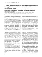
Calcium deposition in osteoarthritis menisci
Images of two normal control menisci and two age-
matched OA menisci are shown in Figure 1. Control
menisci have a smooth, white and glistening surface, with
no signs of degeneration (Figure 1a, b). In contrast, OA
menisci have a rough surface and apparent degeneration
(Figure 1c, d). Alizarin red staining demonstrated that
calcium deposits existed in all OA menisci derived from
eight consecutive unselected OA patients, but not in the
normal control menisci (Figure 1e to 1j).
We observed three distinctive patterns of calcium
deposition in the OA menisci. The first pattern of cal-
cium deposition was limited numbers of small-sized or
medium-sized single calcium deposit (Figure 1g) or small
clusters of small-sized and medium-sized calcium depos-
its (Figure 1h). This type of calcium deposition was
almost always found at the edges of the sections of OA
menisci and appeared to be associated with meniscal
degeneration (Figure 1g, h).
Sun et al. Arthritis Research & Therapy 2010, 12:R56
/>Page 4 of 9
The second pattern of calcium deposition was clusters
of small-sized calcium deposits inside the meniscus (Fig-
ure 1i). This type of calcium deposition was found in
about 65% of the sections among all of the sections of OA
menisci.
The third pattern of calcium deposition was wide-
spread clusters of medium-sized and large-sized calcium
deposits. This type of calcium deposition was found in
about 35% of the sections among all of the sections of OA
menisci.
We graded the alizarin red staining according to these
patterns as described in Materials and methods. The
results along with demographic patient information are
presented in Table 1. As shown, calcium deposits existed
in the transverse and longitudinal sections of all OA
menisci, but not in any sections of the normal control
menisci.
Expression of genes implicated in calcification
We examined and compared the expression of ENPP1
and ANKH in OA meniscal cells and in normal meniscal
cells. Both microarray and real-time RT-PCR analyses
indicated that the expression of ENPP1 and ANKH was
upregulated in OA meniscal cells (Table 2). In addition,
microarray and real-time RT-PCR analyses indicated that
the expression of matrix Gla protein and serglycin, which
are putative endogenous calcification inhibitors [42,43],
was also upregulated in OA meniscal cells.
Meniscal cell-mediated calcium deposition
Five OA meniscal cell cultures and three normal meniscal
cell cultures were investigated using an ATP-induced cal-
cium deposition assay. As shown in Figure 2, ATP
induced only a small amount of calcium deposition in the
monolayer cultures of normal meniscal cells after treat-
ment with ATP for 48 hours (left-hand group, P = 0.006).
In contract, ATP induced a large amount of calcium
deposition in the monolayer cultures of OA meniscal
cells under the same condition (right-hand group, P =
0.003). β-Glycerophosphate only induced a small amount
of calcium deposition when it was used as an alternative
source of phosphate (data not shown). In fact, the ATP-
induced calcium deposition in the monolayer cultures of
OA meniscal cells derived from five OA patients was
more than sixfold greater than that seen in the monolayer
cultures of normal meniscal cells derived from three con-
trol subjects. The difference between OA meniscal cell-
mediated and normal control meniscal cell-mediated cal-
cium deposition was statistically significant (P < 0.005).
The detailed results of the calcium deposition assay are
presented in Table 3.
Comparison of osteoarthritis meniscal cell and
osteoarthritis hyaline articular chondrocyte
We compared cell-mediated calcium deposition between
OA meniscal cells and OA hyaline articular chondrocytes
derived from four OA patients. As shown in Figure 3a,
both monolayer cultures of OA meniscal cells and OA
hyaline articular chondrocytes produced large amounts
of calcium deposition after the treatment with ATP for 72
hours. Collectively, OA meniscal cells produced more
calcium deposition than OA hyaline articular chondro-
cytes. Finally, we found that CaPC, a potent anti-calcifi-
cation agent, inhibited the OA meniscal cell-mediated
calcium deposition in a dose-dependent manner (Figure
3b; P < 0.0001).
Discussion
We demonstrated for the first time that calcium deposi-
tion was common in the menisci of end-stage OA
patients. The ages of two OA patients were similar to the
ages of two control subjects, and the ages of five among
the eight OA patients were below the age of 60 years.
Only OA meniscal specimens contained calcium depos-
its. This result indicates that meniscal calcification in OA
is mainly a disease-related phenomenon. It is worth not-
Figure 1 Normal and osteoarthritis menisci, stained with alizarin
red. Menisci were derived from (a) a 39-year-old female osteosarcoma
patient, (b) a 43-year-old male osteosarcoma patient, (c) a 42-year-old
male osteoarthritis (OA) patient, and (d) a 49-year-old female OA pa-
tient. The normal menisci exhibited a smooth, white and glistening
surface, with no signs of degeneration (a and b). OA menisci showed
discoloration and a rough surface. Degeneration was apparent (c and
d). (e), (f) There were no calcium depositions in the normal menisci; (g)
to (j) calcium deposition was present in all OA menisci. Representative
images of grade 0 (e and f), grade 1 (g), grade 2 (h), grade 3 (i) and
grade 4 (j) of alizarin red staining are shown.
Sun et al. Arthritis Research & Therapy 2010, 12:R56
/>Page 5 of 9
ing that no meniscal calcification is detected in nonse-
lected cadavers before the age of 60 years [32].
Brandes and Muller examined meniscal chondrocalci-
nosis and found three types of meniscal calcification [44].
Type 1A was disseminated calcification, which affected
all four menisci equally. Type 1B was calcification occur-
ring in limited areas, which was associated with meniscal
degeneration. Type 2 was a cloud-like diffuse calcifica-
tion, which contained fine granular amorphous materials.
The investigators concluded that type 1A calcification
represented primary chondrocalcinosis, that type 1B cal-
cification corresponded to secondary chondrocalcinosis,
and that type 2 calcification was dystrophic and postne-
crotic calcification.
In our study, we found three distinctive patterns of cal-
cium deposition in the OA menisci. The first pattern of
calcium deposition was calcification occurring in limited
amounts, associated with meniscal degeneration (Figure
1g, h). This type of meniscal calcification is similar to the
type 1B meniscal calcification described by Brandes and
Muller [44]. The second pattern of calcium deposition
was clusters of small-sized calcium deposits inside the
meniscus (Figure 1i). This type of meniscal calcification
appears to correspond to the type 2 meniscal calcification
observed by Brandes and Muller [44]. The third pattern
of calcium deposition was widespread clusters of
medium-sized and large-sized calcium deposits. This
type of calcification is probably a combination of the type
Table 1: Grade of alizarin red staining
Grade Normal group Osteoarthritis group
12 F 39 F 43 M 42 M 49 F 54 F 55 M 58 F 65 F 66 F 70 F
Transverse
section
00022331344
Longitudinal
section
00031331444
Average
a
0002.51.53313.544
12F, 12-year-old female; 42 M, 42-year-old male; and so forth. The difference of alizarin red staining grades between the osteoarthritis group
and the normal control group was statistically significant with P < 0.02.
a
Average of the transverse section grade and the longitudinal section
grades.
Table 2: Genes differentially expressed in osteoarthritis meniscal cells compared with normal control cells
Gene
name
Gene ID
Real-time
RT-PCRa
Differential gene expressionb (fold)
Description
OA1 OA2 OA3 OA4 OA5
ENPP1 BF057080 2.1 1.7 2.0 2.8 1.9 2.0 Ectonucleotide
pyrophosphatase 1
ANKH AL833238 1.9 2.4 1.8 1.7 1.5 1.8 Ankylosis,
progressive
homolog
MGP NM_000900 5.3 11.8 4.7 21.6 0 17.6 Matrix Gla protein
SRGN NM_002727 3.3 2.2 3.1 8.3 3.1 1.9 Serglycin
a
The ratio of the relative expression level of a specific gene in osteoarthritis (OA) meniscal cells derived from five OA patients (RNA mixture)
to the relative expression level of the specific gene in the control meniscal cells derived from three normal control subjects (RNA mixture),
which were determined by real-time RT-PCR analysis with P < 0.01.
b
Microarray analysis of the differential expression of a specific gene in the
individual OA cells derived from five different OA patients compared with three normal control meniscal cells as a group. OA1, derived from
a 65-year-old female OA patient; OA2, derived from a 56-year-old female OA patient; OA3, derived from a 50-year-old female OA patient; OA4,
derived from a 52-year-old female OA patient; OA5, derived from a 61-year-old male OA patient; Normal 1, derived from a 36-year-old female
osteosarcoma patient; Normal 2, derived from a 43-year-old female osteosarcoma patient; Normal 3, derived from a 12-year-old female
osteosarcoma patient. Microarray analyses were carried out as described in Materials and methods. The raw microarray data can be found in
the Gene Expression Omnibus [GEO:GSE19060] [49].
Sun et al. Arthritis Research & Therapy 2010, 12:R56
/>Page 6 of 9
1B calcification and type 2 calcification in the more
severe degenerative areas. Taken together, our findings
suggest that meniscal calcification in OA may mainly cor-
respond to dystrophic and secondary chondrocalcinosis
rather than to primary chondrocalcinosis.
Calcium crystals were frequently found in the hyaline
articular cartilage of end-stage OA patients [15-17]. It
was believed that the hyaline articular cartilage was the
most likely source of knee joint fluid crystals in OA
patients. Degeneration of the hyaline articular cartilage
would release the calcium crystals embedded in the carti-
lage into the knee joint fluid. In this study, we found that
medium-sized and large-sized calcium deposits were
commonly present at the degenerative edges (Figure 1g,
h) or at the areas adjacent to the degenerative edges of
OA menisci (Figure 1j). Because of their locations, these
calcium deposits can be readily released into the knee
joint fluid during joint articulation. Our findings suggest
that degenerative menisci may be one of the sources of
joint fluid crystals in OA.
Elevated gene expression of ANKH and ENPP1 causes
crystal deposition in cartilage [25,45]. In the present
study, we found that the expression of several genes
implicated in the biomineralization biological process
including ENPP1 and ANKH was upregulated in OA
Figure 2 ATP-induced calcium deposition. ATP-induced calcium
deposition in monolayer cultures of osteoarthritis (OA) meniscal cells
derived from five OA patients (right-hand group) was significantly
higher than that in the monolayer cultures of normal control meniscal
cells derived from three osteosarcoma patients (left-hand group). The
difference between the two groups was statistically significant (*P <
0.005). Count per minute (CPM) data were normalized against total
protein levels.
Table 3: Calcium deposition in monolayer cultures of meniscal cells
Normal meniscal cells Osteoarthritis meniscal cells
Age/gender of
patients
Control (CPM) ATP (CPM) Age/gender of
patients
Control (CPM) ATP (CPM)
12 years, F 27.5 ± 4.7 202.8 ± 61.0 42 years, M 31.0 ± 2.1 1,648.5 ± 243.3
39 years, F 33.8 ± 15.3 173.5 ± 26.2 49 years, F 53.5 ± 32.2 882.0 ± 151.9
43 years, M 27.5 ± 5.1 170.0 ± 25.3 50 years, F 56.8 ± 22.8 1,537.0 ± 376.3
65 years, F 27.3 ± 2.5 1,008.0 ± 198.6
67 years, F 59.8 ± 35.2 874.3 ± 154.0
CMP, count per minute normalized against total protein levels; F, female; M, male.
Figure 3 Comparison of osteoarthritis meniscal cell-mediated
and osteoarthritis chondrocyte-mediated calcium deposition. (a)
ATP-induced calcium deposition in the monolayer cultures of osteoar-
thritis (OA) meniscal cells derived from four OA patients (OA-M) (see
Table 3) was 60% greater than present in the monolayer cultures of OA
hyaline articular chondrocytes (OA-C). The difference was statistically
significant (*P < 0.05). (b) Calcium phosphocitrate (CaPC) inhibited
ATP-induced calcium deposition in the monolayer cultures of OA me-
niscal cells in a dose-dependent manner (*P < 0.001). Count per min-
ute (CPM) data were normalized against total protein levels.
Sun et al. Arthritis Research & Therapy 2010, 12:R56
/>Page 7 of 9
meniscal cells. This finding was consistent with the previ-
ous finding that ENPP1 was upregulated in the calcified
regions of OA menisci [46] and that ANKH was upregu-
lated in OA articular cartilage [47]. Our findings indicate
that OA meniscal cells may play an active role in the path-
ological meniscal calcification. Indeed, OA meniscal cells
induced much more calcium deposition than normal
control meniscal cells in the monolayer cultures. This
finding was consistent with a recent finding that OA hya-
line articular chondrocytes produced calcium deposition
in cell culture, whereas normal control hyaline articular
chondrocytes derived from the hyaline articular cartilage
of osteosarcoma patients did not [17]. The activities and
protein levels of ENPP1, ANKH and matrix Gla protein
in OA meniscal cells were not obtained in the present
work. This information would certainly be interesting
and important. We look forward to future study supply-
ing this information.
The findings that calcium deposits were present in all
OA menisci and that OA meniscal cells induced much
more calcium deposition than normal meniscal cells will
have significant impact on our understating of OA and
the development of disease-modifying drugs for OA ther-
apy. Recently, it was reported that CaPC, a potent anti-
calcification agent, inhibited meniscal calcification in
Hartley guinea pigs and that the inhibition was accompa-
nied by a significant reduction in the degeneration of hya-
line articular cartilage [48]. Our finding that CaPC
inhibited OA meniscal cell-mediated calcium deposition
was consistent with this report. Although our findings
provide no support for the notion that calcium deposi-
tion in OA joint tissues is a causative factor to OA, patho-
logical calcification in OA may still be a valid therapeutic
target for OA therapy. Our study demonstrates clearly
that meniscal calcification is a disease-related phenome-
non in OA. Theoretically, inhibition of meniscal calcifica-
tion can be achieved either by targeting the calcium
deposits (physical target) or by targeting the cells (biolog-
ical target). Targeting the calcium deposits directly will
inhibit the growth of the calcium deposits and reduce the
detrimental downstream biological effects of these cal-
cium deposits. Targeting the cells at the cellular, genetic
or epigenetic levels will not only inhibit the formation
and growth of calcium deposits, but may also convert the
altered OA meniscal cells to more normal-like meniscal
cells, thereby eliminating an important disease compo-
nent of OA.
Our study has some limitations that should be consid-
ered. The first limitation is that the normal control
meniscal cells were not optimal normal meniscal cells. To
minimize this limitation, we only collected overtly nor-
mal-appearing meniscal specimens from osteosarcoma
patients whose tumors were located distant from the
knee. Another limitation is the small size of our speci-
mens; the exact contribution of aging to meniscal calcifi-
cation could therefore not be determined. It is likely that
an age-associated increase of meniscal calcification may
account for some of the calcification in the clinical speci-
mens. It is difficult to obtain age-matched control menis-
cal specimens because osteosarcoma occurs often in
younger patients while OA occurs mostly in older
patients. We will continue this line of study when more
age-matched normal control meniscal specimens become
available in the future.
Conclusions
Our findings suggest that OA is not merely a hyaline
articular cartilage disease, but also a meniscal disease.
Pathological meniscal calcification mediated by OA
meniscal cells, which may alter the biomechanical prop-
erties of the meniscus and the expression of extracellular
matrix-degrading enzymes, is potentially an important
contributory factor to OA.
Abbreviations
ANKH: ankylosis, progressive homolog; ATP: adenosine-5'-triphosphate; CaPC:
calcium phosphocitrate; DMEM: Dulbecco's modified Eagle's medium; ENPP1:
ectonucleotide pyrophosphatase/phosphodiesterase 1; GAPDH: glyceralde-
hyde-3-phosphate dehydrogenase; OA: osteoarthritis; PCR: polymerase chain
reaction; RT: reverse transcription.
Competing interests
The authors declare that they have no competing interests.
Authors' contributions
YS, HEG and ENH conceived the study and participated in its design and coor-
dination. YS wrote the manuscript, and analyzed the microarrays. DRM and JSK
provided surgical tissues and participated in the discussion of experimental
results. HJN assisted with statistical analysis. NZ performed histologic embed-
ding, sectioning and staining. YS and PRH graded alizarin red staining. PRH pre-
pared cell cultures, performed the calcium deposition assay and extracted
RNA. HEG assisted with manuscript preparation.
Acknowledgements
The present study is supported in part by a Charlotte-Mecklenburg Education
and Research Foundation Grant and a Mecklenburg County Medical Society
Smith Arthritis Fund Grant (to YS). This study was performed at Carolinas Medi-
cal Center, Charlotte, NC, USA.
Author Details
1
Department of Orthopaedic Surgery, Carolinas Medical Center, PO Box 32861,
Charlotte, NC 28232, USA and
2
Department of Biostatistics, Carolinas Medical
Center, PO Box 32861, Charlotte, NC 28232, USA
References
1. Goldring MB, Goldring SR: Osteoarthritis. J Cell Physiol 2007, 213:626-634.
2. Samuels J, Krasnokutsky S, Abramson SB: Osteoarthritis: a tale of three
tissues. Bull NYU Hosp Jt Dis 2008, 66:244-250.
3. Kato H, Matsumine A, Wakabayashi T, Hasegawa M, Sudo A, Shintani K,
Fukuda A, Kato K, Ide N, Orita S, Hasegawa T, Matsumura C, Furukawa M,
Tasaki T, Sonoda H, Uchida A: Large-scale gene expression profiles,
differentially represented in osteoarthritic synovium of the knee joint
using cDNA microarray technology. Biomarkers 2007, 12:384-402.
4. Sun Y, Mauerhan DR, Firestein GS, Loeffler BJ, Hanley EN, Gruber HE:
Telomerase transduced osteoarthritis fibroblast-like synoviocytes
Received: 5 October 2009 Revised: 22 February 2010
Accepted: 30 March 2010 Published: 30 March 2010
This article is available from: 2010 Sun et al.; licensee BioMed Central Ltd. This is an open access article distributed under the terms of the Creative Commons A ttribution License ( which permits unrestricted use, distribution, and reproduction in any medium, provided the original work is properly cited.Arthritis R esearch & Thera py 2010, 12:R56
Sun et al. Arthritis Research & Therapy 2010, 12:R56
/>Page 8 of 9
display a distinct gene expression profile. J Rheumatol 2009,
36:141-155.
5. Hopwood B, Tsykin A, Findlay DM, Fazzalari NL: Microarray gene
expression profiling of osteoarthritic bone suggests altered bone
remodelling, WNT and transforming growth factor-beta/bone
morphogenic protein signalling. Arthritis Res Ther 2007, 9:R100.
6. Noble J, Hamblen DL: The pathology of the degenerate meniscus
lesion. J Bone Joint Surg Br 1975, 57:180-186.
7. Englund M, Guermazi A, Gale D, Hunter DJ, Aliabadi P, Clancy M, Felson
DT: Incidental meniscal findings on knee MRI in middle-aged and
elderly persons. N Engl J Med 2008, 359:1108-1115.
8. Chan WP, Lang P, Stevens MP, Sack K, Majumdar S, Stoller DW, Basch C,
Genant HK: Osteoarthritis of the knee: comparison of radiography, CT,
and MR imaging to assess extent and severity. AJR Am J Roentgenol
1991, 157:799-806.
9. Bennett LD, Buckland-Wright JC: Meniscal and articular cartilage
changes in knee osteoarthritis: a cross-sectional double-contrast
macroradiographic study. Rheumatology 2002, 41:917-923.
10. Englund M: Meniscal tear - a feature of osteoarthritis. Acta Orthop Scand
Suppl 2004, 75:1-45.
11. Hunter DJ, Zhang YQ, Tu X, Lavalley M, Niu JB, Amin S, Guermazi A, Genant
H, Gale D, Felson DT: Change in joint space width: hyaline articular
cartilage loss or alteration in meniscus? Arthritis Rheum 2006,
54:2488-2495.
12. Felson DT, Anderson JJ, Naimark A, Kannel W, Meenan RF: The prevalence
of chondrocalcinosis in the elderly and its association with knee
osteoarthritis: the Framingham Study. J Rheumatol 1989, 16:1241-1245.
13. Carroll GJ, Stuart RA, Armstrong JA, Breidahl PD, Laing BA: Hydroxyapatite
crystals are a frequent finding in osteoarthritic synovial fluid, but are
not related to increased concentrations of keratan sulfate or
interleukin 1 beta. J Rheumatol 1991, 18:861-866.
14. Nalbant S, Martinez JA, Kitumnuaypong T, Clayburne G, Sieck M,
Schumacher HR Jr: Synovial fluid features and their relations to
osteoarthritis severity: new findings from sequential studies.
Osteoarthritis Cartilage 2003, 11:50-54.
15. Gordon GV, Villanueva T, Schumacher HR, Gohel V: Autopsy study
correlating degree of osteoarthritis, synovitis and evidence of articular
calcification. J Rheumatol 1984, 11:681-686.
16. Kirsch T, Swoboda B, Nah H: Activation of annexin II and V expression,
terminal differentiation, mineralization and apoptosis in human
osteoarthritic cartilage. Osteoarthritis Cartilage 2000, 8:294-302.
17. Fuerst M, Lammers L, Schafer F, Niggemeyer O, Steinhagen J, Lohmann
CH, Ruther W: Investigation of calcium crystals in OA knees. Rheumatol
Int 2009, 30:623-631.
18. McCarty DJ: Crystal-induced inflammation of the joints. Annu Rev Med
1970, 21:357-366.
19. McCarthy GM, Augustine JA, Baldwin AS, Christopherson PA, Cheung HS,
Westfall PR, Scheinman RI: Molecular mechanism of basic calcium
phosphate crystal-induced activation of human fibroblasts. Role of
nuclear factor kappab, activator protein 1, and protein kinase C. J Biol
Chem 1998, 273:35161-35169.
20. Zeng XR, Sun Y, Wenger L, Cheung HS: Basic calcium phosphate crystal-
induced Egr-1 expression stimulates mitogenesis in human fibroblasts.
Biochem Biophys Res Commun 2005, 330:658-664.
21. McCarthy GM, Mitchell PG, Struve JA, Cheung HS: Basic calcium
phosphate crystals cause coordinate induction and secretion of
collagenase and stromelysin. J Cell Physiol 1992, 153:140-146.
22. Sun Y, Wenger L, Brinckerhoff CE, Misra RR, Cheung HS: Basic calcium
phosphate crystals induce matrix metalloproteinase-1 through the
Ras/mitogen-activated protein kinase/c-Fos/AP-1/metalloproteinase 1
pathway. Involvement of transcription factor binding sites AP-1 and
PEA-3. J Biol Chem 2002, 277:1544-1552.
23. Morgan MP, Whelan LC, Sallis JD, McCarthy CJ, Fitzgerald DJ, McCarthy
GM: Basic calcium phosphate crystal-induced prostaglandin E
2
production in human fibroblasts: role of cyclooxygenase 1,
cyclooxygenase 2, and interleukin-1beta. Arthritis Rheum 2004,
50:1642-1649.
24. Sun Y, Firestein G, Wenger L, Wang CY, Cheung H: Telomera se
transduced osteoarthritic fibroblast-like synoviocyte cell line. Biochem
Biophys Res Commun 2004, 323:1287-1292.
25. Johnson K, Pritzker K, Goding J, Terkeltaub R: The nucleoside
triphosphate pyrophosphohydrolase isozyme PC-1 directly promotes
cartilage calcification through chondrocyte apoptosis and increased
calcium precipitation by mineralizing vesicles. J Rheumatol 2001,
28:2681-2691.
26. Pendleton A, Johnson MD, Hughes A, Gurley KA, Ho AM, Doherty M, Dixey
J, Gillet P, Loeuille D, McGrath R, Reginato A, Shiang R, Wright G, Netter P,
Williams C, Kingsley DM: Mutations in ANKH cause chondrocalcinosis.
Am J Hum Genet 2002, 71:933-940.
27. Ali SY: Apatite-type crystal deposition in arthritic cartilage. Scan
Electron Microsc 1985:1555-1566.
28. Heinkel D, Gohr CM, Uzuki M, Rosenthal AK: Transglutaminase
contributes to CPPD crystal formation in osteoarthritis. Front Biosci
2004, 9:3257-3261.
29. Canhao H, Fonseca JE, Leandro MJ, Romeu JC, Pimentao JB, Costa JT,
Queiroz MV: Cross-sectional study of 50 patients with calcium
pyrophosphate dihydrate crystal arthropathy. Clin Rheumatol 2001,
20:119-122.
30. Dufauret-Lombard C, Vergne-Salle P, Simon A, Bonnet C, Treves R, Bertin P:
Ultrasonography in chondrocalcinosis. Joint Bone Spine 2010 in press.
31. Hough AJ Jr, Webber RJ: Pathology of the meniscus. Clin Orthop Relat Res
1990:32-40.
32. Mitrovic D, Stankovic A, Morin J, Borda-Iriarte O, Uzan M, Quintero M,
Memin Y, Bard M, de SS, Richewaert A: Anatomic incidence of
meniscochondrocalcinosis of the knee. Rev Rhum Mal Osteoartic 1982,
49:495-499.
33. Mitrovic DR, Stankovic A, Iriarte-Borda O, Uzan M, Quintero M, Miravet L,
Kuntz D: The prevalence of chondrocalcinosis in the human knee joint.
An autopsy survey. J Rheumatol 1988, 15:633-641.
34. Turhanen PA, Demadis KD, Peraniemi S, Vepsalainen JJ: A novel strategy
for the preparation of naturally occuring phosphocitrate and its
partially esterified derivatives. J Org Chem 2007, 72:1468-1471.
35. Demadis KD, Sallis JD, Raptis RG, Baran P: A crystallographically
characterized nine-coordinate calcium-phosphocitrate complex as
calcification inhibitor in vivo. J Am Chem Soc 2001, 123:10129-10130.
36. Gruber HE, Mauerhan D, Chow Y, Ingram JA, Norton HJ, Hanley EN Jr, Sun
Y: Three-dimensional culture of human meniscal cells: extracellular
matrix and proteoglycan production. BMC Biotechnol 2008, 8:54.
37. Rosenthal AK, Gohr CM, Uzuki M, Masuda I: Osteopontin promotes
pathologic mineralization in articular cartilage. Matrix Biol 2007,
26:96-105.
38. Rosenthal AK, Mattson E, Gohr CM, Hirschmugl CJ: Characterization of
articular calcium-containing crystals by synchrotron FTIR.
Osteoarthritis Cartilage 2008, 16:1395-1402.
39. Sun Y, Mauerhan DR, Firestein GS, Loeffler BJ, Hanley EN, Gruber HE:
Telomerase transduced osteoarthritis fibroblast-like synoviocytes
display a distinct gene expression profile. J Rheumatol 2009,
36:141-155.
40. Sun Y, Mauerhan DR, Honeycutt PR, Kneisl JS, Norton JH, Hanley EN Jr,
Gruber HE: Analysis of meniscal degeneration and meniscal gene
expression. BMC Musculoskelet Disord 2010, 11:19.
41. Pfaffl M, Meyer HH, Sauerwein H: Quantification of insulin-like growth
factor-1 (IGF-1) mRNA: development and validation of an internally
standardised competitive reverse transcription-polymerase chain
reaction. Exp Clin Endocrinol Diabetes 1998, 106:506-513.
42. Yagami K, Suh JY, Enomoto-Iwamoto M, Koyama E, Abrams WR, Shapiro
IM, Pacifici M, Iwamoto M: Matrix GLA protein is a developmental
regulator of chondrocyte mineralization and, when constitutively
expressed, blocks endochondral and intramembranous ossification in
the limb. J Cell Biol 1999, 147:1097-1108.
43. Theocharis AD, Seidel C, Borset M, Dobra K, Baykov V, Labropoulou V,
Kanakis I, Dalas E, Karamanos NK, Sundan A, Hjerpe A: Serglycin
constitutively secreted by myeloma plasma cells is a potent inhibitor of
bone mineralization in vitro. J Biol Chem 2006, 281:35116-35128.
44. Brandes A, Muller KM: Calcinosis of the meniscus. Morphologic and
roentgenographic findings for zonal classification. Pathologe 1995,
16:269-277.
45. Netter P, Bardin T, Bianchi A, Richette P, Loeuille D: The ANKH gene and
familial calcium pyrophosphate dihydrate deposition disease. Joint
Bone Spine 2004, 71:365-368.
46. Johnson K, Hashimoto S, Lotz M, Pritzker K, Goding J, Terkeltaub R: Up-
regulated expression of the phosphodiesterase nucleotide
pyrophosphatase family member PC-1 is a marker and pathogenic
Sun et al. Arthritis Research & Therapy 2010, 12:R56
/>Page 9 of 9
factor for knee meniscal cartilage matrix calcification. Arthritis Rheum
2001, 44:1071-1081.
47. Ijiri K, Zerbini LF, Peng H, Otu HH, Tsuchimochi K, Otero M, Dragomir C,
Walsh N, Bierbaum BE, Mattingly D, van FG, Komiya S, Aigner T, Libermann
TA, Goldring MB: Differential expression of GADD45β in normal and
osteoarthritic cartilage: potential role in homeostasis of articular
chondrocytes. Arthritis Rheum 2008, 58:2075-2087.
48. Cheung HS, Sallis JD, Demadis KD, Wierzbicki A: Phosphocitrate blocks
calcification-induced articular joint degeneration in a guinea pig
model. Arthritis Rheum 2006, 54:2452-2461.
49. Gene Expression Omnibus [ />]
doi: 10.1186/ar2968
Cite this article as: Sun et al., Calcium deposition in osteoarthritic meniscus
and meniscal cell culture Arthritis Research & Therapy 2010, 12:R56