Báo cáo y học: "A crucial role for tumor necrosis factor receptor 1 in synovial lining cells and the reticuloendothelial system in mediating experimental arthritis" pps
Bạn đang xem bản rút gọn của tài liệu. Xem và tải ngay bản đầy đủ của tài liệu tại đây (1022.99 KB, 11 trang )
Arntz et al. Arthritis Research & Therapy 2010, 12:R61
/>Open Access
RESEARCH ARTICLE
BioMed Central
© 2010 Arntz et al.; licensee BioMed Central Ltd. This is an open access article distributed under the terms of the Creative Commons
Attribution License ( which permits unrestricted use, distribution, and reproduction in
any medium, provided the original work is properly cited.
Research article
A crucial role for tumor necrosis factor receptor 1 in
synovial lining cells and the reticuloendothelial
system in mediating experimental arthritis
Onno J Arntz, Jeroen Geurts, Sharon Veenbergen, Miranda B Bennink, Ben T van den Brand, Shahla Abdollahi-
Roodsaz, Wim B van den Berg and Fons A van de Loo*
Abstract
Introduction: Rheumatoid arthritis (RA) is an autoimmune inflammatory disease that mainly affects synovial joints.
Biologics directed against tumor-necrosis-factor (TNF)-α are efficacious in the treatment of RA. However, the role of TNF
receptor-1 (TNFR1) in mediating the TNFα effects in RA has not been elucidated and conflicting data exist in
experimental arthritis models. The objective is to investigate the role of TNFR1 in the synovial lining cells (SLC) and the
reticuloendothelial system (RES) during experimental arthritis.
Methods: Third generation of adenovirus serotype 5 were either injected locally in the knee joint cavity or systemically
by intravenous injection into the retro-orbital venous sinus to specifically target SLC and RES, respectively. Transduction
of organs was detected by immunohistochemistry of the eGFP transgene. An adenoviral vector containing a short
hairpin (sh) RNA directed against TNFR1 (HpTNFR1) was constructed and functionally evaluated in vitro using a nuclear
factor-kappaB (NF-κB) reporter assay and in vivo in streptococcal cell wall-induced arthritis (SCW) and collagen-induced
arthritis (CIA). Adenoviruses were administered before onset of CIA, and the effect of TNFR1 targeting on the clinical
development of arthritis, histology, quantitative polymerase chain reaction (qPCR), cytokine analyses and T-cell assays
was evaluated.
Results: Systemic delivery of Ad5.CMV-eGFP predominantly transduced the RES in liver and spleen. Local delivery
transduced the synovium and not the RES in liver, spleen and draining lymph nodes. In vitro, HpTNFR1 reduced the
TNFR1 mRNA expression by three-fold resulting in a 70% reduction of TNFα-induced NF-κB activation. Local treatment
with HpTNFR1 markedly reduced mRNA and protein levels of interleukin (IL)-1β and IL-6 in SLC during SCW arthritis and
ameliorated CIA. Systemic targeting of TNFR1 in RES of liver and spleen by systemic delivery of Ad5 virus encoding for a
small hairpin RNA against TNFR1 markedly ameliorated CIA and simultaneously reduced the mRNA expression of IL-1β,
IL-6 and Saa1 (75%), in the liver and that of Th1/2/17-specific transcription factors T-bet, GATA-3 and RORγT in the
spleen. Flow cytometry confirmed that HpTNFR1 reduced the numbers of interferon (IFN)γ (Th1)-, IL-4 (Th2)- and IL-17
(Th17)-producing cells in spleen.
Conclusions: TNFR1-mediated signaling in both synovial lining cells and the reticuloendothelial system independently
played a major pro-inflammatory and immunoregulatory role in the development of experimental arthritis.
Introduction
Rheumatoid arthritis (RA) is a chronic and systemic auto-
immune disease that mainly affects synovial joints and is
characterized by inflammatory synovitis, ultimately lead-
ing to the destruction of cartilage and bone. The central
role for tumor necrosis factor-alpha (TNFα) in RA patho-
genesis has been extensively demonstrated in experimen-
tal arthritis by successful treatment of murine collagen-
induced arthritis (CIA) with TNFα antibodies [1,2] and
development of arthritis in transgenic mice overexpress-
ing human TNF [3]. Most importantly, TNFα has been
identified as a key cytokine in human RA [4], which has
* Correspondence:
1
Rheumatology Research and Advanced Therapeutics, Department of
Rheumatology, Radboud University Nijmegen Medical Centre, 6525 GA
Nijmegen, The Netherlands
Full list of author information is available at the end of the article
Arntz et al. Arthritis Research & Therapy 2010, 12:R61
/>Page 2 of 11
led to the development of effective treatment of disease
by administration of neutralizing TNF antibodies [5,6].
TNFα signaling is mediated via two distinct receptors
encoded by the genes Tnfrsf1a (TNFR1) and Tnfrsf1b
(TNFR2). The TNF receptors are transmembrane glyco-
proteins and share only 28% homology, predominantly
between their extracellular domains. Both TNFR1 and
TNFR2 activate a wide range of proinflammatory signal
pathways, leading to activation of nuclear factor-kappa-B
(NF-κB) and c-Jun N-terminal kinase, via recruitment of
TNF receptor-associating factors (reviewed in [7]).
Attenuation of CIA in TNFR1-deficient mice has demon-
strated a dominant role of this receptor in disease [8,9].
Recent investigations on the cell-specific contribution of
TNFR1-mediated signaling in RA pathogenesis have
revealed remarkably different functions of TNFR1 in
mesenchymal or hematopoietic compartments. Cells
from the prior compartment - in particular, synovial
fibroblasts (SFs) - have been identified as the primary tar-
gets for TNFα in the development of arthritis [10]. In
contrast, TNFR1-mediated signaling in cells from the lat-
ter compartment, such as leukocytes, exerts an anti-
inflammatory function [11,12].
This cell specificity of TNFR1 function is highly rele-
vant to the safety and efficacy of treatments that target
TNFα signaling. Scintigraphic imaging of the biodistribu-
tion of radiolabeled anti-TNF after systemic administra-
tion in RA patients has shown that antibodies accumulate
not only in inflamed joints but also in the liver and spleen
[13]. However, the function of TNFR1 expression in these
secondary lymphoid organs and its contribution to RA
pathogenesis remain to be elucidated.
In this study, we investigated the effects of TNFR1-
mediated signaling in synovial lining cells (SLCs) and the
reticuloendothelial system (RES) during experimental
arthritis. To this end, we used cell-specific RNA interfer-
ence (RNAi)-mediated silencing of TNFR1 based on ade-
noviral delivery of a short hairpin RNA (shRNA)-
expressing construct.
Materials and methods
Animals
Male 10- to 12-week-old DBA/1J and C57BL/6 mice were
obtained from Janvier (Janvier, Elavage, France). During
viral experiments, mice were housed in HEPA-filtered
individually ventilated cages. The animals were fed a
standard diet with food and water ad libitum. All in vivo
studies complied with national legislation and were
approved by the local authorities on the care and use of
animals.
Induction of collagen-induced arthritis
Bovine collagen type II (bCII) was dissolved in 0.05 M
acetic acid to a concentration of 2 mg/mL and was emul-
sified in equal volumes of Freund's complete adjuvant (2
mg/mL of Mycobacterium tuberculosis strain H37Ra;
Difco Laboratories, now part of Becton Dickinson and
Company, Franklin Lakes, NJ, USA). DBA1/J mice were
immunized intradermally at the base of the tail with 100
μL of emulsion (100 μg of bCII). On day 21, the mice were
given an intraperitoneal booster injection of 100 μg of
bCII dissolved in phosphate-buffered saline (PBS). Mice
were killed on day 31 by cervical dislocation.
Streptococcal cell wall (SCW) preparation and induction of
SCW arthritis
Streptococcus pyogenes T12 organisms were cultured
overnight in Todd-Hewitt broth. Cell walls were prepared
as described previously [14]. The resulting supernatant
obtained after centrifugation at 10,000 g contained 11%
muramic acid. Unilateral arthritis was induced by intra-
articular (i.a.) injection of 5 μg of streptococcal cell wall
(SCW) fragments (rhamnose content) in 6 μL of PBS.
Cell culture
Mouse embryonic fibroblasts (NIH 3T3) stably trans-
fected with a 5 × NF-κB-luciferase reporter were culti-
vated in Dulbecco's modified Eagle's medium (DMEM)
supplemented with 1 mM pyruvate, penicillin-streptomy-
cin (Lonza, Basel, Switzerland), and 5% fetal calf serum
(FCS). Cells were kept at 37°C in a humid atmosphere
containing 5% CO
2
.
Plasmids
For cloning, we used Pfu DNA polymerase (Stratagene,
La Jolla, CA, USA) and T4 DNA Ligase (New England
Biolabs, Inc., Ipswich, MA, USA). All generated con-
structs were verified by sequencing. The U6 promoter
was polymerase chain reaction (PCR)-cloned from
mouse genomic DNA into XbaI/SalI sites of pShuttle
(kind gift of Bert Vogelstein, Howard Hughes Medical
Institute, Baltimore, MD, USA) to give pShuttle-U6 using
primers forward 5'-TCTAGAGATCCGACGCCGCCA-
TCTCTA-3' and reverse 5'-GTCGACGTTAA-
CAAGGCTTTTCTCCA-3'. The target sequence for
silencing the Tnfrsf1a gene [EMBL:M60468
] was ATCT-
TCGGTCCTAGTAACT (base pairs 1095 to 1113), and
we used ACTCATGTCTTGATCAGCT (no complemen-
tary sequence in murine genome) as scrambled control
sequence. The silencing cassette was constructed using
the following oligonucleotides: forward 5'-TG-target-
TTCAAGAGA
-target reverse complimentary-TTTT-
TGCA-3' and reverse 5'-AAAA-target-TCTCTTGAA
-
target reverse complimentary-CA-3', where the loop and
polyA sequences are underlined and bold, respectively.
Oligonucleotides (4.5 nM) were mixed in annealing buf-
fer (100 mM potassium acetate, 2 mM magnesium ace-
tate, 30 mM HEPES pH 7.4), heated for 5 minutes at 95°C,
Arntz et al. Arthritis Research & Therapy 2010, 12:R61
/>Page 3 of 11
and gradually cooled to room temperature. Annealed
DNA fragments were ligated in HpaI/SalI sites of pShut-
tle-U6.
Adenoviral vectors
Replication-deficient adenoviral vectors (E1/E3 deleted)
Ad5.U6-HpTNFR1 and Ad5.U6-HpNS (hairpin non spe-
cific control, scrambled RNA from TNFR1) were pre-
pared according to the AdEasy system [15], with the
exception that replication-competent recombinant free
viral particles were produced in E1 transformed N52E6
amniocyte cells [16]. Ad5.CMV-eGFP was a kind gift of
Jay Kolls (Department of Pediatrics, Children's Hospital
of Pittsburgh, PA, USA).
Viruses were purified by two consecutive CsCl
2
gradi-
ent purifications and stored in small aliquots at -80°C in
buffer containing 25 mM Tris, pH 8.0, 5 mM KCl, 0.2 mM
MgCl
2
, 137 mM NaCl, 730 μM Na
2
HPO
4
, 0.1% ovalbu-
min, and 10% glycerol. The infectious particle titer (ffu)
was determined by titrating vector stocks on 911 indica-
tor cells and measuring viral capsid protein immunohis-
tochemically 20 hours after transduction.
Study design and histology
To study which organs are transduced after systemic or
local treatment, Ad5.CMV-eGFP was injected into naïve
DBA/1J mice intravenously or intra-articularly with 3 ×
10
8
or 10
7
ffu adenovirus, respectively. One day later, liver,
spleen, lung, knee joints, draining lymph nodes, blood,
and bone marrow cells (BMCs) were isolated. They were
fixated in 4% paraformaldehyde for 4 days for immuno-
histochemistry (IHC). After decalcification in 5% formic
acid, specimens were processed for paraffin embedding.
Tissue sections (7 μm) were stained with anti-GFP anti-
body. For mRNA measurement with reverse transcrip-
tion-quantitative PCR (RT-qPCR), all parts were isolated.
DBA/1J mice were injected intravenously or intra-artic-
ularly 1 day after bCII booster (day 22) with 3 × 10
8
or 10
7
ffu adenovirus, respectively. For the siRNA (short inter-
fering RNA) hairpin-treated mice, mice were sacrificed 3
days post-transduction, and synovium (i.a.), spleen, and
liver (intravenous, i.v.) were isolated. Development of
arthritis in front and hind paws was macroscopically
monitored (scores between 0 and 2) until day 31. The
macroscopic arthritis score is based on the clinical signs
of inflammation in each paw and ankle; the maximum
score is 8 (1 for each hind paw and 1 for the ankle). Mice
were killed at day 26 or 31 by cervical dislocation. At day
26, synovial tissue explants (i.a.), spleen, and liver (i.v.)
were removed. At day 31, ankle and knee joints (all
groups) were removed and fixed in 4% paraformaldehyde
for 4 days. After decalcification in 5% formic acid, speci-
mens were processed for paraffin embedding. Tissue sec-
tions (7 μm) were stained with hematoxylin and eosin
(cell influx) or safranin-O (cartilage proteoglycan deple-
tion). Histological changes were scored in the patella/
femur region on five semi-serial sections of the knee
joint, spaced 70 μm apart. Scoring was performed by two
observers without knowledge of the group, as described
before. Histopathological changes were scored using the
following parameters. Cartilage depletion, defined as the
loss of proteoglycan content, was scored on a scale rang-
ing from 0 to 3 per region, depending on the intensity of
staining in the cartilage. Infiltration of cells was scored on
a scale of 0 to 3 (0 = no cells, 1 = mild cellularity, 2 = mod-
erate cellularity, 3 = maximal cellularity), depending on
the number of inflammatory cells in the synovial cavity
(exudate) or synovial tissue (infiltrate). Cartilage erosion
was graded on a scale of 0 to 3, ranging from no damage
to compete loss of articular cartilage.
Immunohistochemistry
Paraffin sections were stained with rabbit anti-GFP
(1:800) (#2555; Cell Signaling Technology, Danvers, MA,
USA) overnight at 4°C. After washing, sections were
incubated for 1 hour with biotinylated secondary anti-
body goat anti-rabbit-BIOT (1:400) (Vector Laboratories,
Burlingame, CA, USA). After washing, sections were
incubated for 30 minutes with Vectastain (1:400) (Vector
Laboratories). Thereafter, sections were stained with 3,3'-
diaminobenzidine and counterstained with hematoxylin,
embedded in Permount (Thermo Fisher Scientific Inc.,
Rockford, IL, USA).
Spleen cell isolation and antigen-presenting cell
stimulation
Spleens were mashed and filtered, and erythrocytes were
removed by osmotic shock. After washing, the splenic
cell fraction was incubated in RPMI 1640 (Invitrogen
Corporation, Carlsbad, CA, USA) at 37°C in 5% CO
2
for 1
hour in order to separate adherent cells from nonadher-
ent cells. The cells in the adherent cell fraction consisting
mainly of macrophages are termed antigen-presenting
cells (APCs). Splenic APCs were stimulated for 24 hours
with 10 ng/mL TNFα (Abcam, Cambridge, UK). Cytokine
production was analyzed using Luminex multianalyte
technology. The Bioplex system in combination with
multiplex cytokine kits (Bio-Rad, Veenendaal, the Neth-
erlands) was used.
Flow cytometry analysis
Total spleen cells obtained as described above were cul-
tured (10
6
/mL) for 2 hours in RPMI 1640 (Invitrogen
Corporation) supplemented with 10% FCS, penicillin-
streptomycin, 1 mM pyruvate, 1 μl/mL Golgiplug inhibi-
tor (BD Biosciences, San Jose, CA, USA), 10 ng/mL PMA
(phorbol 12-myristate 13-acetate), and 1 μg/mL ionomy-
cin. Thereafter, cells were labeled for 30 minutes at 4°C
Arntz et al. Arthritis Research & Therapy 2010, 12:R61
/>Page 4 of 11
with antibody TCRβ-FITC (T-cell receptor beta-fluores-
cein isothiocyanate) (1:200) and CD4-APC (1:100) or
their respective isotype control antibodies. Cells were
washed and consecutively fixed and permeabilized using
cytofix/cytoperm solution (BD Biosciences). Thereafter,
cells were incubated with phycoerythrin (PE)-labeled
antibodies interferon-gamma (IFNγ)-PE (1:200), interleu-
kin-4 (IL-4)-PE (1:200), or IL-17-PE (1:500) (BioLegend,
San Diego, CA, USA) or appropriate isotype controls for
30 minutes at 4°C in PBS containing 1% bovine serum
albumin (BSA), 2% FCS, and 0.1% saponin. Analyses were
performed on a BD FACSCalibur (BD Biosciences).
Cytokine measurements
Synovial tissue explants were incubated for 1 hour at
room temperature in 200 μL of RPMI 1640 supplemented
with 0.1% BSA, penicillin-streptomycin, and 1% pyruvate.
Subsequently, supernatant was harvested and centrifuged
for 5 minutes at 1,000 g. Murine IL-1β, IL-6, and TNFα
levels were determined using the Luminex multianalyte
technology and the BioPlex system in combination with
BioPlex Mouse Cytokine Assays (Bio-Rad Laboratories,
Inc., Hercules, CA, USA). Cytokines were measured in 50
μL of washout medium. The sensitivities were 5, less than
3, and 5 pg/mL for IL-1β, TNFα, and IL-6, respectively.
Luciferase measurements
NIH-3T3-5 × NF-κB-luciferase cells were seeded at 5 ×
10
4
cells per well in a Krystal 2000 96-well plate (Thermo
Labsystems, Brussels, Belgium). The day after, cells were
transduced with adenovirus at the indicated multiplicity
of infection (MOI) in 50 μL of DMEM for 4 hours at 37°C.
Two days post-transduction, cells were stimulated with
10 ng/mL recombinant murine TNFα or IL-1β (R&D Sys-
tems, Abingdon, UK) for 6 hours and subsequently lysed
in ice-cold lysis buffer (0.5% NP-40, 1 mM DTT, 1 mM
EDTA, 5 mM MgCl
2
, 100 mM KCl, 10 mM Tris-HCl pH
7.5). Alternatively, TNFα was antagonized by preincubat-
ing cells for 1 hour with 10 μg/mL Enbrel (Wyeth Phar-
maceuticals, Hoofddorp, The Netherlands). Luciferase
activity was quantified using the Bright-Glo luciferase
assay system (Promega Corporation, Madison, WI, USA)
by adding an equal volume of Bright-Glo to the cell lysate.
Luminescence was quantified in a luminometer
(Lumistar; BMG Labtech GmbH, Offenburg, Germany),
expressed as relative light units, and normalized to total
protein content of the cell/tissue extracts using a BCA
(bicinchoninic acid) protein assay kit (Thermo Fisher Sci-
entific, Inc.).
RNA isolation
Synovial and liver tissue was snap-frozen in liquid nitro-
gen and homogenized using a MagNa Lyser (Roche,
Basel, Switzerland). Total RNA was extracted using TRI
reagent (Sigma-Aldrich, St. Louis, MO, USA). Isolated
RNA samples were treated with RNase-free DNase I
(Qiagen, Venlo, The Netherlands) for 15 minutes. Synthe-
sis of cDNA was accomplished by reverse transcription-
PCR using an oligo(dT) primer and Moloney murine leu-
kemia virus reverse transcriptase (Invitrogen Corpora-
tion).
Quantitative polymerase chain reaction
qPCR was performed using SYBR Green PCR Master mix
and the ABI 7000 Prism Sequence Detection system
(Applied Biosystems Inc., Foster City, CA, USA) in accor-
dance with the instructions of the manufacturer. Primers
were designed over exon-exon junctions in Primer
Express (Applied Biosystems Inc.) and used at 300 nM in
the PCR (Supplementary methods in Additional file 1).
PCR conditions were as follows: 2 minutes at 50°C and 10
minutes at 95°C, followed by 40 cycles of 15 seconds at
95°C and 1 minute at 60°C. Gene expression (cycle
threshold, Ct) values were normalized using glyceralde-
hyde-3-phosphate dehydrogenase (Gapdh) as a reference
gene (ΔCt = Ct
gene
- Ct
Gapdh
).
Statistical analysis
Data are represented as mean ± standard error of the
mean, and significant differences were calculated using
Student t test, one-way analysis of variance, or Mann-
Whitney U test, as indicated (GraphPad Prism; GraphPad
Software, Inc., San Diego, CA, USA). P values of less than
0.05 were regarded as significant.
Results
Biodistribution after local and systemic administration of
adenoviruses in mice
Ad5.CMV-eGFP was injected intravenously or intra-
articularly 1 day after the bCII booster immunization in
mice that had no clinical signs of arthritis. One day later,
liver, spleen, lung, blood, BMCs, and synovium of the
knee joints were isolated and prepared for IHC or pro-
cessed for mRNA isolation. As expected, the systemically
administered adenoviruses were scavenged by the RES
primarily in liver and spleen. IHC detection of eGFP
transgene expression, after systemic delivery of adenovi-
ruses encoding for eGFP showed that in liver the Kupffer
cells were predominantly transduced [17] and in the
spleen the marginal metallophilic macrophages around
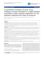
the white pulpa [18] (Figure 1a, c, e, g, i). The synovium,
draining lymph nodes, and lung remained negative on
IHC. A more sensitive detection method is RT-qPCR,
and at the mRNA level, the spleen, liver, but also blood
and BMCs were positive for eGFP, whereas the synovium
remained negative (Figure 1k). One day after i.a. injec-
Arntz et al. Arthritis Research & Therapy 2010, 12:R61
/>Page 5 of 11
tion, only SLCs, probably type B cells (based upon their
morphology), were transduced as shown by RT-qPCR
and IHC, whereas lung, liver, spleen, draining lymph
nodes, blood, and BMCs were negative on IHC (Figure
1b, d, f, h, j).
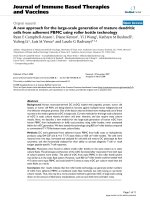
HpTNFR1 expression decreased TNFR1 mRNA expression
and TNFα signaling in vitro
RNAi-mediated downregulation of gene expression
involves both translational repression and accelerated
mRNA turnover [19]. To investigate the efficiency of
TNFR1 gene silencing by shRNA expression, we trans-
duced murine NF-κB-luciferase reporter fibroblasts with
adenoviral vector encoding a hairpin construct targeting
TNFR1 (HpTNFR1) or a scrambled control sequence
(HpNS). After 2 days, cells were stimulated with TNFα
for 6 hours, and TNFα-induced NF-κB activation and
TNFR1 expression were quantified using a luciferase
assay or qPCR, respectively (Figure 2a, b). At MOIs 1 and
10, we observed a strong reduction of NF-κB activation
(70%) in the HpTNFR1-treated group as compared with
the HpNS group. This was accompanied by two- and
three-fold reductions (2
ΔΔCt
) of TNFR1 mRNA levels at
MOIs 1 and 10, respectively. Next, we investigated the
specificity of the TNFR1-targeting construct (Figure 2c).
NF-κB-luciferase reporter fibroblasts were either trans-
duced with HpTNFR1 or preincubated with a specific
TNF antagonist (Enbrel) and then stimulated with TNFα
or IL-1β. Both HpTNFR1 and Enbrel showed a strong
reduction (90%) of TNFα-induced NF-κB activation. In
contrast, HpTNFR1 treatment did not affect IL-1β-
induced NF-κB activation, indicating the specific target-
ing of TNFR1-mediated signal transduction.
TNFR1 silencing in synovial lining cells ameliorated arthritis
Previously, it was demonstrated that TNFR1 in SFs is
essential to the development of strictly TNF-driven
arthritis [10]. Therefore, we sought to investigate whether
this mechanism also holds for arthritis models that are
known to be partly TNF-dependent, including SCW [20]
and CIA [1]. SLCs from knee joints of naïve C57BL/6
were transduced by i.a. injection with adenoviral vectors
encoding HpTNFR1 or HpNS. One day thereafter, joints
were challenged with 5-μg SCW fragments, and after 24
hours, synovial cytokine mRNA expression and protein
levels were measured by qPCR and Luminex, respectively
(Figure 3a, b). TNFR1, but not TNFR2, mRNA level was
decreased (twofold) in synovial tissue explants from the
HpTNFR1-treated group. In addition, we observed a
strong reduction (more than threefold) in mRNA levels of
IL-1β, IL-6, and TNFα. Corresponding with these results,
protein levels of IL-1β and IL-6 were significantly
reduced in the HpTNFR1 group compared with the
HpNS group. Next, knee joints of CIA-negative mice
were transduced with HpTNFR1 or HpNS at day 1 after
booster (day 22). RT-qPCR analysis at day 26 showed a
strong (more than fourfold) reduction in synovial mRNA
levels of IL-1β, IL-6, and TNFα (Figure 4a). Arthritis
development was monitored until day 31 (Figure 4b).
While CIA incidence was equal between treatments,
Figure 1 Localization of transgene expression after local or sys-
temic administration of adenoviral reporter vector in mice. One
day after collagen booster, nonarthritic DBA/1J mice were injected in-
tra-articularly with 10
7
ffu or intravenously with 3 × 10
8
ffu Ad-eGFP. Af-
ter administration, eGFP was assessed by immunohistochemistry in
lung (a, b), liver (c, d), spleen (e, f), lymph nodes (LNs) (g, h), and syn-
ovium (i, j) after local (right frames) or systemic (left frames) treatment.
Sites of eGFP-positive cells are indicated by arrows. (k) The expression
of eGFP mRNA levels in each organ, blood, and bone marrow cells
(BMCs). Draining LNs were negative on immunohistochemistry and
quantitative polymerase chain reaction (not detected). Data are repre-
sented as the difference in cycle threshold (ΔCt) values compared with
the housekeeping gene GAPDH (glyceraldehyde-3-phosphate dehy-
drogenase). mRNA levels that could not be detected are noted by 'nd'
(not detectable). Background mean mRNA levels of eGFP are as low as
the negative control. Bars represent mean ± standard error of the
mean, and statistical differences were determined using Student t test.
*P < 0.01. IA, intra-articular; IV, intravenous.
A
B
C
D
EF
GH
I
J
K
F
F
*
Background
Lung Liver Spleen Synovium LN Blood BMC
-10.0
-8.0
-6.0
-4.0
-2.0
0.0
2.0
4.0
6.0
IV
IA
nd nd
eGFP expression
D
Ct(corrected for G A PDH)
Arntz et al. Arthritis Research & Therapy 2010, 12:R61
/>Page 6 of 11
TNFR1 silencing clearly reduced macroscopic arthritis
severity. Histology taken at day 31 revealed protection
against cartilage destruction and a significant reduction
in the amount of synovial inflammatory cell infiltrate and
joint space inflammatory cell exudate (Figure 4c).
TNFR1 silencing in the reticuloendothelial system
prevented collagen-induced arthritis development
Recently, it was shown that TNFR1 silencing in the radio-
sensitive hematopoietic compartment aggravates disease
in CIA [12]. Secondary lymphoid organs, such as liver
and spleen, are rich in mature and functional cells of
hematopoietic origin, such as lymphocytes, monocytes,
and APCs. To delineate the function of TNFR1 in hepatic
and splenic cells during arthritis, CIA-negative mice (col-
lagen type II immunized mice without macroscopic signs
of arthritis) were injected intravenously with HpTNFR1
or HpNS at day 1 after booster injection (day 22). We
monitored arthritis development until day 31 (Figure 5a).
Up to day 30, the incidence of arthritis in the paws of
mice treated with HpTNFR1 (40%) was considerably
reduced compared with HpNS treatment (83%) (data not
shown). In addition, macroscopic arthritis scores were
significantly reduced in the TNFR1 group. Histology of
knee joints taken on day 31 confirmed a significant
reduction in joint inflammation and revealed a strong
suppression of cartilage proteoglycan depletion (Figure
5b, c).
Figure 2 Validation of hairpin construct targeting tumor necrosis
factor receptor 1 (HpTNFR1) in vitro. (a) NIH-3T3-5 × NF-κB-lu-
ciferase cells were transduced at indicated multiplicity of infection
(MOI) HpTNFR1 or hairpin non specific (HpNS) and, after 2 days, stimu-
lated with 10 ng/mL mTNFα for 6 hours. Nuclear factor-kappa-B (NF-
κB)-driven luciferase activity is represented as mean ± standard error of
the mean (SEM) (n = 4) of percentages compared with the HpNS
group. The numbers of HpNS transduced cells (doses MOI 10) with or
without TNFα stimulation were 164,232 ± 864 and 21,555 ± 864 rela-
tive light units (RLU)/mg protein, respectively. (b) Expression of TNFR1
in NIH-3T3-5 × NF-κB-luciferase cells transduced at indicated MOI with
HpTNFR1. Data are represented as the mean (n = 10) of the difference
in TNFR1 ΔCt values compared with HpNS-treated group (ΔΔCt). (c)
NIH-3T3-5 × NF-κB-luciferase cells were transduced at MOI 10 with
HpTNFR1 or HpNS or left untreated. After 2 days, untreated cells were
preincubated for 1 hour with 10 μg/mL Enbrel, and thereafter all
groups were stimulated with 10 ng/mL mTNFα or mIL-β for 6 hours.
Luciferase activity is represented as mean ± SEM (n = 4). Statistical dif-
ferences were determined using analysis of variance with Bonferroni
post-test. *P < 0.05; ***P < 0.001. Ct, cycle threshold; IL, interleukin; TNF,
tumor necrosis factor.
0.1 1 10
0
25
50
75
100
MOI HpTNFR1
NF-
B activity
(% of HpNS)
A
B
C
*
*
0.1 1 10
-3.0
-2.0
-1.0
0.0
MOI HpTNFR1
Ct (vs. HpNS)
0
2000
4000
6000
8000
10000
12000
HpNS
HpTNFR1
Enbrel
TNF IL-1
RLU/pg protein
***
***
Figure 3 Effects of silencing tumor necrosis factor receptor 1
(TNFR1) in synovial lining cells during streptococcal cell wall
(SCW) arthritis. Knee joints of naïve C57BL/6 mice were injected with
10
7
ffu hairpin construct targeting TNFR1 (HpTNFR1) or hairpin non
specific (HpNS), and 2 days post-transduction, joints were challenged
with 5 μg of SCW fragments. (a) Expression of indicated genes in syn-
ovial tissue at 24 hours after SCW challenge. Data are represented as
mean ± standard error of the mean (SEM) (n = 3-6) of the difference in
ΔCt values compared with the HpNS group (ΔΔCt). (b) Cytokine pro-
tein levels in 1-hour cultures of synovial tissue explants isolated at 24
hours after challenge. Bars represent mean ± SEM (n = 7), and statistical
differences were determined using Student t test. *P < 0.05. Ct, cycle
threshold; IL, interleukin; TNF, tumor necrosis factor.
IL-1
IL-6
0
50
100
150
200
250
HpNS
HpTNFR1
Concentration (pg/ml)
A
B
TNFR1 TNFR2 IL-1
IL-6 TNF
-4
-3
-2
-1
0
1
Ct (vs. HpNS)
*
*
Arntz et al. Arthritis Research & Therapy 2010, 12:R61
/>Page 7 of 11
Figure 4 Silencing of tumor necrosis factor receptor 1 (TNFR1) in
synovial lining cells ameliorates collagen-induced arthritis (CIA).
One day after collagen booster, knee joints of CIA-negative mice were
injected with 10
7
ffu hairpin construct targeting TNFR1 (HpTNFR1) or
hairpin non specific (HpNS). (a) Expression of indicated genes in syn-
ovial tissue at day 26 of CIA. Data are represented as mean ± standard
error of the mean (SEM) (n = 6) of the difference in ΔCt values com-
pared with the HpNS group (ΔΔCt). (b) Appearance of arthritis in fore
and hind paws was monitored at indicated time points and scored for
severity. (c) Histological analysis of inflammation ('infiltrate' and 'exu-
date') and proteoglycan depletion in patellar and femoral cartilage ('PG
loss') from knee joints isolated at day 31. Data are represented as mean
± SEM (n = 9 mice), and statistical differences were calculated using
Mann-Whitney U test. *P < 0.05, **P = 0.01. Ct, cycle threshold; IL, inter-
leukin; TNF, tumor necrosis factor.
21 23 25 27 29 31
0
1
2
3
4
5
HpNS
TNF R1
Days after immunization
Macroscopic a rthritis score
A
B
*
**
**
C
Infiltrate Exudate PG loss
0.0
0.5
1.0
1.5
2.0
HpNS
TNF R1
Histological score (0-3)
*
*
*
TNFR1 TNFR2 TNF
IL-1
IL-6
-4
-3
-2
-1
0
Ct (vs. HpNS)
Figure 5 Tumor necrosis factor receptor 1 (TNFR1) silencing in
the hepatic and splenic reticuloendothelial system ameliorates
collagen-induced arthritis (CIA). One day after collagen booster,
CIA-negative mice were injected intravenously with 3 × 10
8
ffu hairpin
construct targeting TNFR1 (HpTNFR1) or hairpin non specific (HpNS).
(a) Appearance of arthritis in fore and hind paws was monitored at in-
dicated time points and scored for severity. (b) Histological analysis of
inflammation ('infiltrate' and 'exudate') and proteoglycan depletion in
patellar and femoral cartilage ('PG loss') from knee joints isolated at day
31. Data are represented as mean ± standard error of the mean (n = 6
mice), and statistical differences were calculated using Mann-Whitney
U test. *P < 0.05, **P < 0.005. (c) Representative picture of safranin-O-
stained tissue sections of knee joints from mice treated systemically
with HpNS or HpTNFR1. Original magnification × 40. C, cartilage; F, fe-
mur; JS, joint space; P, patella; S, synovium.
21 23 25 27 29 31
0
1
2
3
4
HpNS
HpTNFR1
Days after immunization
Macrosc opic arthritis score
A
B
Infiltrate Exudate PG loss
0.0
0.5
1.0
1.5
2.0
2.5
3.0
HpNS
HpTNFR1
Histological score (0-3)
P
F
JS
C
C
S
C
HpNS HpTNFR1
**
*
*
*
*
*
Arntz et al. Arthritis Research & Therapy 2010, 12:R61
/>Page 8 of 11
TNFR1 silencing in antigen-presenting cells reduced the
number of T helper cells in spleen and dampened the
acute-phase response in liver
To elaborate on the mechanisms behind HpTNFR1-
mediated prevention of CIA, we analyzed proinflamma-
tory gene expression in liver at disease endpoint by qPCR
(Figure 6a). This showed a significantly reduced (>3-fold)
expression of TNFR1, IL-1β, IL-6 and the acute phase
gene Saa1. To study the effects of TNFR1 silencing in
spleen, we performed FACS and qPCR analyzes on the
splenocytes (Figure 6b, c) and cytokine measurements on
the APC fraction (Figure 6d). FACS (fluorescence-acti-
vated cell sorting) analysis showed a significant reduction
in the number of CD4
+
/TCRβ T cells, stained intracellu-
larly for T helper 1 (Th1) (IFNγ), Th2 (IL-4), or Th17 (IL-
17) cytokine expression. This was accompanied by a
strong decrease (more than fourfold) in mRNA expres-
sion of their respective transcription factors (T-bet,
GATA-3, and RoRγT). Since both IL-1 and IL-6 have
been described as crucial cytokines in T-cell expansion
and differentiation [21,22], we measured their production
by TNFα-stimulated APCs in HpTNFR1- and HpNS-
treated groups. Indeed, secreted IL-6 protein levels were
significantly reduced in HpTNFR1-treated as compared
with HpNS-treated groups. Together, these data demon-
strate a clear proinflammatory role of TNFR1 in SFs and
splenic APCs.
Discussion
The pleiotropic biological and immunological activities
of TNFα are determined by its cellular localization (trans-
membrane or soluble) [23-26] and the cell-specific rela-
tive abundance of its respective receptors, TNFR1 and
TNFR2 [9,12,27,28]. The role of TNF as pivotal mediator
of the cytokine cascade in inflammation and RA patho-
genesis has been unequivocally established, but the rela-
tive contributions of specific cell types and TNF
receptors have not been fully elucidated. Delineating the
role of TNF and its receptors in different tissues and cell
types relevant to disease may contribute to a better and
safer TNF-targeting strategy in RA patients. While a
number of studies using TNFR1-deficient mice have
established the global contribution of signal transduction
through this receptor in CIA [8,9,11,29], cell-specific
functions of TNFR1 have thus far been studied only in
SFs, bone marrow-derived macrophages, and radiosensi-
tive hematopoietic cells [10,12,30,31]. In this study, we
have demonstrated that, after local treatment in the knee
joint, only the SLCs were transduced and that there was
no spillover to other organs. Gouze and colleagues [32]
have shown that, after i.a. adenovirus delivery, 75% to
90% of the transduced cells are positive for fibroblast
markers (CD90, CD29, and VCAM-1) and no transduced
cells were positive for the macrophage marker CD11b.
Ten percent of the transduced cells are positive for the
APC marker CD86. After systemic delivery, predomi-
nantly liver and spleen were transduced, while synovium
remained negative. It is well documented that systemic
i.v. delivery of adenoviruses targets the Kupffer cells in
the liver [33] and marginal zone macrophages in spleen
[34]. Stone and colleagues [35] demonstrated that adeno-
viruses in the circulation become opsonized by blood
platelets and that these aggregates are sequestered in the
RES. Interestingly, depletion of synovial tissue mac-
rophages [36] or the macrophages in spleen and liver [37]
after local or systemic administration of clodronate-
encapsulated liposomes demonstrates the crucial role of
both the local and systemic macrophages in mediating
experimental arthritis. For this, we can conclude that
TNFR1-mediated signaling in joint, liver, and spleen RES
compartments contributes to the local joint inflamma-
tion and the development of autoimmunity during exper-
imental arthritis.
The contribution of TNFR1-mediated signaling in
SLCs to joint inflammation was investigated after SCW
challenge. In the acute phase, SCW arthritis represents
Figure 6 Effects of tumor necrosis factor receptor 1 (TNFR1) si-
lencing in the hepatic and splenic reticuloendothelial system.
One day after collagen booster, mice negative for collagen-induced ar-
thritis were injected intravenously with 3 × 10
8
ffu hairpin construct tar-
geting TNFR1 (HpTNFR1) or hairpin non specific (HpNS). (a) Expression
of indicated genes in liver isolated at day 26. Data are represented as
mean (n = 5) of the difference in ΔCt values compared with the HpNS
group (ΔΔCt). (b) Analysis of intra-cellular cytokine expression in T cells
isolated from spleen at day 26. Data are represented as mean ± stan-
dard error of the mean (SEM) (n = 4) of the percentage of positive cells
compared with the HpNS group. (c) Expression of indicated genes in
isolated splenic T cells. Data are represented as mean (n = 5) of the dif-
ference in ΔCt values compared with the HpNS group (ΔΔCt). (d) Se-
creted cytokine levels from splenic antigen-presenting cells stimulated
for 24 hours with 10 ng/mL mTNFα. Data are represented as mean ±
SEM (n = 5). Statistical differences were calculated using analysis of
variance with Bonferroni post-test. *P < 0.05, **P < 0.01. Ct, cycle
threshold; IL, interleukin; TNF, tumor necrosis factor.
A
TNFR1 Saa1 IL-1
IL-6 TNF
-2.5
-2.0
-1.5
-1.0
-0.5
0.0
Ct (vs. HpNS)
*
*
*
*
B
IFN
IL-4 IL-17
0
25
50
75
Positive cel ls (% of HpNS)
*
*
C
T-bet GATA-3 RoR
T
-4.0
-3.0
-2.0
-1.0
0.0
Ct (vs. HpNS)
*
*
D
0
5
10
15
20
25
HpNS
HpTNFR1
Concentration (pg/ml)
**
IL-1b
IL-6
Arntz et al. Arthritis Research & Therapy 2010, 12:R61
/>Page 9 of 11
an innate immune response against SCW fragments that
is driven by direct activation of macrophages [38,39].
TNFR1 silencing in SLCs resulted in a significant reduc-
tion of secreted IL-6 and IL-1β levels in the joint, which
indicates an inhibition of the local cytokine cascade. The
reduction of IL-6 is most likely a direct effect of TNFR1
silencing in SLCs since previous studies demonstrated
that TNF-induced IL-6 secretion in human RA SFs is
mediated exclusively through TNFR1 [40,41]. In contrast,
hematopoietic cells (neutrophils and monocytes), but not
mesenchymal cells (SFs), were identified as the main
source of IL-1 in TNF-driven joint pathology [42]. The
observed reduction of IL-1β suggests that TNF signaling
in SLCs plays an important role in chemoattraction of
inflammatory cells. Indeed, histological analysis of
HpTNFR1-treated joints in CIA showed almost complete
prevention of IL-1-induced cartilage proteoglycan loss,
which was accompanied by an impressive reduction of
inflammatory cell influx. We have revealed, in line with
the study of Armaka and colleagues [10], a dominant role
of TNFR1-mediated signaling in SLCs in joint inflamma-
tion.
Remarkably, we found that TNFR1 silencing in knee
joints also protected the ipsilateral ankle joints in CIA
mice. While such distal effects have been described
before in local gene therapy approaches [43-45], the
underlying mechanism is still not fully understood. How-
ever, such an effect might point toward a role of local
TNFR1-mediated signaling in the development of auto-
immunity. In support of this, previous investigations
using periarticular delivery of secreted transgenes, vIL-10
and TNFR, in CIA showed that distant anti-arthritic
effects coincided with a reduction of specific collagen
antibody titers and modulation of T-cell responses,
respectively [45-47]. Notably, the beneficial systemic
effects of periarticular TNFR gene therapy correlated well
with circulating levels of the transgene [45]. In the
absence of transgene spillover to the circulation, distal
effects have been attributed to antigen-primed APCs
exposed to the therapeutic transgene traveling from
treated to untreated joints [47-49]. In our approach,
TNFR1 silencing was restricted to SLCs that would
exclude transgene spillover or direct modulation of APCs
as a causative for systemic effects. However, local TNFR1
treatment reduced local levels of IL-6 and IL-1β. Both
cytokines are implicated in APC function, which is in
turn a prerequisite for induction of auto-reactive CD4
+
T
cells and autoimmunity. Eriksson and colleagues [50]
demonstrated that IL-1 receptor type I is required for
efficient activation of dendritic cells (DCs). IL-6 switches
the differentiation of monocyte-derived APCs from DCs
to macrophages [51]. The observed reduction of both IL-
1β and IL-6 synthesis in the inflamed joint may result in
the development of immature DCs, a differentiation state
associated with a tolerogenic function of these cells.
Alternatively, tolerogenic DCs can be induced by IL-10, a
cytokine that inhibits the synthesis of IL-1 and IL-6 in
monocytes and other cell types [52]. Alternatively,
TNFR1 treatment might have affected the APC-like func-
tion of SLCs. Although SFs are not considered to be pro-
fessional APCs, approximately 60% to 70% of SFs in the
rheumatic joint express MHC (major histocompatibility
complex) class II molecules and have the capacity to serve
as accessory cells for superantigen-mediated T-cell acti-
vation [53-55]. Importantly, the interaction between
cytokine-activated T cells and SFs was found to be depen-
dent on transmembrane TNFα on the surface of T cells
and resulted in increased production of IL-6 and
chemokine IL-8 [56]. Indeed, we found strongly
decreased IL-6 production in HpTNFR1-treated joints,
which may abrogate the ability of SLCs to present auto-
antigens found within joint tissues.
Strikingly, systemic treatment with HpTNFR1 amelio-
rated CIA almost to the same extent as local treatment.
We have previously shown that SOCS3 (suppressor of
cytokine signaling-3) overexpression in splenic APCs
ameliorates CIA via a general suppression of Th subsets
[18]. Similarly, we observed a reduction in the number of
Th1 (IFNγ, T-bet), Th2 (IL-4, GATA-3), and Th17 (IL-17,
RoRγT) cells upon TNFR1 in splenic APCs after antigen
booster injection. In line with these similar findings, it
has been demonstrated that TNFR1-deficient murine
myocardiocytes show increased expression of SOCS3 and
reduced IL-6 secretion upon TNF infusion [57]. We have
confirmed that the splenic APCs from HpTNFR1-treated
mice produce markedly less IL-6 and IL-1β after TNF
stimulation. As these cytokines are crucially involved in
Th17 differentiation [21,22], the observed large reduction
of Th17 numbers in spleen is not unexpected. Thus,
TNFR1 modulation in the RES has a clear-cut effect on
immunity in CIA.
In a side-by-side comparison, we have demonstrated
equal efficacies of local and systemic RNAi-mediated
TNFR1-targeting gene therapy in alleviating CIA. Impor-
tantly, cell-specific gene therapeutic targeting of TNFR1
clearly modulated proinflammatory effects of TNFα
without interfering with protective effects of TNF signal-
ing that have been described in hematopoietic cells
[11,12]. It will be interesting to investigate whether local
or systemic TNFR1 knockdown gives a different outcome
in CIA when using a therapeutic regimen.
Conclusions
Specific silencing of TNFR1 in SLCs, hepatic and splenic
RES by respectively local or systemic delivery of Ad5
virus encoding for small hairpin RNA against TNFR1
revealed a dominant and clear proinflammatory role of
TNF signaling in these cells during CIA. Systemic treat-
Arntz et al. Arthritis Research & Therapy 2010, 12:R61
/>Page 10 of 11
ment dampened the liver acute-phase response and
reduced proliferation of Th subsets in spleen. Local treat-
ment inhibited the proinflammatory cytokine cascade in
the joint. Gene therapeutic targeting of TNFR1 may be a
promising and safer approach for TNFα blockade in RA
patients.
Additional material
Abbreviations
APC: antigen-presenting cell; bCII: bovine collagen type II; BMC: bone marrow
cell; BSA: bovine serum albumin; CIA: collagen-induced arthritis; Ct: cycle
threshold; DC: dendritic cell; DMEM: Dulbecco's modified Eagle's medium; FCS:
fetal calf serum; Gapdh: glyceraldehyde-3-phosphate dehydrogenase; HpNS:
hairpin non specific; HpTNFR1: hairpin construct targeting tumor necrosis fac-
tor receptor 1; i.a.: intra-articular; IFNγ: interferon-gamma; IHC: immunohis-
tochemistry; IL: interleukin; i.v.: intravenous; MOI: multiplicity of infection; NF-
κB: nuclear factor-kappa-B; PBS: phosphate-buffered saline; PCR: polymerase
chain reaction; PE: phycoerythrin; qPCR: quantitative polymerase chain reac-
tion; RA: rheumatoid arthritis; RES: reticuloendothelial system; RNAi: RNA inter-
ference; RT-qPCR: reverse transcription-quantitative polymerase chain reaction;
SCW: streptococcal cell wall; SF: synovial fibroblast; shRNA: short hairpin RNA;
SLC: synovial lining cell; SOCS3: suppressor of cytokine signaling-3; TCRβ: T-cell
receptor beta; Th: T helper; TNFα: tumor necrosis factor-alpha; TNFR: tumor
necrosis factor receptor.
Competing interests
The authors declare that they have no competing interests.
Authors' contributions
OJA helped to acquire data and contributed to the study design, statistical and
data analysis, interpretation of data, and drafting of the manuscript. SV, BTvdB,
and MBB helped to acquire data. JG, SA-R, and FAvdL contributed to the study
design, statistical and data analysis, interpretation of data, and drafting of the
manuscript. WBvdB conceived of the study and helped draft the manuscript.
All authors read and approved the final manuscript.
Acknowledgements
This research was supported by a VIDI grant (917.46.363) to FAJvdL from the
Netherlands Organization for Scientific Research. This research was performed
within the framework of TI-Pharma, project number D1-101.
Author Details
Rheumatology Research and Advanced Therapeutics, Department of
Rheumatology, Radboud University Nijmegen Medical Centre, 6525 GA
Nijmegen, The Netherlands
References
1. Joosten LA, Helsen MM, Loo FA van de, Berg WB van den: Anticytokine
treatment of established type II collagen-induced arthritis in DBA/1
mice. A comparative study using anti-TNF alpha, anti-IL-1 alpha/beta,
and IL-1Ra. Arthritis Rheum 1996, 39:797-809.
2. Williams RO, Feldmann M, Maini RN: Anti-tumor necrosis factor
ameliorates joint disease in murine collagen-induced arthritis. Proc
Natl Acad Sci USA 1992, 89:9784-9788.
3. Keffer J, Probert L, Cazlaris H, Georgopoulos S, Kaslaris E, Kioussis D, Kollias
G: Transgenic mice expressing human tumour necrosis factor: a
predictive genetic model of arthritis. EMBO J 1991, 10:4025-4031.
4. Brennan FM, Chantry D, Jackson A, Maini R, Feldmann M: Inhibitory effect
of TNF alpha antibodies on synovial cell interleukin-1 production in
rheumatoid arthritis. Lancet 1989, 2:244-247.
5. Elliott MJ, Maini RN, Feldmann M, Long-Fox A, Charles P, Katsikis P,
Brennan FM, Walker J, Bijl H, Ghrayeb J, Woody JN: Treatment of
rheumatoid arthritis with chimeric monoclonal antibodies to tumor
necrosis factor alpha. Arthritis Rheum 1993, 36:1681-1690.
6. Elliott MJ, Maini RN, Feldmann M, Long-Fox A, Charles P, Bijl H, Woody JN:
Repeated therapy with monoclonal antibody to tumour necrosis factor
alpha (cA2) in patients with rheumatoid arthritis. Lancet 1994,
344:1125-1127.
7. MacEwan DJ: TNF receptor subtype signalling: differences and cellular
consequences. Cell Signal 2002, 14:477-492.
8. Mori L, Iselin S, De LG, Lesslauer W: Attenuation of collagen-induced
arthritis in 55-kDa TNF receptor type 1 (TNFR1)-IgG1-treated and
TNFR1-deficient mice. J Immunol 1996, 157:3178-3182.
9. Tada Y, Ho A, Koarada S, Morito F, Ushiyama O, Suzuki N, Kikuchi Y, Ohta A,
Mak TW, Nagasawa K: Collagen-induced arthritis in TNF receptor-1-
deficient mice: TNF receptor-2 can modulate arthritis in the absence of
TNF receptor-1. Clin Immunol 2001, 99:325-333.
10. Armaka M, Apostolaki M, Jacques P, Kontoyiannis DL, Elewaut D, Kollias G:
Mesenchymal cell targeting by TNF as a common pathogenic principle
in chronic inflammatory joint and intestinal diseases. J Exp Med 2008,
205:331-337.
11. Notley CA, Inglis JJ, Alzabin S, McCann FE, McNamee KE, Williams RO:
Blockade of tumor necrosis factor in collagen-induced arthritis reveals
a novel immunoregulatory pathway for Th1 and Th17 cells. J Exp Med
2008, 205:2491-2497.
12. Williams-Skipp C, Raman T, Valuck RJ, Watkins H, Palmer BE, Scheinman RI:
Unmasking of a protective tumor necrosis factor receptor I-mediated
signal in the collagen-induced arthritis model. Arthritis Rheum 2009,
60:408-418.
13. Barrera P, Oyen WJ, Boerman OC, van Riel PL: Scintigraphic detection of
tumour necrosis factor in patients with rheumatoid arthritis. Ann
Rheum Dis 2003, 62:825-828.
14. Broek MF van den, Berg WB van den, Putte LB van de, Severijnen AJ:
Streptococcal cell wall-induced arthritis and flare-up reaction in mice
induced by homologous or heterologous cell walls. Am J Pathol 1988,
133:139-149.
15. He TC, Zhou S, da Costa LT, Yu J, Kinzler KW, Vogelstein B: A simplified
system for generating recombinant adenoviruses. Proc Natl Acad Sci
USA 1998, 95:2509-2514.
16. Schiedner G, Hertel S, Kochanek S: Efficient transformation of primary
human amniocytes by E1 functions of Ad5: generation of new cell lines
for adenoviral vector production. Hum Gene Ther 2000, 11:2105-2116.
17. Shayakhmetov DM, Li ZY, Ni S, Lieber A: Analysis of adenovirus
sequestration in the liver, transduction of hepatic cells, and innate
toxicity after injection of fiber-modified vectors. J Virol 2004,
78:5368-5381.
18. Veenbergen S, Bennink MB, de Hooge AS, Arntz OJ, Smeets RL, Berg WB
van den, Loo FA van de: Splenic suppressor of cytokine signaling 3
transgene expression affects T cell responses and prevents
development of collagen-induced arthritis. Arthritis Rheum 2008,
58:3742-3752.
19. Wu L, Belasco JG: Let me count the ways: mechanisms of gene
regulation by miRNAs and siRNAs. Mol Cell 2008, 29:1-7.
20. Berg WB van den, Joosten LA, Kollias G, Loo FA van de: Role of tumour
necrosis factor alpha in experimental arthritis: separate activity of
interleukin 1beta in chronicity and cartilage destruction. Ann Rheum
Dis 1999, 58(Suppl 1):I40-I48.
21. Ben-Sasson SZ, Hu-Li J, Quiel J, Cauchetaux S, Ratner M, Shapira I, Dinarello
CA, Paul WE: IL-1 acts directly on CD4 T cells to enhance their antigen-
driven expansion and differentiation. Proc Natl Acad Sci USA 2009,
106:7119-7124.
22. Hirota K, Hashimoto M, Yoshitomi H, Tanaka S, Nomura T, Yamaguchi T,
Iwakura Y, Sakaguchi N, Sakaguchi S: T cell self-reactivity forms a
cytokine milieu for spontaneous development of IL-17+ Th cells that
cause autoimmune arthritis. J Exp Med 2007, 204:41-47.
23. Cowley SC, Sedgwick JD, Elkins KL: Differential requirements by CD4+
and CD8+ T cells for soluble and membrane TNF in control of
Francisella tularensis live vaccine strain intramacrophage growth. J
Immunol 2007, 179:7709-7719.
24. Muller S, Rihs S, Schneider JM, Paredes BE, Seibold I, Brunner T, Mueller C:
Soluble TNF-alpha but not transmembrane TNF-alpha sensitizes T cells
for enhanced activation-induced cell death. Eur J Immunol 2009,
39:3171-3180.
Additional file 1 Supplemental Methods. Primerdesign.
Received: 16 October 2009 Revised: 8 March 2010
Accepted: 6 April 2010 Published: 6 April 2010
This article is available from: 2010 Arntz et al.; licensee BioMed Central Ltd. This is an open access article distributed under the terms of the Creative Commons Attribution License ( which permits unrestricted use, distribution, and reproduction in any medium, provided the original work is properly cited.Arthritis R esearch & Therapy 2010, 12:R61
Arntz et al. Arthritis Research & Therapy 2010, 12:R61
/>Page 11 of 11
25. Oikonomou N, Harokopos V, Zalevsky J, Valavanis C, Kotanidou A,
Szymkowski DE, Kollias G, Aidinis V: Soluble TNF mediates the transition
from pulmonary inflammation to fibrosis. PLoS One 2006, 1:e108.
26. Zalevsky J, Secher T, Ezhevsky SA, Janot L, Steed PM, O'Brien C, Eivazi A,
Kung J, Nguyen DH, Doberstein SK, Erard F, Ryffel B, Szymkowski DE:
Dominant-negative inhibitors of soluble TNF attenuate experimental
arthritis without suppressing innate immunity to infection. J Immunol
2007, 179:1872-1883.
27. Butler DM, Feldmann M, Di PF, Brennan FM: p55 and p75 tumor necrosis
factor receptors are expressed and mediate common functions in
synovial fibroblasts and other fibroblasts. Eur Cytokine Netw 1994,
5:441-448.
28. Douni E, Kollias G: A critical role of the p75 tumor necrosis factor
receptor (p75TNF-R) in organ inflammation independent of TNF,
lymphotoxin alpha, or the p55TNF-R. J Exp Med 1998, 188:1343-1352.
29. Yamaguchi N, Ohshima S, Umeshita-Sasai M, Nishioka K, Kobayashi H,
Mima T, Kishimoto T, Saeki Y: Synergistic effect on the attenuation of
collagen induced arthritis in tumor necrosis factor receptor I (TNFRI)
and interleukin 6 double knockout mice. J Rheumatol 2003, 30:22-27.
30. Ermolaeva MA, Michallet MC, Papadopoulou N, Utermohlen O, Kranidioti
K, Kollias G, Tschopp J, Pasparakis M: Function of TRADD in tumor
necrosis factor receptor 1 signaling and in TRIF-dependent
inflammatory responses. Nat Immunol 2008, 9:1037-1046.
31. Zakharova M, Ziegler HK: Paradoxical anti-inflammatory actions of TNF-
alpha: inhibition of IL-12 and IL-23 via TNF receptor 1 in macrophages
and dendritic cells. J Immunol 2005, 175:5024-5033.
32. Gouze E, Gouze JN, Palmer GD, Pilapil C, Evans CH, Ghivizzani SC:
Transgene persistence and cell turnover in the diarthrodial joint:
implications for gene therapy of chronic joint diseases. Mol Ther 2007,
15:1114-1120.
33. Smith JS, Xu Z, Tian J, Stevenson SC, Byrnes AP: Interaction of
systemically delivered adenovirus vectors with Kupffer cells in mouse
liver. Hum Gene Ther 2008, 19:547-554.
34. Hiltunen MO, Turunen MP, Turunen AM, Rissanen TT, Laitinen M, Kosma
VM, Yla-Herttuala S: Biodistribution of adenoviral vector to nontarget
tissues after local in vivo gene transfer to arterial wall using
intravascular and periadventitial gene delivery methods. FASEB J 2000,
14:2230-2236.
35. Stone D, Liu Y, Shayakhmetov D, Li ZY, Ni S, Lieber A: Adenovirus-platelet
interaction in blood causes virus sequestration to the
reticuloendothelial system of the liver. J Virol 2007, 81:4866-4871.
36. van Lent PL, Holthuysen AE, Bersselaar LA van den, van RN, Joosten LA,
Loo FA van de, Putte LB van de, Berg WB van den: Phagocytic lining cells
determine local expression of inflammation in type II collagen-induced
arthritis. Arthritis Rheum 1996, 39:1545-1555.
37. Richards PJ, Williams AS, Goodfellow RM, Williams BD: Liposomal
clodronate eliminates synovial macrophages, reduces inflammation
and ameliorates joint destruction in antigen-induced arthritis.
Rheumatology (Oxford) 1999, 38:818-825.
38. Abdollahi-Roodsaz S, Joosten LA, Helsen MM, Walgreen B, van Lent PL,
Bersselaar LA van den, Koenders MI, Berg WB van den: Shift from toll-like
receptor 2 (TLR-2) toward TLR-4 dependency in the erosive stage of
chronic streptococcal cell wall arthritis coincident with TLR-4-
mediated interleukin-17 production. Arthritis Rheum 2008,
58:3753-3764.
39. Joosten LA, Koenders MI, Smeets RL, Heuvelmans-Jacobs M, Helsen MM,
Takeda K, Akira S, Lubberts E, Loo FA van de, Berg WB van den: Toll-like
receptor 2 pathway drives streptococcal cell wall-induced joint
inflammation: critical role of myeloid differentiation factor 88. J
Immunol 2003, 171:6145-6153.
40. Alsalameh S, Amin RJ, Kunisch E, Jasin HE, Kinne RW: Preferential
induction of prodestructive matrix metalloproteinase-1 and
proinflammatory interleukin 6 and prostaglandin E2 in rheumatoid
arthritis synovial fibroblasts via tumor necrosis factor receptor-55. J
Rheumatol 2003, 30:1680-1690.
41. Kunisch E, Gandesiri M, Fuhrmann R, Roth A, Winter R, Kinne RW:
Predominant activation of MAP kinases and pro-destructive/pro-
inflammatory features by TNF alpha in early-passage synovial
fibroblasts via TNF receptor-1: failure of p38 inhibition to suppress
matrix metalloproteinase-1 in rheumatoid arthritis. Ann Rheum Dis
2007, 66:1043-1051.
42. Zwerina J, Redlich K, Polzer K, Joosten L, Kronke G, Distler J, Hess A, Pundt
N, Pap T, Hoffmann O, Gasser J, Scheinecker C, Smolen JS, van den BW,
Schett G: TNF-induced structural joint damage is mediated by IL-1.
Proc Natl Acad Sci USA 2007, 104:11742-11747.
43. Bakker AC, Joosten LA, Arntz OJ, Helsen MM, Bendele AM, Loo FA van de,
Berg WB van den: Prevention of murine collagen-induced arthritis in
the knee and ipsilateral paw by local expression of human interleukin-
1 receptor antagonist protein in the knee. Arthritis Rheum 1997,
40:893-900.
44. Lubberts E, Joosten LA, van den BL, Helsen MM, Bakker AC, Xing Z,
Richards CD, Berg WB van den: Intra-articular IL-10 gene transfer
regulates the expression of collagen-induced arthritis (CIA) in the knee
and ipsilateral paw. Clin Exp Immunol 2000, 120:375-383.
45. Mukherjee P, Wu B, Mayton L, Kim SH, Robbins PD, Wooley PH: TNF
receptor gene therapy results in suppression of IgG2a anticollagen
antibody in collagen induced arthritis. Ann Rheum Dis 2003, 62:707-714.
46. Mukherjee P, Yang SY, Wu B, Song Z, Myers LK, Robbins PD, Wooley PH:
Tumour necrosis factor receptor gene therapy affects cellular immune
responses in collagen induced arthritis in mice. Ann Rheum Dis 2005,
64:1550-1556.
47. Whalen JD, Lechman EL, Carlos CA, Weiss K, Kovesdi I, Glorioso JC, Robbins
PD, Evans CH: Adenoviral transfer of the viral IL-10 gene periarticularly
to mouse paws suppresses development of collagen-induced arthritis
in both injected and uninjected paws. J Immunol 1999, 162:3625-3632.
48. Whalen JD, Thomson AW, Lu L, Robbins PD, Evans CH: Viral IL-10 gene
transfer inhibits DTH responses to soluble antigens: evidence for
involvement of genetically modified dendritic cells and macrophages.
Mol Ther 2001, 4:543-550.
49. Kim SH, Lechman ER, Kim S, Nash J, Oligino TJ, Robbins PD: Ex vivo gene
delivery of IL-1Ra and soluble TNF receptor confers a distal synergistic
therapeutic effect in antigen-induced arthritis. Mol Ther 2002,
6:591-600.
50. Eriksson U, Kurrer MO, Sonderegger I, Iezzi G, Tafuri A, Hunziker L, Suzuki S,
Bachmaier K, Bingisser RM, Penninger JM, Kopf M: Activation of dendritic
cells through the interleukin 1 receptor 1 is critical for the induction of
autoimmune myocarditis. J Exp Med 2003, 197:323-331.
51. Chomarat P, Banchereau J, Davoust J, Palucka AK: IL-6 switches the
differentiation of monocytes from dendritic cells to macrophages. Nat
Immunol 2000, 1:510-514.
52. de Waal MR, Abrams J, Bennett B, Figdor CG, de Vries JE: Interleukin 10(IL-
10) inhibits cytokine synthesis by human monocytes: an
autoregulatory role of IL-10 produced by monocytes. J Exp Med 1991,
174:1209-1220.
53. Boots AM, Wimmers-Bertens AJ, Rijnders AW: Antigen-presenting
capacity of rheumatoid synovial fibroblasts. Immunology 1994,
82:268-274.
54. Tran CN, Davis MJ, Tesmer LA, Endres JL, Motyl CD, Smuda C, Somers EC,
Chung KC, Urquhart AG, Lundy SK, Kovats S, Fox DA: Presentation of
arthritogenic peptide to antigen-specific T cells by fibroblast-like
synoviocytes. Arthritis Rheum 2007, 56:1497-1506.
55. Zimmermann T, Kunisch E, Pfeiffer R, Hirth A, Stahl HD, Sack U, Laube A,
Liesaus E, Roth A, Palombo-Kinne E, Emmrich F, Kinne RW: Isolation and
characterization of rheumatoid arthritis synovial fibroblasts from
primary culture primary culture cells markedly differ from fourth-
passage cells. Arthritis Res 2001, 3:72-76.
56. Tran CN, Lundy SK, White PT, Endres JL, Motyl CD, Gupta R, Wilke CM,
Shelden EA, Chung KC, Urquhart AG, Fox DA: Molecular interactions
between T cells and fibroblast-like synoviocytes: role of membrane
tumor necrosis factor-alpha on cytokine-activated T cells. Am J Pathol
2007, 171:1588-1598.
57. Wang M, Markel T, Crisostomo P, Herring C, Meldrum KK, Lillemoe KD,
Meldrum DR: Deficiency of TNFR1 protects myocardium through
SOCS3 and IL-6 but not p38 MAPK or IL-1beta. Am J Physiol Heart Circ
Physiol 2007, 292:H1694-H1699.
doi: 10.1186/ar2974
Cite this article as: Arntz et al., A crucial role for tumor necrosis factor recep-
tor 1 in synovial lining cells and the reticuloendothelial system in mediating
experimental arthritis Arthritis Research & Therapy 2010, 12:R61