Báo cáo y học: "B cells from rheumatoid arthritis patients show important alterations in the expression of CD86 and FcgRIIb, which are modulated by anti-tumor necrosis factor therapy" potx
Bạn đang xem bản rút gọn của tài liệu. Xem và tải ngay bản đầy đủ của tài liệu tại đây (665.99 KB, 11 trang )
RESEA R C H ART I C L E Open Access
B cells from rheumatoid arthritis patients show
important alterations in the expression of CD86
and FcgRIIb, which are modulated by anti-tumor
necrosis factor therapy
Diego Catalán
1
, Octavio Aravena
1
, Francisca Sabugo
2
, Pamela Wurmann
2
, Lilian Soto
2
, Alexis M Kalergis
3
,
Miguel Cuchacovich
2
, Juan C Aguillón
1*
, Millenium Nucleus on Immunology and Immunotherapy P-07-088-F
Abstract
Introduction: Several molecules help preserve peripheral B cell tolerance, but when altered, they may predispose
to autoimmunity. This work studied the expression of the costimulatory molecule CD86 and the inhibitory receptor
for IgG immune complexes FcgRIIb (CD32b), on B cells from rheumatoid arthritis (RA) patients, and the influence of
anti-tumor necrosis factor (TNF) therapy.
Methods: Peripheral B cells from 18 RA patients and 13 healthy donors were characterized using flow cytometry.
Eleven patients who underwent a six-month adalimumab therapy were further assessed for phenotypic changes
on their B cells.
Results: RA patients exhibited a high percentage of naïve and memory B cells expressing CD86. In contrast,
expression of FcgRIIb was significantly reduced on RA memory B cells and plasmablasts as compa red to healthy
donors, probably due to downregulation of this receptor when differentiating from naïve to memory cells. These
alterations on FcgRIIb were associated with high levels of anti-citrullinated vimentin autoantibodies. In addition,
treatment with adalimumab normalized the expression of CD86 on memory B cells and reduced the expression of
FcgRIIb, mainly on naïve B cells.
Conclusions: Our findings show that peripheral B cells from RA patients have an altered expression of key
molecules, such as CD86 and FcgRIIb. Because this latter receptor is required for feedback in hibition, a deficient
expression might contribute to humoral autoimmune responses. Furthermore, these molecules are likely to be
influenced by inflammatory factors, since they were modu lated by TNF inhibition.
Introduction
Rheumatoid arthritis (RA) is a chronic, inflammatory,
and autoimmune disease that affects mainly synovial
joints, leading to progressive destruction, pain, and dis-
ability. It is well known from mouse models that B cells
play a pivotal role in the development of the autoim-
mune process as a precursor of antibody-secreting cells
but also as antigen-presenting cells (APCs) [1,2].
Immune cells express an array of receptors that bind the
Fc portion of IgG-containing immune complexes (FcgRs).
Particularly, it has been stated that B cells and plasma cells
express only the low-affinity receptor FcgRIIb,which,in
contrast to FcgRIIa, has an immunoreceptor tyrosine-
based inhibitory motif on the cytoplasmic domain. This
characteristic confers an inhibitory function to the recep-
tor which is essential in several checkpoint stages in which
abnormal humoral responses are quenched by mechan-
isms that include the deletion of autoreactive clones and
feedback inhibition of IgG secretion [3].
Given this property, it is not surprising that these
molecules have been involved in autoimmune processes.
* Correspondence:
1
Programa Disciplinario de Inmunología, Instituto de Ciencias Biomédicas
(ICBM), Facultad de Medicina, Universidad de Chile, Avenida Independencia
1027, Santiago, Chile
Catalán et al. Arthritis Research & Therapy 2010, 12:R68
/>© 2010 Catalán et al; licensee BioMed Central Ltd. This is an Open Access article distributed under the terms of the Creative Commons
Attribution License ( y/2.0), which permits unrestricted use, distribution, and reproduction in
any medium, provided the original work is properly cited.
Autoimmune-susceptible mice present several poly-
morphisms in the regulatory regions of the FcgRIIb
gene, which result in a reduced expression of the recep-
tor on germinal center B cells [4]. Moreove r, depending
on the strain, mice deficient in FcgRIIb can sponta-
neously develop a lupus-like syndrome, become suscep-
tible to collagen- induced arthritis (CIA), or develop a
severe phenotype of CIA or experimental autoimmune
encephalomyelitis [5-8]. In contrast, overexpression of
FcgRIIb on B cells, but not on macrophages, leads to
an early resolution of CIA and reduced spontaneous
lupus [9].
On the other hand, human autoimmune diseases char-
acterized by a deregulated secretion of autoantibodies,
such as systemic lupus erythematosus (SLE) and RA,
have been associated with abnormalities in FcgRIIb reg-
ulation. Polymorphisms in the promoter region as well
as in the transmembrane domain of the FcgRI Ib gene
have been described to affect the expression and func-
tion of this receptor, respectively [10-12]. While bo th
polymorphisms in FcgRIIb are associated with SLE
occurrence [ 10,13], the one on the transmembrane
dom ain is also associated with joint dama ge in RA [14].
Although alterations in the expression of FcgRIIb on B
cells have been described for other autoimmune diseases
[15-18], no data about RA are available.
The aim of our study was to evaluate the phenotype of
B cells fr om RA patients , focusing on their activation
status and their expression of FcgRIIb. These parameters
were compared with those obtained from B cells of
healthy individuals. In addition, we followed up on these
patients during ant i-tumor necrosis factor (anti-TNF)
therapy and assessed the phenotype of their B cells after
6 m onths of treatment. Our findings show that B cells
from RA patie nts are activated, as reflected by the
expression of CD86. We have also observed an altered
expression of FcgRIIb, which is associated with the pre-
sence of autoantibodies. These abnormalities were
shown to be partially reverted by anti-TNF therapy.
Materials and methods
Patients
We recruited 18 patients meeting the Am erican College
of Rheumatology criteria for RA [19]. All of the patients
were women, with a mean ± standard deviation (SD)
age of 52.8 ± 10.5 years and disease duration of 16.3 ± 7
years at study entry. All of them exhibited an active dis-
ease defined as a t least six swollen joints, at least nine
tender joints, and morning stiffness for more than 1
hour, regardless of being under treatment with disease-
modifying antirheumatic drugs. Disease activity was
determined based on t he disease a ctivity score for 28
joints (DAS-28) [20]. Thirteen patients received 40 mg
of adalimumab (kindly provided by Abbott Laboratories,
Abbott Park, IL, USA) subcutaneously every other week
during 24 weeks. The European League Again st Rheu-
matism (EULAR) respons e criteria wer e used to estab-
lish the degree of response to treatment [21]. Thirteen
healthy women were recruited as a control group, with
a mean ± SD age of 39.4 ± 9.7 years. For the analy ses of
CD86 expression, we had available samples of only 8 of
the 13 healthy donors. Blood samples for flow cytometry
analyses and serum determinations were drawn from RA
patients at study ent ry and 6 months after beginning
adalimumab administration. The study was approved by
the Ethica l Committee of the Hosp ital Clínico Universi-
dad de Chile, and all patients and controls gave their
written informed consent.
Serum antibody determination
Serum antibodies against modified and citrullinated
vimentin (anti-MCV) were measured using a commer-
cial enzyme-linked immunosorbent assay (ELISA) kit
(Orgentec, Mainz, Germany) in accordance with the
instructions of the manufacturer. The cutoff level was
set at 20 U/mL. Serum anti-cyclic citrullinated peptide
(ccp) antibodies were detected using a commercial sec-
ond-generation ELISA (Axis-Shield, Dundee, Scotland)
in accordance with the instructions of the manufacturer.
A cutoff level of 5 U/mL was considered. Total serum
IgG levels were measured by ELISA at the Immunology
Laboratory of the Hospital Clínico Univ ersidad de Chile,
and the range of concentrations considered normal was
639 to 1,349 mg/dL.
B-lymphocyte phenotyping
We characterized B cells using the following monoclo-
nal anti-human antibodies: anti-CD19 phycoerythrin
(PE) cyanine 5 (Cy5), anti-CD27 fluorescein isothiocya-
nate (FITC), anti-CD27 PE, anti-CD86 FI TC (BD Bios-
ciences, San Jose, CA, USA), a nd anti-FcgRII PE (clone
7.3; Fitzgerald Industries International, Acton, MA,
USA). For the staining procedure, anticoagulated whole
blood was incubated with fluorescent antibodies for 30
minutes at 4°C. Subsequently, red cells were lysed with
ammonium chloride potassium (ACK) buffer, washed,
and fixed for flow cytometry (FACSCalibur; BD Bios-
ciences, San Jose, CA, USA). Data were analyzed with
WinMDI 2.9 Software (TSRI Flow Cytometry Core
Facility, La Jolla, CA, USA). A region was set to define
the lymphocytic population according to forward and
side scatter patterns. B c ells were defined as CD19-
expressing cells, a nd a second region was set for t hem,
while CD27 was used to discriminate memory from
naïve subsets. Plasmablasts w ere defined as CD19
low
CD27
high
-expressing cells (Figure 1a). Mean fluores-
cence inten sity (MFI) was used as the analy sis para-
meter for the expression of FcgRIIb.
Catalán et al. Arthritis Research & Therapy 2010, 12:R68
/>Page 2 of 11
Statistical analyses
All of the study variables were tested for normality with
the S hapiro-Wilk test. Differences between patient and
control groups were analyzed using the two-tailed
unpaired Student t test or Mann-Whitney U test, when
appropriate. For comparisons betw een different B-cell
subsets and for different adalimumab therapy time
points, the two-tailed paired Student t test or Wilcoxon
signed-rank test were used, when appropriate. Correla-
tions were evaluated with a two-tailed Spearman corre-
lation test. For contingency analyses, a two-sided Fisher
exact test was us ed. P values of less than 0. 05 were con-
sidered significant. For statistic analyses and graphics,
GraphPad Prism 4 software (GraphPad Software, Inc.,
La Jolla, CA, USA) was used.
Results
Rheumatoid arthritis patients show peripheral B-cell
frequencies similar to those of healthy controls
Abnormalities in the frequency of B cells and in the
proportion of different B-cell subsets in autoimmune
diseases have been previously reported [22,23]. Despite a
broad dispersion, we found no significant differences
between RA and control subjects when comparing the
percentage of B cells in the lymphocytic population
(mean percentage ± SD 7.5 ± 3.1 and 9 ± 2.2, respec-
tively). Furthermore, when we discriminat ed B cells in
naïve cells, memory cells and plasmablasts by means of
CD19 and CD27 staining, we detected similar percen-
tages of these subsets in the two groups of individuals
(Figure 1).
Higher frequency of activated naïve and memory B cells
in rheumatoid arthritis patients than in healthy controls
To assess the activation level of circula ting B ly mpho-
cytes from RA patients, we analyzed CD86, a co-stimu-
latory molecule that increases its expression on B-cell
surface upon activation [24,25]. We observed that RA
patients have more CD86-expressing cells on the naïve
and memory subsets than healthy controls do (P =
0.042 and P = 0.017, respectively), while no significant
differences were observed for plasmablasts (Figure 2).
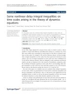
Figure 1 Characterization of B-cell subpopulations from rheumatoi d arthritis (RA ) patients and healthy cont rols (HCs). (a) Dot plot
representing the distribution of naïve B cells (R3), memory B cells (R4), and plasmablasts (R5) which was used on further analyses. No differences
in the percentages of naïve B cells (b), memory B cells (c), and plasmablasts (d) between 18 RA patients and 13 HCs were detected. P > 0.05,
two-tailed unpaired Student t test. Horizontal lines represent mean values.
Catalán et al. Arthritis Research & Therapy 2010, 12:R68
/>Page 3 of 11
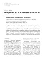
Figure 2 Increased expression of CD86 on naïve and memory B cells from rheumatoid arthritis (RA) patients. (a) Dot plots
representative of the expression of CD86 on B cells from an RA patient (left) and a healthy control (HC) (right). The number in the quadrant
represents the percentage of CD19
+
CD86
+
cells. Graphics summarizing the percentages of CD86
+
cells among naïve B cells (b), memory B cells
(c), and plasmablasts (d) from 18 RA patients and 9 HCs. *P < 0.05, two-tailed Mann-Whitney U test. Horizontal lines represent mean values.
Catalán et al. Arthritis Research & Therapy 2010, 12:R68
/>Page 4 of 11
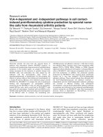
Reduced FcgRIIb expression on memory B cells and
plasmablasts from rheumatoid arthritis patients
As for CD86, the evaluation of FcgRIIb expression was
carried out by analyzing each B-cell subset individually.
We found that, although naïve B cells from R A patients
and controls expressed similar levels of this receptor, its
expression was significantly lower on RA memory B
cells and plasmablasts (P = 0.0005 and P = 0.0013,
respectively) (Figure 3). No correlations were o bserved
between Fcg RIIb expression on B cells and disease activ-
ity or percentage of CD86 -expressing B cells (data not
shown).
There is evide nce of a normal upregulation of FcgRIIb
fol lowing naïve B-cell activatio n and differentiation to a
memory cell [15]. In our sample of healthy subjects, we
detected that most individuals upregulate the expression
of FcgRIIb from naïve to memor y B cells (8/13), in con-
trast with the RA group, in which 15 out of 18 patients
were downregulators (ΔMFI of greater than 10 betw een
naïve and memory populations) (P = 0.029). This
decrease of FcgRIIb expression on memory B cells as
compared w ith naïve B cells from RA patients was sta-
tistically significant ( P = 0.0001) (Figure 4b). We also
noticed that RA patients show a further decrease in
FcgRIIb expression on plasmablasts in comparison with
memory B cells (P = 0.0001) (Figure 4b). A similar
reduction was observed in healthy controls (P = 0.0002)
(Figure 4c), a lthough the expression levels reached by
the healthy controls were still higher than those exhib-
ited by RA patients (Figure 3c).
FcgRIIb expression on B cells is associated with
autoantibody levels
As one of the main functions of Fc gRIIb on B cells is to
control the development of autoimmunity by providing
feedback inhibition in order to limit the secretion of
autoantibodies, we assessed whether the levels of auto-
antibodies on RA patients were related to the expression
of this inhibitory receptor on B cells. For this purpose,
we measured serum anti-MCV antibodies since they
have been described to be highly specific for RA [26].
Interestingly, RA patients negative for serum anti-MCV
antibodies or with low levels (less than 50 U/mL) dis-
played a higher expression of FcgRIIb but only on mem-
ory B cells (P = 0.048) (Figure 5a). A lso, we found that
all three patients who did not downregulate FcgRIIb
from naïve to memory B cells exhibited no or very low
titers of anti-MCV antibodies (P = 0.033) ( Figure 5b).
We obtained similar results when we measured anti-ccp
antibodies (data not shown). To evaluate whether this
association was restricted to autoantibodies, we com-
pared FcgRIIb expression on memory B cells in patients
with normal (less than 1,350 mg/dL) o r high (at least
1,350 mg/dL) l evels of total serum IgG without
Naïve B cells
RA HC
0
200
400
600
MFI Fc
γ
RIIb
Memory B cells
RA HC
0
200
400
600
***
MFI Fc
γ
RIIb
Plasmablasts
RA HC
0
200
400
600
**
MFI Fc
γ
RIIb
A
B
C
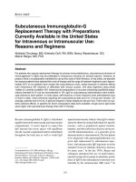
Figure 3 Decreased expression of FcgRIIb on memory B cells
and plasmablasts from rheumatoid arthritis (RA) patients.
Graphics summarize the expression of FcgRIIb on naïve B cells (a),
memory B cells (b), and plasmablasts (c) from 18 RA patients and
13 healthy controls (HCs). Expression was quantified as mean
fluorescence intensity (MFI). **P < 0.01, ***P < 0.001, two-tailed
unpaired Student t test. Horizontal lines represent mean values.
Catalán et al. Arthritis Research & Therapy 2010, 12:R68
/>Page 5 of 11
detecting significant differ ences between the two groups
(Figure 5c).
Anti-tumor necrosis factor therapy can influence B-cell
phenotype in rheumatoid arthritis patients
Next, we wanted to evaluate whether the alterations
observed on RA patients’ B cells could be reverted by
the treatment with adalimumab. Of the 13 RA patients
who completed 6 months of treatment with an anti-
TNF antibody (adalimumab), only 11 exhibited at least a
moderate response according to the EULAR response
criteria. In these patients, the percentage of total B cells
as well as the proportion of naïve, memory, or plasma-
blast subsets remained unchanged (data not shown).
However, the anti-TNF therapy caused a decrease in the
proportion of memory B cells expressing CD86 after 6
months of therapy (P = 0.032) (Figure 6a). Notably, the
change affecting CD86 paralleled a reduction in the
intensity of FcgRIIb expression, but this decrease
reached significance on the naïve B-cell subpopulation
only (P = 0.003) (Figure 6b). Consequently, there was an
attenuation of the receptor downregulation observed
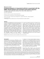
Figure 4 Altered regulation of FcgRIIb on B cells from rheumatoid arthritis (RA) patients. (a) Representative histograms of FcgRIIb (CD32b)
expression on naïve B cells (gray line), memory B cells (black line), and plasmablasts (dotted line) from an RA patient (left) and a healthy control
(right). The shaded curve represents the isotype control. Graphics show a comparison of FcgRIIb expression between naïve B cells, memory B
cells, and plasmablasts from 18 RA patients (b) and 13 healthy controls (c). Expression was quantified as mean fluorescence intensity (MFI). The
differences in the FcgRIIb expression levels between B-cell subpopulations were analyzed with the two-tailed paired Student t test; ***P < 0.001.
Horizontal lines represent mean values. PE, phycoerythrin.
Catalán et al. Arthritis Research & Therapy 2010, 12:R68
/>Page 6 of 11
before adalimumab treatment was started (Figure 4b),
but the difference b etween naïve and memory B cells
was still significant (P = 0.046) (Figure 6c). In addition,
anti-MCV antibody titers remained stable throughout
this period (Figure 6d).
Discussion
In the present work, we provide evidence that phenoty-
pic alterations on B cells from RA patients affect key
molecules involved in the regulation of antigen presen-
tation and antibody secretion functions. A major role of
B cells in the development and perpetuation of RA has
been consistently demonstrated with the appearance of
B cell-depleting therapy and its impressive results in
reducing symptoms and preventing disease progression
[27]. Studies on murine models have suggested that
antigen-specific B cells are required as APCs for the
induction of autoimmune arthritis, owing to their
expression of MHC (major histocompatibility complex)
class II and co-stimulatory molecules CD80 and CD86
[28,29]. C D86 is upregulated on activated B cells upon
B-cell receptor (BCR) and CD19/CD21 complex engage-
ment [24,25]. Our results show that RA patients have a
higher proportion of naïve and memory B cells expres-
sing CD86 than healthy controls, reflecting an expanded
activated status within these subpopulations, which is
likely to favor a more productive interaction with patho-
genic T cells. Analogous results have been reported for
other inflammatory diseases, such a s SLE [30- 33], sys-
temic sclerosis [34], a sthma [35], and irritable bowel
syndrome [36]. Presumably, constant stimulatio n of
autoantigens through BCR and the influence of other
proinflammatory signals, such as stimulation through
Toll-like receptors (TLRs) [37], give rise to this activated
phenotype. Also, it is p robable that the effect of these
activation stimuli could be attenuated by the cross-link-
ing of IgG-containing immune complexes to their inhi-
bitor receptor, FcgRIIb, since studies on dendritic cells
(DCs)havedemonstratedthatexposuretoimmune
complexes together with a blockage of FcgRIIb is suffi-
cient to induce an increase in the expression of CD86
[38,39], whereas the overexpression of this receptor on
murine B cells reverses the induction of CD86 triggered
via BCR [9]. However, in a multifactorial complex dis-
ease such as RA, it is expected that the expression of
this or other activation markers is influenced by a vari-
ety of factors as it is suggested by the absence of corre-
lation between CD86-expressing B cells and FcgRIIb
expression levels found in our patients.
Our data show that RA patients present reduced levels
of FcgRIIb on memory B cells and plasmablasts com-
pared with healthy donor s. This phenomeno n can b e
explained by the abnormal downregulation of this recep-
tor from naïve to memory B cells which we observed in
Non-downregulators Downregulators
0
300
600
900
1200
*
Anti-MCV (U/ml)
< 50 U/ml
≥
50 U/ml
0
100
200
300
400
*
Anti-MCV
MFI Fc
γ
RIIb
< 1350 mg/dl
≥
1350 mg/dl
0
100
200
300
400
Total IgG
MFI Fc
γ
RIIb
A
B
C
Figure 5 FcgRIIb expression levels on rheumatoid arthritis (RA)
patients’ memory B cells are inversely associated with anti-
modified and citrullinated vimentin (anti-MCV) titers. (a) FcgRIIb
expression on memory B cells from RA patients with high titers (at
least 50 U/mL) and from those with no or low titers (less than 50
U/mL) of serum anti-MCV antibodies. Expression was quantified as
mean fluorescence intensity (MFI). *P < 0.05, two-tailed Mann-
Whitney U test. (b) Anti-MCV antibody titers in patients who
downregulated the expression of FcgRIIb (the difference between
MFI of naïve B cells and MFI of memory B cells was greater than 10
for downregulators) and in those who upregulated or maintained it
almost invariable (the difference between MFI of naïve B cells and
MFI of memory B cells was not greater than 10 for non-
downregulators). *P < 0.05, two-tailed Mann-Whitney U test. (c)
FcgRIIb expression on memory B cells from RA patients with normal
levels (less than 1,350 mg/dL) and high levels (at least 1,350 mg/dL)
of total serum IgG. Expression was quantified as MFI. P > 0.05, two-
tailed Mann-Whitney U test. Horizontal lines represent mean values.
Catalán et al. Arthritis Research & Therapy 2010, 12:R68
/>Page 7 of 11
Memory B cells
pre anti-TNF post anti-TNF
0
5
10
15
20
25
*
% CD86
Naïve B cells
pre anti-TNF post anti-TNF
0
3
6
9
12
% CD86
Plasmablasts
pre anti-TNF post anti-TNF
0
25
50
75
100
% CD86
Plasmablasts
pre anti-TNF post anti-TNF
0
100
200
300
400
MFI Fc
γ
RIIb
Memory B cells
pre anti-TNF post anti-TNF
0
100
200
300
400
MFI Fc
γ
RIIb
Naïve B cells
pre anti-TNF post anti-TNF
0
200
400
600
**
MFI Fc
γ
RIIb
Naïve Memory Plasmablasts
0
100
200
300
400
*
***
**
MFI Fc
γ
RIIb
pre anti-TNF post anti-TNF
0
300
600
900
1200
Anti-MCV (U/ml)
A
B
C
D
Figure 6 B-cell phenotype and anti-modified and citrullinated vimentin (anti-MCV) titers on rheumatoid arthritis (RA) patients after 6
months of adalimumab therapy. After 6 months of anti-tumor necrosis factor (anti-TNF) therapy, B-cell phenotype and serum anti-MCV
antibodies from 11 RA patients, who exhibited at least a moderate response to the treatment, were reassessed. (a) Graphics summarizing the
percentages of CD86
+
cells among naïve B cells, memory B cells, and plasmablasts from RA patients before and after 6 months of anti-TNF
therapy. *P < 0.05, two-tailed Wilcoxon signed-rank test. (b) Graphics summarizing FcgRIIb expression on naïve B cells, memory B cells, and
plasmablasts from RA patients before and after 6 months of anti-TNF therapy. Expression was quantified as mean fluorescence intensity (MFI). **P
< 0.01, two-tailed paired Student t test. (c) Comparison of FcgRIIb expression between naïve B cells, memory B cells, and plasmablasts from RA
patients after anti-TNF therapy. The differences in FcgRIIb expression levels between B-cell subpopulations were analyzed with the two-tailed
paired Student t test; *P < 0.05, **P < 0.01, ***P < 0.001. (d) Comparison of serum anti-MCV antibody levels before and after 6 months of anti-
TNF therapy. P > 0.05, two-tailed paired Student t test. Horizontal lines represent mean values.
Catalán et al. Arthritis Research & Therapy 2010, 12:R68
/>Page 8 of 11
our RA group. These results are concordant with those
seen in SLE and chronic inflammatory demyelinating
polyneuropathy, other autoimmune diseases character-
ized by uncontrolled secretion of autoantibodies [15-18].
It has been postulated that FcgRIIb upregulation might
constitute a critical checkpoint in peripheral tolerance
by providing an inhibit ory feedback that limits the
ongoing humoral response to self-antigens. In fact, in
lupus-prone mice, the restoration of FcgRIIb levels on B
cells can revert the secretion of autoantibodies and renal
disease [40]. Furthermore, the expression of this inhibi-
tory receptor specifically on B cells, but not on macro-
phages, can be determinant for controlling
autoimmunity in models of arthritis and lupus [9].
Through this work, we provide new evidence that may
help to reinforce t his concept as we have revealed, f or
the first time, an association between high levels of
FcgRIIb on memory B cells and no or low titers o f spe-
cific autoantibodies, anti-MCV antibodies. This interest-
ing result appears to be exclusive for autoimmune
responses since we did not find a similar association
when analyzing total IgG le vels. It is noteworthy to con-
sider that in this study we have examined only the
expression of FcgRIIb, but not its function, which if
altered could also affect the regulatory ability over the
humoral response against citrullinated proteins. Like-
wise, a recent publication has demonstrated an associa-
tion between a functional polymorphism for FcgRIIb and
anti-ccp (+) RA in an Asian population [41].
After patients underwent 6 months of therapy with an
anti-TNF antibody, we observed a decrease in CD86-
expressing memory B cells, reflecting an attenuation of
the B-cell activated status. Paradoxically, FcgRIIb expres-
sion on the naïve B-cell subset also decreased signifi-
cantly. It has been demonstrated that FcgRIIa and
FcgRIIb on human monocytes are differentially regulated
by Th1/Th2 cytokines, with interferon-gamma favoring
the activator receptor and interleukin-4 favoring the
inhibitory receptor [42]. On the other han d, TNF down-
modulates FcgRIIb and FcgRIIa on monocytes, not
affecting FcgRIIa but reducing FcgRIIb expression on
DCs, while increasing FcgRIIa without changing FcgRIIb
on neutrophils, indicating a tight cell-specific regulation
[43-47]. To our knowledge, no studies addressing the
effect of TN F over FcgRIIb on human B cells have been
published, but our results strongly suggest t hat TNF or
other downstream cytokines may influence the expres-
sion of this receptor on B lymphocytes. In regard to RA,
exposure o f DCs to synovial fluid from RA patients has
been shown to lead to an upregulation of FcgRIIb [14]
and an elevated expressi on of FcgRIIb has been demon-
strated on RA synovial tissue, probably counteracting
the upregulation of other activating receptors [48].
Some studies have reported that FcgR e xpression levels
on leukocytes can vary with anti-rheumatic drug s, which
wouldpromoteamoreinhibitoryprofile[48-52].Itis
possible that the reduction in FcgRIIb expression that we
observed on n aïve B cells as a consequence of anti-TNF
therapy is accompanied by a decrease in the expression
of activating receptors on the se cells, like TLR9 and
CD21, which w ould determine a restoration of the pro-
tectiv e activator/inhibitor balance, but this issue needs to
be investigated. The effect of TNF blockage, however,
was not sufficient to prevent the downregulation of
FcgRIIb from naïve to memory B cells. These results are
in accordance with the fact that our group of patients did
not achieve a reduction in anti-MCV titers after 6
mont hs of therapy . Others have repor ted that changes in
these antibodies become significant after 18 months of
anti-TNF therapy [53], so it is conceivable that a full nor-
malization of B-cell phenotype may become apparent
only over longer follow-up periods.
Conclusions
Our data demonstrate the existence of important altera-
tions in the phenotype of peripheral B cells from RA
patients, involving the expression of the co-stimulatory
molecule CD86 and the inhibitory receptor FcgRIIb, the
latter being associated with hig h titers of autoantibodie s.
We consider that our study contributes relevant evi-
dence to a better comprehension of the molecular
mechanisms that are implied in the regulation of B cells
and the role that they play in the autoimmune response
elicited in RA.
Abbreviations
APC: antigen-presenting cell; BCR: B-cell receptor; ccp: cyclic citrullinated
peptide; CIA: collagen-induced arthritis; DC: dendritic cell; ELISA: enzyme-
linked immunosorbent assay; EULAR: European League Against Rheumatism;
FcgR: Receptor for the Fc region of IgG-containing immune complexes; FITC:
fluorescein isothiocyanate; MCV: modified and citrullinated vimentin; MFI:
mean fluorescence intensity; PE: phycoerythrin; RA: rheumatoid arthritis; SD:
standard deviation; SLE: systemic lupus erythematosus; TLR: Toll-like receptor;
TNF: tumor necrosis factor.
Acknowledgements
We thank Nancy Fabres and Juana Orellana for their excellent technical
assistance and Abbott Laboratories for providing the adalimumab doses.
This work was supported by Fondecyt-Chile (grant 1090174) and Millenium
Nucleus of Immunology and Immunotherapy (P07/088-F). DC is a recipient
of a Conicyt Doctoral Fellowship.
Author details
1
Programa Disciplinario de Inmunología, Instituto de Ciencias Biomédicas
(ICBM), Facultad de Medicina, Universidad de Chile, Avenida Independencia
1027, Santiago, Chile.
2
Sección de Reumatología, Departamento de Medicina,
Hospital Clínico, Universidad de Chile, Santos Dumont 999, Santiago, Chile.
3
Departamento de Genética Molecular y Microbiología, Facultad de Ciencias
Biológicas, Pontificia Universidad Católica de Chile, Av. Bernardo O’Higgins
340, Santiago, Chile.
Authors’ contributions
DC participated in the design of the study, carried out the acquisition and
analysis of data, and drafted the manuscript. OA participated in B-cell
Catalán et al. Arthritis Research & Therapy 2010, 12:R68
/>Page 9 of 11
phenotyping. FS coordinated the recruitment of patients, performed the
clinical evaluations, and helped with data analysis. PW participated in the
recruitment and clinical evaluations of patients. LS participated in the
recruitment of patients and clinical data analysis. MC participated in the
design and coordination of the study. AMK participated in the conception
and design of the study and critically revised the manuscript. JCA
participated in the conception and design of the study and in the
interpretation of data and helped to draft the manuscript. All authors read
and approved the final manuscript.
Competing interests
The authors declare that they have no competing interests.
Received: 23 January 2010 Revised: 25 March 2010
Accepted: 15 April 2010 Published: 15 April 2010
References
1. Korganow AS, Ji H, Mangialaio S, Duchatelle V, Pelanda R, Martin T,
Degott C, Kikutani H, Rajewsky K, Pasquali JL, Benoist C, Mathis D: From
systemic T cell self-reactivity to organ-specific autoimmune disease via
immunoglobulins. Immunity 1999, 10:451-461.
2. Takemura S, Klimiuk PA, Braun A, Goronzy JJ, Weyand CM: T cell activation
in rheumatoid synovium is B cell dependent. J Immunol 2001,
167:4710-4718.
3. Nimmerjahn F, Ravetch JV: Fcgamma receptors as regulators of immune
responses. Nat Rev Immunol 2008, 8:34-47.
4. Jiang Y, Hirose S, Abe M, Sanokawa-Akakura R, Ohtsuji M, Mi X, Li N, Xiu Y,
Zhang D, Shirai J, Hamano Y, Fujii H, Shirai T: Polymorphisms in IgG Fc
receptor IIB regulatory regions associated with autoimmune
susceptibility. Immunogenetics 2000, 51:429-435.
5. Bolland S, Ravetch JV: Spontaneous autoimmune disease in Fc(gamma)
RIIB-deficient mice results from strain-specific epistasis. Immunity 2000,
13:277-285.
6. Yuasa T, Kubo S, Yoshino T, Ujike A, Matsumura K, Ono M, Ravetch JV,
Takai T: Deletion of fcgamma receptor IIB renders H-2(b) mice
susceptible to collagen-induced arthritis. J Exp Med 1999, 189:187-194.
7. Kleinau S, Martinsson P, Heyman B: Induction and suppression of
collagen-induced arthritis is dependent on distinct fcgamma receptors. J
Exp Med 2000, 191:1611-1616.
8. Iruretagoyena MI, Riedel CA, Leiva ED, Gutierrez MA, Jacobelli SH,
Kalergis AM: Activating and inhibitory Fcgamma receptors can
differentially modulate T cell-mediated autoimmunity. Eur J Immunol
2008, 38:2241-2250.
9. Brownlie RJ, Lawlor KE, Niederer HA, Cutler AJ, Xiang Z, Clatworthy MR,
Floto RA, Greaves DR, Lyons PA, Smith KG: Distinct cell-specific control of
autoimmunity and infection by FcgammaRIIb. J Exp Med 2008,
205:883-895.
10. Blank MC, Stefanescu RN, Masuda E, Marti F, King PD, Redecha PB,
Wurzburger RJ, Peterson MG, Tanaka S, Pricop L: Decreased transcription
of the human FCGR2B gene mediated by the -343 G/C promoter
polymorphism and association with systemic lupus erythematosus. Hum
Genet 2005, 117:220-227.
11. Floto RA, Clatworthy MR, Heilbronn KR, Rosner DR, MacAry PA, Rankin A,
Lehner PJ, Ouwehand WH, Allen JM, Watkins NA, Smith KG: Loss of
function of a lupus-associated FcgammaRIIb polymorphism through
exclusion from lipid rafts. Nat Med 2005, 11:1056-1058.
12. Kono H, Kyogoku C, Suzuki T, Tsuchiya N, Honda H, Yamamoto K,
Tokunaga K, Honda Z: FcgammaRIIB Ile232Thr transmembrane
polymorphism associated with human systemic lupus erythematosus
decreases affinity to lipid rafts and attenuates inhibitory effects on B cell
receptor signaling. Hum Mol Genet 2005, 14:2881-2892.
13. Kyogoku C, Dijstelbloem HM, Tsuchiya N, Hatta Y, Kato H, Yamaguchi A,
Fukazawa T, Jansen MD, Hashimoto H, Winkel van de JG, Kallenberg CG,
Tokunaga K: Fcgamma receptor gene polymorphisms in Japanese
patients with systemic lupus erythematosus: contribution of FCGR2B to
genetic susceptibility. Arthritis Rheum 2002, 46:1242-1254.
14. Radstake TR, Franke B, Wenink MH, Nabbe KC, Coenen MJ, Welsing P,
Bonvini E, Koenig S, Berg van den WB, Barrera P, van Riel PL: The
functional variant of the inhibitory Fcgamma receptor IIb (CD32B) is
associated with the rate of radiologic joint damage and dendritic cell
function in rheumatoid arthritis. Arthritis Rheum 2006, 54:3828-3837.
15. Mackay M, Stanevsky A, Wang T, Aranow C, Li M, Koenig S, Ravetch JV,
Diamond B: Selective dysregulation of the FcgammaIIB receptor on
memory B cells in SLE. J Exp Med 2006, 203:2157-2164.
16. Su K, Yang H, Li X, Gibson AW, Cafardi JM, Zhou T, Edberg JC, Kimberly RP:
Expression profile of FcgammaRIIb on leukocytes and its dysregulation
in systemic lupus erythematosus. J Immunol 2007, 178:3272-3280.
17. Isaak A, Gergely P Jr, Szekeres Z, Prechl J, Poor G, Erdei A, Gergely J:
Physiological up-regulation of inhibitory receptors Fc gamma RII and
CR1 on memory B cells is lacking in SLE patients. Int Immunol 2008,
20:185-192.
18. Tackenberg B, Jelcic I, Baerenwaldt A, Oertel WH, Sommer N, Nimmerjahn F,
Lunemann JD: Impaired inhibitory Fcgamma receptor IIB expression on B
cells in chronic inflammatory demyelinating polyneuropathy. Proc Natl
Acad Sci USA 2009, 106:4788-4792.
19. Arnett FC, Edworthy SM, Bloch DA, McShane DJ, Fries JF, Cooper NS,
Healey LA, Kaplan SR, Liang MH, Luthra HS, Medsger TA, Mitchell DM,
Neustadt DH, Pinals RS, Schaller JG, Sharp JT, Wilder RL, Hunder GG: The
American Rheumatism Association 1987 revised criteria for the
classification of rheumatoid arthritis. Arthritis Rheum 1988, 31:315-324.
20. Prevoo ML, van ‘t Hof MA, Kuper HH, van Leeuwen MA, Putte van de LB,
van Riel PL: Modified disease activity scores that include twenty-eight-
joint counts. Development and validation in a prospective longitudinal
study of patients with rheumatoid arthritis. Arthritis Rheum 1995, 38:44-48.
21. van Gestel AM, Haagsma CJ, van Riel PL: Validation of rheumatoid arthritis
improvement criteria that include simplified joint counts. Arthritis Rheum
1998, 41:1845-1850.
22. Odendahl M, Jacobi A, Hansen A, Feist E, Hiepe F, Burmester GR, Lipsky PE,
Radbruch A, Dorner T: Disturbed peripheral B lymphocyte homeostasis in
systemic lupus erythematosus. J Immunol 2000, 165:5970-5979.
23. Lindenau S, Scholze S, Odendahl M, Dorner T, Radbruch A, Burmester GR,
Berek C: Aberrant activation of B cells in patients with rheumatoid
arthritis. Ann N Y Acad Sci 2003, 987:246-248.
24. Mongini PK, Tolani S, Fattah RJ, Inman JK: Antigen receptor triggered
upregulation of CD86 and CD80 in human B cells: augmenting role of
the CD21/CD19 co-stimulatory complex and IL-4. Cell Immunol 2002,
216:50-64.
25. Good KL, Avery DT, Tangye SG: Resting human memory B cells are
intrinsically programmed for enhanced survival and responsiveness to
diverse stimuli compared to naive B cells. J Immunol 2009, 182:890-901.
26. Dejaco C, Klotz W, Larcher H, Duftner C, Schirmer M, Herold M:
Diagnostic
value of antibodies against a modified citrullinated vimentin in
rheumatoid arthritis. Arthritis Res Ther 2006, 8:R119.
27. Edwards JC, Szczepanski L, Szechinski J, Filipowicz-Sosnowska A, Emery P,
Close DR, Stevens RM, Shaw T: Efficacy of B-cell-targeted therapy with
rituximab in patients with rheumatoid arthritis. N Engl J Med 2004,
350:2572-2581.
28. O’Neill SK, Shlomchik MJ, Glant TT, Cao Y, Doodes PD, Finnegan A:
Antigen-specific B cells are required as APCs and autoantibody-
producing cells for induction of severe autoimmune arthritis. J Immunol
2005, 174:3781-3788.
29. O’Neill SK, Cao Y, Hamel KM, Doodes PD, Hutas G, Finnegan A: Expression
of CD80/86 on B cells is essential for autoreactive T cell activation and
the development of arthritis. J Immunol 2007, 179:5109-5116.
30. Folzenlogen D, Hofer MF, Leung DY, Freed JH, Newell MK: Analysis of
CD80 and CD86 expression on peripheral blood B lymphocytes reveals
increased expression of CD86 in lupus patients. Clin Immunol
Immunopathol 1997, 83:199-204.
31. Bijl M, Horst G, Limburg PC, Kallenberg CG: Expression of costimulatory
molecules on peripheral blood lymphocytes of patients with systemic
lupus erythematosus. Ann Rheum Dis 2001, 60:523-526.
32. Dolff S, Wilde B, Patschan S, Durig J, Specker C, Philipp T, Kribben A,
Witzke O: Peripheral circulating activated b-cell populations are
associated with nephritis and disease activity in patients with systemic
lupus erythematosus. Scand J Immunol 2007, 66:584-590.
33. Chang NH, McKenzie T, Bonventi G, Landolt-Marticorena C, Fortin PR,
Gladman D, Urowitz M, Wither JE: Expanded population of activated
antigen-engaged cells within the naive B cell compartment of patients
with systemic lupus erythematosus. J Immunol 2008, 180:1276-1284.
34. Sato S, Fujimoto M, Hasegawa M, Takehara K: Altered blood B lymphocyte
homeostasis in systemic sclerosis: expanded naive B cells and diminished
but activated memory B cells. Arthritis Rheum 2004, 50:1918-1927.
Catalán et al. Arthritis Research & Therapy 2010, 12:R68
/>Page 10 of 11
35. Hofer MF, Jirapongsananuruk O, Trumble AE, Leung DY: Upregulation of
B7.2, but not B7.1, on B cells from patients with allergic asthma. J Allergy
Clin Immunol 1998, 101:96-102.
36. Ohman L, Lindmark AC, Isaksson S, Posserud I, Strid H, Sjövall H, Simrén M:
B-cell activation in patients with irritable bowel syndrome (IBS).
Neurogastroenterol Motil 2009, 21:644-650, e27.
37. Hanten JA, Vasilakos JP, Riter CL, Neys L, Lipson KE, Alkan SS, Birmachu W:
Comparison of human B cell activation by TLR7 and TLR9 agonists. BMC
Immunol 2008, 9:39.
38. Dhodapkar KM, Kaufman JL, Ehlers M, Banerjee DK, Bonvini E, Koenig S,
Steinman RM, Ravetch JV, Dhodapkar MV: Selective blockade of inhibitory
Fcgamma receptor enables human dendritic cell maturation with IL-
12p70 production and immunity to antibody-coated tumor cells. Proc
Natl Acad Sci USA 2005, 102:2910-2915.
39. Kalergis AM, Ravetch JV: Inducing tumor immunity through the selective
engagement of activating Fcgamma receptors on dendritic cells. J Exp
Med 2002, 195:1653-1659.
40. McGaha TL, Sorrentino B, Ravetch JV: Restoration of tolerance in lupus by
targeted inhibitory receptor expression. Science 2005, 307:590-593.
41. Chen JY, Wang CM, Ma CC, Hsu LA, Ho HH, Wu YJ, Kuo SN, Wu J: A
transmembrane polymorphism in FcgammaRIIb (FCGR2B) is associated
with the production of anti-cyclic citrullinated peptide autoantibodies in
Taiwanese RA. Genes Immun 2008, 9:680-688.
42. Pricop L, Redecha P, Teillaud JL, Frey J, Fridman WH, Sautes-Fridman C,
Salmon JE: Differential modulation of stimulatory and inhibitory Fc
gamma receptors on human monocytes by Th1 and Th2 cytokines. J
Immunol 2001, 166:531-537.
43. Liu Y, Masuda E, Blank MC, Kirou KA, Gao X, Park MS, Pricop L: Cytokine-
mediated regulation of activating and inhibitory Fc gamma receptors in
human monocytes. J Leukoc Biol 2005, 77:767-776.
44. Wijngaarden S, Winkel van de JG, Jacobs KM, Bijlsma JW, Lafeber FP, van
Roon JA: A shift in the balance of inhibitory and activating Fcgamma
receptors on monocytes toward the inhibitory Fcgamma receptor IIb is
associated with prevention of monocyte activation in rheumatoid
arthritis. Arthritis Rheum 2004, 50:3878-3887.
45. Liu Y, Gao X, Masuda E, Redecha PB, Blank MC, Pricop L: Regulated
expression of FcgammaR in human dendritic cells controls cross-
presentation of antigen-antibody complexes. J Immunol 2006,
177:8440-8447.
46. Belostocki K, Park MS, Redecha PB, Masuda E, Salmon JE, Pricop L:
FcgammaRIIa is a target for modulation by TNFalpha in human
neutrophils. Clin Immunol 2005, 117:78-86.
47. Guriec N, Daniel C, Le Ster K, Hardy E, Berthou C: Cytokine-regulated
expression and inhibitory function of FcgammaRIIB1 and -B2 receptors
in human dendritic cells. J Leukoc Biol 2006, 79:59-70.
48. Magnusson SE, Engstrom M, Jacob U, Ulfgren AK, Kleinau S: High synovial
expression of the inhibitory FcgammaRIIb in rheumatoid arthritis.
Arthritis Res Ther 2007, 9:R51.
49. Wijngaarden S, van Roon JA, Winkel van de JG, Bijlsma JW, Lafeber FP:
Down-regulation of activating Fcgamma receptors on monocytes of
patients with rheumatoid arthritis upon methotrexate treatment.
Rheumatology (Oxford) 2005, 44:729-734.
50. Wijngaarden S, Winkel van de JG, Bijlsma JW, Lafeber FP, van Roon JA:
Treatment of rheumatoid arthritis patients with anti-TNF-alpha
monoclonal antibody is accompanied by down-regulation of the
activating Fcgamma receptor I on monocytes. Clin Exp Rheumatol 2008,
26:89-95.
51. Belostocki K, Pricop L, Redecha PB, Aydin A, Leff L, Harrison MJ, Salmon JE:
Infliximab treatment shifts the balance between stimulatory and
inhibitory Fcgamma receptor type II isoforms on neutrophils in patients
with rheumatoid arthritis. Arthritis Rheum 2008, 58:384-388.
52. Torsteinsdottir I, Arvidson NG, Hallgren R, Hakansson L: Monocyte
activation in rheumatoid arthritis (RA): increased integrin, Fc gamma
and complement receptor expression and the effect of glucocorticoids.
Clin Exp Immunol 1999, 115:554-560.
53. Nicaise Roland P, Grootenboer Mignot S, Bruns A, Hurtado M, Palazzo E,
Hayem G, Dieude P, Meyer O, Chollet Martin S: Antibodies to mutated
citrullinated vimentin for diagnosing rheumatoid arthritis in anti-CCP-
negative patients and for monitoring infliximab therapy. Arthritis Res Ther
2008, 10:R142.
doi:10.1186/ar2985
Cite this article as: Catalán et al.: B cells from rheumatoid arthritis
patients show important alterations in the expression of CD86 and
FcgRIIb, which are modulated by anti-tumor necrosis factor therapy.
Arthritis Research & Therapy 2010 12:R68.
Submit your next manuscript to BioMed Central
and take full advantage of:
• Convenient online submission
• Thorough peer review
• No space constraints or color figure charges
• Immediate publication on acceptance
• Inclusion in PubMed, CAS, Scopus and Google Scholar
• Research which is freely available for redistribution
Submit your manuscript at
www.biomedcentral.com/submit
Catalán et al. Arthritis Research & Therapy 2010, 12:R68
/>Page 11 of 11