Báo cáo y học: "Inhibitory effects of ZSTK474, a novel phosphoinositide 3-kinase inhibitor, on osteoclasts and collagen-induced arthritis in mice" pot
Bạn đang xem bản rút gọn của tài liệu. Xem và tải ngay bản đầy đủ của tài liệu tại đây (1.94 MB, 11 trang )
Toyama et al. Arthritis Research & Therapy 2010, 12:R92
/>Open Access
RESEARCH ARTICLE
© 2010 Toyama et al.; licensee BioMed Central Ltd. This is an open access article distributed under the terms of the Creative Commons
Attribution License ( which permits unrestricted use, distribution, and reproduction in
any medium, provided the original work is properly cited.
Research article
Inhibitory effects of ZSTK474, a novel
phosphoinositide 3-kinase inhibitor, on osteoclasts
and collagen-induced arthritis in mice
Shoko Toyama
1
, Naoto Tamura*
1
, Kazuhiko Haruta
2
, Takeo Karakida
3
, Shigeyuki Mori
2
, Tetsuo Watanabe
2
,
Takao Yamori
4
and Yoshinari Takasaki
1
Abstract
Introduction: Targeting joint destruction induced by osteoclasts (OCs) is critical for management of patients with
rheumatoid arthritis (RA). Since phosphoinositide 3-kinase (PI3-K) plays a critical role in osteoclastogenesis and bone
resorption, we examined the effects of ZSTK474, a novel phosphoinositide 3-kinase (PI3-K)-specific inhibitor, on murine
OCs in vitro and in vivo.
Methods: The inhibitory effect of ZSTK474 on OC formation was determined and compared with other PI3-K inhibitors
by counting tartrate-resistant acid phosphatase (TRAP)-positive multinucleated cells after culturing murine bone
marrow monocytic OC precursors, and RAW264.7 cells. Activation of Akt and expression of nuclear factor of activated T
cells (NFAT) c1 in cultured RAW264.7 cells were examined. The suppressing effect of ZSTK474 on bone resorption was
assessed by the pit formation assay. The in vivo effects of ZSTK474 were studied in collagen-induced arthritis (CIA) in the
mouse. Oral daily administration of ZSTK474 was started either when more than half or when all mice developed
arthritis. Effects of ZSTK474 were evaluated using the arthritis score and histological score of the hind paws.
Results: ZSTK474 inhibited the differentiation of bone marrow OC precursors and RAW264.7 cells in a dose-dependent
manner. The inhibitory effect of ZSTK474 was much stronger than that of LY294002, the most commonly used PI3-K
inhibitor. In addition, ZSTK474 suppressed the bone resorbing activity of mature OCs. Moreover, oral daily
administration of ZSTK474, even when begun after the development of arthritis, ameliorated CIA in mice without
apparent toxicity. Histological examination of the hind paw demonstrated noticeable reduction of inflammation and of
cartilage destruction in ZSTK474-treated mice. ZSTK474 also significantly decreased OC formation adjacent to the tarsal
bone of the hind paw.
Conclusions: These findings suggest that inhibition of PI3-K with ZSTK474 may potentially suppress synovial
inflammation and bone destruction in patients with RA.
Introduction
Rheumatoid arthritis (RA) is a systemic autoimmune dis-
ease characterized by chronic inflammation of the syn-
ovium as well as by destruction of inflamed joints
through bone erosion. The management of patients with
RA consists of both reduction of inflammation and pro-
tection of the joints from structural damage [1]. Some
anti-rheumatic drugs, including biologics, are quite use-
ful but are not effective in all patients; hence, new thera-
peutic agents are required.
It has been speculated that joint destruction is directly
caused by osteoclasts (OCs) [2], which differentiate from
monocytic precursors that have infiltrated the inflamed
joints. After this infiltration, monocytic precursors con-
vert to tartrate -resistant acid phosphatase (TRAP)-posi-
tive cells and fuse with each other, eventually forming
giant multinucleated OCs. Although the growth and dif-
ferentiation of OCs mainly depend on receptor activator
of nuclear factor κB ligand (RANKL) and macrophage-
colony stimulating factor (M-CSF), proinflammatory
* Correspondence:
1
Department of Internal Medicine and Rheumatology, Juntendo University
School of Medicine, 2-1-1 Hongo, Bunkyo-ku, Tokyo, 113-8421, Japan
Full list of author information is available at the end of the article
Toyama et al. Arthritis Research & Therapy 2010, 12:R92
/>Page 2 of 11
cytokines, such as tumor necrosis factor (TNF)-α, which
are over-expressed in the inflamed joints, promote this
process [3]. After differentiation, ανβ3 integrins on differ-
entiated OCs engage with the bone extracellular matrix;
this process is followed by bone resorption [4,5]. It has
been demonstrated that this increased resorbing activity
of OCs results not only in bone erosion and further joint
destruction but also in systemic osteoporosis in patients
with RA. Therefore, suppressing OCs is a major aspect of
RA therapy [6,7].
Signal transduction via the phosphoinositide 3-kinase
(PI3-K)/Akt pathway is essential for regulating cellular
responses, such as proliferation, survival, migration,
motility and tumorigenesis, in a variety of cell types [8],
not just OCs. Class I PI3-Ks are heterodimers and are
found in four isoforms. Class IA PI3-Ks (PI3-Kα, PI3-Kβ
and PI3-Kδ) are composed of a catalytic subunit p110 (α,
β, or δ) and a regulatory subunit p85 (α or β), and acti-
vated through tyrosine kinase signaling. The class IB PI3-
K (PI3-Kγ) is a heterodimer consisting of a catalytic sub-
unit p110γ associated with one of two regulatory sub-
units, p101 and p84, and activated via seven-
transmembrane G-protein-coupled receptors (GPCRs)
[9]. Whereas the expression of PI3-Kα and PI3-Kβ is
ubiquitous, that of PI3-Kδ and PI3-Kγ is mainly restricted
to hematopoietic cells [8].
Many signal transduction molecules are involved in dif-
ferent phases of growth and development in OCs, such as
Src homology-2 (SH2)-containing inositol-5-phosphatase
(SHIP), Vav3, Gab2, extracellular signal-regulated kinase
(ERK) and p38 mitogen-activated protein kinase (MAPK)
[10-14]. In OCs, PI3-K is a major downstream effecter of
the M-CSF receptor, RANK, and αβν3 integrin. The
importance of PI3-K for differentiation, survival and
motility of OCs has been demonstrated by using the PI3-
K inhibitors wortmannin and LY294002 [15-22], and also
by studying mice deficient in the expression of the p85α
subunit of class IA PI3-K [23]. In addition, several tran-
scription factors, including NF-kB, c-fos, AP-1, PU.1, and
CREB, are involved in regulating osteoclastogenesis in its
early or late phase, and expression of NFATc1 is specific
to the RANKL induced-signaling pathway and essential
for terminal differentiation of OCs [24,25].
Wortmannin and LY294002, potent inhibitors of PI3-K
that have been extensively used for studying ex vivo PI3-
K-driven signal pathways, also inhibit other related
enzymes [9,26]. LY294002 causes severe dermal toxicity
[27], and wortmannin and its analog has shown hepatic
toxicity [28] when administered in mice. ZSTK474, a syn-
thesized s-triazine derivative that strongly inhibited the
growth of tumor cells, was subsequently identified as a
novel PI3-K-specific inhibitor [29-33]. Furthermore,
ZSTK474 is suitable for oral administration, and demon-
strated marked in vivo antitumor activity in mice grafted
with human cancer cells without showing toxicity to
major organs [29].
Since the action of ZSTK474 on OCs is unknown, we
examined the effects of ZSTK474 in an in vitro OC cul-
ture system and found strong inhibitory effects on the
differentiation and bone resorbing activity of OCs. More-
over, daily administration of ZSTK474 ameliorated colla-
gen-induced arthritis (CIA) in mice, remarkably reducing
the migration of inflammatory cells and OCs in the syn-
ovial tissue.
Materials and methods
PI3-K inhibitors
ZSTK474 and IC87114 (a PI3-Kδ-selective inhibitor)
were synthesized at Central Research Laboratories of
Zenyaku Kogyo Co. Ltd. (Tokyo Japan). LY294002 was
purchased from Sigma Chemical Co. (St Louis, MO,
USA). AS605240 (a PI3-Kδ-selective inhibitor) was pur-
chased from Calbiochem (Schwalbach, Germany). In in
vivo experiments, ZSTK474 was prepared as a solid dis-
persion [34].
Animals
Male DBA/1 mice (eight weeks old) were purchased from
Charles River Laboratories Japan (Kanagawa, Japan).
They were maintained at approximately 22°C with a 12-
hour light/dark cycle and given standard chow and tap
water ad libitum. Newborn ddY mice were obtained from
the Japan SLC, Inc. (Shizuoka, Japan). All animal experi-
ments were approved by the local ethical committees of
each institution.
Osteloclast formation
In vitro OC formation was examined as previously
described [35]. Briefly, primary osteoblasts derived from
growing calvarial cells of newborn ddY mice at three- to
four-days of age were suspended in alpha-minimum
essential medium (α-MEM, Sigma) supplemented with
10% (v/v) fetal bovine serum (FBS, Gibco BRL, Gaithers-
burg, MD, USA), 100 U/ml penicillin and 100 μg/ml
streptomycin, and plated at a density of 2 × 10
4
cells/well
in 24-well plates (Corning Incorporated, Corning, NY,
USA) overnight. Mouse bone marrow cells containing
monocytic OC precursors were removed aseptically from
the tibiae of four- to six-week old ddY male mice, and co-
cultured on adherent osteoblasts at a density of 1.0 ×
10
6
cells/well in medium containing 10
-7
M 1α,25-
(OH)
2
D
3
(Wako Pure Chemical Industries, Ltd., Osaka,
Japan) for five to six days in the presence or absence of
varying concentrations of ZSTK474 or other PI3-K inhib-
itors. Otherwise, non-adherent bone marrow cells were
cultured alone with 10 ng/ml of M-CSF (R & D Systems,
Minneapolis, MN, USA) for two days, and then adherent
cells were cultured with 100 ng/ml of soluble RANKL
Toyama et al. Arthritis Research & Therapy 2010, 12:R92
/>Page 3 of 11
(sRANKL) (R & D Systems) for three days. In some
experiments, RAW264.7 cells (American Type Culture
Collection, Manassas, VA, USA) were plated at a density
of 2.5 × 10
4
cells/well in a 24-well tissue culture plate
overnight, and sRANKL (100 ng/ml), TNF-α (50 ng/ml)
and ZSTK474 were added. The medium was changed
every two to three days. The cells were fixed with 3.7%
formalin, permeabilized with 0.1% Triton X-100, and
stained with TRAP. OC formation was determined by
counting TRAP-positive multinucleated cells having
three or more nuclei, and OCs were counted in each set
of duplicated wells.
Real time-polymerase chain reaction (PCR) for the
quantification of RANKL expression
The osteoblasts were plated at a density of 2 × 10
5
cells/
well in six-well plates, and cultured with or without
1α,25-(OH)
2
D
3
for 24 hours in the presence or absence of
ZSTK474. Total RNA was extracted using a total RNA
isolation kit (Ambion Inc., Austin. TX, USA), and 3 μg of
the total RNA was reverse transcribed using a You-prime
Fast-Strand Breads kit (Amersham Pharmacia Biotech,
Inc., Piscataway, NJ, USA). The primers used in PCR
were 5'-GACTCGACTCTGGAGAGT-3' (sense primer)
and 5'-GAGAACTTGGGATTTTGATGC-3' (antisense
primer) for RANKL and 5'-AGCCATGTACGTAGCCA-
TCC (sense primer) and 3'-CTCTCAGCTGTGGTGGT-
GAA (antisense primer) for β-actin. Real-time PCR was
performed using 1 μg of cDNA and Power SYBR Green
Master Mix (Applied Bio Systems, Foster City, CA, USA)
on an ABI PRISM 7500 Sequence Detection System
(Applied Bio Systems) with conditions at 95°C for 10 min-
utes, followed by 40 cycles at 95°C for 15 seconds and
60°C for one minute. The expression of RANKL was
quantified using the comparative C
T
, applying the for-
mula X
n
= 2
-ΔCT
, where X
n
is the relative amount of target
gene in question and ΔC
T
is the difference between the
C
T
of the house keeping gene for a given sample [36].
Western blotting for Akt and NFATc1
RAW264.7 cells were plated at a density of 2.5 × 10
5
cells/
well in a six-well tissue culture plate overnight, and
ZSTK474 was added. After incubation for 30 minutes, 50
to 100 ng/ml of sRANKL, or sRANKL plus TNF-α (50
ng/ml), was added and the cells were incubated for the
indicated time. Cells were washed twice with ice-cold
phosphate-buffered saline (PBS) containing 1% phos-
phatase inhibitor cocktail (Sigma), detached with a cell
scraper, centrifuged, and lysed with lysis buffer (1% Tri-
ton X-100, 1% phosphatase inhibitor cocktail and 1 mM
of PMSF in Tris-buffer, pH 7.6). The lysates were boiled
with sodium dodecyl sulfate (SDS) -sample buffer and
run on SDS-PAGE followed by blotting with a 1:1000
dilution of anti-phospholylated Akt (anti-phospho Akt),
anti-Akt, anti-IκB, anti-phospho cJun, anti-phospho p42/
p44, anti-β-actin (Cell Signal Technology, Inc., Beverly,
MA, USA) and anti-NFAT1c monoclonal antibody (Santa
Cruz Biotechnology, Santa Cruz, CA, USA).
Immunofluorescence microscopy
RAW264.7 cells (200 μl, 2.5 × 10
5
/ml) were plated onto
Lab Tek Chamber slide (Thermo Fisher Scientific, Roch-
ester, NY, USA) overnight. After treatment with 0.1 μM of
ZSTK474 for 30 minutes, 100 ng/ml of sRANKL and 50
mg/ml of TNF-α were added, and the cells were cultured
for 48 hours. Then, the cells were fixed with 4% para-
formaldehyde, washed with PBS three times, permeabi-
lized with 0.1% Triton X-100 in PBS, and blocked with
10% normal goat serum (Nichrei, Tokyo, Japan). The cells
were incubated with anti-NFATc1 antibody diluted in
PBS (1:50) for one hour, washed with PBS, and followed
with phycoerythrin-conjugated goat anti-rabbit IgM+IgG
(H+L chain specific, Beckman Coulter) for another one
hour. The cells were postfixed in Aqua-Poly/Mount
(Polysciences, Washington, PA, USA) and viewed using
fluorescence microscope (Nikon ECLIPSE E600/Y-FL).
Bone resorbing activity of OC
A 10 cm culture dish (Corning) was coated with 5 ml of
type I collagen mixture at 4°C. The dish was placed in a
CO
2
incubator at 37°C for 10 minutes to render the aque-
ous type I collagen gelatinous. Primary osteoblasts (5 ×
10
5
cells/dish) and bone marrow cells (6 × 10
6
cells/dish)
were co-cultured on the collagen gel-coated dish for five
days. The dish was then treated with 4 ml of 0.2% collage-
nase solution (Nitta Gelatin Co., Osaka, Japan) for 20
minutes at 37°C in a shaking water bath (60 cycles/min-
ute). The cells were collected by centrifugation at 600
rpm for three minutes, then washed and suspended with
α-MEM containing 10% FBS (OC preparation). Dentine
slices (Immunodiagnostic Systems, Ltd., Boldon, UK)
were cleaned by ultrasonication in distilled water, steril-
ized using 70% ethanol, dried under ultraviolet light, and
placed in 96-well plates. A 0.1-ml aliquot of the OC prep-
aration was transferred onto the slices. After incubation
for 72 hours in the presence or absence of the PI3-K
inhibitors, the medium was removed and 1 ml of 1 M
NH
4
OH was added to each well and incubated for 30
minutes. The dentin slices were then cleaned by ultrason-
ication, stained with hematoxylin (Wako Pure Chemical
Industries) for 35 to 45 seconds, and washed with dis-
tilled water. The area of resorption pits that formed on
dentine slices was observed under a light microscope and
measured.
Toyama et al. Arthritis Research & Therapy 2010, 12:R92
/>Page 4 of 11
CIA in mice
Male DBA/1 mice, eight-weeks of age, were injected
intradermally in the base of the tail with 200 μg of bovine
type II collagen (Collagen Gijutsu Kenshu-Kai., Tokyo,
Japan) emulsified in complete Freund's adjuvant (Difco,
Detroit, MI, USA) on Day 1, and the same amount of the
antigen emulsified in incomplete Freund's adjuvant
(Difco) on Day 22. When half of the mice had developed
arthritis (Day 28), the mice were randomly divided into
four groups of eight mice. Each group orally received
vehicle or 25, 50, 100 mg/kg of ZSTK474, once/day. In
another therapeutic protocol, 100 mg/kg of ZSTK474 was
administered from the day when all mice developed
arthritis (Day 31). Total arthritis score was defined as the
sum of the paw swelling scores for each paw (0 to 4 per
paw), with a maximum score of 16. In the semi-therapeu-
tic protocol, the mice were killed on Day 50, and the right
hind paws were removed, fixed in paraformaldehyde,
decalcified in Kalkitox (Wako Pure Chemical Industries,
Ltd.), embedded in paraffin and sectioned. The sections
were then stained with hematoxylin and eosin (H&E) or
safranin O to assess hyperplasia of synovial tissue, infil-
tration of leukocytes, and destruction of cartilage. Each
parameter was graded separately and assigned a severity
score as follows: grade 0, no detectable change: 1 to 4,
slight to severe changes. The number of OC in talus was
counted in every third 6 μm section. To examine in vivo
OC formation in CIA mice, the hind paws were removed
on Day 52 and rapidly frozen in the therapeutic protocol.
The frozen tissue was sectioned according to the method
described previously [37] and the sections were stained
with H&E or TRAP. Plasma TRACP5b levels were mea-
sured using a mouse TRAP™ Assay (Immunodiagnostic
System Ltd).
Statistical analysis
Statistical significance of differences was assessed by one-
way analysis of variance (ANOVA) followed by Dunnett's
test or the Student's t-test for comparison of two samples.
Statistical tests were performed using Kaleida graph 3.6
(Synergy Software, Reading, PA, USA). In all analyses, P <
0.05 was considered statistically significant.
Results
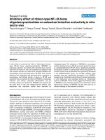
Inhibitory effects of ZSTK474 on OC formation in co-culture
system
To determine whether ZSTK474 could inhibit osteoclas-
togenesis in vitro, mouse bone marrow monocytic pre-
cursors were co-cultured with osteoblasts together with
1α,25-(OH)
2
D
3
in the presence or absence of various con-
centrations of ZSTK474 or other PI3-K inhibitors. The
effect was also examined in OC differentiation of the
bone marrow precursors in response to M-CSF and
sRANKL. OC formation was significantly inhibited by
ZSTK474 in both culture systems, and this inhibitory
effect was much stronger than that of LY294002 (Figure
1a), the most commonly used PI3-K inhibitor at present.
IC87114 also inhibited OC formation similarly to
LY294002, whereas AS605240 had virtually no effect on
the OC differentiation, indicating that PI3-Kδ might play
a more important role in OC formation in these culture
systems. ZSTK474 suppressed OC formation in a dose-
dependent manner at lower concentrations (Figure 1b
and 1c). No TRAP-positive cells were observed with 0.2
μM of ZSTK474, suggesting that differentiation of OCs
was completely suppressed at this concentration. On the
other hand, 0.04 μM of ZSTK474 were likely to allow the
monocytic precursors to differentiate into small TRAP-
positive cells, but not to form large OCs (Figure 1b). In
addition, ZSTK474, even at 1 μM, did not decrease the
expression of RANKL mRNA in osteoblasts cultured
with 1α,25-(OH)
2
D
3
(Figure 1d), indicating that RANKL
expression on osteoblasts might not be involved in sup-
pressing effect of ZSTK474 on OC differentiation.
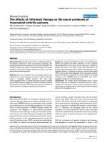
Inhibition of Akt phosphorylation and NFATc1 expression in
RAW264.7 cells by ZSTK474
To confirm that ZSTK474 affected the monocytic precur-
sors but not the osteoblasts, we examined its effect on the
phosphorylation of Akt in RAW264.7 cells. Phosphoryla-
tion of Akt induced by sRANKL (100 ng/ml) was abol-
ished by ZSTK474 (Figure 2a). However, ZSTK474 did
not inhibit the degradation of IκB and phosophorylation
of JNK and ERK1/2 induced by sRANKL. On the other
hand, the expression of NFATc1, which occurs in the late
phase of OC differentiation and promotes terminal osteo-
clastogenesis in association with a complex of cJun and
cFos [38,39], was attenuated in RAW264.7 cells treated
with sRANKL by 0.1 μM of ZSTK474, although ZSTK474
did not apparently affect the expression of cFos (Figure
2b). We further analyzed translocation of NFATc1 by
immunofluorescence microscopy. Calcium entry to OC
precursor cells activates the calcium/calmodulin-depen-
dent pathway, leading to NFATc1 translocation into the
nucleus. ZSTK474 repressed the translocation of NFATc1
to the nucleus in response to sRANKL and TNF-α (Figure
2c). These results indicated that ZSTK474 at least
blocked the RANK/RANKL-PI3-K/Akt cascade in mono-
cytic precursors, resulting in inhibition of OC differentia-
tion.
Inhibitory effects of ZSTK474 on OC formation induced by
both RANKL and TNF-α
We next examined the effects of ZSTK474 on OC forma-
tion induced by RANKL and TNF-α, since it was specu-
lated that TNF-α enhanced OC formation in RA. In fact,
RANKL-induced phosphorylation of Akt was enhanced
by the addition of TNF-α (Figure 2d). ZSTK474 (0.03, 0.1,
Toyama et al. Arthritis Research & Therapy 2010, 12:R92
/>Page 5 of 11
and 0.3 μM) inhibited the phosphorylation of Akt
induced by RANKL (100 ng/ml) and TNF-α (50 ng/ml) in
RAW264.7 cells (Figure 2d). Moreover, the OC formation
induced by RANKL (100 ng/ml) and TNF-α (50 ng/ml)
was inhibited by ZSTK474 in a dose-dependent manner.
OC formation was completely inhibited by ZSTK474 (0.3
μM, Figure 2e).
Inhibition of bone resorbing activity of OC by ZSTK474
We next examined whether ZSTK474 also inhibited the
bone-resorbing activity of mature OCs. The OCs that had
matured on the collagen-gel were transferred onto den-
tine slices, the total areas of the resorbed pits were mea-
sured after three days culture. This experiment revealed
that 0.1 μM of ZSTK474 completely prevented pit forma-
tion by OCs (Figure 3a, b). LY294002 and IC87114, but
not AS605240, also inhibited the bone resorption more
weakly (Figure 3b). Because PI3-K is important for OC
survival [19], it was supposed that PI3-K inhibited the
survival of mature OCs and consequently suppressed the
bone resorption. Therefore, we tested whether ZSTK474
affected the survival of mature OCs. Complete and par-
tial inhibition of OC survival was observed in the pres-
ence of 1 μM and 0.1 μM of ZSTK474, respectively
(Figure 3c).
Figure 1 Inhibitory effect of ZSTK474 on OC formation. Mouse bone marrow cells containing OC precursors were cultured with osteoblasts in the
presence of 1α,25-(OH)
2
D
3
for five days. Indicated concentrations of ZSTK474, LY294002, AS605240 (a PI3-Kγ-selective inhibitor), or IC87114 (a PI3-Kδ-
selective inhibitor) were added at the initiation of cultures. Mouse bone marrow cells were also cultured with M-CSF (10 ng/ml) for two days and then
with M-CSF and sRANKL (100 ng/ml) for another three days. The inhibitors were added simultaneously with sRANKL. a) and c) TRAP-positive multinu-
cleated cells were counted as OC. The columns and bars indicate the mean and standard deviation (S.D.) from duplicated wells. b) OC formation in
co-culture with osteoblast (upper) and culture with M-CSF and sRANKL (lower). Representative results were shown in a, b, and c. d) RANKL mRNA was
measured using real-time PCR, with results normalized the value of β-actin. The columns and bars indicate the mean and S.D. of three independent
experiments. * = P > 0.05, ** = P < 0.01, *** = P < 0.001.
Toyama et al. Arthritis Research & Therapy 2010, 12:R92
/>Page 6 of 11
Amelioration of CIA in mice with oral administration of
ZSTK474
To determine whether interference with PI3-K activity by
ZSTK474 reduces joint destruction in vivo, we examined
the effects of ZSTK474 on CIA in mice. ZSTK474 was
administered from the day when more than 50% of the
mice developed arthritis (Day 28). While vehicle-treated
mice developed active arthritis, administration of daily
oral ZSTK474 ameliorated joint inflammation in a dose-
dependent manner. The arthritis score reached 7.5 ± 0.9
by Day 50 in the vehicle-treated group, whereas oral
administration of ZSTK474 reduced the arthritis score to
4.1 ± 1.2 (25 mg/kg, P < 0.05), 1.3 ± 0.6 (50 mg/kg, P <
0.001), and 0.5 ± 0.5 (100 mg/kg, P < 0.001, Figure 4a).
Histological staining of the affected synovial tissues dem-
onstrated that administration of ZSTK474 (50 mg/kg)
markedly attenuated infiltration of inflammatory cells,
proliferation of synovial fibroblasts and cartilage/bone
destruction (Figure 4b, Table 1). Especially, the number of
OCs in talus decreased significantly in ZSTK474 (50 mg/
kg)-treated group (Table 1). Furthermore, a remarkable
reduction was observed in the arthritis score even in the
therapeutic protocol in which ZSTK474 administration
was begun (100 mg/kg) after development of arthritis. At
Day 52, there were highly significant differences between
the vehicle-treated group and the ZSTK474 (100 mg/kg)-
treated group (mean arthritis score: 6.8 ± 1.0 versus 2.4 ±
0.5, Figure 4c). TRAP-staining of the joint section con-
firmed numerous OCs adjacent to the tarsal bones of
vehicle-treated mice, whereas TRAP-positive OC forma-
tion in ZSTK474-treated mice was markedly decreased
(Figure 5a). In addition, plasma levels of TRACP5b, a bio-
marker of systemic bone resorption, raised significantly
in vehicle-treated, 25 mg/kg, and 50 mg/kg ZSTK474-
treated mice, compared to intact mice. In contrast, the
Figure 2 Suppressive effect of ZSTK474 on OC precursor cells. a) Inhibition of Akt phosphorylation by ZSTK474. RAW264.7 cells were incubated
with or without 0.1 μM of ZSTK474 for 30 minutes and for another 15 minutes in the presence of soluble RANKL. The phosphorylated Alt (p-Akt) and
whole Akt (Akt) were examined by the Western blotting. b) The expression of NFATc1 was determined in RAW264.7 cells cultured for 48 hours in the
presence of soluble RANKL with or without ZSTK474 pretreatment. c) NFATc1 localization was visualized using immunofluorescence microscopy in
RAW264.7 cells cultured with sRANKL and TNF-α for 24 hours. Treated with vehicle (left) and 0.1 μM of ZSTK474 (right). d) The phosphorylation of Akt
in RAW264.7 cells cultured with soluble RANKL and TNF-α was inhibited by ZSTK474. e) RAW264.7 cells were cultured in the presence of RANKL and
TNF-α in the presence or absence of ZSTK474. The number of TRAP staining-positive multinucleated cells was counted.
Toyama et al. Arthritis Research & Therapy 2010, 12:R92
/>Page 7 of 11
TRACP5b levels were sustained in 100 mg/kg ZSTK474-
treated mice (Figure 5b).
Discussion
In this study, we demonstrated that ZSTK474, a novel
PI3-K-specific inhibitor, suppressed osteoclastogenesis
and bone resorption. The in vitro inhibitory effect of
ZSTK474 on OC formation, observed by culturing bone
marrow cells, was much stronger than that of LY294002.
Although both inhibit all isoforms of class I PI3-K, the
inhibitory activities of ZSTK474 (IC
50
: PI3-Kα: 1.6 × 10
-8
M; PI3-Kβ: 4.4 × 10
-8
M; PI3-Kγ: 4.9 × 10
-8
M; PI3-Kδ: 4.6
× 10
-9
M) were much stronger than those of LY294002
(IC
50
: PI3-Kα: 5.5 × 10
-7
M; PI3-Kβ: 1.1 × 10
-5
M; PI3-Kγ:
1.2 × 10
-5
M; PI3-Kδ: 1.6 × 10
-6
M) on all isoforms, espe-
cially PI3-Kδ [30]. A PI3-Kδ-selective inhibitor, IC87114
(1 μM), completely inhibited OC formation, while a PI3-
Kγ-selective inhibitor, AS605240, had no inhibitory effect
Figure 3 Blocking bone resorbing activity of OCs with ZSTK474. Mouse bone marrow derived monocytic OC precursors were co-cultured with
osteoblasts in the presence of 1α,25-(OH)
2
D
3
on a collagen gel-coated dish. The matured OCs were collected and transferred onto dentin slices and
incubated for 72 hours in the presence or absence of ZSTK474 and other PI3-K inhibitors. The dentin slices were stained with hematoxylin, and the
pits formed in the resorbed area on the slices were observed (a) and measured (b) under a light microscope. (c) Matured OCs, differentiated from
bone marrow cells as described above, were further cultured with 5 ng/ml of TNF-α and the PI3-K inhibitors. After 24 hours, TRAP-positive multinu-
cleated cells were counted, and the percentages of surviving cells were calculated. The columns and bars indicate the mean and S.D. from duplicated
wells in a) and c).
Table 1: Histological score and osteoclast number
Synovium Leukocyte Cartilage/bone Osteoclast
Vehicle (n = 6) 2.3 ± 0.8 1.7 ± 0.9 2.7 ± 0.6 62.0 ± 38.6
ZSTK474
(n = 6, 50 mg/kg)
0.0 ± 0.0 0.0 ± 0.0 0.8 ± 0.3 0.3 ± 0.2
P-value 0.009 0.073 0.036 0.024
Toyama et al. Arthritis Research & Therapy 2010, 12:R92
/>Page 8 of 11
on OC formation. These results indicate the involvement
of PI3-Kδ in the OC culture system, consistent with a
previous report which implicated a critical role of class
IA PI3-K in OC formation by demonstrating that OC
progenitor cells from mice lacking p85α, a regulatory
subunit of class IA PI3-K, showed impaired growth and
differentiation [23].
Blocking of the phosphorylation of Akt by ZSTK474 in
RAW264.7 cells indicated that the inhibitory effect on
OC formation observed in the bone marrow monocytic
cells was due at least in part to suppression of PI3-K/Akt
signal pathway in the OC precursors. This suggestion is
supported by the observation that the consequent expres-
sion of NFATc1, an essential factor for terminal RANKL-
induced differentiation of OCs [25,38], was also pre-
vented by ZSTK474. The reduced expression of NFATc1
was dependent on neither NFkB nor cFos in the condi-
tion of this study. Additionally, translocation of NFATc1
into the nucleus was also inhibited by ZSTK474, implying
that ZSTK474 might suppress the autoamplification, cal-
cium-signal-mediated persistent activation [40], of
NFATc1. Moreover, ZSTK474 inhibited the phosphoryla-
tion of Akt and OC differentiation induced by both
RANKL and TNF-α, which are fundamental factors for
OC formation in RA, implying that ZSTK474 might
inhibit OC formation in patients with RA.
ZSTK474 also suppressed the bone resorbing activity of
OCs as assessed in an in vitro pit formation assay. This
could be explained by the inhibitory effect of ZSTK474
on survival of mature OCs in part. Likewise, signaling via
PI3-K is crucial for remodeling and assembly of actin fila-
ments, cell spreading and adhesion [41]. Furthermore,
blocking PI3-K with ZSTK474 inhibited the membrane
ruffling induced by platelet-derived growth factor
Figure 4 Oral administration of ZSTK474 ameliorated CIA in mice. In vitro effect of ZSTK474 was examined in mice CIA. On Day 28, when half of
the mice had developed arthritis, oral administration of ZSTK474 was commenced once a day. a) Arthritis scores were compared among the groups.
b) Synovial tissues from the hindpaws of vehicle-treated CIA mice, 50 mg ZSTK474-treated CIA mice and normal age-matched DBA mice were stained
with hematoxylin and eosin (H&E) or with safranin O. Representative results are shown. c) In the therapeutic protocol, 100 mg/kg of ZSTK474 was start-
ed on Day 31, when all mice had developed arthritis.
Toyama et al. Arthritis Research & Therapy 2010, 12:R92
/>Page 9 of 11
(PDGF) in murine embryonic fibroblasts [29]. In OCs,
the SH3 domain of tyrosine kinase, c-src, interacts with
the p85 regulatory domain of PI3-K, and this signaling
pathway is crucial for colony-stimulating factor-1-
induced OC spreading [22]. Therefore, ZSTK474 might
suppress the cytoskeletal change of OCs, resulting in the
reduced bone resorption observed in this study.
ZSTK474 suppressed inflammation and also protected
against joint destruction in CIA in mice. Although it is
difficult to ascertain the direct effect of ZSTK474 on OCs
in this model, the TRAP-staining of the synovial tissue
sections demonstrated marked reduction of OC forma-
tion. In addition, plasma levels of TRACP5b, that
reported to correspond with systemic but not localized
bone resorption [42], were not increased in 100 mg/kg
ZSTK474-treated mice. This result implied that 100 mg/
kg of ZSTK474 possibly prevented the systemic bone
resorption.
Both the semi-therapeutic and therapeutic treatments
of ZSTK474 ameliorated joint inflammation in a mouse
model of RA. This anti-rheumatic effect might be
explained by contribution of PI3-K to activation, prolifer-
ation and migration of inflammatory cells, such as lym-
phocytes, macrophages, neutrophils, mast cells and
synovial fibroblasts [9]. However, the titers of antibody to
type II collagen were not significantly different between
vehicle- and ZSTK474-treated mice in this experiment
(data not shown). Regarding migration, chemokine
receptors, such as the MCP-1 receptor and the RANTES
receptor, are GPCRs that associate with PI3-Kγ and
induce signals for chemotaxis of the inflammatory cells
[9]. It was reported that the PI3-Kγ-selective inhibitor
suppressed joint inflammation in mouse CIA by inhibit-
ing migration of neutrophils to the joints [43]. This inhib-
itory process might occur in the ZSTK474-treated mice.
Additionally, synovial pannus tissues of patients with RA
express phosphorylated Akt [44] and exhibit tumor-like
behaviors, such as angiogenesis, proliferation and inva-
sion. A recent report demonstrated potent antiangiogenic
activity for ZSTK474, which could be attributed to both
inhibition of VEGF secretion by cancer cells and inhibi-
tion of PI3-K in endothelial cells [45]. These findings also
account for the effects of ZSTK474 on CIA mice. In addi-
tion, ZSTK474 did not affect the count of peripheral
white blood cells and red blood cells (data not shown).
Further studies are underway to evaluate how ZSTK474
exerts anti-inflammatory activity in vivo.
Clinical studies have demonstrated that the degree of
inflammation and the progression of joint destruction do
not always correspond with each other [46,47]. In current
therapy for RA, anti-rheumatic drugs are required not
only to control the inflammation but also to suppress the
joint destruction. On the other hand, recent reports have
shown convincing pathogenic evidence for the involve-
ment of class I PI3-K and Akt signaling pathways in syn-
ovial fibroblasts [44,48-52] and other cells [43,53,54] in
patients with RA. Synovial tissue from patients with RA
expressed higher levels of phosphorylated Akt than that
Figure 5 Administration of ZSTK474 inhibited in vivo OC formation and bone resorption in CIA mice. a) The synovial sections described above
were stained with H&E and also with TRAP to examine in vivo OC formation. Representative results are shown. b) Plasma levels of TRACP5b were mea-
sured. The levels of TRACP5b in vehicle- 25 mg/kg, and 50 mg/kg ZSTK474-treated mice, but not 100 mg/kg ZSTK474-treated mice, were significantly
raised in comparison with that of intact mice (*P < 0.05, **P < 0.01).
Toyama et al. Arthritis Research & Therapy 2010, 12:R92
/>Page 10 of 11
from patients with osteoarthritis [44]. Moreover, block-
ing the PI3-K/Akt pathway by intracellular gene transfer
of phosphatate and tensin homolog deleted on chromo-
some 10 (PTEN), which dephosphorylates phosphati-
dylinositol - 3,4,5 - tris - phosphate (Ptdlns(3,4,5)P
3
) and
attenuates the downstream signals of PI3-K, CIA in rats
[52]. Taken together, the present results indicate that PI3-
K could be a potent target for RA therapy.
Conclusions
We have demonstrated inhibitory effects of ZSTK474 on
in vitro OC formations and CIA in mice. Inhibition of
PI3-K with ZSTK474 may potentially have an anti-rheu-
matic effect in patients with RA.
Abbreviations
CIA: collagen-induced arthritis; ERK: extracellular signal-regulated kinase; FBS:
fetal bovine serum; GCPRs: G-protein-coupled receptors; MAPK: mitogen-acti-
vated protein kinase; M-CSF: macrophage-colony stimulating factor; NFATc1:
nuclear factor of activated T cells c1; OCs: osteoclasts; PDGF: platelet-derived
growth factor; PI3-K: phosphoinositide 3-kinase; PTEN: phosphatate and tensin
homolog deleted chromosome 10; RA: rheumatoid arthritis; RANK: receptor
activator of nuclear factor κB; RANKL: RANK ligand; SHIP: Src homology-2 (SH2)-
containing inositol-5-phosphatase; TNF: tumor necrosis factor; TRAP: tartrate-
resistant acid phosphatase; α-MEM: alpha-minimum essential medium
Competing interests
KH, SM and TW were employed by Zenyaku Kogyo Co., Ltd (Tokyo, Japan),
which is the proprietary company of ZSTK474. TY has a research fund from
Zenyaku Kogyo Co., Ltd. ST, NT, TK and YT declare that they have no competing
interests.
Authors' contributions
ST performed data acquisition and was involved in drafting of the manuscript.
NT contributed to the study design and did most of the drafting of the manu-
script. KH designed the in vivo and part of the in vitro experiments, and carried
out the analysis and interpretation of data; he was also involved in drafting of
the manuscript. TK participated in the in vitro experiments and gave helpful
advice. SM contributed essentially to the animal experiments. TW provided the
synthesized PI3-K inhibitors used in this study. TY fundamentally participated
in the concept of the study using ZSTK474. YT supervised conception and
design of the study. All authors read and approved the final manuscript.
Acknowledgements
We thank Asako Sasaki (Central Research Laboratory, Zenyaku Kogyo Co., Ltd,
Tokyo, Japan) and Naoki Ishihara (Department of Internal Medicine and Rheu-
matology, Juntendo University School of Medicine, Tokyo, Japan) for technical
support. We also owe thanks to Shin-ichi Yaguchi (Zenyaku Kogyo Co., Ltd)
who gave insightful comments and Makoto Fukae and Shinichiro Oida
(Department of Biochemistry, School of Dental Medicine, Tsurumi University,
Yokohama, Japan) who supported the set up of the in vitro experiments.
Author Details
1
Department of Internal Medicine and Rheumatology, Juntendo University
School of Medicine, 2-1-1 Hongo, Bunkyo-ku, Tokyo, 113-8421, Japan,
2
Central
Research Laboratory, Zenyaku Kogyo Co., Ltd, 2-33-7 Oizumimachi, Nerima-ku,
Tokyo, 178-0062, Japan,
3
Department of Biochemistry, School of Dental
Medicine, Tsurumi University, 2-1-3 Tsurumi, Tsurumi-ku, Yokohama,
Kanagawa, 230-8501, Japan and
4
Division of Molecular Pharmacology, Cancer
Chemotherapy Center, Japanese Foundation for Cancer Research, 3-10-6
Ariake, Koto-ku, Tokyo, 135-8550, Japan
References
1. Combe B, Landewe R, Lukas C, Bolosiu HD, Breedveld F, Dougados M,
Emery P, Ferraccioli G, Hazes JM, Klareskog L, Machold K, Martin-Mola E,
Nielsen H, Silman A, Smolen J, Yazici H: EULAR recommendations for the
management of early arthritis: report of a task force of the European
Standing Committee for International Clinical Studies Including
Therapeutics (ESCISIT). Ann Rheum Dis 2007, 66:34-45.
2. Redlich K, Hayer S, Ricci R, David J, Tohidast-Akrad M, Kollias G, Steiner G,
Smolen J, Wagner E, Schett G: Osteoclasts are essential for TNF-alpha-
mediated joint destruction. J Clin Invest 2002, 110:1419-1427.
3. Teitelbaum S: Osteoclasts; culprits in inflammatory osteolysis. Arthritis
Res Ther 2006, 8:201.
4. Gravallese E, Manning C, Tsay A, Naito A, Pan C, Amento E, Goldring S:
Synovial tissue in rheumatoid arthritis is a source of osteoclast
differentiation factor. Arthritis Rheum 2000, 43:250-258.
5. Shigeyama Y, Pap T, Kunzler P, Simmen B, Gay R, Gay S: Expression of
osteoclast differentiation factor in rheumatoid arthritis. Arthritis Rheum
2000, 43:2523-2530.
6. Schett G: Osteoimmunology in rheumatic diseases. Arthritis Res Ther
2009, 11:210.
7. Schett G, Stach C, Zwerina J, Voll R, Manger B: How antirheumatic drugs
protect joints from damage in rheumatoid arthritis. Arthritis Rheum
2008, 58:2936-2948.
8. Rückle T, Schwarz M, Rommel C: PI3Kgamma inhibition: towards an
'aspirin of the 21st century'? Nat Rev Drug Discov 2006, 5:903-918.
9. Rommel C, Camps M, Ji H: PI3K delta and PI3K gamma: partners in crime
in inflammation in rheumatoid arthritis and beyond? Nat Rev Immunol
2007, 7:191-201.
10. Faccio R, Teitelbaum S, Fujikawa K, Chappel J, Zallone A, Tybulewicz V,
Ross F, Swat W: Vav3 regulates osteoclast function and bone mass. Nat
Med 2005, 11:284-290.
11. Faccio R, Zou W, Colaianni G, Teitelbaum S, Ross F: High dose M-CSF
partially rescues the Dap12-/- osteoclast phenotype. J Cell Biochem
2003, 90:871-883.
12. Wada T, Nakashima T, Oliveira-dos-Santos A, Gasser J, Hara H, Schett G,
Penninger J: The molecular scaffold Gab2 is a crucial component of
RANK signaling and osteoclastogenesis. Nat Med 2005, 11:394-399.
13. Takeshita S, Namba N, Zhao J, Jiang Y, Genant H, Silva M, Brodt M,
Helgason C, Kalesnikoff J, Rauh M, Humphries R, Krystal G, Teitelbaum S,
Ross F: SHIP-deficient mice are severely osteoporotic due to increased
numbers of hyper-resorptive osteoclasts. Nat Med 2002, 8:943-949.
14. Lee S, Woo K, Kim S, Kim H, Kwack K, Lee Z, Kim H: The
phosphatidylinositol 3-kinase, p38, and extracellular signal-regulated
kinase pathways are involved in osteoclast differentiation. Bone 2002,
30:71-77.
15. Nakamura I, Takahashi N, Sasaki T, Tanaka S, Udagawa N, Murakami H,
Kimura K, Kabuyama Y, Kurokawa T, Suda T: Wortmannin, a specific
inhibitor of phosphatidylinositol-3 kinase, blocks osteoclastic bone
resorption. FEBS Lett 1995, 361:79-84.
16. Nakagawa N, Kinosaki M, Yamaguchi K, Shima N, Yasuda H, Yano K,
Morinaga T, Higashio K: RANK is the essential signaling receptor for
osteoclast differentiation factor in osteoclastogenesis. Biochem Biophys
Res Commun 1998, 253:395-400.
17. Pilkington M, Sims S, Dixon S: Wortmannin inhibits spreading and
chemotaxis of rat osteoclasts in vitro. J Bone Miner Res 1998, 13:688-694.
18. Palacio S, Felix R: The role of phosphoinositide 3-kinase in spreading
osteoclasts induced by colony-stimulating factor-1. Eur J Endocrinol
2001, 144:431-440.
19. Lee S, Chung W, Kwak H, Chung C, Kwack K, Lee Z, Kim H: Tumor necrosis
factor-alpha supports the survival of osteoclasts through the
activation of Akt and ERK. J Biol Chem 2001, 276:49343-49349.
20. Gingery A, Bradley E, Shaw A, Oursler M: Phosphatidylinositol 3-kinase
coordinately activates the MEK/ERK and AKT/NFkappaB pathways to
maintain osteoclast survival. J Cell Biochem 2003, 89:165-179.
21. Bradley E, Ruan M, Vrable A, Oursler M: Pathway crosstalk between Ras/
Raf and PI3K in promotion of M-CSF-induced MEK/ERK-mediated
osteoclast survival. J Cell Biochem 2008, 104:1439-1451.
22. Grey A, Chen Y, Paliwal I, Carlberg K, Insogna K: Evidence for a functional
association between phosphatidylinositol 3-kinase and c-src in the
spreading response of osteoclasts to colony-stimulating factor-1.
Endocrinology 2000, 141:2129-2138.
Received: 30 November 2009 Revised: 1 May 2010
Accepted: 18 May 2010 Published: 18 May 2010
This article is available from: 2010 Toyama et al.; licensee BioMed Central L td. This is an open access article distributed under the terms of the Creative Commons Attribution License ( which permits unrestricted use, distribution, and reproduction in any medium, provided the original work is properly cited.Arthritis R esearch & Therapy 2010, 12:R92
Toyama et al. Arthritis Research & Therapy 2010, 12:R92
/>Page 11 of 11
23. Munugalavadla V, Vemula S, Sims E, Krishnan S, Chen S, Yan J, Li H, Niziolek
P, Takemoto C, Robling A, Yang F, Kapur R: The p85alpha subunit of class
IA phosphatidylinositol 3-kinase regulates the expression of multiple
genes involved in osteoclast maturation and migration. Mol Cell Biol
2008, 28:7182-7198.
24. Asagiri M, Takayanagi H: The molecular understanding of osteoclast
differentiation. Bone 2007, 40:251-264.
25. Takayanagi H, Kim S, Koga T, Nishina H, Isshiki M, Yoshida H, Saiura A, Isobe
M, Yokochi T, Inoue J, Wagner E, Mak T, Kodama T, Taniguchi T: Induction
and activation of the transcription factor NFATc1 (NFAT2) integrate
RANKL signaling in terminal differentiation of osteoclasts. Dev Cell
2002, 3:889-901.
26. Kong D, Dan S, Yamazaki K, Yamori T: Inhibition profiles of
phosphatidylinositol 3-kinase inhibitors against PI3K superfamily and
human cancer cell line panel JFCR39. Eur J Cancer 2010, 46:1111-1121.
27. Hu L, Zaloudek C, Mills G, Gray J, Jaffe R: In vivo and in vitro ovarian
carcinoma growth inhibition by a phosphatidylinositol 3-kinase
inhibitor (LY294002). Clin Cancer Res 2000, 6:880-886.
28. Ihle N, Williams R, Chow S, Chew W, Berggren M, Paine-Murrieta G, Minion
D, Halter R, Wipf P, Abraham R, Kirkpatrick L, Powis G: Molecular
pharmacology and antitumor activity of PX-866, a novel inhibitor of
phosphoinositide-3-kinase signaling. Mol Cancer Ther 2004, 3:763-772.
29. Yaguchi S, Fukui Y, Koshimizu I, Yoshimi H, Matsuno T, Gouda H, Hirono S,
Yamazaki K, Yamori T: Antitumor activity of ZSTK474, a new
phosphatidylinositol 3-kinase inhibitor. J Natl Cancer Inst 2006,
98:545-556.
30. Kong D, Yamori T: ZSTK474 is an ATP-competitive inhibitor of class I
phosphatidylinositol 3 kinase isoforms. Cancer Sci 2007, 98:1638-1642.
31. Kong D, Yamori T: Phosphatidylinositol 3-kinase inhibitors: promising
drug candidates for cancer therapy. Cancer Sci 2008, 99:1734-1740.
32. Kong D, Yaguchi S, Yamori T: Effect of ZSTK474, a novel
phosphatidylinositol 3-kinase inhibitor, on DNA-dependent protein
kinase. Biol Pharm Bull 2009, 32:297-300.
33. Kong D, Yamori T: Advances in development of phosphatidylinositol 3-
kinase inhibitors. Curr Med Chem 2009, 16:2839-2854.
34. Sugama T, Ishihara N, Tanaka Y, Takahashi M, Yaguchi S, Watanabe T:
Amorphous form of heterocyclic compound, solid dispersion and
medicinal preparation each comprising the same, and process for
production of the same. Patents. WO2009066775 2009.
35. Suda T, Jimi E, Nakamura I, Takahashi N: Role of 1 alpha,25-
dihydroxyvitamin D3 in osteoclast differentiation and function.
Methods Enzymol 1997, 282:223-235.
36. Tsubaki M, Kato C, Manno M, Ogaki M, Satou T, Itoh T, Kusunoki T,
Tanimori Y, Fujiwara K, Matsuoka H, Nishida S: Macrophage inflammatory
protein-1alpha (MIP-1alpha) enhances a receptor activator of nuclear
factor kappaB ligand (RANKL) expression in mouse bone marrow
stromal cells and osteoblasts through MAPK and PI3K/Akt pathways.
Mol Cell Biochem 2007, 304:53-60.
37. Kawamoto T: Use of a new adhesive film for the preparation of multi-
purpose fresh-frozen sections from hard tissues, whole-animals,
insects and plants. Arch Histol Cytol 2003, 66:123-143.
38. Ikeda F, Nishimura R, Matsubara T, Tanaka S, Inoue J, Reddy S, Hata K,
Yamashita K, Hiraga T, Watanabe T, Kukita T, Yoshioka K, Rao A, Yoneda T:
Critical roles of c-Jun signaling in regulation of NFAT family and RANKL-
regulated osteoclast differentiation. J Clin Invest 2004, 114:475-484.
39. Matsuo K, Galson D, Zhao C, Peng L, Laplace C, Wang K, Bachler M, Amano
H, Aburatani H, Ishikawa H, Wagner E: Nuclear factor of activated T-cells
(NFAT) rescues osteoclastogenesis in precursors lacking c-Fos. J Biol
Chem 2004, 279:26475-26480.
40. Takayanagi H: Osteoimmunology: shared mechanisms and crosstalk
between the immune and bone systems. Nat Rev Immunol 2007,
7:292-304.
41. Rivard N: Phosphatidylinositol 3-kinase: a key regulator in adherens
junction formation and function. Front Biosci 2009, 14:510-522.
42. Stolina M, Adamu S, Ominsky M, Dwyer D, Asuncion F, Geng Z, Middleton
S, Brown H, Pretorius J, Schett G, Bolon B, Feige U, Zack D, Kostenuik P:
RANKL is a marker and mediator of local and systemic bone loss in two
rat models of inflammatory arthritis. J Bone Miner Res 2005,
20:1756-1765.
43. Camps M, Rückle T, Ji H, Ardissone V, Rintelen F, Shaw J, Ferrandi C,
Chabert C, Gillieron C, Françon B, Martin T, Gretener D, Perrin D, Leroy D,
Vitte P, Hirsch E, Wymann M, Cirillo R, Schwarz M, Rommel C: Blockade of
PI3Kgamma suppresses joint inflammation and damage in mouse
models of rheumatoid arthritis. Nat Med 2005, 11:936-943.
44. Zhang H, Wang Y, Xie J, Liang X, Liu D, Yang P, Hsu H, Ray R, Mountz J:
Regulation of tumor necrosis factor alpha-mediated apoptosis of
rheumatoid arthritis synovial fibroblasts by the protein kinase Akt.
Arthritis Rheum 2001, 44:1555-1567.
45. Kong D, Okamura M, Yoshimi H, Yamori T: Antiangiogenic effect of
ZSTK474, a novel phosphatidylinositol 3-kinase inhibitor. Eur J Cancer
2009, 45:857-865.
46. Smolen J, van der Heijde D, St Clair E, Emery P, Bathon J, Keystone E, Maini
R, Kalden J, Schiff M, Baker D, Han C, Han J, Bala M: Predictors of joint
damage in patients with early rheumatoid arthritis treated with high-
dose methotrexate with or without concomitant infliximab: results
from the ASPIRE trial. Arthritis Rheum 2006, 54:702-710.
47. Smolen J, Han C, van der Heijde D, Emery P, Bathon J, Keystone E, Maini R,
Kalden J, Aletaha D, Baker D, Han J, Bala M, St Clair E: Radiographic
changes in rheumatoid arthritis patients attaining different disease
activity states with methotrexate monotherapy and infliximab plus
methotrexate: the impacts of remission and tumour necrosis factor
blockade. Ann Rheum Dis 2009, 68:823-827.
48. Kim G, Jun J, Elkon K: Necessary role of phosphatidylinositol 3-kinase in
transforming growth factor beta-mediated activation of Akt in normal
and rheumatoid arthritis synovial fibroblasts. Arthritis Rheum 2002,
46:1504-1511.
49. Chen Q, Casali B, Pattacini L, Boiardi L, Salvarani C: Tumor necrosis factor-
alpha protects synovial cells from nitric oxide induced apoptosis
through phosphoinositide 3-kinase Akt signal transduction. J
Rheumatol 2006, 33:1061-1068.
50. Morel J, Audo R, Hahne M, Combe B: Tumor necrosis factor-related
apoptosis-inducing ligand (TRAIL) induces rheumatoid arthritis
synovial fibroblast proliferation through mitogen-activated protein
kinases and phosphatidylinositol 3-kinase/Akt. J Biol Chem 2005,
280:15709-15718.
51. Traister R, Fabre S, Wang Z, Xiao X, Hirsch R: Inflammatory cytokine
regulation of transgene expression in human fibroblast-like
synoviocytes infected with adeno-associated virus. Arthritis Rheum
2006, 54:2119-2126.
52. Wang C, Shiau A, Chen S, Lin L, Tai M, Shieh G, Lin P, Yo Y, Lee C, Kuo S, Liu
M, Jou I, Yang C, Shen P, Lee H, Wu C: Amelioration of collagen-induced
arthritis in rats by adenovirus-mediated PTEN gene transfer. Arthritis
Rheum 2008, 58:1650-1656.
53. Liu H, Huang Q, Shi B, Eksarko P, Temkin V, Pope R: Regulation of Mcl-1
expression in rheumatoid arthritis synovial macrophages. Arthritis
Rheum 2006, 54:3174-3181.
54. Kim K, Cho M, Park M, Yoon C, Park S, Lee S, Kim H: Increased interleukin-
17 production via a phosphoinositide 3-kinase/Akt and nuclear factor
kappaB-dependent pathway in patients with rheumatoid arthritis.
Arthritis Res Ther 2005, 7:R139-148.
doi: 10.1186/ar3019
Cite this article as: Toyama et al., Inhibitory effects of ZSTK474, a novel phos-
phoinositide 3-kinase inhibitor, on osteoclasts and collagen-induced arthritis
in mice Arthritis Research & Therapy 2010, 12:R92