Báo cáo y học: " JNK activation is responsible for mucus overproduction in smoke inhalation injury" potx
Bạn đang xem bản rút gọn của tài liệu. Xem và tải ngay bản đầy đủ của tài liệu tại đây (586.89 KB, 8 trang )
RESEARC H Open Access
JNK activation is responsible for mucus
overproduction in smoke inhalation injury
Won-II Choi
1,2,3
, Olga Syrkina
2,3
, Kun Young Kwon
4
, Deborah A Quinn
2
, Charles A Hales
2,3*
Abstract
Background: Increased mucus secretion is one of the important characteristics of the response to smoke
inhalation injuries. We hypothesized that gel-forming mucins may contribute to the increased mucus production in
a smoke inhalation injury. We investigated the role of c-Jun N-terminal kinase (JNK) in modulating smoke-induced
mucus secretion.
Methods: We intubated mice and exposed them to smoke from burning cotton for 15 min. Their lungs were then
isolated 4 and 24 h after inhalation injury. Three groups of mice were subjected to the smoke inhalation injury:
(1) wild-type (WT) mice, (2) mice lacking JNK1 (JNK1-/- mice), and (3) WT mice administered a JNK inhibitor. The
JNK inhibitor (SP-600125) was injected into the mice 1 h after injury.
Results: Smoke exposure caused an increase in the production of mucus in the airway epithelium of the mice
along with an increase in MUC5AC gene and protein expression, while the expression of MUC5B was not increased
compared with cont rol. We found increased MUC5AC protein expression in the airway epithelium of the WT mice
groups both 4 and 24 h after smoke inhalation injury. However, overproduction of mucus and increased MUC5AC
protein expression induced by smoke inhalation was suppressed in the JNK inhibitor-treated mice and the JNK1
knockout mice. Smoke exposure did not alter the expression of MUC1 and MUC4 proteins in all 3 groups
compared with cont rol.
Conclusion: An increase in epithelial MUC5AC protein expression is associated with the overproduction of mucus
in smoke inhalation injury, and that its expression is related on JNK1 signaling.
Introduction
Smoke inhalation injury is a serious threat to victims of
house fires, explos ions, and other disasters involving fire
and smoke. This type of injury alone can be lethal as
shown in the Cocoanut Grove fire, in which 492 people
died, most without burns [1]. In the Rhode Island night-
clubfire,95peopledied(outof350victimsandsurvi-
vors of this tragedy), and 187 people were treated for
smoke inhalation lung injury and bu rns [2]. Autopsy
series from fire victims s how sloughed mucosal cells
and a collection of proteinaceous debris obstructing the
airways [3]. There are multiple case reports in adults
and children of airway obstruction due to these tracheo-
bronchial casts [3]. The airway microenvironment is sig-
nificantly altered by smoke inhalation with lung
parenchymal damage occurring because of surfactant
denaturation, loss of endothelial and epithelial barrier
functions, and influx of inflammatory cells [4-7]. Pre-
viously we demonstrated smoke-induced mucus over-
production in a small animal model [8].
In the healthy lung, MUC1 and MUC4 are expressed
on the apical surface of the respiratory epithelium.
MUC5AC and MUC2 are expressed in the goblet ce lls
of the superficial airway epithelium, whereas MUC5B is
expressed in the mucosal cells of the submucosal glands
[9]. Among them, MUC5AC is considered to be the pre-
dominant mucin in airway mucus [10]. Although mu cus
overproduction is one of the characteristics of the
response to smoke inhalation airway injury, there is only
limited information available on the regulation of mucus
secretion in such injuries.
c-Jun N-terminal kinase (JNK) activation is required
for the in vitro transcriptional up- regulation of
MUC5AC in response to tobacco smoke [11]. However,
* Correspondence:
2
Pulmonary/Critical Care Unit, Department of Medicine, Massachusettes
General Hospital and Harvard Medical School, Boston, MA, USA
Full list of author information is available at the end of the article
Choi et al. Respiratory Research 2010, 11:172
/>© 2010 Choi et al; lic ensee BioMed Central Ltd. This is an Open Access article distributed under the terms of the Creative Commons
Attribution License ( which permits unrestricted use, distribution, and re production in
any medium, provided the original work is properly cited.
the in vivo activation of JNK in the case of smoke inha-
lation has not yet been studied. In the present study, we
used our previously established small-animal model of
smoke inhalation injury [7] to determine whether t he
mucin genes wer e regulated by cotton smoke inhalation,
and to test the hypothesis that smoke inhalation induces
airway mucus overproduction through activation of the
JNK pathway and that treatment with a JNK inhibitor
could diminish airway mucus overproduction.
Materials and methods
Animal Preparation
This stu dy was approved by the Massachusetts General
Hospital Subcommittee on Research Animal Care and
conducted in compliance with guidelines of United
States Department of Agriculture Animal Welfare Act,
PublicHealthServicePolicyonHumaneCareandUse
of Laboratory Animals.
Materials
The JNK inhibitor II (SP-600125) was purchased from
Calbiochem (San Diego, CA). The dose was chosen on the
basis of previous i n vivo studies that showed 30 mg/kg
inhibited JNK activity [12,13]. The mice were treated with
SP600125 in dimethyl sulfoxide (Sigma Chemical, St.
Louis, MO) or an equivalent amount of dimethyl sulfoxide
without inhibitors 1 h after injury.
Experimental animals
We used a modification of the established rodent model
of smoke inhalation injury model as described pre-
viously [8]. Male C57BL/6, either wild-type JNK+/+ or
JNK1-/- that have been backcrossed for five generations
on a C57BL/6 background, weighing between 20 and
25 g were obtained from Jackson Laboratories (Bar Har-
bor, ME). The constructs pJNK1-/- was transfected into
W9.5 embryonic stem (ES) cells. Chimeras were gener-
ated by injecting these ES cells into C57BL/6 (B6) blas-
tocysts. Heterozygotes (+/-) were intercrossed to
generate homozygous mutant mice (-/-) [14].
Animals were orally intubated with a polyethylene
catheter under general anesthesia with intraperitoneal
ketamine (50 mg/kg) and diazepam (5 mg/kg) while
spontaneously breathing room air and then placed in
the smoke chamber for 15 min. Following 15 min of
smoke inhalation, animals were allowed to recover. Ani-
mals were extubated 10 min after smoke. Intubation
lasted for 30 min. One hour after smoke exposure, some
animals received an injection of JNK inhibitor or
Dimethyl sulfoxide (DMSO) as a vehicle subcutaneously.
Experimental design
Wild-type JNK1 -/- mice and the wild-type mice i njected
with the JNK inhibitor were assigned to one of 3 groups:
onewasthecontrolgroup;miceinthesecondgroup
were subjected to cotton smoke inhalation for 15 min fol-
lowed by a 4-h recovery period; and mice in the third
group were subje cted to cotton smoke inhalation for
15 min followed by a 24-h recovery period. A JNK inhibi-
tordoseof30mg/kgwasselectedonthebasisofpre-
vious in vivo studies that showed that th is do se inhibi ts
JNK activity [ 8,15,1 6]. Four and tw enty-fo ur hours afte r
exposure, the animals were anesthetized and killed by
exsanguination. The mice in the control group were
killed 4 h after extubation, and their lungs were removed
en bloc. The control group mice were further divided
into 3 groups: wil d-type, WC; JNK1-/-, JKOC; a nd wild-
type administered the JNK inhibitor, JIC. In addition, the
mice subjected to 15 min of smoke inhalation followed
by a 4-h recovery period were divided into 3 groups:
wild-type, WS4; JNK1-/-, JKOC4 ; and wild type adminis-
tered the JNK inhibitor, JIS4. The thir d group of mice
subjected to 15 min of smoke inhalation followed b y a
24-h recovery period were also divided into 3 groups:
wild-type, WS24; JNK-1-/-, JKOC24;andwildtype
administered the JNK inhibitor, JIS24. Each group was
assigned 7 mice, and a total of 63 mice were studied.
Western blot analysis
For determination of MUC1, MUC4, MUC5AC, and
JNK protein expression, Western blot analysis was per-
formed with MUC1 (Abcam, Cambridge, UK), MUC 4
(Invitrogen, Carlsbad, California), MUC 5AC antibody
and JNK antibodies (Santa Cruz Biot echnology, Santa
Cruz, CA, and Cell Signaling Technology, Beverly, MA).
Blots were developed by enha nced chemiluminescence
(NEN Life Science Products, Boston, MA).
Assessment of mucus
Paraffin-embedded samples were sectioned at 5 μmand
stained with Alcian blue (AB)atpH2.5andperiodic
acid-Schiff (PAS) for the localization of acidic and neu-
tral mucin distribution in the airway epithelium of con-
trol mice ( anesthetized and intubated for 30 min while
spontaneously breathing room air witho ut smoke expo-
sure) and in mice with smoke injury (anesthet ized, intu-
bated, and exposed to smoke for 15 min). Both wild
type and JNK-1 -/- mice were allowed to recover from
smoke inhalation and they were killed 4 h or 24 h after
exposure. Intubation lasted 30 min in both groups. For
quantitat ive analysis of the airway mucous secretion, all
histological slides of the left lung were randomly sorted
and masked b efore observation. The quantity of mucin
production in the airway was assessed by measuring the
percentage of PAS-positive ce lls in the air way epithe-
lium. The numbers of PAS-positive cells were counted
on longitudinal lung sections of the proximal to distal
airways. Each section had 4 randomly selected regions
Choi et al. Respiratory Research 2010, 11:172
/>Page 2 of 8
evaluated, two segments of the proximal airway and two
segments from the distal airway. A minimum of 100
sequential airway epithelial cells were coun ted from
each region and the total number of PAS positive cells
per total epithelial cells was determined for each region.
These regio nal values were then averaged to give a final
PAS score per a nimal. For quantitation of airway
obstruction, each slide was systematically scanned using
× 4 objective magnification, and for each cross-sectioned
airway, a score of 0-100% was made as an estimate of
the degree of luminal obstruction for each cross-sec-
tioned airway present. A mean obstruction score was
determined for each animal and then for each group [6].
Pathology scoring
The pathol ogical changes were compared using a modi-
fication of a previously described scoring system for
pathological changes after smoke inhalation [ 8]. Briefly,
we examined four fields (2 periphera l and 2 central) for
five injurious variables on each slide. Injurious variables
included 1) airway epithelial shedding, 2) airway epithe-
lial edema, 3) increased cellularity in the airway and par-
enchymal tissues, 4) increased peribronchial and
perivascular cuff area, and 5) alveolar atelectasis. The
total lung injury score was calculated as the sum of each
variable (0 for none or normal to 3 for severe).
Lung immunohistochemistry
The paraffin sections were cut to 5 μminthickness,
mounted on silane-coated glass slides, and stored for 1
h at 60°C. The slides were deparaffinized with xylene,
three times, 5 min each, and were rehydrated with
graded alcohols (100, 95, 70 and 50%) for 5 min, respec-
tively. After washing with 0.01 M phosphate buffered
saline (PBS) for 5 min, the sections were digested with
Proteinase K (20 μg/ml)atroomtemperaturefor
20 min, and were washed twice with distilled water for
2 min each. The endogenous peroxidase activity was
blocked with 3% hydrogen peroxide (H2O2) in PBS for
5 min; the slides were rinsed twice with PBS for 5 min.
Sections for positive control were treated with 3%
H2O2, then washed twice with PBS. For negative con-
trols, sections were covered with reaction buffer alone
and incubated following same conditions. The sections
were incubated 1.5 h with monoclonal antibody against
MUC5AC (Santa Cruz Biotechnology, Santa Cruz, CA)
at a concentration of 10 μg/ml. The sections were then
incubated with biotinylated goat anti-mouse Ig antibody
as the secondary antibody, a nd the antibody reactions
were vi sualized by using diaminobenzidine as chroma-
gen (DAKO, Ca rpinteria, CA). For microscopic observa-
tion, the sections were counterstained lightly wit h
hematoxylin for one min. The quantity of MUC5AC
protein production in the airway was assessed by
measuring the percentage of MUC5AC positive cells in
the airway epithelium. The method for evaluating the
numbers of MUC5AC positive cells was same as PAS
positive cell counting.
Quantitative real-time PCR
Total RNA was isolated by the phenol and guanidine iso-
thiocyanate method using Trizol® (Invitrogen, Carlsbad,
CA). Genomic DNA was removed from the extr acted
total RNA using the RNeasy kit (Quiagen, Austin, TX).
cDNA was made with equal amounts of mRNA (2 μg),
using the Superscript III reverse transcrip tase (Invitro-
gen, Carlsbad, CA), as per manufacture r’sinstructions.
The primer sequence for mucin genes were as follows:
MUC5AC,5’ -ACTGTTACTATGCGATGTGTAGCCA-
3’ (sense) and 5’ -GAGGAAACACATTGCACCGA-3’
(antisense) (GenBank accession no. NM_010844);
MUC5B,5’-GAACGCCATATTCCCGACACT-3’ (sense)
and 5’-GCCCCAGGTGGAGGGACATAA-3’ (antisense)
(GenBank accession no. NM_028801); MUC2,5’ -
ACGATGCCTACACCAAGGTC-3’ (sense) and 5’ -
CCATGTTATTGGGGCATTTC-3’ (antisense) (Gen-
Bank accession no. NM_023566); MUC6,5’ -C ACACA
ACCAACACCAATTC-3’ (sense) and 5’-TGAGAAAGG-
TAGGAAGTAGAGG-3’ (antisense) (GenBank accession
no. NM_181729); GAPDH,5’-CAACTACATGGTCTA-
CATGTTC-3’ (sense) and 5’ -CGCCAGTAGACTCCAC-
GAC-3’ (antisense) (GenBank accession no. NC_000072).
Quantitative real-time reverse transcription polymerase
chain reaction (qRT-PCR) was performed on the samples
by using Applied Biosystems Assays-On-Demand pri-
mer/probe sets and TaqMan Universal PCR Mix (PE
Applied Biosystems, Foster City, CA). The samples were
analyzed on the Stratagene MX3000P sequence detection
system under the following conditions: 94°C for 3 min,
45 cycles at 94°C for 30 s, 50°C. The fold change was
deter mined as described in the Applied Biosystems man-
ufacturer’s instructions (4371095 Rev A, PE Applied Bio-
systems, Foster City, CA). Briefly, the average crossing
threshold (CT) of the target genes for each group minus
the average housekeeping gene (GAPDH) CT was used
to determine the relative expression (ΔCT). Th e average
ΔCT of the experimental animals (smoke inhalation) was
subtracted from the average control (intubation only)
ΔCT to determine the ΔΔCT. The ΔΔCT was then used
in the formula 2
ΔΔCT
to de termine the f old change in
mRNA expression. The upper and lower limits of fold
change were determined by taking the averaged standard
deviat ions of each experimental group through the above
calculations [17,18].
Immunofluorescence
Paraffin-embedded lung tissue sa mples were de-waxed in
xylene twice for 5 min each time, rehydrated in an ethanol
Choi et al. Respiratory Research 2010, 11:172
/>Page 3 of 8
series (100-70%) for 3 min each followed by rehydration in
phosphate-buffered saline (PBS) for 30 min. The rack is
transferred into 200 ml of pre-warmed (94°C-96°C) Dako
(DAKO, Carpinteria, CA) target retrieval solution. Follow-
ing antigen retrieval, the sections were washed three times
with PBS, blocked in 4% skimmed milk for 1 hr, and then
stained using the kit mentioned below according to the
manufacturer’s recommendations but with the following
mod ifications. Sections were incubated with the pr imary
antibody pJNK (1 : 400, Cell Signaling Technolo gy, Bev-
erly, MA) at 4°C overnight and secondary antibody,
Alexa488-cojugated goat anti-mouse IgG
1
(1:2000. Invitro-
gen, Carlsbad, CA) for 60 minutes prior to viewing with a
Nikon Eclipse E600 microscope using an NCF Fluor 40
objective lens. Visualization of th e nuclei was by 4’,6-dia-
midine-2’-phenylindole, dihydrochloride (DAPI) staining.
Statistical analysis
Analyses were performed using SPSS (Version 13.0 soft-
ware). For comparison between groups, analy sis of var-
iance(ANOVA) followed by multiple comparisons by
Scheffé’s test with Bonferroni post hoc analysis. Signifi-
cance was set at P < 0.05. All values were expressed as
means ± SE.
Results
Pathologic score and airway obstruction
Fifteen minutes smoke inhalation caused an i ncrease in
pathologic score in wild type mice either 4 h or 24 h
recovery compared with control. The pathological
scores 4 h and 24 h after smoke inhalation was signifi-
cantly decreased by use of the JNK inhibitor or
JNK -/ Although the score was decreased with 24 h
after recovery compared with 4 h in wild type mice,
the results did not reach to statistical significant
(Table 1). Mucous plugging was assessed periodic acid-
Schiff (PAS) staining. The average percentage of airway
obstruction with mucous plugging was decreased in
JNK inhibitor treatment and JNK -/- m ice. Although
three was a trend to less obstruction in JNK -/- mice
than JNK inhibitor, the results did not reach to statisti-
cal significant (Table 1).
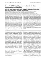
Smoke-induced mucus production in the airway of
mice through JNK activation
Since smoke inhalation duringfiresisassociatedwith
mucus hypersec retion, we evaluated mucin secretion i n
theairwayofmicebyusingthePASstain.ThePAS
stain is mainly used for stai ning str uctures containing a
high pro portion of carbohydrate macromolecules (glyco-
gen, glycoprotein, and proteoglycans), typically found in
mucus. Four and twenty-four hours after smok e inhala-
tion, the wild-type mice clearly showed increased PAS
stained cells in their airways (Figure 1). We observed
minim um or no PAS staining in the mice in the co ntrol
group, JNK1 KO group, and JNK inh ibitor group. Semi-
quantitative scale val ues for the percentage of PAS-posi-
tive cells were significantly increased in the WS4 and
WS24 mice compared with the WC, JIC, and JKOC
mice (Table 1).
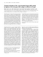
Mucin gene and protein expression
MUC1 and MUC4 are important membrane-bound
mucins. These mucins generate the sol layer of mucus.
In the present smoke inhalation mouse model, we
observed no difference in MUC1 a nd MUC4 protein
expression between mice in the control and smoke inha-
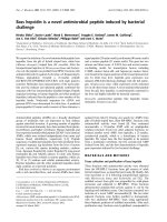
lation groups (Figure 2). Gel-forming mucin genes such
as MUC2, MUC5AC, MUC5B, and MUC6 were evalu-
ated by quantitative PCR. Only MUC5AC gene expres-
sion, which was also evaluated by immunoblotting
(Figure 3) and immunohistochemistry (Figure 4), was
found to be increased in the wild-type mice subjected to
smoke inhalation. Semi-quantitative scale values for the
percentage of MUC5AC-positive cells were significantly
increased in the WS4 and WS24 mice compared with
the WC, JIC, and JKOC mice (Table 1).
Smoke-induced activation of JNK
Immunoblotti ng data suggested that p JNK was activated
in the mice 4 and 24 h after smoke exposure (Figure 5).
Immunofluorescence imagingfurthercontributedto
these results by showing that smoke induced the phos-
phorylation of J NK, especial ly in the small airway
epithelium. Smoke-induced phosphorylation of JNK
Table 1 Pathologic score, airway obstruction, PAS and MUC 5AC positive cells in the airway epithelium
Intubation only Smoke 15 min and 4 h recovery Smoke 15 min and 24 h recovery
Group Wild type
(WC)
JNK inhibitor
(JIC)
JNK -/-
(JKOC)
Wild type
(WS4)
JNK inhibitor
(JIS4)
JNK -/-
(JKOS4)
Wild type
(WS24)
JNK inhibitor
(JIS24)
JNK -/-
(JKOS24)
Pathologic score 0.5 ± 0.1 0.4 ± 0.1 0.4 ± 0.2 7.8 ± 1.6* 2.1 ± 0.4 1.1 ± 0.3 6.4 ± 1.2* 1.8 ± 0.4 0.9 ± 0.2
Airway obstruction (%) 9.4 ± 2.1 8.1 ± 1.5 9.1 ± 1.3 36.8 ± 9.1* 15.1 ± 3.4 12.1 ± 4.3 28.4 ± 5.7* 12.6 ± 4.4 11.0 ± 3.7
PAS positive cells (%) 0.4 ± 0.2 0.3 ± 0.2 0.3 ± 0.1 25.8 ± 7.8* 3.1 ± 1.4 1.9 ± 1.2 18.8 ± 3.7* 2.4 ± 1.6 1.1 ± 0.4
MUC5AC positive cells (%) 0.3 ± 0.1 0.3 ± 0.1 0.2 ± 0.1 23.0 ± 7.3* 2.8 ± 1.6 2.2 ± 0.9 17.8 ± 3.1* 3.4 ± 1.3 1.7 ± 0.9
Values are means ± SE.
* P < 0.05 versus control, JNK inhibitor, and JNK -/
Choi et al. Respiratory Research 2010, 11:172
/>Page 4 of 8
suggested that this kinase might participate in the
inductionofMUC5ACgeneexpressioninthelung
cells. To investigate this possibility, we manipulated JNK
activity and assessed the effect s of this treatment on the
responsiveness of MUC5AC to smoke. JNK -/- or mice
injected with the J NK inhibitor SP600125 attenuated
both MUC5AC protein expression and JNK activity
(Figure 5).
Figure 1 Representative images of the airway wall stained with peri odic acid-Schiff to quantify the mucin-containing goblet cells.
Histologic sections were accessed at 4 and 24 h after smoke inhalation injury. (Magnification, 400×). There was an increase in the amount of
PAS-stained cells (purple-magenta color) in the small airway epithelium in the wild type mice. However, there was only minimal or no PAS
staining in the mice of the control group, JNK -/- group, or JNK inhibitor treated group. A, WC; B, WS4; C, WS24; D, JKOC; E, JKOS4; F, JKOS24; G,
JIC; H, JIS4; I, JIS24.
Figure 2 Immunoblot of the airway and lung tissues of the
mice subjected to smoke inhalation. No difference in MUC1 (A)
and MUC4 (B) (membrane-bound mucins) protein expression was
observed among the 3 groups.
Figure 3 MUC5AC RNA and protein expression. MUC 5AC mRNA
expression (A) was significantly increased in the smoke inhalation
mice groups compared to the control groups. * P < 0.01 versus
Control. Mucin protein, 170 kDa MUC5AC, expression was increased
at 4 (WS4) and 24 h (WS24) after smoke inhalation injury compared
with control (WC) in wild type mice (B).
Choi et al. Respiratory Research 2010, 11:172
/>Page 5 of 8
Discussion
Airway mucus production is observed in burn trauma
victims [19] and also in a combined burn and smoke
inhalation injury model [6] , but the mechanism by
which smoke damages the airway still remains unclear.
In our mouse model of smoke inhalation injury, we
found that smoke inhalation induced the mucus over-
production was associated with an increase in epithelial
MUC5AC protein expression, and this was dependent
on the activation of the JNK pathway.
Four and twenty-four hours after exposure to smoke
from burning cotton, we observed that MUC5AC mRNA
levels were elevated in the mouse lungs, and MUC5AC
protein was expressed predominantly in the surface cells
of the mouse airway. This elevated expression was abro-
gated by JNK1 mutation and the JNK inhibitor, indicating
the dependence of MUC5 AC expression on JNK activity.
JNK activation was prominent in the airway epithelial cells
(Figure 5). Although the JNK inhibitor was introduced 1 h
after smoke inhalation injury, we still observed a decrease
in mucus production. These results suggested that the
JNK pathway can be a potential target for regulating
mucus overproduction in smoke inhalation injury.
In the present study, MUC5AC protein expression was
increased within 4 hour after 15 min smoke inhalation.
The expression was sustained after 24 hour recovery.
Similar to the present study, MUC5AC can be induced
within 24 hour of inflammatory or bacterial stimulation.
Intratracheal instillation of IL-13 elicited huge amount
of induction of MUC5AC mRNA within 24 hour in
wild-type mouse lung [ 20]. Up-regulation of MUC5AC
mucin transcr iption was induced by 7 hour of S trepto-
coccus pneumoniae incubation [21]. Twelve hour of
human neutrophil peptide-1 or lipopolysaccharide incu-
bation caused an increase in MUC5AC mRNA levels
[22]. However, MUC5AC can be up-regulated different
time course in relation to different stimulatio n. In mur-
ine asthma model, airway MUC5AC gene was over-
expressed after 24 hour sensitization of ovalbumin [23].
In the present mouse model of smoke inhalation,
MUC5AC was the predominant gel-forming muci n gene
that was expressed. We observed no differences in
Figure 4 Immunohistochemistry of the MUC 5AC protein. Wild-type smoke inhalation mice showed increased MUC5AC protein expression in
their airway epithelium 4 and 24 h after injury, whereas the JNK inhibitor and JNK -/- mice groups did not (MUC5AC protein staining: A-G, 100×;
H- and I, 400×). A, WC; B, WS4; C, WS24; D, JKOS4; E, JKOS24; F, JIS4; G, JIS24; H, WS4 400×; I, WS24 400×.
Choi et al. Respiratory Research 2010, 11:172
/>Page 6 of 8
MUC5B, MUC2, or MUC6 mRNA expression between
mice in the control and the smoke injury groups (data not
shown). The membrane-associated mucins, MUC1 and
MUC4, were found to be highly expressed in both the
control and smoke inhalation group mice. MUC5AC gene
expression was found to be increased 4 h after smoke
exposure, and it remaine d elevated throughout the 24-h
recovery period. This suggested that in the case of smoke
inhalation exposure, even for short periods of time, mucus
overproduction may persist for more than 24 h after initial
exposure. Hence, we conc luded that MUC5AC c an be a
potential target for reducing mucus overproduction after
smoke inhalation injuries.
Conclusions
In this study, we showed that MUC5AC protein over-
expression in response to cotton smoke inhalation is
tightly regulated via the JNK signaling pathways.
These findings suggested that smoke inhalation
can cause the overall up-regulation of MUC5AC
production by JNK activation in the bronchial muco-
sal cells. These findings can contribute to the devel-
opment of new therapeutic strategies to treat smoke
inhalation injuries.
Abbreviations
JNK: c-Jun N-terminal kinase; DMSO: Dimethyl sulfoxide; WT: wild-type; AB:
Alcian blue; PAS: periodic acid-Schiff; QRT-PCR: Quantitative real-time reverse
transcription polymerase chain reaction; CT: crossing threshold; GAPDH:
glyceraldehyde-3-phosphate dehydrogenase; PBS: phosphate buffered saline;
DAPI: 4’,6-diamidine-2’-phenylindole, dihydrochloride; ANOVA: Analysis of
variance.
Acknowledgements
This study was supported by funds from Shriners Hospital, Boston #8620 and
Susannah Wood fou ndation (CAH).
Author details
1
Department of Internal Medicine, Dongsan Hospital, Keimyung University
School of Medicine, Daegu, Korea.
2
Pulmonary/Critical Care Unit, Department
of Medicine, Massachusettes General Hospital and Harvard Medical School,
Boston, MA, USA.
3
Shriners Burn Hospital, Boston, MA, USA.
4
Department of
Pathology, Dongsan Hospital, Keimyung University School of Medicine,
Daegu, Korea.
Figure 5 Smoke-induced JNK activation. Western blotting performed with a n antibody that recognizes the phosphorylated form of JNK. Mice
were exposed to cotton smoke for 15 min, which was followed by a recovery period of 4 and 24 h. JNK inhibitor (SP-600125) treated and JNK
-/- mice did not show pJNK protein expression after smoke inhalation. Immunofluorescence (IF) showing JNK activation (D-F) in response to
smoke. Green, phosphorylated JNK; blue, nuclei (4’-6-diamidino-2-phenylindole (DAPI). (Magnification, 400×). Wild-type control group mice did
not show expression of pJNK. pJNK activation was observed predominantly in the small airway epithelium of the mice subjected to smoke
inhalation at 4 and 24 h after recovery. A, WC DAPI; B, WS4 DAPI; C, WS24 DAPI; D, WC JNK IF; E, WS4 JNK IF; F, WS24 JNK IF.
Choi et al. Respiratory Research 2010, 11:172
/>Page 7 of 8
Authors’ contributions
WIC was responsible for carrying out the experiments, for data analysis, and
for drafted this manuscript; KYK was responsible for the analysis and design
for the histologic study; OS oversaw the animal experiments, instructed WIC
in his implementation; DAQ and CAH are experts in sepsis experiment and
assisted in the experimental design and the data analysis and interpretation.
All authors contributed to the drafting and revisions of the manuscript.
Competing interests
The authors declare that they have no competing interests.
Received: 11 August 2010 Accepted: 7 December 2010
Published: 7 December 2010
References
1. Saffle JR: The 1942 fire at Boston’s Cocoanut Grove nightclub. Am J Surg
1993, 166(6):581-591.
2. Dacey MJ: Tragedy and response- the Rhode Island nightclub fire. N Engl
J Med 2003, 349(21):1990-1992.
3. Nakae H, Tanaka H, Inaba H: Failure to clear casts and secretions
following inhalation injury can be dangerous: report of a case. Burns
2001, 27(2):189-191.
4. Fukuda T, Kim DK, Chin MR, Hales CA, Bonventre JV: Increased group IV
cytosolic phospholipase A2 activity in lungs of sheep after smoke
inhalation injury. Am J Physiol 1999, 277(3 Pt 1):L533-542.
5. Hales CA, Elsasser TH, Ocampo P, Efimova O: TNF-alpha in smoke
inhalation lung injury. J Appl Physiol 1997, 82(5):1433-1437.
6. Cox RA, Burke AS, Soejima K, Murakami K, Katahira J, Traber LD,
Herndon DN, Schmalstieg FC, Traber DL, Hawkins HK: Airway obstruction
in sheep with burn and smoke inhalation injuries. Am J Respir Cell Mol
Biol 2003, 29(3 Pt 1):295-302.
7. Quinn DA, Moufarrej R, Volokhov A, Syrkina O, Hales CA: Combined smoke
inhalation and scald burn in the rat. J Burn Care Rehabil 2003,
24(4):208-216.
8. Syrkina OL, Quinn DA, Jung W, Ouyang B, Hales CA: Inhibition of JNK
activation prolongs survival after smoke inhalation from fires. Am J
Physiol Lung Cell Mol Physiol 2007, 292(4):L984-991.
9. Voynow JA, Gendler SJ, Rose MC: Regulation of mucin genes in chronic
inflammatory airway diseases. Am J Respir Cell Mol Biol 2006,
34(6):661-665.
10. Rose MC, Voynow JA: Respiratory tract mucin genes and mucin
glycoproteins in health and disease. Physiol Rev 2006, 86(1):245-278.
11. Gensch E, Gallup M, Sucher A, Li D, Gebremichael A, Lemjabbar H,
Mengistab A, Dasari V, Hotchkiss J, Harkema J, et al: Tobacco smoke
control of mucin production in lung cells requires oxygen radicals AP-1
and JNK. J Biol Chem 2004, 279(37):39085-39093.
12. Han Z, Boyle DL, Chang L, Bennett B, Karin M, Yang L, Manning AM,
Firestein GS: c-Jun N-terminal kinase is required for metalloproteinase
expression and joint destruction in inflammatory arthritis. J Clin Invest
2001, 108(1):73-81.
13. Minutoli L, Altavilla D, Marini H, Passaniti M, Bitto A, Seminara P, Venuti FS,
Famulari C, Macri A, Versaci A, et al: Protective effects of SP600125 a new
inhibitor of c-jun N-terminal kinase (JNK) and extracellular-regulated
kinase (ERK1/2) in an experimental model of cerulein-induced
pancreatitis. Life Sci 2004,
75(24):2853-2866.
14. Dong C, Yang DD, Wysk M, Whitmarsh AJ, Davis RJ, Flavell RA: Defective T
cell differentiation in the absence of Jnk1. Science 1998,
282(5396):2092-2095.
15. Yatsushige H, Yamaguchi M, Zhou C, Calvert JW, Zhang JH: Role of c-Jun
N-terminal kinase in cerebral vasospasm after experimental
subarachnoid hemorrhage. Stroke 2005, 36(7):1538-1543.
16. Okuno S, Saito A, Hayashi T, Chan PH: The c-Jun N-terminal protein kinase
signaling pathway mediates Bax activation and subsequent neuronal
apoptosis through interaction with Bim after transient focal cerebral
ischemia. J Neurosci 2004, 24(36):7879-7887.
17. Bas A, Forsberg G, Hammarstrom S, Hammarstrom ML: Utility of the
housekeeping genes 18 S rRNA, beta-actin and glyceraldehyde-3-
phosphate-dehydrogenase for normalization in real-time quantitative
reverse transcriptase-polymerase chain reaction analysis of gene
expression in human T lymphocytes. Scand J Immunol 2004,
59(6):566-573.
18. Heid CA, Stevens J, Livak KJ, Williams PM: Real time quantitative PCR.
Genome Res 1996, 6(10):986-994.
19. Cox RA, Mlcak RP, Chinkes DL, Jacob S, Enkhbaatar P, Jaso J, Parish LP,
Traber DL, Jeschke MG, Herndon DN, et al: Upper airway mucus
deposition in lung tissue of burn trauma victims. Shock 2008,
29(3):356-361.
20. Thai P, Chen Y, Dolganov G, Wu R: Differential regulation of MUC5AC/
Muc5ac and hCLCA-1/mGob-5 expression in airway epithelium. Am J
Respir Cell Mol Biol 2005, 33(6):523-530.
21. Ha U, Lim JH, Jono H, Koga T, Srivastava A, Malley R, Pages G, Pouyssegur J,
Li JD: A novel role for IkappaB kinase (IKK) alpha and IKKbeta in ERK-
dependent up-regulation of MUC5AC mucin transcription by
Streptococcus pneumoniae. J Immunol 2007, 178(3):1736-1747.
22. Ishimoto H, Mukae H, Sakamoto N, Amenomori M, Kitazaki T, Imamura Y,
Fujita H, Ishii H, Nakayama S, Yanagihara K, et al: Different effects of
telithromycin on MUC5AC production induced by human neutrophil
peptide-1 or lipopolysaccharide in NCI-H292 cells compared with
azithromycin and clarithromycin. J Antimicrob Chemother 2009,
63(1):109-114.
23. Young HW, Williams OW, Chandra D, Bellinghausen LK, Perez G, Suarez A,
Tuvim MJ, Roy MG, Alexander SN, Moghaddam SJ, et al: Central role of
Muc5ac expression in mucous metaplasia and its regulation by
conserved 5’ elements. Am J Respir Cell Mol Biol 2007, 37(3):273-290.
doi:10.1186/1465-9921-11-172
Cite this article as: Choi et al.: JNK activation is responsible for mucus
overproduction in smoke inhalation injury. Respiratory Research 2010
11:172.
Submit your next manuscript to BioMed Central
and take full advantage of:
• Convenient online submission
• Thorough peer review
• No space constraints or color figure charges
• Immediate publication on acceptance
• Inclusion in PubMed, CAS, Scopus and Google Scholar
• Research which is freely available for redistribution
Submit your manuscript at
www.biomedcentral.com/submit
Choi et al. Respiratory Research 2010, 11:172
/>Page 8 of 8