Báo cáo y học: " A novel small molecule target in human airway smooth muscle for potential treatment of obstructive lung diseases: a staged highthroughput biophysical screening" pps
Bạn đang xem bản rút gọn của tài liệu. Xem và tải ngay bản đầy đủ của tài liệu tại đây (1.22 MB, 9 trang )
RESEARC H Open Access
A novel small molecule target in human airway
smooth muscle for potential treatment of
obstructive lung diseases: a staged high-
throughput biophysical screening
Steven S An
1*†
, Peter S Askovich
2†
, Thomas I Zarembinski
2
, Kwangmi Ahn
3
, John M Peltier
2
,
Moritz von Rechenberg
2
, Sudhir Sahasrabudhe
2
, Jeffrey J Fredberg
4
Abstract
Background: A newly identified mechanism of smooth muscle relaxation is the interaction between the small
heat shock protein 20 (HSP20) and 14-3-3 proteins. Focusing upon this class of interactions, we describe here a
novel drug target screening approach for treating airflow obstruction in asthma.
Methods: Using a high-throughput fluorescence polarization (FP) assay, we screened a library of compounds that
could act as sm all molecule modu lators of HSP20 signals. We then applied two quantitative, cell-based biophysical
methods to assess the functional efficacy of these molecules and rank-ordered their abilities to relax isolated
human airway smooth muscle (ASM). Scaling up to the level of an intact tissue, we confirmed in a concentration-
responsive manner the potency of the cell-based hit compounds.
Results: Among 58,019 compound tested, 268 compounds caused 20% or more reduction of the polarized
emission in the FP assay. A small subset of these primary screen hits, belonging to two scaffo lds, caused relaxation
of isolated ASM cell in vitro and attenuated active force development of intact tissue ex vivo.
Conclusions: This staged biophysical screening paradigm provides proof-of-principle for high-throughput and
cost-effective discovery of new small molecule therapeutic agents for obstructive lung diseases.
Background
For treatment of bronchospasm in asthma, a well known
target is the b
2
-adrenergic receptor (b
2
-AR) on smooth
muscle that wraps circumferentially around the con-
ducting airways [1]. By triggering relaxation of this air-
waysmoothmuscle(ASM),the conventional inhaled
b
2
-agonists alleviate constriction of the airway lumen
driven by ASM contraction and thereby relieve airflow
obstruction. However, not all asthmatic patients respond
equally well to inhaled b
2
-agonists [2-4], and some even
experience accelerated lung function decline [5,6]. The
primary pathway by which b
2
-agonists modulate ASM
contraction is through activation of adenylyl c yclase,
resulting in accumulation of intracellular 3’,5’-cyclic ade-
nosine monophosphate (cAMP) and subsequent activa-
tion of cAMP-dependent protein kinase (PKA) [1,7].
PKA then mediates multiple downstream signals that
culminate in ASM relaxation [7-9].
One of the major protein substrates for PKA is the
smal l heat shock protein 20 (HSP2 0) [10-12], and phos-
phorylation of HSP20 is now linked to relaxation of
both airway a nd vascular smooth mu scle [10-15]. Th e
mechanistic action of HSP20 phosphorylation is incom-
pletel y understo od, however [11,16-18]. Recently, Dreiza
and colleagues [19] h ave demonstrated that the phos-
phorylated form of HSP20 (pHSP20) interacts with 14-
3-3 proteins, which are considered the “gatekeepers” of
actin depolymerizing protein cofilin [20-22]. Hence,
mounting evidence points to the molecular interaction
between pHSP20 and a class of 14-3-3 proteins as a
* Correspondence:
† Contributed equally
1
Division of Physiology, Department of Environmental Health Sciences, Johns
Hopkins Bloomberg School of Public Health, Baltimore, MD 21205, USA
Full list of author information is available at the end of the article
An et al . Respiratory Research 2011, 12:8
/>© 2011 An et al; licensee BioMed Central Ltd. This is an Open Access article distributed under the terms of the Creative Commons
Attribution License (http://creativecommons .org/licenses/by/2.0), which pe rmits unrestricted use, distribution, and reproduction in
any medium, provided the original work is properly cited.
critical determinant of cofilin-mediated disruption of
actin stress fibers and smooth muscle relaxation
[15,19,23].
Here we focu sed on pHSP20 and 14-3-3 g protein
interactions as molecular t argets. We designed a staged
high-throughput screen in human ASM for the discov-
ery of potential small molecule therapeutic agents
against airflow obstruction in asthma. First, we screened
a library of compounds that could act as small molecule
modulators of pHSP20-14-3-3 g protein interactions
using a high-throughput fluorescence polarization (FP)
assay. We then tested the effects of these small molecule
analogs of pHSP20 on cell stiffness and cell traction
force exercised by human ASM. At the level of a single
ASM cell, w e measured changes in cell stiffness using
magnetic twisting cytometry (MTC) and changes in cell
traction force using Fourier transform traction micro-
scopy (FTTM). Finally, scaling up to the level of an
intact tissue, we validated the potency of the cell-based
hit compounds using experimental animals in ex vivo
setting.
Methods
Materials
Bovine trachea were collected from a local slaughter-
house (Dale T Smith & Sons Inc., Draper, UT) and trans-
ported to the laboratory in cold (4°C) bicarbonate buffer
containing 120 mM NaCl, 4.7 mM KCl, 1.0 mM MgSO
4
,
1.0 mM NaH
2
PO
4
,10mMglucose,1.5mMCaCl
2
,and
25 mM Na
2
HCO
3
(pH 7.4). Tissue culture reagents were
obtained from Sigma (St. Louis, MO) with the exception
of Dulbecco’s modified Eagles’s medium (DMEM)-Ham’s
F-12 (1:1) which was purchased from GIBCO (Grand
Island,NY).Thesyntheticarginine-glycine-aspartic acid
(RGD) containing peptide was purchased from American
Peptide Company (Sunnyvale,CA).Primaryantibodies
against HSP20, cofilin, phospho rylated cofilin and 14-3-3
g proteins, as well as the appropriate secondary antibo-
dies, were obtained from Millipore (Billeri ca, MA).
Unless otherwise noted, all other reagents were obtained
from Sigma. Acetylcholine, histamine, serotonin, isopro-
terenol, and N
6
,2’ -O- dibutyryladenosi ne 3’ ,5’ -cyclic
monophosphate (db-cAMP) were reconstituted in steri le
distilled water, frozen in aliquots, and diluted appropri-
ately in serum-free media on the day of use.
Statement on animal welfare
Fischer 344 rat strains (male, 7-9 wk-old) were pur-
chased from Harlan Sprague-Dawley, Inc. (Indianapolis,
IN) and housed in a conventional animal facility at Har-
vard School of Public Health (Boston, MA). All experi-
mental protocols conducted on animals were performed
in accordance with the standards established by the US
Animal Welfare Acts, as well as the Policy and
Procedures Manual of the Harvard University School of
Public Health Animal Care and Use Committee.
Isometric force measurements
As described previously by us and others [14, 24], b ovine
tracheal strips and rat tracheal rings (i.e. transverse
rings, 1.0 m m in width) were prepared and mounted in
organ bath containing a bicarbonate buffer. Tissue
strips/rings were tied with surgical silk and attached at
one end to a force transducer (Kent Scientific, Litchfield,
CT). The other end of tissue strips/rings were connected
to a length manipulator. Tissue strips/rings were pro-
gressively stretched t o a tota l force of ~10 g and then
released to a passive force of ~0.5 g. Subsequently, the
isometric force in response to a contracting agonist
acetylcholine was determined until a consistent maximal
force was produced. Here we used sub-maximally acti-
vated tissue strips/rings (~80% of the maximal contrac-
tion with 3 μM acetylcholine) and used 5% w/v
cyclodextrin as a vehi cle for the delivery of compounds.
For each pre-contracted tissue, compounds were added
directly to the organ bath. To ens ure the viability of the
tissue, the isometric force in re sponse to 110 mM KCl
(with equimolar replacement of NaCl in bicarbonate
buffer) was measured after each experiment.
Cell isolation and culture
Smooth muscle (i.e. vascular and airway) cells were iso-
lated from either the aorta or the trachealis of the highly
inbred Fischer 344 rat strains (male, 7-9 wk-old) as
described previously [15,25]. Human ASM cells were
isolated, characterized and provided by Dr. Reynold A.
Panett ieri, Jr. (Univers ity of Pennsylva nia). Cells wer e
grown until confluence at 37°C in humidified air con-
taining 5% CO
2
and passaged with 0.25% trypsin-0.02%
EDTA solution every 10-14 days. ASM cells in culture
were elongated and spindle shaped, grew with the typi-
cal hill-and-valley appearance, and showed positive
staining for the smooth muscle -specific protein a-actin
and calponin. In the present study, we used cells in pas-
sages 3-7. Unless otherwise specified, serum-deprived
post-confluent cells were plated at 30,000 cells/cm
2
on
plastic wells (96-well Removawell, Immunlon II: Dyne-
tech) previously coated with type I collagen (Vitrogen
100; Cohesion, Palo Alto, CA) at 500 ng/cm
2
. Cells were
maintained in serum-free media for 24 h at 37°C in
humidified air containing 5% CO
2
. These conditions
have been optimized for seeding cultured cells on col-
lagen matrix and for assessing their mechanical proper-
ties [25-31].
Magnetic twisting cytometry (MTC)
Stiffness of the adherent A SM cell was measured as
described by us in d etail elsewhere [25,29,32]. In brief,
An et al . Respiratory Research 2011, 12:8
/>Page 2 of 9
an RGD-coated ferrimagnetic microbead (4.5 μmin
diameter) bound to the surface of the cell was magne-
tized horizontally and then twisted in a vertically aligned
homogenous magnetic field that varied sinusoidally in
time. The sinusoidal twisting magnetic field causes both
a rotation and a pivoting displacement of the bead: as the
bead moves, the cell develops internal stresses which in
turn resist bead motions [29]. Lateral bead displacements
in response to the resulting oscillatory torque were
detected optically (with a spatial resolution of ~5 nm),
and the ratio of specific torque to bead displacements
was computed and expressed here as the cell stif fness in
units of Pascal per nm (Pa/nm).
For each individual cell, stiffness was measured c on-
tinuously for the duration of 600 s (Additional file 1,
Figure S1): baseline stiffness was measured for the first
0-60 s and stiffness changes in re sponse to compoun ds
were measured up to the indicated time (60-600 s). In
general, changes in cell stiffness approached a steady-
state level within 600 s. In the present study, we
reported this steady-state cell stiffness (540-600 s) upon
treatment with various compounds. Moreover, to adjust
for cell-to-cell and day-to-day variability in baseline stiff-
ness, we normalized stiffness changes to respective base-
line stiffness of an individual ASM cell.
Fourier transform traction microscopy (FTTM)
The contractile stress arising at the interface between
each adherent cell and its substrate was measured with
traction microscopy [25,27] . Cells w ere plated spar sely
on elastic gel blocks (Young’smodulusof8kPawitha
Poisson’s ratio of 0.48), and allowed to adhere and stabi-
lize for 24 h. For each adherent cell, the traction field
was computed using Fourier transform traction cytome-
try as described previously [33,34]. The computed trac-
tion field was used to obtain the net c ontractile
moment, which is a scalar measure of the cell’s co ntrac-
tile strength [33]. The net contractile moment is
expressed in units of pico-Newton meters (pNm).
Protein expression/phosphorylation detection
The expression of HSP20, cofilin, and phosphorylated
cofilin was detected as previo usly described [19,35]. For
each well of confluent ASM cell s (on 6-well plates),
total cell protein was quantified by the Bradford method
(using Bio-Rad dye reagent, Richmond, CA), and equal
amounts of protein were resolved by SDS-PAGE and
transferred to nitrocellulose membrane. Membranes
were blocked and then prob ed with primary antibodies
to HSP20, cofilin or phosphorylated cofilin. Immunor-
eactive proteins were detected with appropriate second-
ary antibodies and visualized by light emission on film
with enhanced chemiluminescent substrate (Cell Signal-
ing, Danvers, MA).
Surface plasmon resonance (SPR) assay
All SPR experiments were performed on a BIAcore 3000
instrument. Phosphorylated HSP20 (p HSP20) peptide
wasimmobilizedtooneflowcellofaCM5chip(BIA-
core) via a standard amino coupling procedure. The
other three flow cells contained immob ilized unpho-
sphorylated HSP20 peptide (HSP20), a phosphorylated
peptide containing a scrambled sequence of the pHSP20
peptide, and an empty surface blocked with ethanola-
mine, respectively. The 5 different 14-3-3 isoforms (b, ζ,
h, ε and ϒ), expressed and purified from E. coli (described
in detail below), were injected separately at equal concen-
trations in HBS (HEPES Buffered Saline, pH 7.4) with a
flow rate of 20 μl/min across the pHSP20 and control
surfaces. The dissociation was monitored for ca. 12 m in
in a HBS flow. Between injections, the surfaces were
regenerated with a 30s pulse of 10 mM NaOH. The sig-
nal obtained from the HSP20 peptide surface were sub-
tracted from that of the pHSP20 peptide surface.
Fluorescence polarization (FP) assay
The 58,019 structurally diverse chemical compounds were
obtained from ChemBridge (San Diego, CA) and ChemDiv
(San Diego, CA). 8-mer peptides containing the recogni-
tion motif for 14-3-3 proteins were synthesized and puri-
fied via HPLC to > 95% purity, and their size confirmed by
mass spectrometry (BioSynthesis, Inc., Lewisville, TX).
The sequences of 8-mer peptides used were: 1) fluoro-
phore-pHSP20 (6-FAM-WLRRApSAP); 2) positive control
(WLRRApSAP); and 3) negative control (WLRRASAP).
The 247-amino acid 14-3-3g coding region was cloned
as a fusion with an N-terminal GST-His tag using the vec-
tor pDEST15 (Life Technologies) with expression under
the control of the T7 promoter. BL21 (DE3) competent
cells were transfo rmed with pDEST15- GST-His14-3-3g.
Transformed bacteria were inoculated in 100 mL of LB
media containing ampicillin at 10 μg/mL and grown over-
night at 37°C. The overnight culture was diluted 1:50 in 4
L of fresh LB with the same concentration of antibiotic as
described above. These cells were allowed to grow at 37°C
for approximately 2-3 h, until the optical density at 600
nm reached 0.4 to 0.8. Induction was started by addition
of IPTG a t a final concentration of 0.1 mM, followed by
incubation at 30°C for 5 h. Cells were harvested by centri-
fuge at 5000 rpm for 30 min. The cell pellet was resus-
pended, sonicated and centrifuged, and the soluble protein
was subjected to two-step GST-His tag affinity purification
according to manufacturer’s instruct ions (Si gma-Aldrich
Inc., St. Louis, MO; Qiagen Inc., Valencia, CA). Fractions
containing GST-His-14-3-3g (determined through SDS-
PAGE) were pooled, and the protein concentration mea-
sured using the Bradford protein assay (Bio-rad, Hercules,
CA). GST-His-14-3-3g purity was assessed by SDS-PAGE
and Coomassie Blue staining. This method was also used
An et al . Respiratory Research 2011, 12:8
/>Page 3 of 9
to prep are the other 14-3-3 isoforms used in the Surface
Plasmon Resonance (SPR) experiments.
For the FP assay, we used 384-well microplates (low-
volume, flat-bot tom, black plates; Greiner-Bio-One
North America Inc., Monroe, NC). First, GST-His-14-3-
3g and FAM-pHSP20 were added to the wells at final
concentrations of 1 μM and 2 nM, respectively, in a
final reaction buffer of 1X HBS-EP (0.01 M HEPES, pH
7.4, 0.15 M NaCl, 0.005% (v/v) polysorbate 20, 3 mM
EDTA, 10 mM MgCl
2
). Com pounds or negative/positive
controls were then added at final concentrations of 10
μMand1μM, respectively. After 4 h incubation at
room temperature, the FP was read using Perkin-Elmer
Fusion Universal Microplate Analyzer (Perkin-Elmer,
Shelton, CT) using 485 nm excitation (light-emitting
diode) and 515 nm emission (20 nm bandwidth) set-
tings. Compounds eliciting a variation of FP greater
than 20% were flagged as optically active. After initial
screening, flagged compounds were verified for inhibi-
tion in a concentration-responsive manner in order to
establish their IC
50
concentrations. All FP reactions
were assayed in triplicate for each compound.
Statistical analysis
For the comparisons among treatments, we used two
sample t-test, the Analysis of Variance (ANOVA) with
adjusting for multiple comparisons by applying the
Tukey’s method, or the Wilcoxon test depending on the
distribution of data. To satisfy the distributional
ass umptions associated with ANOVA, cell stiffness data
were converted to log scale prior to analyses. All ana-
lyses were performed in SAS V.9.1, and the 2-sided
P-values less than 0.05 were considered significant.
Results and Discussion
Targeting HSP20 signals in the end-effector of airway
constriction
Under basal conditions, human ASM cells expressed
HSP20 and the actin-depolymerizing protein cofilin
(Figure 1A) , the latter of which was predominantly in its
CFL
pCFL
HSP20
123
CFL
pCFL
HSP20
123
Polarized
Emission
Non-Polarized
Emission
Protein-Bound
Fluor-Labeled Peptide
Plane
Polarized
Light
Small Molecule
Interaction Inhibitor
Fluor-Labeled
Peptide
Plane
Polarized
Light
Polarized
Emission
Non-Polarized
Emission
Protein-Bound
Fluor-Labeled Peptide
Plane
Polarized
Light
Small Molecule
Interaction Inhibitor
Fluor-Labeled
Peptide
Plane
Polarized
Light
0
20
40
60
80
100
120
140
160
180
0.1 1 10 100 1000
Com
p
ounds
[
μ
M
]
Fluorescence Polarization (FP) Signal
pHSP20 peptide
Compound 85062
Compound 85064
Compound 85065
Compound 85067
Compound 85069
Compound 85070
(PRLX24905)
N
N
+
S
N
H
R
3
R
2
R
1
Cl
-
0
20
40
60
80
100
120
140
160
180
0.1 1 10 100 1000
Com
p
ounds
[
μ
M
]
Fluorescence Polarization (FP) Signal
pHSP20 peptide
Compound 85062
Compound 85064
Compound 85065
Compound 85067
Compound 85069
Compound 85070
(PRLX24905)
N
N
+
S
N
H
R
3
R
2
R
1
Cl
-
N
N
+
S
N
H
R
3
R
2
R
1
Cl
-
AB
CD
0
12001000800600400200
Time (s)
100
200
300
400
500
600
0
-100
700
Response (RU)
0
12001000800600400200
Time (s)
100
200
300
400
500
600
0
-100
700
Response (RU)
0
12001000800600400200
Time (s)
100
200
300
400
500
600
0
-100
700
Response (RU)
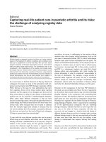
Figure 1 Targe ting pHSP20-14-3-3 protein interactions. A. A representative Western blot (n = 3 separate experiments) using antibodies to
HSP20 (lane 1), phosphorylated cofilin (lane 2), and cofilin (lane 3). B. A representative SPR-based evaluation of HSP20 binding to a class of 14-3-
3 proteins. Synthesized peptides containing a partial sequence of phosphorylated HSP20 were immobilized via amine-coupling to a BIAcore chip,
and GST-HIS-14-3-3 isoforms (YWHAG, g; YWHAH, h; YWHAZ, ζ; YWHAB, b; and YWHAE, ε) were injected at 20 μg/ml. Experiments were
conducted in triplicate. C. A schematic drawing of the principle behind the fluorescence polarization (FP) assay. FP signals of a flourophore is
defined here as, FP = (V-H)/(V+H); where V is the vertical component and H is the horizontal component of the emitted light when excited by
vertical plane polarized light. D. Changes in FP signals in response to a number of compounds belonging to the PRLX24905 scaffold (USA Patent
& Trademark, Publication 20090136561: “Non-peptidyl agents with pHSP20-like activity, and uses thereof”). Data are presented as mean ± SE (n =
3 separate experiments).
An et al . Respiratory Research 2011, 12:8
/>Page 4 of 9
inactive phosphorylated form as reported earlier [12].
Phosphorylated cofilin is bound to 14-3-3 proteins [20-22]
and, in human ASM, PKA-activated phosphorylation of
HSP20 is associated with dephosphorylation of cofilin and
subsequent loss of actin stress fibers [12]. Dreiza and col-
leagues [19] have demonstrated that phosphopeptide ana-
logs of HSP20 (pHSP20) co-precipitate with a class of 14-
3-3 proteins and, moreover, competitively inhibit the bind-
ing of phosphorylated cofilin to 14-3-3 proteins. Using
SPR-based evaluation of protein interactions, we found
that pHSP20 exhibited the highest binding affinity for the
g isoform of 14-3-3 proteins (Figure 1B). Hence, we
focused on pHSP20-14-3-3 g protein interactions in
human ASM as a potential molecular target against exces-
sive constriction of the airways in asthma.
Screening small molecule modulators of pHSP20-14-3-3 g
protein interactions
Using a high-throughput in vitro FP assay, we screened
a library of compounds that could act as small molecule
modulators of HSP20 signals (Figure 1C). To this end,
we employed a fluorophore-conjugated 8-mer peptide
fragment of pHSP20 (6-FAM-WLRRApSAP) containing
the recognition motif for 14-3-3 proteins; compared
with the full-length pHSP20, this peptide fragment has a
higher binding affinity for 14-3-3 g proteins [19].
Among 58,019 compounds tested, 268 compounds
caused 20% or more reduction of the polarized emis sion
in FP assay (data not shown). Using the FP assay, there-
fore, we were able to quickly screen compounds that
could modulate molecular interactions between pHSP20
and 14-3-3 g proteins and find a number of promising
scaffolds that could act as small molecule analogs of
pHSP20. Here we limited our observations to a number
of these tested scaffolds (both positive and negative).
Compounds belonging to one of the scaffolds
(i.e. PRLX24905) showed a range of modulation of
pHSP20-14-3-3 g protein interactions in the FP assay (Fig-
ure 1D). For example, compounds 85065 and 85067
caused no reduction of the polarized emission, whereas
compound 85070 induced maximal reductio n with an
IC
50
of approximately 50 μM. These compounds, together
with structurally related scaffolds readily available from
the supplier’s catalogue, were re-ordered and re-tested for
activity in a concentration-response manner. From these
primary screen hits, we selected seven scaffolds and
assessed their functional effects on cell stiffness and cell
traction force exercised by human ASM. As previously
demonstrated by us elsewhere [27], ASM cells maintain
relatively high basal tone in culture that is attributable in
large part to the dynamic interactions between acti n and
myosin. Unless otherwise noted, we assessed the effects of
compounds on their abilities to decrease cell stiffness and
cell traction force in the absence of contracting agonists.
Testing functional efficacy of small molecule analogs of
pHSP20
At the level of a single ASM cell, we measured temporal
changes in cell stiffness using MTC (Additional file 1,
FigureS1).Overthecourseof10min,humanASM
cells treated with either the b
2
-agonist isoproterenol or
the cell-permeable cAMP analog dibutyryl-cAMP
(db-cAMP) showed marked decreases in cell stiffness
(Figure 2A). Cells treated with a buffer blank (0.1%,
0.5% or 2.0% w/v cyclodextrin) exhibited statistically
0.0
0.2
0.4
0.6
0.8
Isoproterenol db-cAMP
Cell Stiffness (Pa/nm)
0.0
0.2
0.4
0.6
0.8
0.1% 0.5% 2.0%
Cell Stiffness (Pa/nm)
baseline
treatment
Relaxing Agonists
Cyclodextrin (% w/v)
*
*
#
*
#
A
B
0.0
0.2
0.4
0.6
0.8
1.0
1.2
1.4
Cyclodextrin
10144
10183
8739
85067
85064
85062
85069
85070
Cell Stiffness (normalized to baseline)
0.0
0.2
0.4
0.6
0.8
1.0
1.2
1.4
85070 (0.02 mM)
85070 (0.1 mM)
85070 (0.2 mM)
db-cAMP (1 mM)
ISO (0.01 mM)
Cell Stiffness (normalized to baseline)
Compounds
(
0.2 mM
)
ns
#
*
*
*
*
DC
*
Figure 2 Testing functional efficacy of small molecules with
magnetic twisting cytometry. A and B. The steady-state, stiffness
prior to (baseline, open bars) and after the respective cell treatment
(closed bars). Human ASM cells were treated for 10 min with
(A) relaxing agonists (10 μM isoproterenol or 1 mM db-cAMP) and
(B) buffer blank (0.1%, 0.5% or 2% w/v cyclodextrin). Stiffness is
expressed as Pascal per nm (Pa/nm). Data are presented by
geometric means, and error bars indicate standard error (SE);
* indicates P < 0.001 and # indicates P < 0.05 from respective
baseline stiffness (n = 152 to 442 cells). C and D. Stiffness responses
of human ASM cells. Human ASM cells were (C) treated with vehicle
control (0.5% w/v cyclodextrin) or a number of small molecules (200
μM) belonging to the PRLX24905 scaffold and (D) treated with an
increasing concentration of compound 85070. For comparison,
stiffness responses to relaxing agonists (10 μM isoproterenol or
1 mM db-cAMP) are shown. Stiffness responses are normalized to
respective baseline stiffness of an individual ASM cell. Data are
presented by geometric means ± SE (n = 314 to 1024 cells); *
indicates P < 0.001 and # indicates P < 0.05 from vehicle control.
An et al . Respiratory Research 2011, 12:8
/>Page 5 of 9
significant increases in cell stiffness; however, the
increases were less than 10% from the respective base-
line stiffness. There were no statistical differences in the
stiffness among cells treated with different cyclodextrin
concentrations (Figure 2B). In this study, we chose 0.5%
w/v cyclodextrin as a vehicle for the delivery of small
molecules.
Among the seven scaffolds which showed activity in
the FP assay as small molecule analogs of pH SP20, only
a small subset of compounds belonging to two scaffolds
caused appreciable decreases in cell stiffness. For
instance, human ASM cells treated for 10 min with
compounds belonging to the PRLX24905 scaffold
exhibited a range of stiffness responses (Figure 2C).
Compared to cells treated with vehicle control (0.5% w/
v cyclodextrin), there were no statistical differences in
stiffness responses of ce lls treated with compounds
10144, 10183, and 8739. On the other hand, cells treated
with compound 85067 showed increases (P < 0.05)
whereas cells treated with compounds 85064, 85062,
85069 and 85070 showed progressive decreases in cell
stiffness (P < 0.001). Most strikingly, however, com-
pound 85070 that caused the greatest reduction of the
polarized emission in the FP assay induced maximal
decreases in cell stiffness (Figure 2C). Compound 85070
also caused c oncentration-dependent decreases in cell
B
A
Pa
C
D
Figure 3 Spatiotemporal changes in cell traction forces. Phase contrast (A) and t raction field images (B, 0 min; C, 5 min; D, 10 min) of a
single human ASM cell treated with compound 85070. Colors show the magnitude of the tractions in Pascal (Pa), and arrows show the direction
and relative magnitude of the tractions. Scale bar, 50 μm. This is a representative of cells (n = 4) treated with 200 μM compound 85070.
An et al . Respiratory Research 2011, 12:8
/>Page 6 of 9
stiffness (Figure 2D). Although the rate of decreases in
cell stiffness by compound 85070 was slower than that
by b
2
-agonist isoproterenol (Additional file 1, Figure S1),
we found that compound 85 070 was more efficacious in
decreasing the stiffness of the human ASM cell than
that by either the b
2
-agonist isoproterenol or the cell-
permeable analog of cAMP (db-cAMP).
Consistent with stiffness responses, human ASM
cells treated with compound 85070 exhibited both
spatial and temporal dec reases in contractile fo rce as
measured by traction microscopy (Figure 3). Over the
course of 10 min, compound 85070 significantl y inhib-
ited the ability of an individual human ASM cell to
generate contractile force. For example, the net con-
tractile moment, which is a scalar measure of cell’ s
contractile strength [33], decreased from 36.2 pNm
(median, n = 4) at time z ero to 7.9 pNm at 5 min and
3.1 pNm by 10 min upon incubation with compound
85070 (P < 0.01; Wilcoxon test). Such decreases were
significant (P < 0.05; Wilcoxon Test) when compared
with time-matched cells treated with vehicle control
(0.5% w/v cyclodextrin). For cells treated with vehicle
control, there were no statistically significant changes
in the net contractile moment (38.4 pNm at time zero
to 40.3 pNm at 5 min and 36.9 pNm by 10 min; med-
ian, n = 3).
Validation of the cell-based hit compounds
Scaling up to the level of an intact t issue, we tested the
potency of these cell-based hit compounds in ex vivo
setting. For these studies, we used trachealis rings pre-
pared from inherently hyper-responsive Fischer rats
[25,36,37]. For each trachealis ring, we measured
responses of the intact tissue to a contracting agonist
acetylcholine in a concentra tion-responsive manner. We
limited o ur observations to compound 85070 belonging
to the PRLX24905 scaffold.
For each tissue pre-contracted with a sub-maximal
concentration of acetylcholine, compound 85070
decreased the force generating capacity of rat trachealis
(Figure 4A). Compound 85070 also decreased the force
generating capacity of muscle strips prepared from
bovine tracheal is (data not shown). Furthermore, as
measured by MTC, compound 8507 0 decreased the
stiffness of ASM cells isolated from the trachealis of
inherently hyper-responsive Fischer rats (Figure 4B).
Such decreases in cell stiffness were concentration
dependent and, when compared with cells isolated
from the respective rat aorta (i.e. vascular smooth
muscle), cells isolated from the trachealis showed
greater decreases. Compound 85070 also decreased the
stiffness of serotonin-stimulated rat ASM cells, as well
as histamine-stimulated human ASM cells (data n ot
shown).
Conclusions
To accelerate discovery, screening, testing and validation
of new drug targets, here we have used a staged strate gy
that begins with a chemiproteomics-based approach [38]
and progresses through quantitative b iophysical assays
at the levels of the isolated cell and then the intact tis-
sue [25,32]. It remains unclear if the same cost-e ffective
synergies of this staged approach might be applicable in
the discovery of drug targets for other common diseases
that involve changes in cell biophysical properties,
including vasospasm, hypertension, heart failure, and
50
P
M 100
P
M
Cyclodextrin
85070
0
20
40
60
80
100
120
3
P
MAch
Force Inhibition
(% Ach-induced Contraction)
Compound 85070
50
P
M 100
P
M
Cyclodextrin
85070
Cyclodextrin
85070
Cyclodextrin
85070
0
20
40
60
80
100
120
3
P
MAch
Force Inhibition
(% Ach-induced Contraction)
Compound 85070
A
B
0.0
0.2
0.4
0.6
0.8
1.0
1.2
1.4
Cell Stiffness (normalized to baseline)
Aortic Smooth Muscle Cells
Airway Smooth Muscle Cells
85070
[200
P
M]
85070
[50
P
M]
85070
[20
P
M]
db-cAMP
[1 mM]
Cyclodextrin
[0.5 % w/v]
**
*
**
#
**
*
**
*
**
*
**
**
*
*
Figure 4 Validation of the cell-based hit compounds. A.Force
inhibition of pre-contracted ASM tissues from inherently hyper-
responsive Fischer rats. Tracheal rings were first contracted for 10
min with acetylcholine (3 μM) and subsequently treated with
increasing concentrations of compound 85070. For control, we used
5% w/v cyclodextrin. Data are presented as mean ± SE (n = 4
separate experiments). B. Stiffness responses of smooth muscle cells
isolated from aorta and trachealis of the inherently hyper-responsive
Fischer rats. Cells were treated with vehicle control (0.5% w/v
cyclodextrin), dibutyryl-cAMP (1 mM), or compound 85070 (20 μM,
50 μM or 200 μM). Stiffness changes are normalized to respective
baseline stiffness of an individual cell. Data are presented by
geometric means ± SE (n = 127 to 505 cells). For each treatment, *
indicates P < 0.001 and # indicates P < 0.05 between the cell types.
For each cell type, ** indicates P < 0.001 when compared with
respective vehicle control.
An et al . Respiratory Research 2011, 12:8
/>Page 7 of 9
cancer. As p roof-of-principle, here we limited attention
to the interaction of pHSP20 with 14-3-3 g proteins,
screened a library of 58,019 compounds, and discovered
novel small molecule analogs of pHSP20 that might pro-
vide a therapeutic regime for obstructive lung diseases.
At this time, we do not know whether these functional
effects of small molecule analogs of pHSP20 are due to
their direct actions of regulating actin filament dynamics
[16,18], or indirect actions of displacing cofilin alone
(Additional file 1, Figure S2) [19,20,22] or other regula-
tory protein kinases/phosphatases that interact with 14-
3-3 proteins [21]. These mechanisms of actions are cur-
rently under investigation.
Additional material
Additional File 1: Figures S1 and S2. Figure S1: Temporal changes in
cell stiffness as measured by magnetic twisting cytometry. Function
efficacy of small molecules on stiffness of ASM at the level of a single
living cell. Figure S2: Modulation of pCofilin-14-3-3 protein intera ctions. A
potential mechanism of action of small molecules on relaxing ASM.
List of abbreviations
ASM: airway smooth muscle; HSP20: heat shock protein 20; FP: fluorescence
polarization; SPR: surface plasmon resonance; MTC: magnetic twisting
cytometry; β
2
-AR: β
2
-adrenergic receptor; cAMP: 3’,5’-cyclic adenosine
monophosphate; PKA: cAMP-dependent protein kinase; db-cAMP: N
6
,2’-O-
dibutyryladenosine 3’,5’-cyclic monophosphate.
Acknowledgements
This work was supported by NIH grants HL59682 (JJF) and HL33009 (JJF); by
NIEHS Center grant (2P30 ES03819-11) pilot grant (SSA); and by Faculty
Research Initiative Fund from Johns Hopkins Bloomberg School of Public
Health (SSA).
Author details
1
Division of Physiology, Department of Environmental Health Sciences, Johns
Hopkins Bloomberg School of Public Health, Baltimore, MD 21205, USA.
2
Prolexys Pharmaceuticals, Inc., Salt Lake City, UT 84116, USA.
3
Division of
Biostatistics, Department of Public Health Sciences, Penn State College of
Medicine, Hershey, PA 17033, USA.
4
Program in Molecular and Integrative
Physiological Sciences, Harvard School of Public Health, Boston, MA 02115,
USA.
Authors’ contributions
JJF, SS, and SSA conceived the high-throughput biophysical screening
project. SSA, PSA, and JMP designed and implemented experimental
protocols. JMP, TIZ, and MR conducted the FP assay. PSA, TIZ, and MR
performed isometric force measurements of experimental animal models in
ex vivo settings. TIZ and MR conducted pull-down assay and protein
detection analysis. SSA isolated and cultured smooth muscle cells, and
designed and performed all single-cell biophysical measurements. KA
performed statistical analysis; KA and SSA analyzed the data. JJF and SS
oversaw the project. SSA wrote the paper. All authors read and approved
the final manuscript.
Competing interests
SS, PSA, TIZ, JMP, and MR are former employees of Prolexys Pharmaceuticals
Inc., and were compensated by the company at the time this work was
performed. These employees have no financial arrangements with Prole xys
at the present time. JJF and SSA received a consulting fee from Prolexys
Pharmaceutical, Inc. At the present time, JJF and SSA have no financial
relationship with Prolexys Pharmaceuticals. A part of this work (NON-
PEPTIDYL AGENTS WITH pHSP20-LIKE ACTIVITY, AND USES THEREOF) has
been applied for U.S. patent. There are no other competing interests or
conflicts of interest.
Received: 5 October 2010 Accepted: 13 January 2011
Published: 13 January 2011
References
1. Barnes PJ: New drugs for asthma. Nature Reviews Drug Discovery 2004,
3:831-844.
2. Green SA, Turki J, Bejarano P, Hall IP, Liggett SB: Influence of Beta(2)-
Adrenergic Receptor Genotypes on Signal-Transduction in Human
Airway Smooth-Muscle Cells. American Journal of Respiratory Cell and
Molecular Biology 1995, 13:25-33.
3. Israel E, Chinchilli VM, Ford JG, Boushey HA, Cherniack R, Craig TJ, Deykin A,
Fagan JK, Fahy JV, Fish J, Kraft M, Kunselman SJ, Lazarus SC, Lemanske RF Jr,
Liggett SB, Martin RJ, Mitra N, Peters SP, Silverman E, Sorkness CA,
Szefler SJ, Wechsler ME, Weiss ST, Drazen JM: Use of regularly scheduled
albuterol treatment in asthma: genotype-stratified, randomised, placebo-
controlled cross-over trial. Lancet 2004, 364:1505-1512.
4. Taylor DR, Drazen JM, Herbison GP, Yandava CN, Hancox RJ, Town GI:
Asthma exacerbations during long term beta agonist use: influence of
beta(2) adrenoceptor polymorphism. Thorax 2000, 55:762-767.
5. Sears MR, Taylor DR, Print CG, Lake DC, Li QQ, Flannery EM, Yates DM,
Lucas MK, Herbison GP: Regular Inhaled Beta-Agonist Treatment in
Bronchial-Asthma. Lancet 1990, 336:1391-1396.
6. Taylor DR, Sears MR, Herbison GP, Flannery EM, Print CG, Lake DC,
Yates DM, Lucas MK, Li Q: Regular Inhaled Beta-Agonist in Asthma -
Effects on Exacerbations and Lung-Function. Thorax 1993, 48:134-138.
7. Deshpande DA, Penn RB: Targeting G protein-coupled receptor signaling
in asthma. Cellular Signalling 2006, 18:2105-2120.
8. Murray KJ: Cyclic-Amp and Mechanisms of Vasodilation. Pharmacology &
Therapeutics 1990, 47:329-345.
9. Popescu LM, Panoiu C, Hinescu M, Nutu O: The Mechanism of Cgmp-
Induced Relaxation in Vascular Smooth-Muscle. European Journal of
Pharmacology 1985, 107:393-394.
10. Beall A, Bagwell D, Woodrum D, Stoming TA, Kato K, Suzuki A,
Rasmussen H, Brophy CM: The small heat shock-related protein, HSP20, is
phosphorylated on serine 16 during cyclic nucleotide-dependent
relaxation. Journal of Biological Chemistry 1999, 274:11344-11351.
11. Rembold CM, Foster DB, Strauss JD, Wingard CJ, Van Eyk JE: cGMP-
mediated phosphorylation of heat shock protein 20 may cause smooth
muscle relaxation without myosin light chain dephosphorylation in
swine carotid artery. Journal of Physiology-London 2000, 524:865-878.
12. Komalavilas P, Penn RB, Flynn CR, Thresher J, Lopes LB, Furnish EJ, Guo M,
Pallero MA, Murphy-Ullrich JE, Brophy CM: The small heat shock-related
protein, HSP20, is a cAMP-dependent protein kinase substrate that is
involved in airway smooth muscle relaxation. American Journal of
Physiology-Lung Cellular and Molecular Physiology 2008, 294:L69-L78.
13. Flynn CR, Komalavilas P, Tessier D, Thresher J, Niederkofler EE, Dreiza CM,
Nelson RW, Panitch A, Joshi L, Brophy CM: Transduction of biologically
active motifs of the small heat shock-re lated protein, HSP20, leads to
relaxation of vascular s mooth muscle. Faseb Journal 2003,
17
:1358-1360.
14.
Tessier DJ, Komalavilas P, Liu B, Kent CK, Thresher JS, Dreiza CM, Panitch A,
Joshi L, Furnish E, Stone W, Fowl R, Brophy CM: Transduction of peptide
analogs of the small heat shock-related protein HSP20 inhibits intimal
hyperplasia. Journal of Vascular Surgery 2004, 40:106-114.
15. Woodrum D, Pipkin W, Tessier D, Komalavilas P, Brophy CM:
Phosphorylation of the heat shock-related protein, HSP20, mediates
cyclic nucleotide-dependent relaxation. Journal of Vascular Surgery 2003,
37:874-881.
16. Brophy CM, Lamb S, Graham A: The small heat shock-related protein-20 is
an actin-associated protein. Journal of Vascular Surgery 1999, 29:326-333.
17. Bukach OV, Marston SB, Gusev NB: Small heat shock protein with
apparent molecular mass 20 kDa (Hsp20, HspB6) is not a genuine actin-
binding protein. Journal of Muscle Research and Cell Motility 2005,
26:175-181.
18. Tessier DJ, Komalavilas P, Panitch A, Joshi L, Brophy CM: The small heat
shock protein (HSP) 20 is dynamically associated with the actin cross-
linking protein actinin. Journal of Surgical Research 2003, 111:152-157.
An et al . Respiratory Research 2011, 12:8
/>Page 8 of 9
19. Dreiza CM, Brophy CM, Komalavilas P, Furnish EJ, Joshi L, Pallero MA,
Murphy-Ullrich JE, von Rechenberg M, Ho YSJ, Richardson B, Xu N, Zhen Y,
Peltier JM, Panitch A: Transducible heat shock protein 20 (HSP20)
phosphopeptide alters cytoskeletal dynamics. Faseb Journal 2004,
18:261-263.
20. Gohla A, Bokoch GM: 14-3-3 regulates actin dynamics by stabilizing
phosphorylated cofilin. Current Biology 2002, 12:1704-1710.
21. Rubio MP, Geraghty KM, Wong BHC, Wood NT, Campbell DG, Morrice N,
Mackintosh C: 14-3-3-affinity purification of over 200 human
phosphoproteins reveals new links to regulation of cellular metabolism,
proliferation and trafficking. Biochemical Journal 2004, 379:395-408.
22. Yaffe MB: How do 14-3-3 proteins work? - Gatekeeper phosphorylation
and the molecular anvil hypothesis. Febs Letters 2002, 513:53-57.
23. Niwa R, Nagata-Ohashi K, Takeichi M, Mizuno K, Uemura T: Control of actin
reorganization by Slingshot, a family of phosphatases that
dephosphorylate ADF/cofilin. Cell 2002, 108:233-246.
24. An SS, Hai CM: Mechanical signals and mechanosensitive modulation of
intracellular [Ca2+] in smooth muscle. American Journal of Physiology-Cell
Physiology 2000, 279:C1375-C1384.
25. An SS, Fabry B, Trepat X, Wang N, Fredberg JJ: Do biophysical properties
of the airway smooth muscle in culture predict airway
hyperresponsiveness? American Journal of Respiratory Cell and Molecular
Biology 2006, 35:55-64.
26. An SS, Laudadio RE, Lai J, Rogers RA, Fredberg JJ: Stiffness changes in
cultured airway smooth muscle cells. American Journal of Physiology-Cell
Physiology 2002, 283:C792-C801.
27. An SS, Kim J, Ahn K, Trepat X, Drake KJ, Kumar S, Ling GY, Purington C,
Rangasamy T, Kensler TW, Mitzner W, Fredberg JJ, Biswal S: Cell stiffness,
contractile stress and the role of extracellular matrix. Biochemical and
Biophysical Research Communications 2009, 382:697-703.
28. Bursac P, Lenormand G, Fabry B, Oliver M, Weitz DA, Viasnoff V, Butler JP,
Fredberg JJ: Cytoskeletal remodelling and slow dynamics in the living
cell. Nature Materials 2005, 4:557-561.
29. Fabry B, Maksym GN, Butler JP, Glogauer M, Navajas D, Fredberg JJ: Scaling
the microrheology of living cells. Physical Review Letters 2001, 87:148102.
30. Trepat X, Deng LH, An SS, Navajas D, Tschumperlin DJ, Gerthoffer WT,
Butler JP, Fredberg JJ: Universal physical responses to stretch in the
living cell. Nature 2007, 447:592-595.
31. Pechkovsky DV, Hackett TL, An SS, Shahen F, Murray LA, Knight DA: Human
lung parenchyma but not proximal bronchi produces fibroblasts with
enhanced TGFβ signaling and αSMA expression. American Journal of
Respiratory Cell and Molecular Biology 2010, 43:641-651.
32. Deshpande DA, Wang WC, Mcllmoyle EL, Robinett KS, Schillinger RM, An SS,
Sham JS, Liggett SB: Bitter taste receptors on airway smooth muscle
bronchodilate by localized calcium signaling and reverse obstruction.
Nature Medicine 2010, 16:1299-1304.
33. Butler JP, Tolic-Norrelykke IM, Fabry B, Fredberg JJ: Traction fields,
moments, and strain energy that cells exert on their surroundings.
American Journal of Physiology-Cell Physiology 2002, 282:C595-C605.
34. Wang N, Tolic-Norrelykke IM, Chen JX, Mijailovich SM, Butler JP, Fredberg JJ,
Stamenovic D: Cell prestress. I. Stiffness and prestress are closely
associated in adherent contractile cells. American Journal of Physiology-Cell
Physiology 2002, 282:C606-C616.
35. An SS, Fabry B, Mellema M, Bursac P, Gerthoffer WT, Kayyali US, Gaestel M,
Shore SA, Fredberg JJ: Role of heat shock protein 27 in cytoskeletal
remodeling of the airway smooth muscle cell. Journal of Applied
Physiology 2004, 96:1701-1713.
36. Dandurand RJ, Wang CG, Phillips NC, Eidelman DH: Responsiveness of
Individual Airways to Methacholine in Adult-Rat Lung Explants. Journal
of Applied Physiology 1993, 75:364-372.
37. Wang CG, Almirall JJ, Dolman CS, Dandurand RJ, Eidelman DH: In vitro
bronchial responsiveness in two highly inbred rat strains. Journal of
Applied Physiology 1997, 82:1445-1452.
38. Peltier JM, Askovic S, Becklin RR, Chepanoske CL, Ho YSJ, Kery V, Lai SP,
Mujtaba T, Pyne M, Robbins PB, von Rechenberg M, Richardson B, Savage J,
Shelfield P, Thompson S, Weir L, Widjaja K, Xu N, Zhen Y, Boniface JJ: An
integrated strategy for the discovery of drug targets by the analysis of
protein-protein interactions. International Journal of Mass Spectrometry
2004, 238:119-130.
doi:10.1186/1465-9921-12-8
Cite this article as: An et al.: A novel small molecule target in human
airway smooth muscle for potential treatment of obstructive lung
diseases: a staged high-throughput biophysical screening. Respiratory
Research 2011 12:8.
Submit your next manuscript to BioMed Central
and take full advantage of:
• Convenient online submission
• Thorough peer review
• No space constraints or color figure charges
• Immediate publication on acceptance
• Inclusion in PubMed, CAS, Scopus and Google Scholar
• Research which is freely available for redistribution
Submit your manuscript at
www.biomedcentral.com/submit
An et al . Respiratory Research 2011, 12:8
/>Page 9 of 9