Báo cáo y học: " KL-6 concentration in pulmonary epithelial lining fluid is a useful prognostic indicator in patients with acute respiratory distress syndrome" ppt
Bạn đang xem bản rút gọn của tài liệu. Xem và tải ngay bản đầy đủ của tài liệu tại đây (308.36 KB, 7 trang )
RESEARC H Open Access
KL-6 concentration in pulmonary epithelial lining
fluid is a useful prognostic indicator in patients
with acute respiratory distress syndrome
Tomohiro Kondo
1
, Noboru Hattori
1*
, Nobuhisa Ishikawa
1
, Hiroshi Murai
1
, Yoshinori Haruta
1
, Nobuyuki Hirohashi
2
,
Koichi Tanigawa
2
, Nobuoki Kohno
1
Abstract
Background: KL-6 is a mucin-like glycoprotein expressed on the surface of alveolar type II cells. Elevated
concentrations of KL-6 in serum and epithelial lining fluid (ELF) in patients with acute respiratory distress syndrome
(ARDS) have been previously reported; however, kinetics and prognostic significance of KL-6 have not been
extensively studied. This study was conducted to clarify these points in ARDS patients.
Methods: Thirty-two patients with ARDS who received mechanical ventilation under intubation were studied for
28 days. ELF and blood were obtained from each patient at multiple time points after the diagnosis of ARDS. ELF was
collected using a bronchoscopic microsampling procedure, and ELF and serum KL-6 concentrations were measured.
Results: KL-6 levels in ELF on days 0 to 3 after ARDS diagnosis were significantly higher in nonsurvivors than in
survivors, and thereafter, there was no differe nce in concentrations between the two groups. Serum KL-6 levels did
not show statistically significant differences between nonsurvivors and survivors at any time point. When the
highest KL-6 levels in ELF and serum sample from each patient were examined, KL-6 levels in both ELF and serum
were significantly higher in nonsurvivors than in survivors. The optimal cut-off values were set at 3453 U/mL for
ELF and 530 U/mL for serum by receiver operating characteristic (ROC) curve analyses. Patients with KL-6
concentrations in ELF higher than 3453 U/mL or serum concentrat ions higher than 530 U/mL had significantly
lower survival rates up to 90 days after ARDS diagnosis.
Conclusions: ELF and serum KL-6 concentrations were found to be good indicators of clinical outcome in ARDS
patients. Particularly, KL-6 levels in ELF measured during the early period after the diagnosis were useful for
predicting prognosis in ARDS patients.
Background
Acute respiratory distress syndrome (ARDS) is character-
ized by the influx of protein-rich edema fluid into air
spaces because of the increased permeability of the alveo-
lar-capillary barrier [1,2]. The important roles of endothe-
lial injury and increased vascular permeability in the
formation of pulmonary edema have been well established
in this disorder [3]. An intact alveolar epithelial barrier is
necessary for preventing alveolar flooding and facilitating
recovery from ARDS; therefore, the degree of alveolar
epithelial injury is an important predictor of the outcomes
in ARDS [4-6]. When epithelial integrity is lost and alveo-
lar type II cells are injured, normal alveolar epithelial fluid
transport and removal of alveolar edema fluid are impaired
[7]. Moreover, injury to alveolar type II cells reduces the
production and turnover of surfactant [8], and may also
cause intrapulmonary bacterial translocation that may lead
to bacteremia or sepsis [9]. If injury to the alveolar epithe-
lium is severe, epithelial repair is impaired, which may
lead to the development of fibrosis [10].
KL-6 is a high-molecular-weight glycoprotein, classified
according to immunohistochemical and flow cytometry
study findings as cluster 9 mucin-1 (MUC1) of lung
tumor and differentiation antigens [11]. After cleavage of
* Correspondence:
1
Department of Molecular and Internal Medicine, Graduate School of
Biomedical Sciences, Hiroshima University, 1-2-3 Kasumi, Minami-ku,
Hiroshima, 734-8551, Japan
Full list of author information is available at the end of the article
Kondo et al. Respiratory Research 2011, 12:32
/>© 2011 Kondo et al; licensee BioMed Central Ltd. This is an Open Access article distributed under the terms of the Creative Commons
Attribution License ( which permits unrestricted use, distribution, and reproduction in
any medium, provided the original work is properly cited.
the S-S bond near the surface of the epithelia l cell mem-
brane, KL-6 can diffuse into pulmonary epithelial lining
fluid (ELF). In the normal lung, this glycoprotein can be
predominantly found on alveolar type II epithelial cells,
and its expression is greatly increased in proliferating,
regenerating, or injured alveolar type II cells [12-14]. Pre-
vious studies have demonstrated that serum levels of
KL-6 are elevated in a variety of interstitial lung diseases
that are characterized by alveolar epithelial cell damage
[12,14-20]. Because serum levels of KL-6 have been
shown to be correlated with indices of alveolar-capillary
permeability [15], elevated levels of circulating KL-6 are
believed to be associated with its increased leakage from
the alveolar space into the circulation.
Previous studies examined KL-6 levels in the serum
and pulmonary ELF or bronchoalveolar l avage fluid
(BALF) of adult patients with ARDS or acute lung injury
(ALI) [13,21-23], and found that the leve ls of KL-6 were
significantly elevated. These studies also reported that
the levels of KL-6 in these samples were significantly
higher in nonsurvivors than in survivors. Their results
suggest that elevated levels of KL-6 may indicate poor
prognosis in ARDS patients; however, whether or not
KL-6 levels in these samples can predict clinical out-
comes in ARDS patients has not yet been studied in
detail. Furthermore, none of these studies have reported
detailed kinetics of KL-6 levels in ELF and serum in
ARDS patients.
In the present study, to further evaluate the clinical sig-
nificance of KL-6 in ARDS patients, concentrations of
KL-6 in ELF and serum were consecutively measured in
32 patients who developed ARDS in our hospital, and the
kinetics of KL-6 levels in ELF and serum during 4 weeks
after the diagnosis of ARDS were determined. In addi-
tion, the associations between KL-6 levels in these sam-
ples and patient clinical outcomes were examined.
Methods
Study population and protocol
This clini cal study was conducted at Hiroshima U niver-
sity Hospital between July 2007 and March 2009. Th e
human research committee of Hiroshima University
approved this study, and written informed consent was
obtained from each study participant or from immediate
family members. Thirty-two patients were prospectively
diagnosed with ARDS according to the definition of the
American-European Consensus Con ference on ARDS.
They were included in the study if they met consensus
conference oxygenation and radiographic criteria for
ARDS, and were followed until death or hospital dis-
charge. The patients who were discharged from the
hospital were considered to be survivors.
Bronchoscopic microsampling (BMS) of ELF was per-
formed on days 0, 1, 3, 5, 7, 10, 14, 21, and 28 in each
patient unless the patient had been extubated or had died.
The first sample was taken on day 0, within 24 hours after
the diagnosis of ARDS. In addition, blood was sampled on
days 0, 1, 3, 5, 7, 10, 14, 21, and 28.
BMS procedure
All studied patients were sedated and preoxygenated (FiO
2
= 1.0). A flexible bronchoscope (BF-6C240; Olympus,
Tokyo, Japan) was inserted into the lung through an intra-
tracheal tube to ex amine the airway, and any excess spu-
tum was suctioned. Another identical bronchoscope was
then inserted and its tip was advanced into a segmental
bronchus of the right middle lobe (S4 or S5), and the BMS
procedure was performed as described previously [24].
The BMS probe (Ol ympu s, Tokyo, Japan), consisted of a
polyethylene outer sheath 1.7 mm in diameter and an
inner fiber rod probe 1.2 mm in diameter a nd 30 mm in
length, attached to a stainless steel guide wire 100 cm in
length.Briefly,theprobewasinsertedintothechannel
and gently advanced. While the outer sheath was set at the
target in the subsegmental bronchus, the inner probe was
advanced slowly into the peripheral airway until it con-
tacted the mucosal surface, and it was held in that position
for 5-7 seconds, thus allowing the fiber rod to absorb
approximately 20 μL of ELF. The inner probe was then
withdrawn into the outer sheath, and they were removed
together. The wet inner probe was cut, placed in a t ube,
and stored in a freezer at -80°C until analysis. The proce-
dure was performed in triplicate from the same subseg-
mental bronchus.
The stored frozen probes were weighed before the ELF
saline suspension was prepared. Diluted ELF sample
solutions were prepared for biochemical analysis by add-
ing the 3 frozen probes that had been sampled from
the same lung subsegment to a 15 mL polyethylene tube
containing 3 mL of saline, which was then vortexed for
1 minut e. The solution was centrifuged for 15 minutes at
3,000 rpm, and the supernatant was collected. The
probes were dried and weighed to calculate the ELF
volume recovered. The dilution factor was calculated as
follows: ELF volume (mL)/(3 mL + ELF volume [mL]).
In vitro experiments have confirmed that the absorp-
tion of 2-20 μL of human serum by the fiber rod probe
allowed a >93% reco very of biochemical constituents.
The recovery was 96.1% for albumin, 93.7% for lactate
dehydrogenase (LDH), and 95.3% for KL-6.
Measurements of KL-6
KL-6 levels in the serum and ELF samples were mea-
sured by a sandwich-type electrochemiluminescent
immunoassay (ECLIA) using a Picolumi 8220 Analyzer
(Sanko Junyaku, Tokyo, Japan), as previously described
[25]. In brief, the sample was incubated with anti-KL-6
antibody-coated magnetic beads and the bea ds were
Kondo et al. Respiratory Research 2011, 12:32
/>Page 2 of 7
then separated using a magnetic rack. Ruthenium-
labeled anti-KL-6 antibody was added to the beads as a
second antibody, following a PBS wash. The reacti on
mixture was placed into an electrode, and the photons
emitted from the ruthenium were measured by a
photomultiplier.
Statistical analysis
Statistical significance was defined as p < 0.05. Differ-
ences in variables between survivors and nonsurvivors
were compared using the nonparametric Mann-Whitney
U-test, since the da ta were not normally distribute d.
The variables at each time point in survivors and non-
survivors during 4 weeks after the diagnosis of ARDS
were compared using both one-way analysis of variance
(ANOVA) and test for linear trend with multiple com-
parisons. Receiver operating characteristic (ROC) curve
analysis was used to assess KL-6 in ELF as a prognostic
indicator in ARDS patients. Survival until 90 days after
the diagnosis was evaluat ed by the Kaplan-Meier
method. The difference in survival between two groups
was analyzed by the log-rank test. All patients included
into the study were followed-up until 90 days after t he
diagnosis of ARDS.
Results
Characteristics of patients
Thirty-two consecutive patients with ARDS who were
treated with controlled mechanical ventilation in the
intensivecareunitwerestudiedbetweenJuly2007and
March 2009. The primary disorders in these patients
were pneumonia (n = 10), sepsis (n = 10), gastric aspira-
tion (n = 5), liver failure (n = 2), alveolar hemorrhage
(n = 1), inter stitial pneumonia (n = 1), hypersensitivity
pneumonia (n = 1), drug-induced pneumonia (n = 1),
and chest trauma (n = 1). The patients with interstitial
pneumonia, hypersensitivity pneumonia, and drug-
induced pneumonia were confirmed to have had st able
respiratory condition before the onset of ARDS and the
apparent superimposition ofpulmonaryinfectionin
these three patients was denied by the analysis of BALF.
The mean age (± SD) was 70.1 ± 11.7 years, and
27 patients were males. The initial mean value (± SD)
for PaO
2
/FIO
2
was 108.6 ± 39.8, and t he in-hospital
mortality rate was 31.3%.
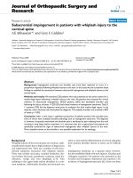
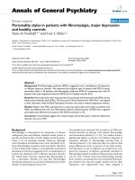
KL-6 levels in ELF and serum samples of survivors and
nonsurvivors
The kinetics of KL-6 levels in ELF and serum samples
were first compared between the survivors and nonsur-
vivors. T he KL-6 levels in ELF were significantly higher
in nonsurvivors than in the survivors on days 0 (p =
0.0087), 1 (p = 0.0421), and 3 (p = 0.0324) (Figure 1a).
The variab les at each time point were compared in the
survivors and nonsur vivors using one-way ANOVA and
no statistical differences were found in each comparison.
However, only in the nonsurvivors, a reducing trend in
ELF levels of KL-6 as time passed after the diagnosis of
ARDS w as observed (test for linear trend, p = 0.0318).
There were no significant differences seen in serum
KL-6 levels between survivors and nonsurvivors at any
time point throughout the clinical courses of the
patients (Figure 1b). To obtain more information on
the clinical significance of KL-6 in ARDS, we selected
the highest ELF and serum KL-6 concentrations among
the series of measurements i n each patient a nd com-
pared the re sults between survivors and nonsurvivors.
The highest concentrations of KL-6 in ELF were
observed on days 2.7 ± 3.3 in the nonsurvivors; whereas
the peak levels in the survivors occurred on days 3.6 ±
4.4. The mean highest concentrations of KL-6 in ELF
were 10733.6 ± 7793.1 U/mL in th e nonsurvivors a nd
3282.3 ± 3474.1 U/mL in the survivors. The highest
concentrations of KL-6 in serum were observed on days
5.8 ± 8.4 in the nonsurvivors; whereas the peak levels in
the survivors occurred on days 2.6 ± 4.5. The mean
highest concentrations of KL-6 in serum were 1060.8 ±
989.8 U/mL in the nonsurvivors and 466.8 ± 602.1 U/mL
U
/mL)
14000
16000
18000
*
**
***
nonsurvivors
survivors
a
ELF KL-6(
U
6000
8000
10000
12000
0
2000
4000
da
y
s
0
1
3
571014
21
28
b
y
28
b
2000
3000
L
-6(U/mL)
1000
Serum K
L
Follow-up days
0
days
0
1
3
57
10 14
21
28
Figure 1 Kinetics of KL-6 levels in ELF (a) and serum (b) in the
nonsurvivors (n = 10) and survivors (n = 22). Data are means ±
SD. *p = 0.0087, **p = 0.0421, ***p = 0.0324 by Mann-Whitney U-
test. A significant reducing trend in ELF levels of KL-6 was observed
in the nonsurvivors (p = 0.0318 by test for linear trend).
Kondo et al. Respiratory Research 2011, 12:32
/>Page 3 of 7
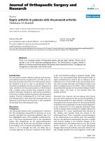
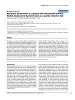
in the survivors. The highest KL-6 levels in ELF and
serum were significantly higher in the nonsurvivors than
in the survivors (p = 0.0025, Figure 2a; and p = 0.0401,
Figure 2b; respectively). In addition to the comparisons
of the KL-6 levels between the survivors and nonsurvi-
vors, the highest KL-6 levels in ELF and serum among
the series of measurements were c ompared between the
patients with primary (n = 20) and secondary (n = 12)
ARDS or between the patients with (n = 3) and without
(n = 29) preexisting interstitial lung disease (ILD). In
each comparison, we found no significant difference
between the two groups of the patients (data not shown).
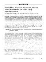
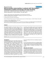
Prognostic values of KL-6 levels in pulmonary ELF and
serum obtained from ARDS patients
To obtain optimal cut-off values for KL-6 in ELF and
serum for prognostic assessment in ARDS patients, recei-
ver operating characteristic (ROC) curve analyses were
performed using the highest concentrations of KL-6 mea-
sured in the serial EL F (Figure 3a) and serum (Figure 3b)
samples. For predicting the risk of mortality, the optimal
cut-off value for KL-6 in ELF was 3453 U/mL, with a sen-
sitivity, specificity, and likelihood ratio of 77.27%, 90.0%,
and 7.73, respectively. Nine out of 14 patients with ELF
KL-6 levels > 3453 U/mL died; whereas only 1 death was
observed in the 18 patients with ELF KL-6 levels < 3453
U/mL died (p = 0.0006). The opti mal cut-off value of
KL-6 in serum was found to be 530 U/mL, with a sensi-
tivity, specificity, and likelihood ratio of 86.36%, 60.0%,
and 2.16, respectively. Whereas 6 out of 9 patients with
serumKL-6levels>530U/mLdied,only4outof23
patients with serum KL-6 levels < 530 U/mL died (p =
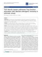
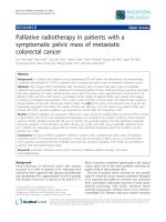
0.0126). Based on these cut-off values, overall survivals
up to 90 days after the diagnosis of the ARDS were deter-
mined using the Kaplan-Meier method. The survival of
patients with concentrations of KL-6 in ELF higher than
3453 U/mL was significantly poorer than the survival of
patients with lower KL-6 concentrations (p = 0.0004,
Figure 4a). Similarly, the survival of patients with higher
serum KL-6 levels (> 530 U/mL) was signif icantly poorer
than the survival of patients with lower serum KL-6 levels
(p = 0,0075, Figure 4b).
Discussion
In this study, we measured KL-6 concentrations in pul-
monary ELF samples and serum samples obtained at
multiple time points from ARDS patients. When the
kinetics of KL-6 levels in ELF and serum were compared
between the survivors and nonsurvivors, only the levels
of KL-6 in ELF on days 0 to 3 after the diagnosis of
ARDS were significantly higher in the nonsurvivors than
in the survivors. There were no differences between sur-
vivors and nonsurvivors in KL-6 concentrations in ELF
samples at other time points, and there were no signifi-
cant differences in serum KL-6 levels between the survi-
vors and nonsurvivors at any time point. However,
when the highest serum KL-6 levels from the serial sam-
ples from each patient were compared between the sur-
vivors and nonsurvivors, statistically significant higher
serum KL-6 levels were seen in the nonsurvivors. In
addition, KL-6 levels in ELF higher than 3453 U/mL
and KL-6 levels in serum higher than 530 U/mL were
a
*
30000
b
**
4000
-6(U/mL)
20000
L-6(U/mL)
2000
3000
ELF KL
10000
Serum K
1000
2000
Survivors
(n=22)
Nonsurvivors
(n=10)
0
Nonsurvivors
(n=10)
Survivors
(n=22)
0
Figure 2 Comparisons of the highest KL-6 levels in ELF (a) and serum (b) from the serial measurements of the nonsurvivors and
survivors. The Box-whisker plots show the 25th and 75th percentiles, the median (horizontal line within the box), and the 10th and 90th
percentiles (whiskers). *p = 0.0025, **p = 0.0401 by Mann-Whitney U- test.
Kondo et al. Respiratory Research 2011, 12:32
/>Page 4 of 7
shown to be significant prognostic factors for predicting
poor overall survival up to 90 days after the diagnosis of
ARDS.
The most important finding in the present study was
that the marked elevation of ELF KL-6 within 3 days
after the diagnosis appeared to correlate with poor prog-
nosis in ARDS patients. This observation was supported
by the following study results: KL- 6 levels in ELF were
significantly elevated in the nonsurvivors on days 0 to 3
after the diagnosis of A RDS compared to the survivors,
and the patients with KL-6 levels in ELF higher than
3453 U/mL had significantly poorer prognosis than those
with lower KL-6 levels in ELF. Lung compartment KL-6
is believed to be produced and released by proliferating
alveolar type II cel ls following injury to alv eolar type I
cells [21], and therefore its level must reflect the severity
of alveolar epithelial injury. The degree of alveolar epithe-
lial injury is believed to be an important predictor of out-
comesinpatientswithARDS[2,26].Basedonthese
concepts, a very high KL-6 level in ELF can be regarded
as an indicator of very severe alveolar epithelial damage,
and a predictor o f poor prognosis in ARDS. In turn, our
data suggest that measurement of KL-6 levels in ELF,
particularly during the early period after ARDS diagnosis,
is useful for assessing the degree of alveolar epithelial
damage and predicting overall clinical outcome.
Another interesting finding was that in the nonsurvi-
vors, the significantly elevated levels of KL-6 in ELF
were only observed on days 0 to 3 after ARDS diagnosis,
and thereafter, the levels of KL-6 in ELF were similar to
the levels in the survivors. In fact, the highest concen-
trations of KL-6 in ELF were observed on days 2.7 ± 3.3
in the nonsurvivors; whereas in the survivors, they
occurred on days 3.6 ± 4.4. Therefore, we can suggest
that at least one BMS procedure within 3 days after the
diagnosis of ARDS is sufficient to predict the clinical
outcome and the KL-6 levels in ELF obtained from 4
days after the diagnosis m ay have less impact on the
prediction of prognosis. Unfortunately, we do not have
conv incing data to explain why levels of KL-6 in ELF in
the nonsurvivors dropped to the same levels as those in
the survivors. It has been suggested that alveolar type II
cells can proliferate when alveolar epithelial cell damage
is mild or moderat e, but when t he damage is very severe,
even type II cells cannot survive and are replaced by the
epithelial cells of bronchial origin [27,28]. Furthermor e, if
100%
a
b
t
y
60
80
S
ensitivi
t
40
60
S
0
20
AUC=0.841
AUC=0.732
0 20 40 60 80 100%
0
100% - Specificity
100% - Specificity
0
20
40
60 80
100%
Figure 3 ROC curve analyses to determine the optimal cutoff values of KL-6 concentrations in ELF (a) and serum (b) for predicting
survival in ARDS patients. The highest KL-6 levels in ELF and serum from each patient were used for the analysis. The vertical axis represents
the number of true-positive responses (sensitivity), and the horizontal axis represents the number of false-positive responses (100%-specificity).
The area under the curve (AUC) represents the fraction of nonsurviving ARDS patients who would have a positive test (high KL-6 concentration
in ELF or serum). The optimal value of KL-6 in ELF was 3453 U/mL, with a sensitivity, specificity, and likelihood ratio of 77.27%, 90.0%, and 7.73,
respectively. The optimal value of KL-6 in serum was 530 U/mL, with a sensitivity, specificity, and likelihood ratio of 86.36%, 60.0%, and 2.16,
respectively.
Kondo et al. Respiratory Research 2011, 12:32
/>Page 5 of 7
the alveolar epithelial injury is too severe for recovery,
insufficient or disorganized epithelial repair occurs,
resulting in the development of fibrosis [2]. Based on
these concepts, we can speculate that in the nonsurvi-
vors, the alveolar type II cells could initially proliferate
during the early stages of ARDS, leading to elevated KL-6
pulmonary ELF concentrations; however, after develop-
ment of severe alveolar epithelial damage, the type II
cells died or disorganized epithelial repair occurred, lead-
ing to decrease in level of KL-6 in ELF.
In contrast to the results of previous re ports [13,22,23],
there were no statistically significant differences in serum
KL-6 levels between the nonsurvivors and survivors
observed at any time points among the serial measure-
ments. Serum K L-6 levels at each time point tended to
be higher in the nonsurvivors than in the survivors;
therefore we believe that if our study would be larger,
statistically significant differences could have bee n seen.
Indeed, when the highest serum level of KL-6 from the
serial measurements in each patient was used for com-
parisons, it was significantly higher in the nonsurvivors
than in the survivors. In addition, the patients with
thehighestserumKL-6levelsthatwerehigherthan
530 U/mL were found to have poorer prognosis than the
other patients. In children with ARDS, circulating levels
of KL-6 were also reported to be higher in the
nonsurvivors than the survivors [29]. These data suggest
that serum KL-6 concentrations also reflect the degree of
alveolar epithelial injury and may be useful for predicting
clinical outcomes in patients with ARDS. However, we
believe that the concentration of KL-6 in ELF is a more
sensitive indicator of alveolar epithelial injury, and is thus
amoreusefulpredictorofclinical outcome than the
serum KL-6 level, because it provides more immediate
information on events taking place in the lung.
Because KL-6 is mainly expressed in alveolar type II
epithelial cells and a sensitive biomarker to detect the
presence of ILD, we questioned whether there was a dif-
ference in KL-6 levels in ELF and serum between the
patients with primary and secondary ARDS or between
the patients with and without preexisting ILD. Interest-
ingly, we found no significant difference in each compari-
son.ThesedatasuggestthatKL-6levelsinELFand
serum were not affected by the cause of ARDS. In addi-
tion, the presence of preexisting ILD seemed not to influ-
ence the KL-6 levels in ELF and serum after developing
ADRS. However, we believe that the number of cases
with preexisting ILD was too small (only three) to reach
the latter conclusion and, t herefor e, further study on this
issue is necessary.
Although promising results were obtained, we are
aware that this study has some limitations. The number
of patients included in the study was not sufficient to
confirm previous observations that circulating KL-6
levels were significantly higher in nonsurvivors than sur-
vivors, particularly during the early period after the
onset of ARDS [13,22,23]. The BMS procedure has an
intrinsic limitation, i n that exploratory sampling in the
lung is limited. Additional study measuring KL-6 in ELF
from different sampling sites in the lungs of each ARDS
patient is necessary.
Conclusion
Concentrations of KL-6 in pulmonary ELF early after
ARDS diagnosis were found to be significantly higher in
nonsurviving patients than in surviving pati ents.
Furthermore, ARDS patients with highe r KL-6 levels in
ELF or serum had significantly poorer prognosis than
those with lower KL-6 levels. The levels of KL-6 in ELF
and serum may reflect the d egree of alveolar epithelial
injury, and may therefore be valuable indicators of out-
come in ARDS. Particularly, the concentrat ion of KL-6
in ELF measured during the early period after the diag-
nosis appears to be a useful marker for pr edicting prog-
nosis in ARDS patients.
List of abbreviations
ALI: acute lung injury; ANOVA: analysis of variance; ARDS: acute respiratory
distress syndrome; AUC: area under the curve; BALF: bronchoalveolar lavage
80
100%
v
ival
Low KL-6 group
a
20
40
60
P
ercent sur
v
High KL-6 group
p
= 0.0004
0 10 20 30 40
50 60 70
80 90
0
20
P
days
b
60
80
100%
u
rvival
Low KL-6 group
p
= 0.0075
b
20
40
60
Percent s
u
High KL-6 group
p
Follow-u
p
da
y
s
0 10 20 30 40 50 60 70 80 90
0
days
Figure 4 Overall survival of ARDS pati ents in relation to KL-6
concentrations in ELF (a) and in serum (b). The survival rate of
patients with a high KL-6 levels in ELF and serum was significantly
lower than that of patients with a low KL-6 levels (ELF: p = 0.0004,
serum: p = 0,0075 by log-rank test).
Kondo et al. Respiratory Research 2011, 12:32
/>Page 6 of 7
fluid; BMS: bronchoscopic microsampling; ELF: epithelial lining fluid; ILD:
interstitial lung disease; ROC: receiver operating characteristic
Acknowledgements
We thank Dr. K. Yoshioka, Department of Molecular and Internal Medicine,
Graduate School of Biomedical Sciences, Hiroshima University; and N. Ohtani
and K. Ohta, department of Emergency and Critical Care Medicine,
Hiroshima University Hospital for their excellent technical assistance and
advice.
This work is supported by grants from Grants-in-Aid for Scientific Research,
and the Ministry of Health, Labour and Welfare of Japan.
Author details
1
Department of Molecular and Internal Medicine, Graduate School of
Biomedical Sciences, Hiroshima University, 1-2-3 Kasumi, Minami-ku,
Hiroshima, 734-8551, Japan.
2
Department of Emergency and Critical Care
Medicine, Hiroshima University Hospital, 1-2-3 Kasumi, Minami-ku, Hiroshima,
734-8551, Japan.
Authors’ contributions
TK designed the study, performed the data analysis and interpretation, and
wrote the manuscript. NH and NI designed the study, interpreted the data,
and edited the manuscript. HM, YH, NH, KT, and NK interpreted the data
and helped to draft the manuscript. All authors read and approved the final
manuscript.
Competing interests
The authors declare that they have no competing interests.
Received: 9 November 2010 Accepted: 22 March 2011
Published: 22 March 2011
References
1. Pugin J, Verghese G, Widmer MC, Matthay MA: The alveolar space is the
site of intense inflammatory and profibrotic reactions in the early phase
of acute respiratory distress syndrome. Crit Care Med 1999, 27:304-312.
2. Ware LB, Matthay MA: Medical progress: the acute respiratory distress
syndrome. N Engl J Med 2000, 342:1334-1349.
3. Brigham KL: Lung edema due to increased vascular permeability. In Lung
Water and Solute Exchange. Edited by: Staub NC. New York: Dekker;
1978:235-276.
4. Matthay MA, Wiener-Kronish JP: Intact epithelial barrier function is critical
for the resolution of alveolar edema in humans. Am Rev Respir Dis 1990,
142:1250-1257.
5. Pittet JF, Mackersie RC, Martin TR, Matthay MA: Biological markers of acute
lung injury: prognostic and pathogenetic significance. Am J Respir Crit
Care Med 1997, 155:1187-1205.
6. Wiener-Kronish JP, Albertine KH, Matthay MA: Differential responses of the
endothelial and epithelial barriers of the lung in sheep to Escherichia
coli endotoxin. J Clin Invest 1991, 88:864-875.
7. Modelska K, Pittet JF, Folkesson HG, Courtney Broaddus V, Matthay MA:
Acid-induced lung injury. Protective effect of anti-interleukin-8
pretreatment on alveolar epithelial barrier function in rabbits. Am J
Respir Crit Care Med 1999, 160:1450-1456.
8. Greene KE, Wright JR, Steinberg KP, Ruzinski JT, Caldwell E, Wong WB,
Hull W, Whitsett JA, Akino T, Kuroki Y, Nagae H, Hudso LD, Martin TR: Serial
changes in surfactant-associated proteins in lung and serum before and
after onset of ARDS. Am J Respir Crit Care Med 1999, 160:1843-1850.
9. Kurahashi K, Kajikawa O, Sawa T, Ohara M, Gropper MA, Frank DW,
Martin TR, Wiener-Kronish JP: Pathogenesis of septic shock in
Pseudomonas aeruginosa pneumonia. J Clin Invest 1999, 104:743-750.
10. Bitterman PB: Pathogenesis of fibrosis in acute lung injury. Am J Med
1992, 92:39S-43S.
11. Stahel RA, Gilks WR, Lehmann HP, Schenker T: Third international
workshop on lung tumor and differentiation antigens: overview of the
results of the central data analysis. Int J CancerSuppl 1994, 8:6-26.
12. Kohno N, Kyoizumi S, Awaya Y, Fukuhara H, Yamakido M, Akiyama M: New
serum indicator of interstitial pneumonitis activity. Sialylated
carbohydrate antigen KL-6. Chest 1989, 96:68-73.
13. Ishizaka A, Matsuda T, Albertine KH, Koh H, Tasaka S, Hasegawa N, Kohno N,
Kotani T, Morisaki H, Takeda J, Nakamura M, Fang X, Martin TR, Matthay MA,
Hashimoto S: Elevation of KL-6, a lung epithelial cell marker, in plasma
and epithelial lining fluid in acute respiratory distress syndrome. Am J
Physiol Lung Cell Mol Physiol 2004, 286:L1088-L1094.
14. Kohno N, Awaya Y, Oyama T, Yamakido M, Akiyama M, Inoue Y,
Yokoyama A, Hamada H, Fujioka S, Hiwada K: KL-6, a mucin-like
glycoprotein, in bronchoalveolar lavage fluid from patients with
interstitial lung disease. Am Rev Respir Dis 1993, 148:637-642.
15. Oyama T, Kohno N, Yokoyama A, Hirasawa Y, Hiwada K, Oyama H, Okuda Y,
Takasugi K: Detection of interstitial pneumonitis in patients with
rheumatoid arthritis by measuring circulating levels of KL-6, a human
MUC1 mucin. Lung 1997, 175(6):379-385.
16. Kohno N, Yokoyama A, Hirasawa Y, Kondo K, Fujino S, Abe M, Hiwada K:
Comparative studies of circulating KL-6, type III procollagen N-terminal
peptide and type IV collagen 7 S in patients with interstitial
pneumonitis and alveolar pneumonia. Respir Med 1997, 91(9):558-561.
17. Hamada H, Kohno N, Yokoyama A, Hirasawa Y, Hiwada K, Sakatani M,
Ueda E: KL-6 as a serologic indicator of Pneumocystis carinii pneumonia
in immunocompromised hosts. Intern Med 1998, 37(3):307-310.
18. Yokoyama A, Kohno N, Kondo K, Ueda S, Hirasawa Y, Watanabe K, Takada Y,
Hiwada K: Comparative evaluation of sialylated carbohydrate antigens,
KL-6, CA19-9 and SLX as serum markers for interstitial pneumonia.
Respirology 1998, 3(3):199-202.
19. Kohno N: Serum marker KL-6/MUC1 for the diagnosis and management
of interstitial pneumonitis. J Med Invest 1999, 46(3-4):151-158.
20. Ohnishi H, Yokoyama A, Kondo K, Hamada H, Abe M, Nishimura K,
Hiwada K, Kohno N: Comparative study of KL-6, surfactant protein-A,
surfactant protein-D, and monocyte chemoattractant protein-1 as serum
markers for interstitial lung disease. Am J Respir Crit Care Med 2002,
165(3):378-381.
21. Inoue Y, Barker E, Daniloff E, Kohno N, Hiwada K, Newman LS: Pulmonary
epithelial cell injury and alveolar-capillary permeability in berylliosis. Am
J Respir Crit Care Med 1997, 156:109-115.
22. Sato H, Callister ME, Mumby S, Quinlan GJ, Welsh KI, duBios RM, Evans TW:
KL-6 levels are elevated in plasma from patients with acute respiratory
distress syndrome. Eur Respir J 2004, 23:142-145.
23. Nathani N, Perkins GD, Tunnicliffe W, Murphy N, Manji M, Thickett DR:
Krebs von Lungen 6 antigen is a marker of alveolar inflammation but
not of infection in patients with acute respiratory distress syndrome. Crit
Care 2008, 12(1):R12.
24. Ishizaka A, Watanabe M, Yamashita T, Ogawa Y, Koh H, Hasegawa N,
Nakamura H, Asano K, Yamaguchi K, Kotani M, Kotani T, Morisaki H,
Takeda J, Kobayashi K, Ogawa S: New bronchoscopic microsample probe
to measure the biochemical constituents in epithelial lining fluid of
patients with acute respiratory distress syndrome. Crit Care Med 2001,
29(4):896-898.
25. Ohshimo S, Yokoyama A, Hattori N, Ishikawa N, Hirasawa Y, Kohno N: KL-6,
a human MUC1 mucin, promotes proliferation and survival of lung
fibroblasts. Biochem Biophys Res Commun 2005, 338(4):1845-1852.
26. Ware LB, Matthay MA: Alveolar fluid clearance is impaired in the majority
of patients with acute lung injury and the acute respiratory distress
syndrome. Am J Respir Crit Care Med 2001, 163(6):1376-1383.
27. Kawanami O, Ferrans VJ, Crystal RG: Structure of alveolar epithelial cells in
patients with fibrotic lung disorders. Lab Invest 1982, 46(1):39-53.
28. Fukuda Y, Ferrans VJ, Schoenberger CI, Rennard SI, Crystal RG: Patterns of
pulmonary structural remodeling after experimental paraquat toxicity.
The morphogenesis of intraalveolar fibrosis. Am J Pathol 1985,
118(3):452-475.
29. Briassoulis G, Mavrikiou M, Margeli A, Lazaropoulou C, Natsi L,
Papassotiriou I, Hatzis T: Circulating levels of KL-6 in acute respiratory
distress syndrome sepsis or traumatic brain injury in critically ill children.
Pediatr Pulmonol 2006, 41(8):790-795.
doi:10.1186/1465-9921-12-32
Cite this article as: Kondo et al.: KL-6 concentration in pulmonary
epithelial lining fluid is a useful prognostic indicator in patients with
acute respiratory distress syndrome. Respiratory Research 2011 12:32.
Kondo et al. Respiratory Research 2011, 12:32
/>Page 7 of 7