Báo cáo y học: " Tenascin-C and alpha-smooth muscle actin positive cells are increased in the large airways in patients with COPD" pot
Bạn đang xem bản rút gọn của tài liệu. Xem và tải ngay bản đầy đủ của tài liệu tại đây (2.11 MB, 11 trang )
RESEARCH Open Access
Tenascin-C and alpha-smooth muscle actin
positive cells are increased in the large airways in
patients with COPD
Magnus Löfdahl
1*
, Riitta Kaarteenaho
2
, Elisa Lappi-Blanco
3
, Göran Tornling
1
and Magnus C Sköld
1
Abstract
Background: Chronic obstructive pulmonary disease (COPD) is characterized by inflammation and remodeling of
the lungs. This results in alterations in extracellular matrix (ECM) and structural changes leading to airflow
obstruction. We studied the expression of tenascin-C (Tn-C) and alpha smooth muscle actin (a-SMA), which act as
a marker of myofibroblasts, in large airways from COPD patients. Our aim was to elucidate whether this expression
correlated with smoking or with disease development.
Methods: Bronchoscopy was performed on 20 COPD patients (mean age 56 years; range 39-61; FEV1/FVC < 70%
and FEV1 median 53% (range 33-69) of predicted). Age and smoking matched smokers (S) without COPD (n = 13)
and age matched non-smokers (NS) (n = 14) served as controls. Bronchial mucosal biopsies were analyzed by
immunohistochemistry. The distribution of Tn-C expression was assessed and graded in three levels, and the
number of spindle shaped cells staining positive for a-SMA were counted.
Results: Biopsies from COPD patients had more (P < 0.001) Tn-C expression than the two control groups. A
significantly (P < 0.05) increased number of spindle shaped cells expressing a-SMA was observed in COPD patients
compared with the controls. Smokers and nonsmokers did not differ in this respect. The expression of Tn-C
correlated positively (P < 0.001) to the number of a-SMA positive cells.
Conclusions: We demonstrate increased expression of Tn-C and a-SMA positive cells in the large airways in COPD.
This was not associated to smoking per se, but to the presence of airway obstruction. Our findings add new
information regarding remodeling characteristics and highlight the large airways as a potential site for airways
obstruction in COPD.
Introduction
Chronic obstructive pulmonary disease (COPD) is
recognized as an important cause of morbidity and mor-
tality [1], affecting 7-14% of all adults in the western
world [2,3]. Tobacco smoking is identified as the most
important risk factor, and the disease is associated with
an abnormal inflammatory response in the lung [4].
Inflammation in COPD has been displayed at different
levels within the bronchial three and in the lung par-
enchyma [5-7]. This inflammatory response is chronic
in nature and has been associated with an increased
level of profibrotic mediators such as transforming
growth factor b (TGF-b) and epidermal growth factor
(EGF) [8]. The major site of the airways obstruction in
COPD is located in the small airways and the obstruc-
tion per se has been found to be associated with struc-
tural changes in the bronchioles and in the pulmonary
parenchyma [9]. Fibrosis observed in the subepithelial
region in the large airways is a hallmark of asthma [10]
and has been shown to correlate to disease severity
[11-13]. In COPD or in chronic bronchitis, some studies
have shown no alteration in reticular basement thick-
ness [14] whereas other studies have shown a thickening
of the reticular basement membrane compared to con-
trols [11,15].
Tenascin-C (Tn-C) is an extracellular matrix glycopro-
tein involved in t issue remodeling. Its expression is
incre ased in the airway wall in diseases characterized by
* Correspondence:
1
Dept Medicine, Division of Respiratory Medicine, Karolinska Institutet,
Karolinska University Hospital Solna. Stockholm Sweden
Full list of author information is available at the end of the article
Löfdahl et al. Respiratory Research 2011, 12:48
/>© 2011 Löfdahl et al; licensee BioMed Central Ltd. This is an Open Access article distributed under the terms of the Crea tive Commons
Attribution License ( which permits unrestricted use, distr ibution, and reproduction in
any medium, provid ed the original work is properly cited.
remodeling such a s asthma [16,17]. In addition, Tn-C
has been shown to be increased in a number of other
lung diseases associated with remodelling of extracellu-
lar matrix (ECM), such as idiopathic pulmonary fibrosis
(IPF), allergic alveolitis, sarcoidosis, asbestosis, crypto-
genic organizing pneumonia (COP), tuberculosis, atypi-
cal mycobacteriosis, lung cancer, mesothelioma and
inflammatory myofibroblastic tumor [18-20], Tn-C has
also been reported to be expressed, during fetal develop-
ment of the human lung [21] but not in healthy human
adult lung. In many lung diseases but also during lung
development, a-smooth muscle positive cells (a-SMA),
which were obviously myofibroblasts, were shown to pro-
duce most of the Tn-C mRNA [22,23]. Myofibroblasts
are fibroblast-like cells that were discovered in the early
70’s [24]. These cells were initially defined in ultrastruc-
tural terms, with the essential features being st ress fibers,
well-developed cell-to-stroma attachment sites i.e. fibro-
nexus and intercellular intermediate and gap junctions
[25]. Microscopic studies demonstrated that these cells
express aalpha-SMA, fibronectin and vimentin [26].
Nowadays myofibroblasts are supposed to be the elemen-
tary factors in the pathogenesis of IPF and cancers [27].
aalpha-SMA is the most commonly used marker for a
myofibroblast, although not specific, since also smooth
muscle and endothelial cells express this marker [28].
Vimentin is an intermediate filament which is virtually
always present in mesenchymal cell line s or n eoplasms.
At the present, the ubiquity of vimentin in soft tissues
limits its diagnostic use in differentiating cell types and it
mostly serves as a positive specimen control [29]. Vimen-
tin is also expressed in inflammatory cells [30].
Studies on Tn-C expression from patients with COPD
are sparse whereas other ECM proteins have been more
extensively analyzed. Krakenberg and co-authors
observed that fibronectin, collagens I, III and IV, lami-
nin and hyaluronan were enhanced in lung tissues of
COPD-patients [ 31]. On the other hand, a recent study
by Gosselink et al revealed that fibronectin is decreased
in small airways of COPD patients [32]. A previous
experimental study using primary human lung fibro-
blasts cultured from patients with COPD and asthma
showed that fluticasone propionate increased the expres-
sion of fibronectin but decreased the expression of Tn-C
whereas salmeterol neither affected fibr onectin or Tn-C
[33]. In our own recently published study precursors of
collagen I and III were shown to have variable expres-
sion profiles in large and small airways of the patients
with different stages of COPD [34].
Given the chronic nature of inflammation in COPD
and the importance of structural changes for lung func-
tion impairment [35], our aim was to quantify measures
of remodeling in the large airways in COPD compared
to smokers and nonsmokers. We therefore hypothesized
that the expression of Tn-C is inc rease d in COPD simi-
larly to many other ECM proteins. Moreover, we wanted
to analyze if the number of a-SMA positive cells, which
probable represent myofibroblasts, a re increased in
COPD. Cell-specific expression of Tn-C and a-SMA
was analyzed in whole bronchial biopsy tissue area, not
only in the area of the basement membrane, and the
immunohistochemical findings were correlated with the
clinical data of the patients.
Materials and methods
Patients and control subjects
Twenty patients with COPD, aged 39-61 years (mean
age 57) were recruited from the Division of Respiratory
Medicine, Karolinska University Hospital Solna, Stock-
holm, Sweden (Table 1). All patients had a post bronch-
odilator FEV
1
/VC <70% and FEV
1
<70% of predicted and
a smoking history of more than ten pack-years. In the
COPD group, three of the patients had quit smoking.
These three ex-smokers had a post bro nchodilator FEV
1
of 1.06, 2.17 and 1.65 (L), and had quit, respectively,
nine years, six months and ten y ears prior to the stud y
entrance. None of the patients in the COPD group had
clinical history or radiological signs of any other pul-
monar y disease than COPD. Age-matched smokers (n =
13) without COPD and non-smokers (n = 14) served as
controls. The COPD patients and the control group of
Table 1 Characteristics and lung function data in COPD
patients, smoking controls (S) and non-smoking
controls (NS)
COPD S NS
N (n) 20 13 14
Males (n) 11 6 7
Age (years) 56 ± 5 55 ± 7 57 ± 4
Pack-years (years) 34 (24-43)### 36 (27-37)††† 0
FEV1/FVC 0.54
(0.38-0.52)
***###
0.80 (0.76-
0.81)
0.84 (0.83-
0.84)
FEV1/VC 0.48
(0.52-0.62)
***###
0.79 (0.75-
0.83)
0.77 (0.76-
0.80)
FEV1 (L) 1.58
(1.22-1.94)
***###
2.89 (2.71-
3.57)
3.32 (2.75-
3.94)
FEV
1
(% predicted) 53 (47-60)***### 98 (95-104)† 109 (106-121)
FEV
1
reversibility (%) 14 (4-19)***### 4 (0-5) 2 (0-3)
FEV
1
reversibility
(mL)
190 (75-265)# 90 (0-170) 70 (8-105)
Data are shown as mean and standard deviation for age, mean and inter
quartile range for pack-years, median and inter quartile range for all others.
Significant difference between groups is marked with * (COPD vs HS), #
(COPD vs NS) and † (HS vs NS). The considered levels of significance were P <
0.05 (*, # or †), P < 0.01 (**, ## or ††) and P < 0.001 (***, ### or †††). FEV1:
Forced expiratory volume in one second, measured post bronchodilation; FVC:
Forced vital capacity; VC: Vital capacity.
Löfdahl et al. Respiratory Research 2011, 12:48
/>Page 2 of 11
smokers was matched regarding smoking history
assessed as pack-years. All had a normal chest X-ray.
No patient or control subject had a history suggesting
allergy or asthma. All patients and controls were in a
stable condition (i.e. none had a respiratory tract infec-
tion within three months prior to the study), and no
participant had received oral or inhaled corticosteroids
during the three months preceding the inclusion. Nine
of the COPD patients used bronchodilator inhalers.
Four of them had a short-acting beta-agonist inhaler,
three had long-acting beta-agonist inhaler and two had
a short-acting antimuscarinic inhaler. In addition, six
patients used oral N-acetylcysteine.
Each participant gave an informed consent and the
study had the approval from the regional ethics commit-
tee, Karolinska University Hospital, Stockholm, Sweden,
approval number: 99-319.
Pulmonary function test
All participants performed a dynamic spirometry in a
standardized manner (Vitalograph
®
, Buckingham, UK).
Both slow vital capacity and forced vital capacity was
performed, before and 10 minutes after inhalation with
2 doses of 0.5 mg terbutalin (Bricanyl
®
Turbuhaler
®
;
AstraZeneca, Södertälje, Sweden), and reversibility was
calculated.
Bronchoscopy and bronchial biopsies
Bronchoscopy was performed as described previously
[36]. Biopsy specimens were taken by use of pulmonary
biopsy forceps with smooth edge jaws (Radial Edge
®
Biopsy Forceps, Boston Scientific, Boston, MA). Four to
six endobronchial mucosal biopsies were taken from
each subject, and they were all collected from lobar or
segmental carinae of the upper left lobe or the apical
segment of the lower left lobe.
Processing and immunohistochemical stainings of
bronchial biopsies
All biopsies were immediately formali n-fixed and
embedded in paraffin. The material was evaluated, and
representative tissue blocks from each case were
selected for immunohistochemistry studies. Immunohis-
tochemical stainings were performed as described pre-
viously [20,22,23,35,37,38]. Negative controls were
obtained by using non-immune serum and PBS as sub-
stitute for the primary antibodies. Information of the
antibodies used is shown in Table 2.
Quantification of Tn-C and a-SMA expression
Two experienced pulmonary pathologists (RK and ELB)
evaluated all biopsies. When analyzing the lung samples,
both pathologists were blinded to t he disease group sta-
tus of the patients. 1-4 biopsy samples of each patient
were analyzed, but for the statistics only one sample of
each case was selected. The average area of the sections
was 1-2 mm
2
, and the whole tissue section was analyzed
by immunohistochemistry in each case. Immunohisto-
chem ical stainings for Tn-C and a-SMA was performed
in serial sections, i.e. in consecutive sections. Staining
for desmin was done in 44 of the most representative
cases for phenotyping the a-SMA positive cells. In addi-
tion, vimentin was evaluated in 22 cases in which the
tissue material was available.
The quantitative expression of Tn-C was assessed in
three categories. Tn-C (a): staining present in basal
epithelial cells and basement membrane of the bronchial
epithelium; Tn-C (b): staining present as in Tn-C (a)
and in the stroma underneath the basement membrane;
Tn-C (c):stainingpresentasinTn-C (b) and in the
wider area of the connective tissue of bronchial walls.
Representative microphotographs for Tn-C are displayed
in Figure 1A-C.
The expression of a-SMA was assessed in spindle
shaped cells which were obvious myofibroblasts. Smooth
muscle cells and cells of vessels were not scored. Quan-
tification of the staining was assessed in four categories.
SMA (a):nocells;SMA (b): 1-4 cells; SMA (c): 5-10;
SMA (d) : >10 cells stained for a-SMA. See F igure 1D-F
for representative microphotographs of the expression
of a-SMA.
Statistical analysis
Descriptive data on the study population were analyzed
by Kruskal-Wallis ANOVA and median test for differ-
ences between the three groups and by Mann-Whitney
test for comparison between two groups.
To analyse differences on immunohistochemistry
between the groups, we employed a proportional odds
analysis for categorical data. For Tn-C, the two odds
ratios Tn-C (a) vs. Tn-C (b+c) and Tn-C (a+b) vs. Tn -
C(c)were assumed to be the same within the pair
wise comparison between the groups. For a-SMA
expression, the three odds ratios SMA (a) vs. SMA (b
+c+d), SMA (a+b) vs. SMA (c+d) and SMA (a+b+c) vs.
SMA (d) were assumed to be the same within the pair
wise comparison between the groups. The proportional
odds model fits data well as demonstrated by the
Table 2 List of the antibodies, concentrations and
antigen-retrieval methods used in the study
Antibody Source Concentration Antigen retrieval
a-SMA Dako 1:1000 MW 19 min in tris-EDTA‡
Desmin Dako 1:300 MW 19 min in tris-EDTA
Tn-C Biohit 1:1000 MW 30 min in tris-EDTA
Vimentin Dako 1:1500 MW 14 min in citrate†
MW = microwave heat treatment; ‡ = tris/EDTA buffer, pH 9.0; † = citrate
buffer, pH 6.0
Löfdahl et al. Respiratory Research 2011, 12:48
/>Page 3 of 11
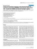
Figure 1 The immunohistochemical expression of Tn-C (1 A-C) and a-SMA (1 D-F) in bronchial biopsies from study subjects.The
figures show representative microphotographs from each category. A: Positivity for Tn-C is seen in tangentially sectioned basal epithelial cells
and along the basement membrane (BM) of bronchial epithelium (arrows); scale bar = 0.05 mm. B: Tn-C positivity in basal epithelial cells and in
the stroma underneath the BM (arrows); scale bar = 0.05 mm. C: Positivity for Tn-C in basal epithelial cells and in a wide area of stromal
connective tissue underneath the BM (arrows); scale bar = 0.05 mm. D: Spindle shaped cells positive for a-SMA (arrows) in a biopsy graded to
the category 1-4 positive cells. Smooth muscle of the bronchial wall (SM) or blood vessels (arrow heads) were not counted; scale bar = 0.05 mm.
E: A bronchial biopsy graded to the category 5-10 a-SMA positive cells (arrows). Blood vessels (arrow heads) or smooth muscle layer of the
bronchial wall (SM) were not counted; scale bar = 0.05 mm. F: More than 10 spindle shaped cells are showing positivity for a-SMA (arrows) in a
bronchial biopsy; scale bar = 0.05 mm.
Löfdahl et al. Respiratory Research 2011, 12:48
/>Page 4 of 11
estimates being close to the observed frequencies. Test
for homogeneity, i.e. no difference between all three
groups, was statistically significant for both Tn-C (p =
0.003) and a-SMA (p = 0.039), but since the difference
between the HS and NS groups was small, we
expressed the main effect by comparing the COPD
group to HS and NS combined (geometric mean of the
odds). Correlations between Tn-C and a-SMA expres-
sion and lung function were calculated with Spear-
man’s rank correlation coefficient.
A significance level of 5% was applied for all statistical
tests, and in case of a statistica lly significant result the
probability value (p-value) is given.
Results
Immunohistochemistry for Tn-C
In general, Tn-C was expressed as extracellular thin and
linear fibers underneath the bronchial epithelium and
also in the wider area of connective tissue of the bron-
chial walls. All evaluated biopsies showed positivity for
Tn-C also in basal epithelial cells but the expression pro-
file varied considerable between different patients. Repre-
sentative microphotographs are shown in Figure 1A-C.
The number and proportion of subjects within each
staining category are presented in Table 3 and Figure 2.
As shown, both the numbers of patients and the propo r-
tion of subjects expressing Tn-C staining beyond basal
epithelial cells and basal membrane, was higher in COPD
patients compared to smokers and nonsmokers (P <
0.001). Of the three ex-smokers in the COPD group, two
were in the lowest staining category (a),andoneinthe
intermediate (b).
Immunohistochemistry for a-SMA
Spindle shaped a-SMA positive cells were present in a
proportion of subjects from all three study groups,
representative microphotographs are shown in
Figure 1D-F and Figure 3A. Out of the 44 cases with
available stainings for desmin, 17 cases were spindl e
shaped cells positive both for a-SMA and desmin,
and 15 cases were spindle shaped cells positive for a-
SMA but negative for desmin. In the remaining 12
cases no spindle shaped cells positive for either a-
SMA or desmin were found which finding indicate
that those cases did not revealed any myofibroblasts.
In the cases with spindle shaped cells positive for
both antibodies, the desmin positive cells were always
very few in numbers (Figure 3B). The a-SMA positive
smooth muscle cells and endothelial cells were
excluded by their different location and morphology
when compared to that of spindle shaped cells (Figure
3C-D).
The number and proportion of subjects within each
stai ning category are present ed in Table 3 and Figure 4.
Presence of a-SMA staining was observed in 83% of the
COPD patients, in 46% of the smokers and in 41% of
the nonsmokers. When data are presented as cumulative
number and proportion of individuals with increasing
number of cells stained positive for a-SMA, COPD
patients had significantly higher (P < 0.05) a-SMA
expression than smokers and nonsmokers.
Of the three ex-smokers in the COPD group, one was
in category (b), and two were in category (c).
Immunohistochemistry for vimentin
Regardless of the presence of a-SMA or desmin positive
spindle shaped cell, all cases expressed vimentin positive
slender stromal cells. Most of them were probably fibro-
blasts of the subepithelial connective tissue (Figure 2E).
In addition to this, all inflammatory cells stained posi-
tively for vimentin.
Correlation between Tn-C and a-SMA
The expression of T n-C correlated positively to t he
expression of a-SMA. The estimate for the correlation
coefficient was 0.6; P < 0.0001 (Figure 5).
Table 3 The expression of Tenascin-C and a-SMA in patients with COPD, smokers (S) and non-smokers (NS)
COPD S NS
Tenascin C (Tn-C)
Number of acceptable biopsies 20 12 14
Subjects expressing Tn-C only in basal epithelial cells and basal membrane, Tn-C (a) 5(25%)9(75%)11(79%)
Subjects expressing Tn-C as Tn-C (a) plus the stroma underneath basement membrane, Tn-C (b) 10 (50%)2(17%)3(21%)
Subjects expressing Tn-C as Tn-C (b) plus wider expression within connective tissue, Tn-C (c) 5(25%)1(8%)0(0%)
a-SMA
Number of acceptable biopsies 18 13 12
Subjects with no cells expressing a-SMA, SMA (a) 3(17%)7(54%)7(59%)
Subjects with 1-5 cells expressing a-SMA, SMA (b) 6(33%)2(15%)3(25%)
Subjects with 5-10 cells expressing a-SMA, SMA
©
4(22%)3(23%)1(8%)
Subjects with >10 cells expressing a-SMA, SMA (d) 5(28%)1(8%)1(8%)
Löfdahl et al. Respiratory Research 2011, 12:48
/>Page 5 of 11
Correlations between Tn-C and a-SMA expression and
lung function parameters
There was no correlation between the expression of Tn-
Cora-SMA and any parameter of pulmonary function
(data not shown).
Discussion
In this study, we invest igated by immunohistochemistry
the expression of Tn-C and a-SMA positive spindle
shaped cells in bronchial mucosal biopsies as measures
of remodeling of large airways in patients with COPD.
We found that COPD patients had more expression of
both Tn-C and a-SMA positive cells compared to con-
trols. In addition, there was a positive correlation
between Tn-C and a-SMA expression. There were,
however, no correlations between the expression of Tn-
Cora-SMA and any lung function parameter.
Due t o the differential immunohistochemical expres-
sion of Tn-C and a-SMA we evaluated them in two dif-
ferent ways: Because the expression of Tn-C was mainly
extracellular and exhibited considerable variations
between individual patients, its expressio n was analyzed
by an applied semiquantitative method which took into
consideration t he specific cellular and histopathological
localizations of the protein also in the areas around
basement membranes. In contrast, a-SMA expression
was mainly intracellular also in those spindle shaped
cells which were quantitatively counted in the present
study. Both these evaluation methods are ea sily applied
in routine clinical diagnostics since no extra equipments
is needed. For further development of our grading sys-
tems the use of computer-assisted tomography might be
beneficial. Our method has not been widely used and its
repeatability may be lesser than 3-dimensional or 2-
dimensional methods described previously [39].
Tn-C is a glycoprotein associated with tissue remodel-
ing. In a study by Liesker et.al.[15],anincreaseinTn-
C expression in the large airways was seen both in
COPD and in asthma patients. There was, however, no
difference between COPD patients and a matched ex-
smokers control group. Th ere are several differences
between the study by Liesker et.al. and the present
study. Firstly, our study has two control groups: smokers
and non-smokers. Since there were no differences in
Tn-C expression between smokers and non smokers in
our study, we believe that the increas ed expression seen
in the COPD group is associated with the disease, i.e.
airways obstruction rather than exposure to tobacco
smoke. Secondly, in our study, quantitatively more
patients wit h a longer duration of smoking were investi-
gated, and the COPD patients had a more severe airway
obstruction. Finally, in our study all COPD patients
except three were current smokers, and all subjects in
the smokers control group were present smokers.
A previous study by Laitinen et.al.showed an increased
expression of Tn-C in the subepithelial layer of the
basement membrane of patients with asthma when
using immunofluorescence and morph ometr ic methods
for analyzing the bronchial biopsy samples [17]. The
quantification method of Tn-C in the present study was
not similar to the study of Laitinen et.al. S ince we ana-
lyzed the immunohistochemical expression of Tn-C in
the specific histological localizations of the airway
mucosa instead of measuring it. F urthermore, w e
Figure 2 Number and proportion of subjects expressing Tenascin C outside the basal epithelial cells and basement membrane. Data is
given for the three groups COPD, smoking controls (S) and non-smoking controls (NS). Tn C (b+c): All subjects with expression outside the basal
epithelial cells and basement membrane. Tn C
©
: Subjects with expression within connective tissue beyond the stroma underneath basal
membrane. The number (n) of individuals in each category is presented underneath corresponding bar. The Odds ratio for a COPD patient to be
in a higher category is statistically increased (P < 0.001) compared to subjects in the control groups.
Löfdahl et al. Respiratory Research 2011, 12:48
/>Page 6 of 11
Figure 3 The immunohistochemical expression of a-SMA, desmin and vimentin in bronchial biopsies from study subjects . The figures
show representative microphotographs from each category. A: High power field of spindle shaped cells positive for a-SMA; scale bar = 0.05
mm. B: High power field of desmin positive spindle shaped cells; scale bar = 0.05 mm. C: Ring like structures of blood vessels positive for a-SMA;
scale bar = 0.05 mm. D: Thick bundles of smooth muscle of the bronchial wall, staining for desmin; scale bar = 0.05 mm. E: Staining for vimentin
from a case in which no a-SMA or desmin positive spindle shaped cells were found. Positive staining pattern in normal fibroblasts and
lymphocytes of the subepithelial connective tissue; scale bar = 0.05 mm. F: A Negative control in which the primary antibody has been
substituted with non-immune mouse serum; scale bar = 0.1 mm.
Löfdahl et al. Respiratory Research 2011, 12:48
/>Page 7 of 11
observed that the staining for Tn-C beyond basal epithe-
lial cells and basement membrane was higher in COPD
patients compared to that of smokers and nonsmokers.
The results of our study are somewhat similar to that
particular study in that respect that in both studies the
increase o f Tn-C seemed to be correlated with the
remodeling proc ess of the airways, and not to its trigger.
To our knowledge, not much attention has previously
been paid on Tn-C expression outside the basement
membrane area. We observed, however, that over 50%
of our COPD-patients showed an increased expression
of Tn-C beyond the basement membrane area. Laitinen
and co-workers analyzed also the number of eosinophils
and lymphocytes, but did not found any correlation
between the amount of these inflammatory cells and the
expression of Tn-C. In the present study we attempted
to compare the number of a-SMA positive spindle
shaped cells, which were obviously myofibroblasts, w ith
the amount of Tn-C and found a positive correlation
between these two markers.
Figure 4 Number and proportions of subjects with cells staining positive for a-SMA. Data is given for the three groups COPD, smoking
controls (S) and non-smoking controls (NS). The numbers (n) of individuals in each category is presented as digits underneath each bar. The
odds for a COPD patient to be in a higher category is statistically increased (P < 0.05) compared to subjects in the control groups.
Figure 5 Correlation between staining for Tenascin C and a-SMA in all subjects. Increasing degree of staining is indicated by categories a-
c and a-d. Number in the circles indicate number of subjects. The estimate for the correlation coefficient was 0.6; P < 0.0001.
Löfdahl et al. Respiratory Research 2011, 12:48
/>Page 8 of 11
We were also ab le to show that biopsies from every
subject displayed a positive immunohistochemical
expression for T n-C at least around basal cells of the
bronchial epithelium, a somewhat novel finding since
Tn-C has not regularly been shown t o be expressed in
normal adult lung tissue. The results of our study might
signify that there is some constitutional expression of
Tn-C in basal epithelial cells of the human bronchus. In
our previo us studies in normal developing human lung
the expressions of Tn- C protein and mRNA were
increased during early developmental stages, and
decreasing in the end of gestation [23]. In the normal
adult human lung Tn-C expression was observ ed to be
very sparse [40], although in our earlier studies we
focused mainly on the alveolar level, not the central air-
ways. However, the enhanced Tn-C expression, co-loca-
lized with the expression of myofibroblasts, has been
observed in small airways i.e. b ronchioles of human
lung in neonatal disorders such as respiratory distress
syndrome (RDS) a nd bronchopulmo nary dysplasia
(BPD) [37].
Both in pulmonary fibrosis and during lung develop-
ment a-SMA positive spindle shaped cells, which were
obviously myofi broblasts, seemed to be the main source
of mRNA of Tn-C by in situ hybridization method
[22,23,37]. Myofibroblasts were in itially defined in ultra-
structural terms, with the essential features of intracellu-
lar fibers, which are positive for a-SMA, which is
nowadays the most common, yet not specific, marker
for a myofibroblast [24,41]. The origin of myofibroblasts
is still unclear. In our earlier studies human lung fibro-
blasts were differentiated into myofibroblasts by expos-
ing cells to transforming growth factor beta (TGF-b).
We observed that ultrastructural features of myofibro-
blasts were detected after exposure, e.g. a -SMA positive
bundles in the cytoplasm of cells, extracellular fibronec-
tin-containing structures on the surface of the cell, and
extracellular Tn-C in the vicinity of the cell [38]. Myofi-
broblasts seemed to have a role of the remodelling pro-
cess o f airways in asthmatic lung at least in animal and
experimental models [42,43], but not much is currently
known about the expression profile and function o f
myofibroblasts in COPD. Toourknowledgethisisthe
first study showing that a -SMA positive cells, which
might be myofibr oblasts, are increased in the airways of
the p atients with COPD, and moreover, the number of
myofibroblasts correlated with the amount of Tn-C. The
results suggest that mos t a-SMA positive cells revealed
typical expression profile of myofibroblast being positive
for a-SMA, vimentin and negative for desmin. In the
minority of cases the a-SMA positive cells were positive
also for desmin, which may suggest the other known
phenotype for myofibroblast [25]. Interestingly, a-SMA
positiv e cells were not present in every patient, whereas
Tn-C positivity, at least in basal epithelial cells, was
observed in every patient studied, which may indicate
that the basal cells might be able to produce Tn-C in
large airways even in healthy lung, and t hat myofibro-
blasts may be responsible for the production of the
excess of Tn-C in patients with COPD.
The clinical relevance of our finding can only be
speculated. Hypothetically, increased ECM deposition
in the large airways in our COPD patients may contri-
bute to airways obstruction. It is, however believed
that the major site of airways obstruction in COPD is
in the small airways and increased airway wall thick-
ness has been shown to correlate with FEV
1
[9,44]. It
is therefore likely to believe that the COPD patients in
thepresentstudyalsohavefeaturesofremodelingin
the small airways and probably also emphysema. Stu-
dies evaluating both large and small airways in a well
characterized patient material should therefore be
encouraged.
In conclusion, patients with COPD, but not smokers,
have signs of airway remodell ing in the large airways as
measured as an increased expression of Tn-C and a-
SMA positive cells which were obviously myofibroblasts.
The finding may represent processes leading to struc-
tural changes in the airway wall causing lung function
impairment in COPD.
Acknowledgements
The authors would like to acknowledge Heléne Blomqvist, Margitha Dahl,
Benita Dahlberg, Gunnel de Forest, Erja Tomperi, Mirja Vahera and Hannu
Wäänänen for excellent technical assistance.
This study was supported by the Swedish Heart-Lung Foundation, King
Gustaf V’s and Queen Victoria’s Freemasons ‘Foundation, King Oscar II
Jubilee Fund, the Hesselmans Foundation, Karolinska Institutet, the
Stockholm City Council, the Academy of Finland, the Jalmari and Rauha
Ahokas Foundation, the Finnish Anti-Tuberculosis Association Foundation,
the Duodecim of Oulu and the state subsidy for the University Hospital of
Oulu.
Author details
1
Dept Medicine, Division of Respiratory Medicine, Karolinska Institutet,
Karolinska University Hospital Solna. Stockholm Sweden.
2
Inst of Clinical
Medicine, Dept of Internal Medicine/Respiratory Research Unit, Centre of
Excellence in Research, University of Oulu and Oulu University Hospital, Oulu,
Finland.
3
Department of Pathology, Oulu University Hospital and Institute of
Diagnostics, Department of Pathology, University of Oulu, Finland.
Authors’ contributions
ML was corresponding author, enrolled and characterized study participants,
performed bronchoscopies and drafted the manuscript, RK and ELB
performed all immunohistochemical analyses and evaluations, and
participated in writing the manuscript, GT performed statistical analyses and
participated in writing the manuscript, MS initiated the project, participated
in its design and coordination, performed bronchoscopies, and participated
in writing the manuscript. All authors read and approved the final
manuscript.
Conflict of Interest disclosures
The authors declare that they have no competing interests.
Received: 29 September 2010 Accepted: 15 April 2011
Published: 15 April 2011
Löfdahl et al. Respiratory Research 2011, 12:48
/>Page 9 of 11
References
1. Murray CJ, Lopez AD: Alternative projections of mortality and disability
by cause 1990-2020: Global Burden of Disease Study. Lancet 1997,
349:1498-1504.
2. Lindberg A, Bjerg A, Ronmark E, Larsson LG, Lundback B: Prevalence and
underdiagnosis of COPD by disease severity and the attributable
fraction of smoking Report from the Obstructive Lung Disease in
Northern Sweden Studies. Respir Med 2006, 100:264-272.
3. Buist AS, McBurnie MA, Vollmer WM, Gillespie S, Burney P, Mannino DM,
Menezes AM, Sullivan SD, Lee TA, Weiss KB, Jensen RL, Marks GB, Gulsvik A,
Nizankowska-Mogilnicka E: International variation in the prevalence of
COPD (the BOLD Study): a population-based prevalence study. Lancet
2007, 370:741-750.
4. Global Initiative for Chronic Obstructive Lung Disease. Global strategy
for the diagnosis, management, and prevention of chronic obstructive
pulmonary disease. [ />5. Di Stefano A, Caramori G, Ricciardolo FL, Capelli A, Adcock IM, Donner CF:
Cellular and molecular mechanisms in chronic obstructive pulmonary
disease: an overview. Clin Exp Allergy 2004, 34:1156-1167.
6. O’Donnell R, Breen D, Wilson S, Djukanovic R: Inflammatory cells in the
airways in COPD. Thorax 2006, 61:448-454.
7. Sullivan AK, Simonian PL, Falta MT, Mitchell JD, Cosgrove GP, Brown KK,
Kotzin BL, Voelkel NF, Fontenot AP: Oligoclonal CD4+ T cells in the lungs
of patients with severe emphysema. Am J Respir Crit Care Med 2005,
172:590-596.
8. Vignola AM, Chanez P, Chiappara G, Merendino A, Pace E, Rizzo A, la
Rocca AM, Bellia V, Bonsignore G, Bousquet J: Transforming growth factor-
beta expression in mucosal biopsies in asthma and chronic bronchitis.
Am J Respir Crit Care Med 1997, 156:591-599.
9. Hogg JC, Chu F, Utokaparch S, Woods R, Elliott WM, Buzatu L,
Cherniack RM, Rogers RM, Sciurba FC, Coxson HO, Pare PD: The nature of
small-airway obstruction in chronic obstructive pulmonary disease. N
Engl J Med 2004, 350:2645-2653.
10. Roche WR, Beasley R, Williams JH, Holgate ST: Subepithelial fibrosis in the
bronchi of asthmatics. Lancet 1989, 1:520-524.
11. Bourdin A, Neveu D, Vachier I, Paganin F, Godard P, Chanez P: Specificity
of basement membrane thickening in severe asthma. J Allergy Clin
Immunol 2007, 119:1367-1374.
12. Minshall EM, Leung DY, Martin RJ, Song YL, Cameron L, Ernst P, Hamid Q:
Eosinophil-associated TGF-beta1 mRNA expression and airways fibrosis
in bronchial asthma. Am J Respir Cell Mol Biol 1997, 17:326-333.
13. Chetta A, Foresi A, Del Donno M, Bertorelli G, Pesci A, Olivieri D: Airways
remodeling is a distinctive feature of asthma and is related to severity
of disease. Chest 1997, 111:852-857.
14. Jeffery PK: Remodeling and inflammation of bronchi in asthma and
chronic obstructive pulmonary disease. Proc Am Thorac Soc 2004,
1:176-183.
15.
Liesker JJ, Ten Hacken NH, Zeinstra-Smith M, Rutgers SR, Postma DS,
Timens W: Reticular basement membrane in asthma and COPD: similar
thickness, yet different composition. Int J Chron Obstruct Pulmon Dis 2009,
4:127-135.
16. Roberts CR, Burke AK: Remodelling of the extracellular matrix in asthma:
proteoglycan synthesis and degradation. Can Respir J 1998, 5:48-50.
17. Laitinen A, Altraja A, Kampe M, Linden M, Virtanen I, Laitinen LA: Tenascin
is increased in airway basement membrane of asthmatics and
decreased by an inhaled steroid. Am J Respir Crit Care Med 1997,
156:951-958.
18. Soini Y, Paakko P, Nuorva K, Kamel D, Linnala A, Virtanen I, Lehto VP:
Tenascin immunoreactivity in lung tumors. Am J Clin Pathol 1993,
100:145-150.
19. Kaarteenaho-Wiik R, Sademies O, Paakko P, Risteli J, Soini Y: Extracellular
matrix proteins and myofibroblasts in granulomas of sarcoidosis,
atypical mycobacteriosis, and tuberculosis of the lung. Hum Pathol 2007,
38:147-153.
20. Kaarteenaho R, Sormunen R, Paakko P: Variable expression of tenascin-C,
osteopontin and fibronectin in inflammatory myofibroblastic tumour of
the lung. APMIS 2010, 118:91-100.
21. Calverley P, Pauwels Dagger R, Lofdahl CG, Svensson K, Higenbottam T,
Carlsson LG, Stahl E: Relationship between respiratory symptoms and
medical treatment in exacerbations of COPD. Eur Respir J 2005, 26:406-413.
22. Paakko P, Kaarteenaho-Wiik R, Pollanen R, Soini Y: Tenascin mRNA
expression at the foci of recent injury in usual interstitial pneumonia.
Am J Respir Crit Care Med 2000, 161:967-972.
23. Kaarteenaho-Wiik R, Kinnula V, Herva R, Paakko P, Pollanen R, Soini Y:
Distribution and mRNA expression of tenascin-C in developing human
lung. Am J Respir Cell Mol Biol 2001, 25:341-346.
24. Gabbiani G, Ryan GB, Majne G: Presence of modified fibroblasts in
granulation tissue and their possible role in wound contraction.
Experientia 1971, 27:549-550.
25. Schurch W, Seemayer TA, Gabbiani G: The myofibroblast: a quarter
century after its discovery. Am J Surg Pathol 1998, 22:141-147.
26. Kawka DW, Kazazis DM, Clark RA: In vivo co-distribution of fibronectin and
actin fibers in granulation tissue: immunofluorescence and electron
microscope studies of the fibronexus at the myofibroblast surface. J Cell
Biol 1984, 98:2091-2106.
27. Vancheri C, Failla M, Crimi N, Raghu G: Idiopathic pulmonary fibrosis: a
disease with similarities and links to cancer biology. Eur Respir J 2010,
35:496-504.
28. Chaponnier C, Gabbiani G: Pathological situations characterized by
altered actin isoform expression. J Pathol 2004, 204:386-395.
29. Wick MR, Hormnick JL:
Immunohistology of soft tissue and osseous
neoplasms. In Diagnostic
Immunohistochemistry 3 edition. Edited by:
Dabbs DJ. Saunders Elsevier; 2010:84.
30. Dellagi K, Brouet JC: Redistribution of intermediate filaments during
capping of lymphocyte surface molecules. Nature 1982, 298:284-286.
31. Kranenburg AR, Willems-Widyastuti A, Moori WJ, Sterk PJ, Alagappan VK, de
Boer WI, Sharma HS: Enhanced bronchial expression of extracellular
matrix proteins in chronic obstructive pulmonary disease. Am J Clin
Pathol 2006, 126:725-735.
32. Gosselink JV, Hayashi S, Elliott WM, Xing L, Chan B, Yang L, Wright C, Sin D,
Pare PD, Pierce JA, Pierce RA, Patterson A, Cooper J, Hogg JC: Differential
expression of tissue repair genes in the pathogenesis of chronic
obstructive pulmonary disease. Am J Respir Crit Care Med 2010,
181:1329-1335.
33. Degen M, Goulet S, Ferralli J, Roth M, Tamm M, Chiquet-Ehrismann R:
Opposite effect of fluticasone and salmeterol on fibronectin and
tenascin-C expression in primary human lung fibroblasts. Clin Exp Allergy
2009, 39:688-699.
34. Harju T, Kinnula VL, Paakko P, Salmenkivi K, Risteli J, Kaarteenaho R:
Variability in the precursor proteins of collagen I and III in different
stages of COPD. Respir Res 2010, 11:165.
35. Lambert RK, Wiggs BR, Kuwano K, Hogg JC, Pare PD: Functional
significance of increased airway smooth muscle in asthma and COPD.
J Appl Physiol 1993, 74:2771-2781.
36. Lofdahl JM, Cederlund K, Nathell L, Eklund A, Skold CM: Bronchoalveolar
lavage in COPD: fluid recovery correlates with the degree of
emphysema. Eur Respir J 2005, 25:275-281.
37. Kaarteenaho-Wiik R, Kinnula VL, Herva R, Soini Y, Pollanen R, Paakko P:
Tenascin-C is highly expressed in respiratory distress syndrome and
bronchopulmonary dysplasia. J Histochem Cytochem 2002, 50:423-431.
38. Kaarteenaho-Wiik R, Paakko P, Sormunen R: Ultrastructural features of lung
fibroblast differentiation into myofibroblasts. Ultrastruct Pathol 2009,
33:6-15.
39. Jeffery P, Holgate S, Wenzel S: Methods for the assessment of
endobronchial biopsies in clinical research: application to studies of
pathogenesis and the effects of treatment. Am J Respir Crit Care Med
2003, 168:S1-17.
40. Kaarteenaho-Wiik R, Tani T, Sormunen R, Soini Y, Virtanen I, Paakko P:
Tenascin immunoreactivity as a prognostic marker in usual interstitial
pneumonia. Am J Respir Crit Care Med 1996, 154:511-518.
41. Eyden B: Electron microscopy in the study of myofibroblastic lesions.
Semin Diagn Pathol 2003, 20:13-24.
42. Michalik M, Pierzchalska M, Legutko A, Ura M, Ostaszewska A, Soja J,
Sanak M: Asthmatic bronchial fibroblasts demonstrate enhanced
potential to differentiate into myofibroblasts in culture. Med Sci Monit
2009, 15:BR194-201.
43. Miller M, Cho JY, McElwain K, McElwain S, Shim JY, Manni M, Baek JS,
Broide DH: Corticosteroids prevent myofibroblast accumulation and
airway remodeling in mice. Am J Physiol Lung Cell Mol Physiol
2006, 290:
L162-169.
Löfdahl et al. Respiratory Research 2011, 12:48
/>Page 10 of 11
44. Hogg JC, Macklem PT, Thurlbeck WM: Site and nature of airway
obstruction in chronic obstructive lung disease. N Engl J Med 1968,
278:1355-1360.
doi:10.1186/1465-9921-12-48
Cite this article as: Löfdahl et al.: Tenascin-C and alpha-smooth muscle
actin positive cells are increased in the large airways in patients with
COPD. Respiratory Research 2011 12:48.
Submit your next manuscript to BioMed Central
and take full advantage of:
• Convenient online submission
• Thorough peer review
• No space constraints or color figure charges
• Immediate publication on acceptance
• Inclusion in PubMed, CAS, Scopus and Google Scholar
• Research which is freely available for redistribution
Submit your manuscript at
www.biomedcentral.com/submit
Löfdahl et al. Respiratory Research 2011, 12:48
/>Page 11 of 11