Báo cáo y học: " Therapeutic efficacy of TBC3711 in monocrotaline-induced pulmonary hypertension" pot
Bạn đang xem bản rút gọn của tài liệu. Xem và tải ngay bản đầy đủ của tài liệu tại đây (1.75 MB, 13 trang )
RESEA R C H Open Access
Therapeutic efficacy of TBC3711 in
monocrotaline-induced pulmonary hypertension
Djuro Kosanovic
1
, Baktybek Kojonazarov
1
, Himal Luitel
1
, Bhola K Dahal
1
, Akylbek Sydykov
1
, Teodora Cornitescu
1
,
Wiebke Janssen
2
, Ralf P Brandes
3
, Neil Davie
4
, Hossein A Ghofrani
1
, Norbert Weissmann
1
, Friedrich Grimminger
1
,
Werner Seeger
1,2
and Ralph T Schermuly
1,2*
Abstract
Background: Endothelin-1 signalling plays an important role in pathogenesis of pulmonary hypertension. Although
different endothelin-A receptor antagonists are developed, a nove l therapeutic option to cure the disease is still
needed. This study aims to investigate the therapeutic efficacy of the selective endothelin-A receptor antagonist
TBC3711 in monocrotaline-induced pulmonary hypertension in rats.
Methods: Monocrotaline-injected male Sprague-Dawley rats were randomized and treated orally from day 21 to
35 either with TBC3711 (Dose: 30 mg/kg body weight/day) or placebo. Echocardiographic measurements of
different hemodynamic and right-heart hypertrophy parameters were performed. After day 35, rats were sacrificed
for invasive hemodynamic and right-heart hypertrophy measurements. Additionally, histologic assessment of
pulmonary vascular and right-heart remodelling was performed.
Results: The novel endoth elin-A receptor antagonist TBC3711 significantly attenuated monocrotaline-induced
pulmonary hypertension, as evident from improved hemodynamics and right-heart hypertrophy in comparison
with placebo group. In addition, muscularization and medial wall thickness of distal pulmonary vessels were
ameliorated. The histologic evaluation of the right ventricle showed a significant reduction in fibrosis and
cardiomyocyte size, suggesting an improvement in right-heart remodelling.
Conclusion: The results of this study suggest that the selective endothelin-A receptor antagonist TBC3711
demonstrates therapeutic benefit in rats with established pulmonary hypertension, thus representing a useful
therapeutic approach for treatment of pulmonary hypertension.
Background
Pulmonary hypertension (PH) is a chronic life-threaten-
ing disease charac terized by a progressive augmentation
of pulmonary arterial pressure that finally leads to right
ventricle failure and death. PH has a multicomplex
pathology that includes a combination of pulmonary
vascular remodelling, vasoconstriction and in situ
thrombosis. The progressive pulmonary vascular remo-
delling is the attribute of PH pathology and is character-
ized by abnormalities of vascular cells, such as increased
proliferation, migration and resistance to apoptosis [1,2].
Although the PH pathology is the subject of intensive
research, the precise molecular mechanisms are not
fully understood and successful therapeutic strategy to
cure the disease is still needed.
An accumulating body of literature clearly underlines
the central role of endothelial dysfunction in the devel-
opment and progression of PH [3-5]. Endothelin (ET)-1
is synthesized by endothelial cells in the human vascula-
ture and causes a strong and potent vasoconstriction
[6,7]. ET-1 is primarily produced by endothelial cells
and manifests effects through 2 G-protein-coupled
receptors ET-A and ET-B. These receptors have a differ-
ent localization and therefore cause the different biologi-
cal responses. The ET -A receptors are mostly express ed
on pulmonary artery smooth muscle c ells (PASMCs),
cardiomyocytes and fibroblasts, whereas the ET-B recep-
tors are presented on endothelial cells and, to a lesser
extent, on PASMCs [8]. After activation by ET-1, both
* Correspondence:
1
University of Giessen Lung Center, Giessen, Germany
Full list of author information is available at the end of the article
Kosanovic et al. Respiratory Research 2011, 12:87
/>© 2011 Kosanovic et a l; licensee BioMed Central Ltd. This is an Open Access article distributed under the terms of the Creative
Commons Attribution License ( 0), which permits unrestricted use, distribution, and
reproduction in any medium, provided the ori ginal work is properly cited.
receptor types located on PASMCs cause a potent
vasoconstriction and pro liferation of PASMCs [9]. The
ET-B receptors expressed on endot helial cells mediate a
vasodilatation through nitric oxide and cyclic guanosine
monophosphate and pr ostacyclin production and ET-B
receptor-mediated ET-1 clearance [10,11]. Additionally,
it is shown that defic iency of the ET-B receptor mark-
edly accelerates the progression of PH in monocrotaline
(MCT)-injected rats [12]. Nishida et al suggest that
ET-A receptor mediated action is exclusively involved in
the pathogenesis of MCT-induced PH, although they
could not rule out a protective role of ET-B receptor
mediated actions [13]. These facts created a novel para-
digm that selective ET-A receptor antagonism is more
favorable than a nonselective ET-A/ET-B approach.
The right-heart failure is the final stage in pro gression
of PH, and it is known that ET receptors are expressed
on cardiomyocytes as well [14]. ET-1 causes cardiac
hypertrophy [15,16], and it was shown that treatment
with an ET-A receptor antagonist improved the hemo-
dynamics and survival in rats with chronic heart failure
[17]. More importantly, the selective ET-A receptor
antagonists, such as LU135252, PD155080, BQ-123,
BMS-193884, significantl y reduced righ t-heart hypertro-
phy and improved heart function i n the MCT model of
PH [16,18-20].
Through the years many selective ET-A receptor
antagonists, such as BQ-123 [16,21,22], YM598 [23],
GF063 [24] and sitaxentan, were developed and
exhibited beneficial therapeutic effects in experimental
models of PH. Sitaxentan, a very potent and selective
ET-A receptor antagonist, successfully prevented and
reversed pulmonary vascular remodelling and right-
heart hypertrophy in rat hypoxic model, whereas only
thepreventiveeffectsintheMCTmodelofPHwere
observed [25,26].
A novel and highly potent ET-A receptor antagonist,
TBC3711 (IC
50
= 0.08 nM) has been reported [27-30].
This compound shows a significantly stronger ET-A/
ET-B selectivity (441.000-fold) as compared with
sitaxentan (6.500-fold) and thus can represent a novel
specific and selective therapeutic approach for the treat-
ment of PH. This study explored, for the first time to
our knowledge, the therapeutic efficacy of TBC3711 in a
well-established MCT model of pulmonary arterial
hypertension in rats and the effects of TBC3711 on (1)
hemodynamics, (2) pulmonary vascular remodelling and
(3) r ight ventricular (RV) hypertrophy and remodelling
on MCT-induced pulmonary hypertension in rats.
Methods
Experimental design
Adult male Sprague-Dawley rats (300-350 g body weight
(BW)) were given a subcutaneous injection of saline
(healthy control, n = 8) or MCT (60 mg/kg BW, n =
30). MCT-injected rats were randomized into 2 groups
and were treated orally by gavage from 21 to 35 days
either with TBC3711 (MCT-TBC3711 group, 30 mg/kg
BW/day, n = 16) or placebo (MCT-placebo group,
methyl-cellulose, n = 14). The BW changes and survival
were monitored from 21 to 35 days for all experimental
groups (Additional file 1, Figure S1). All a nimal studies
were performed according to the guidelines of the
University of Giessen and were approved by the local
authorities.
Echocardiographic measurements
Echocardiographic measurements of pulmonary artery
acceleration time (ACT), tricuspid annular plane systolic
excursion (TAPSE), cardiac output (CO), heart rate
(HR), right ventricular dimensions (RVD) and right ven-
tricular wall thickness (RVWT) were performed for all
experimental groups. Anesthesia was induced with 3%
isoflurane gas and maintained with 1.0% to 1.5% isoflur-
ane in room air supplemented with 100% O
2
. Rats were
laid supine on a heating platform with all legs taped to
electrocardiogram electrodes for monitoring heart rate
(HR). Body temperature was monitored via a rectal
thermometer (Indus Instruments, Houston, TX, USA)
and maintained at 36.5°C to 37.5°C using a heating pad
and lamp. The rat’s chest was shaved and treated with a
chemical hair remover to reduce ultrasound attenuation.
To provide a coupling medium for the transducer, a
warm ultrasound gel was spread over the chest wall.
Transthoracic 2-dimensional, M-mo de and Dop pler
imagi ng were performed with a high-re solution imaging
system equipped with a 25-MHz transducer (Visual-
Sonics, Toronto, ON, Canada). Right ventricular wall
thickness (RVWT) was measured in the m odified para-
sternal long-axis view. R ight ventricular dimension
(RVD) was measured from the right ventricle (RV) out-
flow tract view at the level of the aortic valve. For deter-
mination of tricuspid annular plane systolic excursion
(TAPSE), M-mode cursor was oriented to the junction
of the tricuspid valve plane with the RV free wall using
the apical 4-chamber view. Pulmonary a rtery accelera-
tion time (ACT) (Additional file 2, F igure S2) w as
measured from the pulsed-wave Doppler flow velocity
profile of the RV outflow tract in the parasternal short-
axis view and d efined as the interval from the onset to
the maximal velocity of forward flow [31,32]. Cardiac
output (CO) was calculated as the product of the velo-
city time integral of the pulsed-Doppler tracing in the
left ventricle (LV) outflow tract, the cross-sectional area
of the LV outflow tract and the HR [33]. All echocar-
diographic parameters were calculated offline using tool
section of the VisualSonics Vevo770 System. All the
studies were performed by an experienced sonographer
Kosanovic et al. Respiratory Research 2011, 12:87
/>Page 2 of 13
who was blinded to results of invasive and morpho-
metric studies.
Invasive hemodynamic measurement of the right
ventricular systolic pressure (RVSP) and systemic
arterial pressure (SAP)
The animals were initially anesthetized intraperitoneally
with a mixture of ketamine ( 50 mg/kg) and medetomi-
din (100 μg/kg) for invasive hemodynamic measure-
ments. A right-heart catheter was inserted through the
right jugular vein for measurement of right ventricular
sys tolic pressure (RVSP) and the left carotid artery can-
nulation was performed for systemic arterial pressure
(SAP) measurement, as described previously [34-36].
The partial arterial oxygen pressure (paO
2
)wasmea-
sured by blood gas analysis for the determination of
oxygenation index (paO
2
/FiO
2
) [36].
Assessment of right ventricular (RV) hypertrophy and
paraffin embedding of the hearts
The RV wall was separated from the left ventricular
(LV) wall and ventricular septum (S). RV hyper trophy
wasexpressedasthewetweightratiooftheRVwall
and free LV wall with ventricular septum (RV/(LV+S)),
as already described [34-36]. RVWT was measured by
using a standard micrometer calliper (RVWT invasive).
At the end of all heart hypertrophy measurements the
RV wall, LV wall and S were fixed with formalin (3.5%-
3.7%) and after dehydration were embedded in paraffin
for histologic analysis.
Morphometric analysis of the lung vessels
The left lungs from all experimental groups were for-
malin-fixed and paraffin embedded for the morpho-
metric analysis of the pulmonary vessels (medial wall
thickness and degree of muscularizati on) and for the
assessment of index of proliferation (IOP), as described
previously [35].
Determination of collagen content and cardiomyocyte
size in right ventricles
Freshly dissected RV tissues were fixed in 3.5% to 3.7%
formalin solution overnight, then dehydrated and
embedded in paraffin and sectioned at a thickness of 3
μm. To detect collagen fibres, the RV sections were
stained with 0.1% Sirius red F3B (Niepoetter, Bürstadt,
Germany) in picric acid (F luka, Buchs, Germany). Photo-
micrographs were quantified to determine the interstitial
collagen fraction by using Leica QWin V3 computer-
assisted image analysis software (Leica Microsystem,
Wetzlar, Germany). Average data reflect results from at
least 4 different hearts in each group, as described pre-
viously [37]. For cardiomyocyte size determination the
transverse section of formalin-fixed paraffin-embedded
RVs were stained with fluorescein isothiocy anate-conju-
gated wheat germ agglutinin (Sigma Aldrich, Steinheim,
Germany). Nuclei were stained with diamidino phenylin-
dole (Invitrogen, Darmstadt, Germany) and mounted
with fluorescent mounting medium (Dako, Hamburg,
Germany). The cross-sectional area per cardiomyocyte
was measured by using Leica QWin software [38].
Data analysis
All data w ere expressed as mean ± SEM. The different
experimental groups were statistically analyzed by 1-way
ANOVA and Newman-Keuls post hoc test for multiple
comparisons. P values of < 0.05 were considere d as
statistically significa nt. The correlations between differ-
ent invasive and echocardiographic parameters were
analyzed using a Spearman analysis.
Results
Effect of TBC3711 on hemodynamics in monocrotaline-
induced pulmonary hypertension
MCT induced a robust and severe PH in rats as
reflected by a significantly increased right ventricular
systolic pressure (RVSP) in the placebo group (74.0 ±
3.6 vs 29.3 ± 1.9 mmHg in healthy group; Figure 1a ).
Oral treatment with TBC3711 showed a significant
decrease in RVSP compared with placebo (54.7 ± 3.8 vs
74.0 ± 3.6 mmHg). SAP was n ot changed in a ny of
experimental groups (Figure 1b).
Effect of TBC3711 on pulmonary artery acceleration time,
cardiac output, total pulmonary resistance and
oxygenation index
Pulmonary artery accelera tion time (ACT) was measu red
as described in Methods by using echocardiography (Addi-
tional file 2, Figure S2). MCT induced a severe PH in rats,
which is reflected by a significant reduction in ACT com-
pared with healthy animals (Figure 2a). Animals treated
with TBC3711 showed a signific ant increase of ACT in
comparison with placebo. CO was decreased in MCT-pla-
cebo rats as compared with healthy controls (53.5 ± 4.8 vs
83.4 ± 3.5 ml/min). TBC3711 caused a significant increase
of CO compared with the placebo group (Figure 2b).
Total pulmonary resistance (TPR) was defined as RVSP
divided by cardiac index (CI) and was significantly aug-
mented in the placebo group compared with healthy rats
(5.98 ± 1.03 vs 1.24 ± 0.04 mmHg × min × ml-¹ × 100 g
BW) and TBC3711 effectively reduced the value of TPR
(3.10 ± 0.29 vs 5.98 ± 1.03 mmHg × min × ml-¹ × 100 g
BW in placebo group; Figure 2c). The o xygenation index
(paO
2
/FiO
2
) was significantly reduced in the MCT-
placebo gr oup compared with healthy animals (320 ± 46
vs 472 ± 20 mmHg). The treatment with TBC3711
improved oxygenation index compared with the placebo
group (477 ± 28 vs 320 ± 46 mmHg) (Figure 2d).
Kosanovic et al. Respiratory Research 2011, 12:87
/>Page 3 of 13
TBC3711 reduced right ventricular hypertrophy and
improved right-heart function
Tricuspid annular plane systolic excursion (TAPSE), as a
measure of RV systolic function was significantly
decreased in the placebo group (1.59 ± 0.14 vs 2.68 ±
0.04 mm in healthy animals; Figure 3a). TBC3711
remarkably improved TAPSE as compared with placebo
(2.51 ± 0.06 vs 1.59 ± 0.14 m m). The RV hypertrophy
was evident from a significantly increased RV/(LV+S)
ratio of MCT-injected rats receiving placebo, compared
with the healthy rats (0.61 ± 0.03 vs 0.25 ± 0.01; Figure
3b). Treatment with TBC3711 significantly reduce d the
RV/(LV+S) ratio in comparison with placebo (0.41 ±
0.02 vs 0.61 ± 0.0 3). RVWT w as significantly increased
in MCT-injected rats as measured by both invasive and
echocardiographic approaches. The oral treatment of
rats with TBC3711 showed a significantly reduced
RVWT compared with placebo group (Figures 3c, d).
RVD were augmented in rats injected with MCT (4.94 ±
0.41 vs 2.49 ± 0.04 mm in healthy controls) and chronic
treatment with TBC3711 caused the significant decrease
of RVD compared with placebo group (3.78 ± 0.11 vs
4.94 ± 0.41 mm; Figure 3e).
The correlation between different parameters measured
by invasive and echocardiographic approaches in
monocrotaline-injected rats
The Spearman analysis demonstrated a significant cor-
relation betw een invasive measuremen ts of different
parameters and echocardiographic (echo) measure-
ments (Figure 4). RV/(LV+S) and RVWT-echo (r =
0.88, P < 0.0001); RVSP and RVWT-echo (r = 0.81, P
<0.0001)andRVWTinvasiveandRVWT-echo(r=
0.81, P < 0.0001) showed the noticeable positive corre-
lation. Here we also demonstrated that RVSP signifi-
cantly negatively correlates with ACT (r = -0.89, P <
0.0001).
Effect of TBC3711 on monocrotaline-induced pulmonary
vascular remodelling
To assess the effect of TBC3711 on pulmonary vascu-
lar remodelling, we analyzed medial wall thickness
(Figure 5a; Additional file 3, Figure S3a) and degree of
muscularization (Figure 5b; Additional file 3, Figure
S3b) of peripheral pulmonary vessels (20-50 μm). MCT
injectionresultedinanenhanced pulmonary artery
remodelling as evident from a significantly increas ed
medial wall thickness in placebo group (26.5 ± 1. 1 vs
11.3 ± 0.3% in healthy control). ET-A receptor antago-
nist TBC3711 showed the effective reduction of medial
wall thickness in the treated group compared with pla-
cebo (16.1 ± 0.7 vs 26.5 ± 1.1%). MCT injection in rats
resulted in augmented pulmonary arterial musculariza-
tion as evident from an increase i n the number of fully
muscularized arteries in placebo group (63.2 ± 1.8 vs
3.0 ± 0.8% in healthy controls). The number of non-
muscularized vessels was significantly reduced in
MCT-placebo group as compared with healthy control
a
)
b)
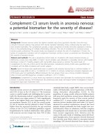
Figure 1 Effect of TBC3711 on hemodynamics in monocrotaline (MCT)-induced pulmonary hypertension. Rats were treated with TBC3711
(n = 14) or vehicle (n = 9) from day 21-35 after MCT-injection. TBC3711 (MCT-TBC3711, 30 mg/kg body weight/day) was administered once per
day orally by gavage. Equal volume of the vehicle (methyl-cellulose) was given to a placebo group (MCT-placebo). After 35 days all animals were
humanely killed for hemodynamic measurements. (a) Right ventricular systolic pressure (RVSP) and (b) Systemic arterial pressure (SAP) are given.
Bars represent mean ± SEM. One-way ANOVA with Newman-Keuls multiple comparison post-hoc test was performed for statistical analysis. ***P
< 0.001.
Kosanovic et al. Respiratory Research 2011, 12:87
/>Page 4 of 13
(2.4 ± 0.6 vs 63.3 ± 4.7%). TBC3711 caused the marked
reduction of pulmonary vascular remodelling as
reflected by a significantly d ecreased fully muscularized
vessels in treated group compared with placebo (18.9 ±
1.5 vs 63.2 ± 1.8%). Also, the number of partially mus-
cularized arteries was significantly increased in
TBC3711 treated group (64.3 ± 1.7 vs 34.4 ± 1.6% in
placebo g roup).
TBC3711 reduced the index of proliferation
MCT caused a strong in situ proliferation of perivascu-
lar cel ls (Additional file 3, Figure S3c) in the sm all pul-
monary vessels (20-50 μm), as evident from increased
IOP (Figure 6), as compared with healthy controls ( 640
± 28 vs 100 ± 22%). TBC3711 showed a significant
reduction in IOP, in c omparison with placebo group
(428 ± 46 vs 640 ± 28%).
a)
b)
c) d)
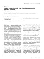
Figure 2 Effect of TBC3711 on pulmonary artery acceleration time (ACT), cardiac output (CO), total pulmonary resistance (TPR) and
oxygenation index (paO
2
/FiO
2
) in monocrotaline (MCT)-induced pulmonary hypertension. Rats were treated with TBC3711 (30 mg/kg
body weight/day, n = 14) or vehicle (n = 9) from day 21-35 after MCT-injection accompanied by echocardiography measurement and blood gas
analysis. (a) Pulmonary artery acceleration time (ACT), (b) Cardiac output (CO), (c) Total pulmonary resistance (TPR) and (d) oxygenation index
(paO
2
/FiO
2
) of different experimental groups are given. Bars represent mean ± SEM. One-way ANOVA with Newman-Keuls multiple comparison
post-hoc test was performed for statistical analysis. *P < 0.05, ***P < 0.001.
Kosanovic et al. Respiratory Research 2011, 12:87
/>Page 5 of 13
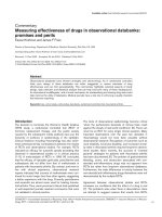
Effect of TBC3711 on collagen content and cardiomyocyte
size in right ventricles of monocrotaline-injected rats
Right ventricle collagen content (Figure 7a, c) was mark-
edly increased in rats with MCT-induced pulmonary
hypertension compared with healthy animals (2.60 ±
0.14 vs 0.54 ± 0.03%). TBC3711 significantly decreased
the collagen area in comparison with placebo group
(1.45 ± 0.05 vs 2.60 ± 0.14%). Cardiomyocytes hypertro-
phy (Figure 7b, d) measurement was expressed as cross -
sectional area (μm
2
) per cardiomyocyte. MCT injection
significantly increased a cardiomyocyte size (484 ± 12 vs
199 ± 8 μm
2
in healthy rats). TBC3711 noticeably
reduced the cardiomyoc yte size compared with placebo
group (261 ± 9 vs 484 ± 12 μm
2
).
Discussion
The main results from the present study are that (a)
TBC3711 significantly improved hemodynamics i n
MCT-induced PH in rats and noticeable reduced pul-
monary vascular remodelling and perivascular proliferat-
ing cells and (b) TBC3711 chronic treatment also
strongly diminished right-heart remodelling as evident
from collagen content and cardiomyocyte size, which was
followed by a significantly decreased RV hypertrophy.
Over the years, many different therapeutic options
have been investigated for the trea tment of pulmonary
hypertension, such as PDE5 inhibitors, prostacyclin ana-
logs and endothelin-receptor antagonists [39,40].
Although these therapeutic approaches improved the
quality of life and prolonged survival of patients with
pulmonary hypertensi on, novel clinical options to
achieve the ultimate goal of reversing the progressive
pulmonary vascular remodelling and RV hypertrophy
are needed.
Although it is not exactly clear what triggers the
initiation of PH, a growing body of literature implicates
a)
b)
c)
d) e)
Figure 3 TBC3711 reduced a right ventricular hypertrophy and improved a right-heart function in monocrotaline model of pulmonary
hypertension. Tricuspid annular plane systolic excursion (TAPSE) was measured by echocardiography. The right ventricle (RV) wall was separated
from the left ventricle (LV) wall and ventricular septum (S). RV hypertrophy (heart ratio) was expressed as the weight ratio of the RV wall and free
LV wall with ventricular septum (RV/(LV+S)) in different experimental groups. Additionally, right ventricular wall thickness (RVWT) and right
ventricular dimensions (RVD) were measured by echocardiography. (a) TAPSE, (b) RV/(LV+S), (c) RVWT measured by echocardiography (RVWT-
echo), (d) RVWT measured by invasive approach (RVWT invasive) and (e) RVD measured by echocardiography are shown for all experimental
groups. Bars represent mean ± SEM. One-way ANOVA with Newman-Keuls multiple comparison post-hoc test was performed for statistical
analysis. ***P < 0.001.
Kosanovic et al. Respiratory Research 2011, 12:87
/>Page 6 of 13
the main role of ET system in the development and pro-
gression of the disease [4,5,41]. ET-1, an extremely
strong vasoconstrictor primarily produced by endothelial
cells, is significantly elevated in both human and MCT-
induced PH [42,43]. ET-1 achieves the biological eff ects
through 2 G p rotein-coupled receptor isoforms ET-A
and ET-B. These receptors have a distinct localization,
unique affinities and locations for binding ET-1 and
therefore cause different biological responses [ 44]. The
ET-A receptors are mostly expressed on PASMCs, car-
diomyocytes and fibroblasts. When expressed on
PASMCs, the ET-B receptors together with ET-A recep-
tors mediate vasoconstrictio n and proliferation of vascu-
lar cells, but when expressed on endothelial cells the
ET-B receptors cause vasodilatation and ET-1 clearance
[9-11,44]. Ivy et al have shown that ET-B recepto r defi-
ciency accelerates the progression of PH in MCT rat
model, and Nishida et al discussed their findings and
suggested that ET-A receptor-mediated action is exclu-
sively involved in the pathogenesis of MCT-induced PH,
although they could not rule out a protective role of
ET-B receptor-mediated actions [12,13]. All these find-
ings pointed toward the requirement of a novel thera-
peutic approach in the direction of selective ET-A
receptor antagonism, altho ugh nonselective approach
(Bosentan) also showed the effective reduction of the
PH in animal models [45,46].
Sitaxentan, a potent and highly selective ET-A recep-
tor antagonist, prevented and reversed pulmonary vascu-
lar remodelling and cardiac hypertrophy in hypoxia
models, whereas only the preventive effects were
observed in MCT and over-circulation models of PH
[3,25,26]. A series of clinical studies were performed
with sitaxentan, named the Sitaxentan to Relieve
a)
b)
c) d)
Figure 4 The correlation betw een different parameters measured by invasive and echocardiogra phic approaches in monocrotaline
(MCT)-injected rats. Correlations between invasive and echocardiographic measurements of different parameters are shown. (a) weight ratio of
the right ventricle (RV) wall and free left ventricle (LV) wall with ventricular septum (RV/(LV+S)) and right ventricular wall thickness measured by
echocardiography (RVWT- echo), (b) right ventricular systolic pressure (RVSP) and pulmonary artery acceleration time (ACT), (c) RVSP and RVWT-
echo and (d) RVWT invasive and RVWT-echo. The Spearman analysis was performed for statistical analysis. All P < 0.0001.
Kosanovic et al. Respiratory Research 2011, 12:87
/>Page 7 of 13
a)
b)
Figure 5 Effect of TBC3711 on monocrotaline (MCT)-induce d pulmonary vascular remodelling. The rat lung sections were stained with
elastica staining for medial wall thickness determination of pulmonary vessels in all experimental groups (a). The rat lung sections were also
immunostained for a-smooth muscle actin and von Willebrand factor. (b) Proportion of non (N), partially (P) or fully (F) muscularized pulmonary
vessels, as a percentage of total pulmonary vessel cross section (sized 20-50 μm) is given for different experimental groups. Bars represent mean
± SEM. One-way ANOVA with Newman-Keuls multiple comparison post-hoc test was performed for statistical analysis. ***P < 0.001.
Kosanovic et al. Respiratory Research 2011, 12:87
/>Page 8 of 13
Impaired Exercise Studies (STRIDE) [3,47]. Collectively,
these studies showed the benefits of selective ET-A
receptor antagonism in the setting of human forms of
PH. Unfortunately, due to a pattern of idiosyncratic liver
function abnormalities, sitaxentan was removed from
the market in December 2010.
In the experiments outlined in the current manuscript,
we used a novel, highly potent ET-A receptor antagonist
(IC
50
= 0.08 nM), named TBC3711. This follow-up
compound was discovered following c hemical substitu-
tion with variety of electron-withdrawing groups at the
ortho position on the anilino ring [28]. TBC3711 shows
a good oral bioavailability in rats and significantly stron-
ger ET-A/ET-B selec tivity (441.000-fold) as compared
with sitaxentan (6500-fold) [27-29]. These advantages of
TBC3711 indicate the need to investigate the therapeu-
tic potency of this compound as a novel therapeutic
option.
TBC3711 improved hemodynamics, reduced pulmon-
ary vascular resistance and increased cardiac output in
MCT rats treated with this compound in comparison
with placebo g roup, as evident from both invasiv e and
noninvasive measurements. Also, rats treated with
TBC3711 had significa ntly higher arterial oxygenation
compared with placebo group. Previously, it was shown
that ACT inversely correlates with mean pulmonary
arterial pres sure [48-50]. In agreem ent with this findi ng
we demonstrat ed that RVSP significantly negatively cor-
relates with ACT, suggesting the importance of a nonin-
vasive approach for prognostic significance in PH.
Corroborating with hemodynamic da ta, TBC3711 signif-
icantly reduced pulmonary vascular remodelling as evi-
dent from decreased medial wall thickness and degree
of muscularization of intra-acinar arteries (20-50 μm
diameter). The decrease of pulmonary vascular remodel-
ling may be associated with notably reduced level of vas-
cular cell proliferation in situ, as evident from IOP. This
is in agreement with literature that suggests t he role of
the E T system in the proliferation of v ascular cells [3].
Additionally, MCT-injected rats treated with TBC3711
exhibited slightly improved survival and body weights
compared with placebo (Additional file 1, Figure S1).
Furthermore, no significant effect was observed on heart
rate, suggesting that impr ovement in cardiac output
after TBC3711 treatment was due to increased stroke
volume (Additional file 4, Figure S4). Our findings that
selective ET-A receptor antagonism in MCT-model of
PH exhibited beneficial effects on hemodynamic changes
and pulmonary vascular remodelling are in line with lit-
erature [21-23,51-53].
Brunner et al demonstrated that ET-A receptor antag-
onism alleviated cardiac dysfunction via improved Ca
2+
handling in MCT-induced RV hypertrophy, suggesting
the role of ET-A receptors in RV dysfunction [18].
TBC3711 therapy significantly improved RV systolic
function in the MCT- induced PH model, as evident from
TAPSE me asurement. TBC3711 strongly reduced the RV
hypertrophy as evident from invasive measurements of
heart ratio (RV/(LV+S)) and RVWT and echocardio-
graphic measurements of RVWT and RV dimensions.
Because remodelling of the right-heart structure is an
attribute of the RV hypertrophy, we also invest igated the
effects of TBC3711 on cardiomyocytes hypertrophy and
cardiac fibrosis and found that both parameters were
significantly diminished in treated rats. The role of the
ET system in pathogenesis of RV hypertrophy is not
clear and very little is known. Jasmin et al have shown
in the MCT model of PH that r ight ventricular ET-1
levels and ET-B receptor density were significantly
increased, whereas ET-A receptors were not affected.
Interestingly, they have shown that these alterations in
the ET system in the RV were noticeably attenuated by
treatment with selective ET-A receptor antagonist [19].
From the other side, Miyauchi et al in 1993 concluded
that inhibition of RV hypertrophy by selective ET-A
receptor antagonist BQ-123 in MCT-inj ected rats may
also be because of the blockade of excessive stimulation
of the heart by ET-1 in additio n to the prevention
of PH [16]. In 2000 Miyauchi et al found that there was
a significant increase of atrial natriuretic peptide
Figure 6 TBC3711 reduced the index of proliferation (IOP) of
perivascular cells in monocrotaline (MCT) model of pulmonary
hypertension. In situ proliferation of perivascular cells in rat lungs
was investigating by using immunostaining of lung tissues with an
anti-proliferating cell nuclear antigen rabbit polyclonal antibody, as
explained in Methods. IOP of different experimental groups is
shown. Bars represent mean ± SEM. One-way ANOVA with
Newman-Keuls multiple comparison post-hoc test was performed
for statistical analysis. ***P < 0.001.
Kosanovic et al. Respiratory Research 2011, 12:87
/>Page 9 of 13
expression in the right-heart of MCT-injected ra ts,
which is a marker for the failing heart and that increase
was strongly prevented by selective ET-A receptor
antagonism [20]. Whether the reduction of right-heart
hypertrophy in our study was solely due t o improved
hemodynamics or to a contribution from direct effect of
TBC3711 on the RV, was not deeply investigated here,
and we do not have data to resolve this question.
In summary, we successfully demonstrated that daily
oral treatment with TBC3711 in the dose of 30 mg/kg
of BW/day impaired the progression of PH, as evident
from significantly improved hemodynamics, RV hyper-
trophy and remodelling and pulmonary vascular remo-
delling in MCT-induced PH model in rats. Although
some manifestations of human PH pathology, such as
neointima formation and plexiform lesions are not
A
B C
a
)
A
B
C
b
)
c) d)
Figure 7 Effect of TBC3711 on collagen content and cardiomyocyte size in right ventricles of monocrotaline (MCT)-injected rats. Right
ventricle (RV) sections were stained with 0.1% Sirius Red in picric acid to assess the collagen content and also with fluorescein isothiocyanate
conjugated wheat germ agglutinin/diamidino phenylindole to assess the cardiomyocyte size. Photomicrographs were quantified to determine
the interstitial collagen fraction and cardiomyocyte size by using computer-assisted image analysis software. (a, b) Representative
photomicrographs are presented (A - healthy control, B - MCT-placebo and C - TBC3711 treated group; arrows indicate collagen formation - red
color). (c) Collagen area and (d) cross sectional area are shown for different experimental groups. Bars represent mean ± SEM. One-way ANOVA
with Newman-Keuls multiple comparison post-hoc test was performed for statistical analysis. ***P < 0.001.
Kosanovic et al. Respiratory Research 2011, 12:87
/>Page 10 of 13
covered by this model, other important pathological
characteristics, such as initial endothelial injury,
increased perivascular inflammation and de novo muscu-
larization of small pulmonary arteries are shared fea-
tures between human disease and MCT model. The
animal models that more closely mimic human PH were
reported in recent years, suc h as combination of MCT
and one-sided pneumonectomy and hypoxia in combi-
nation with vascular endothelial growth factor receptor
(VEGFR)-2 inhibitor SU5416. TBC3711 can thus be
anticipated to yield therapeutic benefit in these animal
models and can be used for further investigation. Addi-
tionally, TBC3711 can be used as a useful tool to further
elucidate the role of ET system in recently found factors
that are implicat ed in pulmonary vascular remodelling
process, such as signal transducers and activators of
transcription-3 (STAT3), nuclear factor of activated T
cell (NFAT) and miR-204 [54,55]. Recently, Nagendran
et al showed the putative contractile and metabolic
effects of ET-1 antagonist on RV functions, which may
have an important clinical value. Therefore, the potential
future studies with TBC3711 may reveal if this antago-
nist has similar effects [56]. Previously it was shown that
TBC3711 ameliorated the hypoxia-induced PH in the
newborn piglet, suggesting the effec tiveness of the com-
pound in pulmonary hyperten sion caused by different
stimulus, like hypoxia [27]. Taken together, the findi ngs
from our study indicate that TBC3711 with its beneficial
properties, such as good oral bioavai lability (~100% in
rats), high potency (ET-A receptor IC
50
= 0.08 nM) and
strong selectivity (441.000-fold) [28] may offer a new
therapeutic approach to cure PH.
Additional material
Additional file 1: Figure S1. Effect of TBC3711 on body weight
changes and survival in monocrotaline (MCT)-induced pulmonary
hypertension. Body weights and survival were monitored each day for
last 2 weeks of experiments and the mean values of those 2 parameters
on day 22 were considered to be 100%. (a) The surviva l curves and (b)
body weight changes of different experimental groups and time points
are shown.
Additional file 2: Figure S2. Representative photographs from
echocardiography. As described in Methods, the pulmonary artery
acceleration time (ACT) was measured by echocardiography. The
representative photographs from different experimental groups are
shown (A - healthy control, B - monocrotaline (MCT)-placebo and C -
MCT-TBC3711).
Additional file 3: Figure S3. Representative photomicrographs from
elastica staining and immunostainings. (a) elastica staining and (b)
double immunostaining (anti-a-smooth muscle actin (purple/violet color)
and anti-von Willebrand factor (brown color) antibodies) were performed
to assess medial wall thickness and degree of muscularization, the 2
well-known measures of pulmonary vascular remodelling.
Immunostaining with anti-proliferating cell nuclear antigen (PCNA)
antibody (c) was performed to assess the in situ proliferation state of
pulmonary vascular cells. Arrows indicate the PCNA-positive nuclei/cells.
The representative photographs from stained lung tissues of different
experimental groups are shown (A - healthy controls, B - monocrotaline
(MCT)-placebo and C - MCT-TBC3711).
Additional file 4: Figure S4. Effect of TBC3711 on heart rate (HR).
The heart rate (beats/min) was measured by echocardiography, as
described in Methods and values of different experimental groups are
given.
Acknowledgements
We would like to acknowledge Diane Warner of Complete Healthcare
Communications, Inc. for her copy-edit and styling assistance, which was
funded by Pfizer Inc.
Author details
1
University of Giessen Lung Center, Giessen, Germany.
2
Max-Planck-Institute
for Heart and Lung Research, Bad Nauheim, Germany.
3
Institute for
Cardiovascular Physiology, J.W. Goethe University, Frankfurt, Germany.
4
Pfizer
Global Research and Development, Sandwich, UK.
Authors’ contributions
DK, HAG, NW, WS, FG and RTS conceived and designed the study. DK, BK
and HL performed experiments. DK, BK, HL, BKD, HAG, NW, FG, WS and RTS
analyzed and interpreted data. AS and TC were involved in interpretation of
data. DK and RTS drafted and finalized the manuscript. BKD, AS, TC, WJ, RPB
and ND were involved in revising the manuscript for important intellectual
content. All authors read and approved the final manuscript.
Competing interests
The authors declare that they have no competing interests.
Received: 19 March 2011 Accepted: 23 June 2011
Published: 23 June 2011
References
1. Ghofrani HA, Barst RJ, Benza RL, Champion HC, Fagan KA, Grimminger F,
Humbert M, Simonneau G, Stewart DJ, Ventura C, Rubin LJ: Future
perspectives for the treatment of pulmonary arterial hypertension. JAm
Coll Cardiol 2009, 54:S108-S117.
2. Humbert M, Morrell NW, Archer SL, Stenmark KR, MacLean MR, Lang IM,
Christman BW, Weir EK, Eickelberg O, Voelkel NF, Rabinovitch M: Cellular
and molecular pathobiology of pulmonary arterial hypertension. JAm
Coll Cardiol 2004, 43:13S-24S.
3. Pullamsetti SS, Schermuly RT: Endothelin receptor antagonists in
preclinical models of pulmonary hypertension. Eur J Clin Invest 2009,
39:3-13.
4. Tuder RM, Cool CD, Yeager M, Taraseviciene-Stewart L, Bull TM, Voelkel NF:
The pathobiology of pulmonary hypertension. Endothelium. Clin Chest
Med 2001, 22:405-18.
5. Lopes AA, Maeda NY, Goncalves RC, Bydlowski SP: Endothelial cell
dysfunction correlates differentially with survival in primary and
secondary pulmonary hypertension. Am Heart J 2000, 139:618-23.
6. Filep JG: Endothelin peptides: biological actions and pathophysiological
significance in the lung. Life Sci 1993, 52:119-33.
7. Yanagisawa M, Kurihara H, Kimura S, Tomobe Y, Kobayashi M, Mitsui Y,
Yazaki Y, Goto K, Masaki T: A novel potent vasoconstrictor peptide
produced by vascular endothelial cells. Nature 1988, 332:411-5.
8. Davenport AP, O’Reilly G, Molenaar P, Maguire JJ, Kuc RE, Sharkey A,
Bacon CR, Ferro A: Human endothelin receptors characterized using
reverse transcriptase-polymerase chain reaction, in situ hybridization,
and subtype-selective ligands BQ123 and BQ3020: evidence for
expression of ETB receptors in human vascular smooth muscle. J
Cardiovasc Pharmacol 1993, 22:S22-S25.
9. Seo B, Oemar BS, Siebenmann R, von SL, Luscher TF: Both ETA and ETB
receptors mediate contraction to endothelin-1 in human blood vessels.
Circulation 1994, 89:1203-8.
10. Dupuis J, Goresky CA, Fournier A: Pulmonary clearance of circulating
endothelin-1 in dogs in vivo: exclusive role of ETB receptors. J Appl
Physiol 1996, 81:1510-5.
Kosanovic et al. Respiratory Research 2011, 12:87
/>Page 11 of 13
11. Hirata Y, Emori T, Eguchi S, Kanno K, Imai T, Ohta K, Marumo F: Endothelin
receptor subtype B mediates synthesis of nitric oxide by cultured
bovine endothelial cells. J Clin Invest 1993, 91:1367-73.
12. Ivy DD, McMurtry IF, Colvin K, Imamura M, Oka M, Lee DS, Gebb S,
Jones PL: Development of occlusive neointimal lesions in distal
pulmonary arteries of endothelin B receptor-deficient rats: a new model
of severe pulmonary arterial hypertension. Circulation 2005, 111:2988-96.
13. Nishida M, Eshiro K, Okada Y, Takaoka M, Matsumura Y: Roles of endothelin
ETA and ETB receptors in the pathogenesis of monocrotaline-induced
pulmonary hypertension. J Cardiovasc Pharmacol 2004, 44:187-91.
14. Kobayashi T, Miyauchi T, Sakai S, Kobayashi M, Yamaguchi I, Goto K,
Sugishita Y: Expression of endothelin-1, ETA and ETB receptors, and ECE
and distribution of endothelin-1 in failing rat heart. Am J Physiol 1999,
276:H1197-H1206.
15. Ito H, Hirata Y, Hiroe M, Tsujino M, Adachi S, Takamoto T, Nitta M,
Taniguchi K, Marumo F: Endothelin-1 induces hypertrophy with
enhanced expression of muscle-specific genes in cultured neonatal rat
cardiomyocytes. Circ Res 1991, 69:209-15.
16. Miyauchi T, Yorikane R, Sakai S, Sakurai T, Okada M, Nishikibe M, Yano M,
Yamaguchi I, Sugishita Y, Goto K: Contribution of endogenous endothelin-
1 to the progression of cardiopulmonary alterations in rats with
monocrotaline-induced pulmonary hypertension. Circ Res 1993, 73:887-97.
17. Sakai S, Miyauchi T, Kobayashi M, Yamaguchi I, Goto K, Sugishita Y:
Inhibition of myocardial endothelin pathway improves long-term
survival in heart failure. Nature 1996, 384:353-5.
18. Brunner F, Wolkart G, Haleen S: Defective intracellular calcium handling in
monocrotaline-induced right ventricular hypertrophy: protective effect
of long-term endothelin-A receptor blockade with 2-benzo[1,3]dioxol-5-
yl-3-benzyl-4-(4-methoxy-phenyl-)- 4-oxobut-2-enoate-sodium (PD
155080). J Pharmacol Exp Ther 2002, 300:442-9.
19. Jasmin JF, Cernacek P, Dupuis J: Activation of the right ventricular
endothelin (ET) system in the monocrotaline model of pulmonary
hypertension: response to chronic ETA receptor blockade. Clin Sci (Lond)
2003, 105:647-53.
20. Miyauchi T, Sato R, Sakai S, Kobayashi T, Ueno M, Kondo H, Kawano S,
Goto K, Yamaguchi I: Endothelin-1 and right-sided heart failure in rats:
effects of an endothelin receptor antagonist on the failing right
ventricle. J Cardiovasc Pharmacol 2000, 36:S327-S330.
21. Bonvallet ST, Zamora MR, Hasunuma K, Sato K, Hanasato N, Anderson D,
Sato K, Stelzner TJ: BQ123, an ETA-receptor antagonist, attenuates
hypoxic pulmonary hypertension in rats. Am J Physiol 1994, 266:
H1327-H1331.
22. DiCarlo VS, Chen SJ, Meng QC, Durand J, Yano M, Chen YF, Oparil S: ETA-
receptor antagonist prevents and reverses chronic hypoxia-induced
pulmonary hypertension in rat. Am J Physiol 1995, 269:L690-L697.
23. Yuyama H, Fujimori A, Sanagi M, Koakutsu A, Sudoh K, Sasamata M,
Miyata K: The orally active nonpeptide selective endothelin ETA receptor
antagonist YM598 prevents and reverses the development of pulmonary
hypertension in monocrotaline-treated rats. Eur J Pharmacol 2004,
496:129-39.
24. Yan LD, Kong LL, Yong Z, Dong HJ, Chi MG, Pan XF, Zhang C, Liang YJ,
Gong ZH, Liu KL: Inhibition of endothelin-1- and hypoxia-induced
pulmonary pressor responses in the rat by a novel selective endothelin
A receptor antagonist, di-n-butylaminocarbamyl-L-Leucyl-D-
Tryptophanyl-D-4-chloro-Phe. J Cardiovasc Pharmacol 2010, 56:246-54.
25. Rondelet B, Kerbaul F, Vivian GF, Hubloue I, Huez S, Fesler P, Remmelink M,
Brimiouille S, Salmon I, Naeije R: Sitaxsentan for the prevention of
experimental shunt-induced pulmonary hypertension. Pediatr Res 2007,
61:284-8.
26. Tilton RG, Munsch CL, Sherwood SJ, Chen SJ, Chen YF, Wu C, Block N,
Dixon RA, Brock TA: Attenuation of pulmonary vascular hypertension and
cardiac hypertrophy with sitaxsentan sodium, an orally active ET(A)
receptor antagonist. Pulm Pharmacol Ther 2000, 13:87-97.
27. Perreault T, Berkenbosch JW, Barrington KJ, Decker ER, Wu C, Brock TA,
Baribeau J: TBC3711, an ET(A) receptor antagonist, reduces neonatal
hypoxia-induced pulmonary hypertension in piglets. Pediatr Res 2001,
50:374-83.
28. Wu C, Decker ER, Blok N, Bui H, You TJ, Wang J, Bourgoyne AR, Knowles V,
Berens KL, Holland GW, Brock TA, Dixon RA: Discovery, modeling, and
human pharmacokinetics of N-(2-acetyl-4,6-dimethylphenyl)-3-(3,4-
dimethylisoxazol-5-ylsulfamoyl)thio phene-2-carboxamide (TBC3711), a
second generation, ETA selective, and orally bioavailable endothelin
antagonist. J Med Chem 2004, 47:1969-86.
29. Kosanovic D, Kojonazarov B, Dahal BK, Ghofrani HA, Weissmann N,
Grimminger F, Seeger W, Schermuly RT: A Highly Selective Endothelin-A
Receptor Antagonist TBC3711 Reverses Monocrotaline Induced
Pulmonary Hypertension [Abstract]. Am J Respir Crit Care Med 2010, 181:
A6316.
30. Mercier O, Sage E, Izziki M, Humbert M, Dartevelle P, Eddahibi S, Fadel E:
Endothelin A receptor blockade improves regression of flow-induced
pulmonary vasculopathy in piglets. J Thorac Cardiovasc Surg 2010,
140:677-83.
31. Hardziyenka M, Campian ME, de Bruin-Bon HA, Michel MC, Tan HL:
Sequence of echocardiographic changes during development of right
ventricular failure in rat. J Am Soc Echocardiogr 2006, 19:1272-9.
32. Lindqvist P, Calcutteea A, Henein M: Echocardiography in the assessment
of right heart function. Eur J Echocardiogr 2008, 9:225-34.
33. Slama M, Susic D, Varagic J, Ahn J, Frohlich ED: Echocardiographic
measurement of cardiac output in rats. Am J Physiol Heart Circ Physiol
2003, 284:H691-H697.
34. Dahal BK, Cornitescu T, Tretyn A, Pullamsetti SS, Kosanovic D, Dumitrascu R,
Ghofrani HA, Weissmann N, Voswinckel R, Banat GA, Seeger W,
Grimminger F, Schermuly RT: Role of epidermal growth factor inhibition
in experimental pulmonary hypertension. Am J Respir Crit Care Med 2010,
181:158-67.
35. Dahal BK, Kosanovic D, Pamarthi PK, Sydykov A, Lai YJ, Kast R, Schirok H,
Stasch JP, Ghofrani HA, Weissmann N, Grimminger F, Seeger W,
Schermuly RT: Therapeutic efficacy of azaindole-1 in experimental
pulmonary hypertension. Eur Respir J 2010, 36:808-18.
36. Schermuly RT, Dony E, Ghofrani HA, Pullamsetti S, Savai R, Roth M,
Sydykov A, Lai YJ, Weissmann N, Seeger W, Grimminger F: Reversal of
experimental pulmonary hypertension by PDGF inhibition.
J Clin Invest
2005, 115:2811-21.
37. Grimm PC, Nickerson P, Gough J, McKenna R, Stern E, Jeffery J, Rush DN:
Computerized image analysis of Sirius Red-stained renal allograft
biopsies as a surrogate marker to predict long-term allograft function. J
Am Soc Nephrol 2003, 14:1662-8.
38. Li HL, Liu C, de CG, Ouzounian M, Sun M, Wang AB, Huang Y, He CW,
Shi Y, Chen X, Nghiem MP, Liu Y, Chen M, Dawood F, Fukuoka M,
Maekawa Y, Zhang L, Leask A, Ghosh AK, Kirshenbaum LA, Liu PP:
Curcumin prevents and reverses murine cardiac hypertrophy. J Clin
Invest 2008, 118:879-93.
39. Boutet K, Montani D, Jais X, Yaici A, Sitbon O, Simonneau G, Humbert M:
Therapeutic advances in pulmonary arterial hypertension. Ther Adv Respir
Dis 2008, 2:249-65.
40. Chin KM, Rubin LJ: Pulmonary arterial hypertension. J Am Coll Cardiol
2008, 51:1527-38.
41. Budhiraja R, Tuder RM, Hassoun PM: Endothelial dysfunction in pulmonary
hypertension. Circulation 2004, 109:159-65.
42. Frasch HF, Marshall C, Marshall BE: Endothelin-1 is elevated in
monocrotaline pulmonary hypertension. Am J Physiol 1999, 276:
L304-L310.
43. Stewart DJ, Levy RD, Cernacek P, Langleben D: Increased plasma
endothelin-1 in pulmonary hypertension: marker or mediator of disease?
Ann Intern Med 1991, 114:464-9.
44. Opitz CF, Ewert R: Dual ET(A)/ET(B) vs. selective ET(A) endothelin receptor
antagonism in patients with pulmonary hypertension. Eur J Clin Invest
2006, 36:1-9.
45. Chen SJ, Chen YF, Meng QC, Durand J, DiCarlo VS, Oparil S: Endothelin-
receptor antagonist bosentan prevents and reverses hypoxic pulmonary
hypertension in rats. J Appl Physiol 1995, 79:2122-31.
46. Clozel M, Hess P, Rey M, Iglarz M, Binkert C, Qiu C: Bosentan, sildenafil,
and their combination in the monocrotaline model of pulmonary
hypertension in rats. Exp Biol Med (Maywood) 2006, 231:967-73.
47. Langleben D, Brock T, Dixon R, Barst R: STRIDE 1: effects of the selective
ET(A) receptor antagonist, sitaxsentan sodium, in a patient population
with pulmonary arterial hypertension that meets traditional inclusion
criteria of previous pulmonary arterial hypertension trials. J Cardiovasc
Pharmacol 2004, 44:S80-S84.
48. Chan KL, Currie PJ, Seward JB, Hagler DJ, Mair DD, Tajik AJ: Comparison of
three Doppler ultrasound methods in the prediction of pulmonary
artery pressure. J Am Coll Cardiol 1987, 9:549-54.
Kosanovic et al. Respiratory Research 2011, 12:87
/>Page 12 of 13
49. Boissiere J, Gautier M, Machet MC, Hanton G, Bonnet P, Eder V: Doppler
tissue imaging in assessment of pulmonary hypertension-induced right
ventricle dysfunction. Am J Physiol Heart Circ Physiol 2005, 289:
H2450-H2455.
50. Bonnet P, Bonnet S, Boissiere J, Le Net JL, Gautier M, Dumas dlR, Eder V:
Chronic hypoxia induces nonreversible right ventricle dysfunction and
dysplasia in rats. Am J Physiol Heart Circ Physiol 2004, 287:H1023-H1028.
51. Bialecki RA, Stinson-Fisher C, Murdoch W, Bertelsen D, Desiato M,
Rumsey W: A novel orally active endothelin-A receptor antagonist,
ZD1611, prevents chronic hypoxia-induced pulmonary hypertension in
the rat. Chest 1998, 114:91S.
52. Chen SJ, Chen YF, Opgenorth TJ, Wessale JL, Meng QC, Durand J,
DiCarlo VS, Oparil S: The orally active nonpeptide endothelin A-receptor
antagonist A-127722 prevents and reverses hypoxia-induced pulmonary
hypertension and pulmonary vascular remodeling in Sprague-Dawley
rats. J Cardiovasc Pharmacol 1997, 29:713-25.
53. Prie S, Leung TK, Cernacek P, Ryan JW, Dupuis J: The orally active ET(A)
receptor antagonist (+)-(S)-2-(4,6-dimethoxy-pyrimidin-2-yloxy)-3-
methoxy-3,3-diphe nyl-propionic acid (LU 135252) prevents the
development of pulmonary hypertension and endothelial metabolic
dysfunction in monocrotaline-treated rats. J Pharmacol Exp Ther 1997,
282:1312-8.
54. Courboulin A, Paulin R, Giguere NJ, Saksouk N, Perreault T, Meloche J,
Paquet ER, Biardel S, Provencher S, Cote J, Simard MJ, Bonnet S: Role for
miR-204 in human pulmonary arterial hypertension. J Exp Med 2011,
208:535-48.
55. Paulin R, Courboulin A, Meloche J, Mainguy V, Dumas dlR, Saksouk N,
Cote J, Provencher S, Sussman MA, Bonnet S: Signal transducers and
activators of transcription-3/pim1 axis plays a critical role in the
pathogenesis of human pulmonary arterial hypertension. Circulation
2011, 123:1205-15.
56. Nagendran J, Sutendra G, Paterson I, Webster L, Haromy A, Ross D,
Rebeyka I, Michelakis E: The endothelin axis is upregulated in human and
rat right ventricular (RV) hypertrophy and its inhibition decreases RV
contractility [Abstract]. Circulation 2009, 120:S805.
doi:10.1186/1465-9921-12-87
Cite this article as: Kosanovic et al.: Therapeutic efficacy of TBC3711 in
monocrotaline-induced pulmonary hypertension. Respiratory Research
2011 12:87.
Submit your next manuscript to BioMed Central
and take full advantage of:
• Convenient online submission
• Thorough peer review
• No space constraints or color figure charges
• Immediate publication on acceptance
• Inclusion in PubMed, CAS, Scopus and Google Scholar
• Research which is freely available for redistribution
Submit your manuscript at
www.biomedcentral.com/submit
Kosanovic et al. Respiratory Research 2011, 12:87
/>Page 13 of 13