Báo cáo y học: "Change in serum KL-6 level from baseline is useful for predicting life-threatening EGFR-TKIs induced interstitial lung disease" pdf
Bạn đang xem bản rút gọn của tài liệu. Xem và tải ngay bản đầy đủ của tài liệu tại đây (1.16 MB, 11 trang )
RESEARC H Open Access
Change in serum KL-6 level from baseline is
useful for predicting life-threatening EGFR-TKIs
induced interstitial lung disease
Shigeo Kawase
1
, Noboru Hattori
1*
, Nobuhisa Ishikawa
1
, Yasushi Horimasu
1
, Kazunori Fujitaka
1
, Osamu Furonaka
2
,
Takeshi Isobe
3
, Seigo Miyoshi
4
, Hironobu Hamada
4,5
, Takashi Yamane
6
, Akihito Yokoyama
6
and Nobuoki Kohno
1
Abstract
Background: A high incidence of interstitial lung disease (ILD) has been reported in patients with advanced non-
small cell lung cancer (NSCLC) treated with epidermal growth factor receptor-tyrosine kinase inhibitors (EGFR-TKIs),
particularly in Japanese populations. A previous report from our laboratory demonstrated that KL-6 was a useful
serum biomarker to assess the severity of drug-induced pneumonitis. Based on these observations, this study was
conducted to evaluate the risk factors of EGFR-TKIs induced ILD and the usefulness of monitoring serum KL-6 levels
in patients who developed EGFR-TKIs induced ILD in a large multi-instituti onal setting.
Methods: We retrospectively reviewed clinical records and radiographies of 341 patients with advanced NSCLCs
who were treated with EGFR-TKIs, and analyzed risk factors for the development of EGFR-TKIs induced ILD.
Changes of circulating levels of KL-6 were also evaluated in the patients who developed EGFR-TKIs induced ILD.
Results: Among the 341 patients included in this study, 20 (5.9%) developed EGFR-TKIs induced ILD, and 9 (2.6%)
died from ILD. Univariate analyses revealed that only preexisting pulmonary fibrosis was a significant risk factor for
the development of EGFR-TKIs induced ILD (p = 0.003). Absolute levels of circulating KL-6 at neither baseline nor
the onset of ILD could discriminate between life-threatening and non-life threatening EGFR-TKIs induced ILDs.
However, we found that the ratios of serum KL-6 levels just after the onset of EGFR-TKIs induced ILD to those at
baseline could quite precisely distinguish survivors from non-survivors (p = 0.006) as well as acute interstitial
pneumonia (AIP) pattern from non-AIP pattern (p = 0.005).
Conclusions: The results of this study strongly support the potential of KL-6 as a diagnostic biomarker for life-
threatening EGFR-TKIs induced ILD. Monitoring of KL-6 is also useful to evaluate the progression and severity of
EGFR-TKIs induced ILD.
Keywords: Lung cancer, KL-6, EGFR-TKI, interstitial lung disease
Background
Gefitinib (ZD1839, Iressa; AstraZeneca) and erlotinib
(Tarceva, OSI-774; OSI Pharmaceuticals) are orally
active epidermal growth factor receptor tyrosin e kinase
inhibitors (EGFR-TKIs) used for the treatment of non-
small cell lung cancer (NSCLC) patients [1]. EGFR-TKIs
sometimes cause drastic tumor regression in specific
subgroups of patients with advanced NSCLC, including
women, non-smokers, patients with lung adenocarci-
noma (ADC) histology, patients of Asian origin and
patients with EGFR mutations [2-6]. On the other hand,
treatment with EGFR-TKIs is associated with serious
side effects, such as life-threatening drug-induced inter-
stitial lung disease (ILD), particularly in Japanese popu-
lations [7-13]. These previous studies have reported that
male gender, smoking history, poor performance status
(PS), and preexisting ILD are risk factors for developing
EGFR-TKIs induced ILD, however, we questioned
whether each of these should be equally considered for
* Correspondence:
1
Department of Molecular and Internal Medicine, Graduate School of
Biomedical Sciences, Hiroshima University, 1-2-3 Kasumi, Minami-ku,
Hiroshima 734-8551, Japan
Full list of author information is available at the end of the article
Kawase et al. Respiratory Research 2011, 12:97
/>© 2011 Kawase et al; licensee BioMed Central Ltd. Th is is an Open Access article distributed under the terms of the Creative Commons
Attribution License ( which permits unrestricted use, dis tribution, and reproduction in
any medium, provided the original work is properly cited.
the risk-benefit assessment to use EGFR-TKIs for the
treatment of NSCLCs in a practical clinical setting. In
addition, we also wondered whether we can assess the
severity of EGFR-TKIs induced ILD when it develops
during EGFR-TKIs treatment.
KL-6 is a mucin-like glycoprotein with a molecular
weight of 200kd and has been classified as human
MUC1 mucin [14-17]. Previous studies have demon-
stratedthatserumlevelsofKL-6areelevatedinavari-
ety of ILDs, such as idiopathic pulmonary fibrosis (IPF),
collagen vascular disease associated interstitial pneumo -
nitis, radiation pneumonitis, pulmonary sarcoi dosis
[18-26]. Furthermore, our laboratory has also demon-
strated that absolute levels of KL-6 at the onset of drug-
induced ILD can predict the clinical outcomes [27].
Although our previous studies have suggested the use-
fulness of KL-6 as a tumor marker [28,29] and a predic -
tor of survival in NSCLC patients treated with EGFR-
TKIs [30], significance of circulating KL-6 level as a
detector of EGFR-TKIs induced ILD or a predictor of
clinical outcome in patients with EGFR-TKIs induced
ILD has not been determined yet.
In the cohort of the present study, to obtain more
information on risk factors for developing EGFR-TKIs
induce d ILD, the characteristics of NSCLC patients who
developed ILD during EGFR-TKIs treatment were ana-
lyzed. In addition, to evaluate whether monitoring
serum KL-6 levels in N SCLC patients during the treat-
ment is useful to detect the development of EGFR-TKIs
induced ILD or predict the clinical outcome of EGFR-
TKIs induced ILD, circulating KL-6 levels were mea-
sured in NSCLC patients included in the cohort before
and during EGFR-TKIs treatment.
Methods
Study subjects
Between August 2002 and August 2010, 341 advanced
NSCLC patients treated with gefitinib (250 mg/day) or
erlotinib (150 mg/day) at Hiroshima University Hospital
(Hiroshima, Japan), Ehime University Hospital (Ehime,
Japan), Shimane University Hospital (Shimane, Japan),
Kochi University Hospital (Kochi, Japan) and Onomichi
General Hospital (Hiroshima, Japan) were consecutively
enrolled in the study. The disease staging was carried
out using computed tomography (CT) scan of t he chest
and abdomen, bone scintigraphy or F-18 fluorodeoxy-
glucose positron emission tomography (FDG-PET/CT),
and magnetic resonance imaging (MRI) of the head. To
obtain information on both the response of tumor to
EGFR-TKIs treatment and the occurrence of EGFR-
TKIs induced ILD, chest radiography and/or CT scans
wereperformedatleastonceamonthateachinstitu-
tion, and the patients were followed-up until 12 weeks
after the administration of EGFR-TKIs. Informed
consent was obtained from all patients. This study com-
plied with the Declaration of Helsinki, and was approved
by the individual institutional Ethical Committees.
Diagnosis of preexisting pulmonary disorder and EGFR-
TKIs induced ILD
The presence of preexisting pulmonary fibrosis was
determined according to the diagnostic criteria set by
the ATS/ERS on the basis of clinical characteristic and/
or chest CT findings, and the types of preexisting pul-
monary fibrosis were classified into idiopathic pulmon-
ary fibrosis (IPF) pattern and non-IPF pattern [31-33].
In addition, the presence of preexisting pulmonary
emphysema was determined by chest CT findings that
show low attenuation areas occupying more than 25% of
the entire lung field in at least one slice [34]. The diag-
nosisofEGFR-TKIsinducedILDwasmadeusingthe
diagnostic algorithm described elsewhere [11,35]. We
defined EGFR-TKIs induced ILD as diffuse pulmonary
infiltrates newly developed during EGFR-TKIs treatment
with lack of evidence for alternative diseases such as
infection, tumor progression, heart failure and pulmon-
ary embolism. When the occurrence of EGFR-TKIs
induced ILDs was suspected, chest CT scans were per-
formed, levels of brain natriuretic peptide (BNP) and D-
dimer in blood were measured, the sputum culture,
blood culture, urine antigen test for Legionella pneumo-
phila and Streptococcus pneumoniae, cytomegalovirus
antigen test, and polymerase chain reaction test for
Pneumocystis jiroveci were conducted. When possible,
bronchoalveolar lavage or lung biopsy was carried out.
Tumor progression was carefully excluded on the basis
of the clinical information including chest CT findings,
physical examinations, and tumor markers. The final
diagnosis of EGFR-TKIs induced ILD was made by the
consensus of at least two independent pulmonologists.
We collected the clinical information of all 341 patients,
such as patient age, sex, histologic type, dise ase stage,
performance status, prior chemotherapy and thoracic
radiation therapy, preexis ting pulmonary fibrosis, preex-
isting pulmonary emphysema, EGFR mutation status,
types of EGFR-TKIs, duration of EGFR-TKIs treatment
and laboratory data.
Subclassification of EGFR-TKIs induced ILD
ThechestradiographyandCTofthepatientswho
developed EGFR-TKIs induced ILD were reviewed sepa-
rately by two independent observers who were not
aware of the patients’ profiles, and were categorized into
four patterns as previously described [27,36]: (1) acute
interstitial pneumonia (AIP) pattern characterized by
extensive bilateral ground glass attenuation or airspace
consolidations with traction bronchiectasis, (2) chronic
interstitial pneumonia (CIP) pattern characterized by
Kawase et al. Respiratory Research 2011, 12:97
/>Page 2 of 11
fibrosis and/or consolidation, (3) cryptogenic organizing
pneumonia/eosinophilic pneumonia (COP/EP) pattern
showing peribronchial or subpl eural consolidation with-
outfibrosis,and(4)hypersensitivity pneumonitis (HP)
pattern with diffuse ground glass opacities without
fibrosis.
EGFR mutation status
In 148 out of 341 NSCLC patients included in the study,
EGFR mutation statuses were assessed using paraffin-
embedded biopsy samples or surgically resected tumor
tissues. To evaluate EGFR mutations, the peptide nucleic
acid-locked nucleic acid polymerase chain reaction
(PNA-LNA PCR) clamp test that can detect G719C,
G719S, G719A, L858R, L861Q, T790M and 7 different
exon 19 deletions [37] was used.
Electrochemiluminescence immunoassay (ECLIA) to
determine circulating levels of KL-6
At least one serum sample was obtained before the EGFR-
TKIs treatment from each patient included in the study.
From 15 out of 20 patients who developed EGFR-TKIs
induced ILD, a total of 2-5 serum samples per patient
were also collected weekly after the occurrence of EGFR-
TKIs induced ILD, and stored at -80°C. Serum KL-6 levels
were measured by sandwich-type electrochemilumines-
cence immunoassay (ECLIA) using a Picolumi 8220 Ana-
lyzer (Eidia, Tokyo, Japan), as previously described [29,30].
Statistical analysis
The data were analyzed with a statistical software pack-
age (JMP, version 7.0.1; SAS Institute Inc.; Cary, North
Carolina) and p < 0.05 indic ated a significant difference.
Data are shown as the mean ± SEM. Differences between
patients with and wit hout preexisting pulmonary fibrosis,
survivors and non-survivors, and patients with AIP pat-
tern and t he other patterns of EGFR-TKIs induced ILD
were analyzed using the Mann-Whitney U-test. We ana-
lyzed differences between patients with preexisting p ul-
monary fibrosis who developed EGFR-TKIs induced ILD
or not using the Fisher’s exact test. In order to test differ-
ences among the variables evaluated prior to and at the
diagnosis of EGFR-TKIs induced ILD, Wilco xon test was
used. The risk factors associated with EG FR-TKIs
induced ILD were evaluated using multiple logistic
regression analysis. The crit erion for removing a variable
was the likelihood ratio statistic, which wa s based on the
maximum partial likelihood estimate (default p-value of
0.05 for removal from the model).
Results
Characteristics of patients
Table 1 shows the characteristics of the 341 patients
enrolled in this study. All patients were Japanese. The
ages of the patients ranged from 30 to 87 years (mean
age 65.2 ± 0.6 SEM). Of the patients, 167 (49.0%) were
female, 296 (86.8%) had adenocarcinomas (ADCs), 171
(50.1%) were never smokers, and 200 (58.7%) were in
good performance status (PS = 0, 1). Forty-seven
(13.8%) patients received thoracic radiations prior to
Table 1 Patients’ characteristics of 341 patients treated
with EGFR-TKIs
Characteristics No. of patients % patients
Total 341 100
Age (years)
Mean (± SEM) 65.2(± 0.6)
< 60 102 29.9
≥ 60 239 70.1
Sex
Female 167 49.0
Male 174 51.0
Histologic type
Adenocarcinoma 296 86.8
Squamous cell carcinoma 34 10.0
Others 11 3.2
Smoking history
Current 60 17.6
Former 110 32.3
Never 171 50.1
Disease stage
IV 206 60.4
IIIB 54 15.8
I-IIIA 18 5.3
Recurrence after surgery 63 18.5
Performance status
≥ 2 141 41.3
0-1 200 58.7
No. of prior chemotherapy regimens
≥ 2 118 34.6
0-1 223 65.4
Prior thoracic radiotherapy
Yes 47 13.8
No 294 86.2
Preexisting pulmonary fibrosis
Yes 48 14.1
No 293 85.9
Preexisting pulmonary emphysema
Yes 82 24.0
No 259 76.0
EGFR mutation status
Wild type 57 16.7
Mutant 91 26.4
Not evaluated 193 56.9
Types of EGFR-TKI
Gefitinib 302 88.6
Erlotinib 39 11.4
Kawase et al. Respiratory Research 2011, 12:97
/>Page 3 of 11
EGFR-TKIs treatment, and preexisting ILDs were identi-
fied in 48 (14.1%) patients. Twenty-six (55.3%) out of
the 47 patients who underwent radiation therapy had
preexisting pulmonary fibrosis. Preexisting pulmonary
emphysema was identified in 82 (24. 0%) patients. PNA-
LNA-PCR clamp tests to det ect EGFR mutations could
be performed in 148 (43.4%) patients, and in 91 patients,
EGFR mutations were detected: L858R mutation in 38
patients, G719S mutation in 2 patients, exon 19 dele-
tions in 45 patients, and other types of mutations in 6
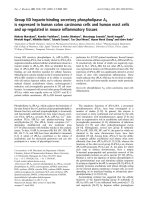
patients. Figure 1 shows the absolute serum KL-6 levels
at the baseline according to the pre sence of preexisting
pulmonary fibrosis. The absolute serum KL-6 levels at
the baseline showed no significant difference between
patients with and without preexisting pulmonary fibrosis
(Mann-Whitney U-test; p = 0.207). Table 2 shows the
characteristics of the 48 patients who had preexisting
pulmonary fibrosis. Eight (16.7%) out of the 48 patients
with preexisting pulmonary fibrosis developed EGFR-
TKIs induced ILD. Statistica l analyses were made to see
the association between the patients’ characteristic and
the development of EGFR-TKIs induced ILD among
these patients (Tab le 2). In the patients who had preex-
isting pulmonary fibrosis, thoracic radiation prior to
EGFR-TKIs treatment was not associated with the
development of E GFR-TKIs induced ILD, however,
there was a weak but statistically significant association
between the development of EGFR-TKIs induced ILD
and EGFR mutation status (p = 0.0498).
Incidence and characteristics of patients with EGFR-TKIs
induced ILD
Among the 341 patients included in this study, 20
(5.9%) developed EGFR-TKIs induced ILD, and 9 (2.6%)
died from ILD. Table 3 shows the characteristics and
cli nical course of these 20 patients. All the patients had
acute onset or exacerbation of respiratory symptoms.
The median interval from the administration of EGFR-
TKI to the occurrence of EGFR-TKIs induced ILD was
19 days (range 5-51 days). The subcla ssifications of
EGFR-TKIs induced ILD categorized by the findings of
chest CT scans in these 20 patients were as follows: AIP
pattern in 5 patients, COP/EP pattern in 9 patie nts, and
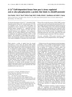
HPpatternin6patients.TheCTimagesof5patients
who demonstrated AIP pattern are shown in Figure 2.
When the occurrence of EGFR-TKIs i nduced ILD was
suspected, the administration of EGFR-TKI was immedi-
ately stopped and high dose me thylprednisolone (1,000
mg daily for 3 days) therapy was started. All of the 5
patients with AIP patterns were refractory to the treat-
ment and eventually died, whereas 7 of 9 patients with
COP/EP pattern and 4 of 6 patients with HP pattern
showed immediate response to the treatment. Postmor-
tem examinations were performed in 3 patients (patient
No. 5, 8 and 11) and diffuse alveolar damage (DAD) was
detected histologically in all of them. In addition, the
presence of preexisting pulmonary fibrosis was sus-
pected in 2 of the 3 patients. Neither infection nor lym-
phangitic spread of cancer cells was pointed out in any
of them.
Risk factors for developing EGFR-TKIs induced ILD
The results of univariate analyses on risk factors for
EGFR-TKIs induced ILD are shown in Table 4. Univari-
ate analyses revealed that only preexisting pulmonary
fibrosis (odds ratio, 4.683; 95% CI, 1.741-12.042; p =
0.003) was a significant risk fac tor for th e development
of EGFR-TKIs induced ILD.
Serum levels of KL-6 in patients who developed EGFR-
TKIs induced ILD
After the administration of t he EGFR-TKIs, measure-
ments o f serum KL-6 levels at least once during and/or
around 4 weeks were achieved in 15 out of 20 patients
who developed EGFR-TKIs induced ILD and 198 out of
321 patients who did not. The ratios of serum KL-6
levels during or around 4 weeks after the start of EGFR-
TKIs to those at baseline were 1.315 ± 0.120 for the for-
mer and 1.000 ± 0.036 for the latter, respectively (mean
± SEM). There was a significant statistical difference
between these ratios (p = 0.004, Mann-Whitney U-tes t).
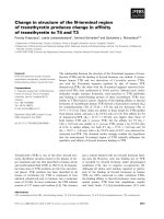
Figure 3 shows the serum levels of KL-6 at the multiple
time points before and after the onset of ILD in 8 survi-
vors (Figure 3A) and 7 non-survivors (Figure 3B). The
S
erum KL-6 level
(
U
/
ml
)
p=0.207
Figure 1 Absolute serum levels of KL-6 at baseline in patients
with and without preexisting pulmonary fibrosis. Each point
represents the absolute serum KL-6 level at baseline in patients with
and without preexisting pulmonary fibrosis. There was no significant
difference between the two groups (p = 0.207, Mann-Whitney U-test).
Kawase et al. Respiratory Research 2011, 12:97
/>Page 4 of 11
serum levels of KL-6 in 7 non-survivors but not in 8
survivors showed consistent trends to increase after the
onset of EGFR-TKIs induced ILD. The absolute serum
KL-6 levels at the onset as well as at baseline showed
no difference between the 7 non-survivors and 8 survi-
vors (Mann-Whitney U-test; p = 0.072 at onset, and p =
0.072 at baseline, respectively). To assess the changes in
serum KL-6 level before and after the onset of ILD, the
ratio of serum KL-6 level just after the onset of ILD to
that at baseline was calculated in 15 of 20 patients who
developed ILD. The differences in t he ratios of serum
KL-6 levels just after the onset of ILD from baseline
were found to be statistically significant between the
survivors and non-survivors (Mann-Whitney U-test; p =
0.006; Figure 4).
Then, we compared the circulating levels of KL-6
according to the patterns of EGFR-TKIs induced ILD sub-
classified by the manifestation on chest CT in 15 of 20
patients who developed EGFR-TKIs induced ILD. The
absolute levels of circulating KL-6 at neither baseline nor
Table 2 Patients’ Characteristics of 48 patients with preexisting pulmonary fibrosis
Characteristics Total EGFR-TKIs induced ILD (+) EGFR-TKIs induced ILD (-) p-value
Total 48 8 40
Age (years)
Mean (± SEM) 67.5(± 3.6) 66.2(± 1.8)
< 60 12 2 10 1.000
≥ 60 36 6 30
Sex
Female 11 3 8 0.361
Male 37 5 32
Histologic type
Adenocarcinoma 37 5 32 0.361
Squamous cell carcinoma/Others 11 3 8
Smoking history
Current/Former 40 5 35 0.116
Never 8 3 5
Disease stage
IV 24 3 21 0.701
I-IIIB/Recurrence after surgery 11 5 19
Performance status
≥ 2 26 5 21 0.710
0-1 22 3 19
No. of prior chemotherapy regimens
≥ 2 20 1 19 0.116
0-1 28 7 21
Prior thoracic radiotherapy
Yes 10 0 10 0.177
No 38 8 30
Pattern of preexisting pulmonary fibrosis
IPF pattern 3 1 2 0.429
Non-IPF pattern 45 7 38
Preexisting pulmonary emphysema
Yes 24 3 21 0.701
No 24 5 19
EGFR mutation status
Wild type 9 5 4 0.0498*
Mutant 11 1 10
(Not evaluated) (28) (2) (26)
Types of EGFR-TKI
Gefitinib 40 5 35 0.116
Erlotinib 8 3 5
*p < 0.05 (Fisher’s exact test)
Kawase et al. Respiratory Research 2011, 12:97
/>Page 5 of 11
Table 3 Characteristics of 20 patients with EGFR-TKIs induced ILD
No Age Sex Histological
type
Smoking
history
Stage PS Prior
CT
Prior
RT
Preexisting
fibrosis
Preexisting
emphysema
EGFR
mutation
Type of EGFR-
TKI
Length of EGFR-
TKI
CT
findings
Prognosis
1 68 M SCC Ex IIIB 1 1 No No Yes N.E. Gefitinib 11 COP/EP Alive
2 80 M SCC Never Rec 2 1 No Yes Yes Wild Gefitinib 17 COP/EP Alive
3 70 F SCC Never IIIA 1 1 Yes No No N.E. Gefitinib 24 HP Alive
4 60 M ADC Never IIIB 1 1 No No No N.E. Gefitinib 35 COP/EP Alive
5 68 F ADC Ex Rec 1 2 No No No Wild Gefitinib 16 AIP Dead
6 60 M ADC Current IIIB 1 2 No No Yes N.E. Gefitinib 26 COP/EP Alive
7 57 M ADC Never Rec 0 3 No No No L858R Gefitinib 51 COP/EP Alive
8 73 M ADC Current IV 4 0 No Yes Yes N.E. Gefitinib 13 AIP Dead
9 65 F ADC Never IV 2 3 No No No N.E. Gefitinib 38 HP Dead
10 69 F ADC Never IIIB 2 1 No Yes No N.E. Gefitinib 14 AIP Dead
11 84 F ADC Ex IIIB 4 0 No Yes No Wild Gefitinib 16 AIP Dead
12 63 F SCC Never IV 1 1 No Yes No N.E. Gefitinib 50 COP/EP Dead
13 67 F ADC Never Rec 0 1 No No No Deletion Gefitinib 48 HP Alive
14 60 M ADC Current IV 4 0 No Yes Yes L858R Gefitinib 17 HP Dead
15 55 M ADC Ex IV 3 4 No No No L858R Gefitinib 47 COP/EP Dead
16 69 M ADC Current IV 3 1 No Yes No Wild Erlotinib 14 AIP Dead
17 56 M SCC Current Rec 0 1 No Yes Yes Wild Erlotinib 21 COP/EP Alive
18 59 M ADC Ex Rec 1 2 No Yes No Wild Erlotinib 5 HP Alive
19 66 M ADC Current IIIB 0 0 No No No L858R Gefitinib 17 COP/EP Alive
20 64 F ADC Never Rec 1 1 No No No L858R Erlotinib 31 HP Alive
Abbreviations: ADC, Adenocarcinoma; SCC, Squamous cell carcinoma; Rec, Recurrence after the surgery; CT, Chemotherapy; RT, Radiation therapy; N.E, Not evaluated; COP/EP, Cryptogenic organizing pneumonia/
Eosinophilic pneumonia; HP, Hypersensitivity pneumonitis; DAD, Diffuse alveolar damage.
Kawase et al. Respiratory Research 2011, 12:97
/>Page 6 of 11
the onset of ILD were found not to be statistically signifi-
cant between the life-threatening pattern (AIP pattern) of
4 patients and the other patterns of 11 patients (Mann-
Whitney U-test; p = 0.648 at onset, and p = 0.845 at base-
line, respectively). When the ratio of serum KL-6 level at
baseline to that at the onset of ILD was compared, this
value was significantly higher in the patients with the life-
threatening pattern (AIP pattern) than that in other pat-
terns (Mann-Whitney U-test; p = 0.005; Figure 5). In addi-
tion, patients whose serum KL-6 levels rose more than 1.5
times h igher than their baseline levels had a high ch ance
of developing the AIP pattern.
Discussion
In this large multi-institutional study, we investigated
the incidence and risk factors for developing ILD in
patients treated with EGFR-TKIs until 12 weeks after
the start of EGFR-TKIs therapy. Univariate analyses
revealed that preexisting pulmonary fibrosis at baseline
was the only risk factor for EGFR-TKIs induced ILD.
Although absolute serum KL-6 levels at neit her baseline
nor the onset of ILD could discriminate between life-
threatening and non-life-threatening EGFR-TKIs
induced ILDs, the ratio of serum KL-6 level at the
occurrence of EGFR-TKIs induced ILD to that at
Table 4 Risk factors for EGFR-TKIs induced ILD at the start of EGFR-TKIs
Variables Odds ratio 95% CI P-value
Univariate analysis
Age (years) ≥ 60/< 60 1.758 0.626-6.256 0.301
Gender Male/Female 1.472 0.593-3.848 0.406
Histological type Non-ADC/ADC 2.342 0.730-6.420 0.142
Smoking history Never/Smoker 1.006 0.402-2.516 0.989
Performance status ≥ 2/0-1 0.942 0.360-2.341 0.899
No. of prior chemotherapy regimens ≥ 2/0-1 0.800 0.277-2.053 0.652
Prior thoracic radiotherapy Yes/No 0.315 0.017-1.575 0.187
Preexisting pulmonary fibrosis Yes/No 4.683 1.741-12.042 0.003*
Preexisting pulmonary emphysema Yes/No 1.382 0.476-3.574 0.531
EGFR mutation Wild type/EGFR mutant 1.667 0.497-5.594 0.400
Types of EGFR-TKI Gefitinib/Erlotinib 2.043 0.562-5.948 0.253
Serum KL-6 level at baseline (U/ml) ≥ 500/< 500 2.096 0.679-7.116 0.199
*P < 0.05
Abbreviation: ADC, Adenocarcinoma.
Case 8
Case 11 Case 16
Case 5 Case 10
Figure 2 Chest CT images of five patients who developed EGFR-TKI induced acute interstitial pneumonia (AIP). Representative chest CT
images of the five patients who developed AIP pattern of EGFR-TKIs induced ILD are shown. Each case number corresponds to the patient’s
number listed in Table 3.
Kawase et al. Respiratory Research 2011, 12:97
/>Page 7 of 11
baseline was found to quite precisely do so. These find-
ings suggest the significance of serum KL-6 level for the
detection of life threatening EGFR-TKIs induced ILD.
The development of molecular targeted agents has
been a key factor in recent advances in cancer therapy,
and some of these agents have been applied in clinical
practice. EGFR-TKIs are one of the representative mole-
cular target agents and, at first, were considered to be
safe agents with mild side effects in comparison to cyto-
toxic agents. However, following the increase in usage of
EGFR-TKIs in lung cancer therapy, a significantly higher
incidence of life-threaten ing drug induced ILD in Japa-
nese patients than that of patients in the rest of the
world was reported [38,39]. In the present study, out of
341 NSCLC patients treated with EGFR-TKIs, 20
patients (5.9%) developed ILD and 9 patients (2.6%) died
from ILD. The incidence and mortality of EGFR-TKIs
induced ILD were relatively higher than those reported
in previous studies from Japan [7-13,39]. This result
might be due to the high incidence of preexisting pul-
monary fibrosis in this study. In this study, the manifes-
tationsofchestCTscansin20patientswhodeveloped
EGFR-TKIs induced ILD were classified as AIP pattern
for 5 patients, COP/EP pattern for 9 patients and HP
pattern for 6 patients. Interestingly, CIP pa ttern was not
observed as was the case in a previous study [36]. All
the patients who demonstrated the AIP pattern died,
whereas the majority of patients with other patterns
recovered from EGFR-TKIs induced ILD. In this study,
the postmortem examination of three patients with AIP
pattern revealed that DAD was the main cause of death
and observations similar to ours have been reported pre-
viously [7,8]. In this study, univariate analysis revealed
that preexisting pulmonary fibrosis was the only risk
factor for developing EGFR-TKIs induced ILD. Although
previous studies reported that male gender, smoking
history and poor PS were also independent risk factors
for developing EGFR-TKIs induced ILD [7-13,39],
neither of them correlated with incidence or mortal ity
of EGFR-TKIs induced ILD in the present study. This
may be due to the small sample size and high incidence
of preexisting pulmonary fibrosis in our studied patients.
0
0.5
1
1.5
2
2.5
HP
㻌
㻌 㻌 㻌 㻌 㻌 㻌 㻌 㻌
㻌 㻌
COP/EP
㻌 㻌 㻌 㻌 㻌 㻌 㻌 㻌 㻌 㻌 㻌 㻌 㻌
AIP
KL-6 ratio
p=0.005
Figure 5 The ratios of the serum levels of KL-6 at the onset of
EGFR-TKI related ILD to those at baseline on the basis of the
sub-classifications of EGFR-TKIs induced ILD. Open, shaded, and
solid bars represent hypersensitivity pneumonitis (HP) pattern,
cryptogenic organizing pneumonia/eosinophilic pneumonia (COP/
EP) pattern, and acute interstitial pneumonia (AIP) pattern,
respectively. There is a significant difference in these ratios between
AIP pattern and the other patterns (p = 0.005).
100
1000
10000
100
1000
10000
500 500
-2 -1 0 1 2
(A) Survivors
㻌 㻌 㻌
㻌
(B) Non-survivors
Levels o
f
serum KL-6
(
U
/
mL
)
-2 -1 0 1 2
W
ee
k W
ee
k
Figure 3 Kinetics of serum KL-6 levels in (A) 8 survivors and (B)
7 non-survivors who developed EGFR-TKIs induced ILD . Week 0
is designated as the week when EGFR-TKIs induced ILD was
diagnosed. Before and after the onset of EGFR-TKIs induced ILD, the
serum levels of KL-6 showed a trend not to change in the survivors
but to increase in the non-survivors.
KL-6 ratio
0
0.5
1
1.5
2
2.5
Su
rviv
o
r
s
㻌 㻌 㻌
㻌 㻌 㻌 㻌 㻌 㻌 㻌 㻌 㻌 㻌
㻌
㻌
N
o
n-
su
rviv
o
r
s
p=
0
.
006
Figure 4 The ratios of the serum levels of KL-6 at the onset of
EGFR-TKIs induced ILD to those at baseline in 8 survivors and
7 non-survivors. Open and solid bars represent survivors and non-
survivors, respectively. There is a significant difference in these ratios
between the survivors and non-survivors (p = 0.006).
Kawase et al. Respiratory Research 2011, 12:97
/>Page 8 of 11
Although a previous study from our l aboratory
reported that serum KL-6 levels at diagnosis increased
only in the life-th rea tening typ es, such as the DAD and
CIP patterns, of drug induced ILDs [27], absolute serum
KL-6 levels at the onset of EGFR-TKIs induced ILD did
not correlate with clinical outcomes in the present
study. The immunohistochem ical analysis of KL -6 using
three postmor tem autopsy specimens showed that KL-6
was expressed at t umor cells in the primary lesions as
well as alveolar epithelial cells in the EGFR-TKIs
induc ed ILDs (data not shown). Therefore, we speculate
that the origin of serum KL-6 at the onset of EGFR-
TKIs induced ILD might be associated with both
NSCLCs and EGFR-TKIs induced ILDs. On the other
hand, we found that the ratios of serum KL-6 levels just
after the onset of ILD to those at baseline could quite
precisely discriminate life-threatening ILD from non-
life-threatening ILD, and correlate well with the disease
progression. We can speculate that a drastic increase in
serum KL-6 levels after the administration of EGFR-
TKIs might be due to severe lung injury accompanied
with both alveolar-capillary destruction and enhance-
ment of alveolar-capillary permeability which allow KL-
6 to leak into the circulation from the alveolar space
[40]. Based on these observations, KL-6 can be regarded
as a good serum biomarker to assess the severity of
alveolar epithelium injury and the clinical outcome of
EGFR-related ILD. Regarding the association between
KL-6 and other serum biomarkers for ILD such as sur-
fact ant protein (SP)-A and SP-D in EGFR-TKIs induced
ILD, we do not have data to discuss. Previous studies,
which measured serum SP-A, SP-D, and KL-6 le vels in
4 patients w ith EGFR-TKIs induced ILD, demonstrate
that serum SP-A and SP-D levels increased in all studied
patients whereas KL-6 levels only elevated in patients
with life-threatening EGFR-TKIs induced ILD [8,41].
This observation is compatible with the findings of the
present study.
In addition to its ability to detect patients who
develop life-threatening ILD, the monitoring of serum
KL-6 levels is also useful to predict survival and progres-
sive disease in NSCLC patients treated with EGFR-TK Is
[30]. As measurement of serum KL-6 level is more
rapid, inexpensive, reproducible, and easier to perform
than CT scans, its monitoring could be quite useful to
assess the condition of NSCLC patients receiving EGFR-
TKIs. The development of EGFR-TKIs induced ILD is
reported to mostly occur within the first 4 weeks after
the start of EGFR-TKIs [11]. In the present study, 5
cases developed ILD within the first 2 weeks (ranged
from 5 to 14 d ays) after the start of EGF-TKIs. There-
fore, based on the results of the present study, once a
week monitoring of serum KL-6 levels in addition to
chest radiography could be recommended for NSCLC
patients receiving EGFR-TKIs particularly for the first 4
weeks after the start of treatment.
Although these promising results were obtained, we
are aware that this study has a n umber of limitations.
First, the numb er of E GFR-TKIs induced ILD patients
included in the study was not sufficient for a valid sta-
tistical analysis. Sec ond, this study was conducted in a
retrospective manner. Therefore, the information on
EGFR mutation statuses in cancer tissue was not
obtained from all the studied patients. Furthermore,
multiple measurements of serum KL-6 levels w ere not
achieved in all patients who developed EGFR-TKIs
induc ed ILD. Third, the enrolled NSCLC patients might
be biased compared with general a dvanced NSCLC
population. We believe that this was caused by our
trend to use EGFR-TKIs for specific subgroups of
NSCLCpatientssuchaswomen,non-smokers,and
patients with EGFR mutations. Finally, the studied
patients were only Japanese. Considering ethnic differ-
ences in the efficacy of EGFR-TKIs treatment and/or
the occurrence of adverse side effects related by EGFR-
TKIs, we should carefully i nterpret the results when this
monitoring system is applied to non-Japanese patients.
A large and prospective study to measur e serum KL-6
levels serially before and after EGFR-TKIs treatment,
also including non-Japanese patients, will be required to
evaluate the utility of monitoring KL-6 in EGFR-TKIs
induced ILDs.
Conclusions
Our results indicate that the change in serum KL-6 level
from baseline should be useful biomarker for the diag-
nosis of life-threatening EGFR-TKIs induced ILD and
for estimating its progress and severity. A risk-benefit
analysis and patient selection should be conside red as
well as close monitoring of serum levels of KL-6, parti-
cularly if using EGFR-TKIs in patients w ith preexisting
pulmonary fibrosis.
List of Abbreviations
EGFR-TKI: epidermal growth factor receptor tyrosine kinase inhi bitor; NSCLC:
non-small cell lung cancer; ADC: adenocarcinoma; ILD: interstitial lung
disease; KL-6: Krebs von den Lungen-6; IPF: idiopathic pulmonary fibrosis;
AIP: acute interstitial pneumonia; CIP: chronic interstitial pneumonia; CT:
computed tomography; FDG-PET: F-18 fluorodeoxyglucose positron emission
tomography; MRI: magnetic resonance imaging; COP/EP: cryptogenic
organizing pneumonia/eosinophilic pneumoni a; HP: hypersensitivity
pneumonitis; PNA-LNA PCR: peptide nucleic acid-locked nucleic acid
polymerase chain reaction; ECLIA: electrochemiluminescence immunoassay.
Acknowledgements
This work was partly supported by Grants-in-Aid for Scientific Research from
the Minister of Education, Culture, Sports, Science and Technology of Japan.
Author details
1
Department of Molecular and Internal Medicine, Graduate School of
Biomedical Sciences, Hiroshima University, 1-2-3 Kasumi, Minami-ku,
Hiroshima 734-8551, Japan.
2
Department of Respiratory Medicine, Onomichi
Kawase et al. Respiratory Research 2011, 12:97
/>Page 9 of 11
General Hospital, 7-19 Kohama, Onomichi, Hiroshima 722-8508, Japan.
3
Department of Clinical Oncology and Respiratory Medicine, Shimane
University, 89-1, Enya-cho, Izumo, Shimane 693-8501, Japan.
4
Department of
Integrated Medicine and Informatics, Ehime University Graduate School of
Medicine, Toon, Ehime 791-0295, Japan.
5
Department of Health and Sports
Medical Sciences, Graduate School of Health Sciences, Hiroshima University,
Hiroshima, Japan.
6
Department of Hematology and Respiratory Medicine,
Kochi Medical School, Kochi University, Nankoku, Kochi 783-8505, Japan.
Authors’ contributions
SK performed part of the statistical analysis and drafted the manuscript. NH
conceived the study, and participated in its design and coordination and
helped to draft the manuscript. NI conceived the study, and participated in
patient recruitment and helped to draft the manuscript. YH performed part
of the statistical analysis and participated in creating the figures. KF, OF, TI,
SM, HH and TY participated in the selection and collection of patient
material. AY conceived the study, and participated in its design and
coordination. NK conceived the study, and participated in its design and
coordination and supervised the study. All authors read and approved the
final manuscript.
Competing interests
Nobuoki Kohno has a personal royalty of KL-6 from a Japanese
pharmaceutical company, Eisai Co., Ltd. The remaining authors have no
conflict of interest.
Received: 9 May 2011 Accepted: 26 July 2011 Published: 26 July 2011
References
1. Modjtahedi H, Essapen S: Epidermal growth factor receptor inhibitors in
cancer treatment: advances, challenges and opportunities. Anticancer
Drugs 2009, 20:851-855.
2. Lynch TJ, Bell DW, Sordella R, Gurubhagavatula S, Okimoto RA,
Brannigan BW, Harris PL, Haserlat SM, Supko JG, Haluska FG, Louis DN,
Christiani DC, Settleman J, Haber DA: Activating mutations in the
epidermal growth factor receptor underlying responsiveness of non-
small-cell lung cancer to gefitinib. N Engl J Med 2004, 350:2129-39.
3. Paez JG, Jänne PA, Lee JC, Tracy S, Greulich H, Gabriel S, Herman P, Kaye FJ,
Lindeman N, Boggon TJ, Naoki K, Sasaki H, Fujii Y, Eck MJ, Sellers WR,
Johnson BE, Meyerson M: EGFR mutations in lung cancer: correlation with
clinical response to gefitinib therapy. Science 2004, 304:1497-1500.
4. Mok TS, Wu YL, Thongprasert S, Yang CH, Chu DT, Saijo N,
Sunpaweravong P, Han B, Margono B, Ichinose Y, Nishiwaki Y, Ohe Y,
Yang JJ, Chewaskulyong B, Jiang H, Duffield EL, Watkins CL, Armour AA,
Fukuoka M: Gefitinib or Carboplatin-Paclitaxel in Pulmonary
Adenocarcinoma. N Engl J Med 2009, 361:947-957.
5. Mitsudomi T, Morita S, Yatabe Y, Negoro S, Okamoto I, Tsurutani J, Seto T,
Satouchi M, Tada H, Hirashima T, Asami K, Katakami N, Takada M,
Yoshioka H, Shibata K, Kudoh S, Shimizu E, Saito H, Toyooka S, Nakagawa K,
Fukuoka M, West Japan Oncology Group: Gefitinib versus cisplatin plus
docetaxel in patients with non-small-cell lung cancer harboring
mutations of the epidermal growth factor receptor (WJTOG3405): an
open label, randomized phase 3 trial. Lancet Oncol 2010, 11:121-128.
6. Maemondo M, Inoue A, Kobayashi K, Sugawara S, Oizumi S, Isobe H,
Gemma A, Harada M, Yoshizawa H, Kinoshita I, Fujita Y, Okinaga S,
Hirano H, Yoshimori K, Harada T, Ogura T, Ando M, Miyazawa H, Tanaka T,
Saijo Y, Hagiwara K, Morita S, Nukiwa T, North-East Japan Study Group:
Gefitinib or chemotherapy for non-small-cell lung cancer with mutated
EGFR. N Engl J Med 2010, 362:2380-2388.
7. Inoue A, Saijo Y, Maemondo M, Gomi K, Tokue Y, Kimura Y, Ebina M,
Kikuchi T, Moriya T, Nukiwa T: Severe acute interstitial pneumonia and
gefitinib. Lancet 2003, 361:137-139.
8. Inomata S, Takahashi H, Nagata M, Yamada G, Shiratori M, Tanaka H,
Satoh M, Saitoh T, Sato T, Abe S: Acute lung injury as an adverse event of
gefitinib. Anticancer Drugs 2004, 15:461-467.
9. Hotta K, Kiura K, Tabata M, Harita S, Gemba K, Yonei T, Bessho A, Maeda T,
Moritaka T, Shibayama T, Matsuo K, Kato K, Kanehiro A, Tanimoto Y,
Matsuo K, Ueoka H, Tanimoto M: Interstitial lung disease in Japanese
patients with non-small cell lung cancer receiving gefitinib: an analysis
of risk factors and treatment outcomes in Okayama Lung Cancer Study
Group. Cancer J 2005, 11:417-424.
10. Ando M, Okamoto I, Yamamoto N, Takeda K, Tamura K, Seto T, Ariyoshi Y,
Fukuoka M: Predictive factors for interstitial lung disease, antitumor
response, and survival in non-small-cell lung cancer patients treated
with gefitinib. J Clin Oncol 2006, 24:2549-2556.
11. Kudoh S, Kato H, Nishiwaki Y, Fukuoka M, Nakata K, Ichinose Y, Tsuboi M,
Yokota S, Nakagawa K, Suga M, Japan Thoracic Radiology Group, Jiang H,
Itoh Y, Armour A, Watkins C, Higenbottam T, Nyberg F: Interstitial lung
disease in Japanese patients with lung cancer: a cohort and nested
case-control study. Am J Respir Crit Care Med 2008, 177:1348-1357.
12. Nakagawa M, Nishimura T, Teramukai S, Tada H, Tanaka F, Yanagihara K,
Furuse K, Wada H, Fukushima M: Interstitial lung disease in gefitinib-
treated Japanese patients with non-small cell lung cancer - a
retrospective analysis: JMTO LC03-02. BMC Res Notes 2009, 2:157.
13. Hotta K, Kiura K, Takigawa N, Yoshioka H, Harita S, Kuyama S, Yonei T,
Fujiwara K, Maeda T, Aoe K, Ueoka H, Kamei H, Umemura S, Moritaka T,
Segawa Y, Kawai H, Bessho A, Kato K, Tabata M, Tanimoto M: Comparison
of the incidence and pattern of interstitial lung disease during erlotinib
and gefitinib treatment in Japanese Patients with non-small cell lung
cancer: the Okayama Lung Cancer Study Group experience. J Thorac
Oncol 2010, 5:179-184.
14. Kohno N, Akiyama M, Kyoizumi S, Hakoda M, Kobuke K, Yamakido M:
Detection of soluble tumor-associated antigens in sera and effusions
using novel monoclonal antibodies, KL-3 and KL-6, against lung
adenocarcinoma. Jpn J Clin Oncol 1988, 18:203-216.
15. Kohno N, Inoue Y, Hamada H, Fujioka S, Fujino S, Yokoyama A, Hiwada K,
Ueda N, Akiyama M: Difference in sero-diagnostic values among KL-6-
associated mucins classified as cluster 9. Int J Cancer 1994, 57(suppl
8):81-83.
16. Hirasawa Y, Kohno N, Yokoyama A, Inoue Y, Abe M, Hiwada K: KL-6, a
human MUC1 mucin, is chemotactic for human fibroblasts. Am J Respir
Cell Mol Biol 1997, 17:501-507.
17. Ohyabu N, Hinou H, Matsushita T, Izumi R, Shimizu H, Kawamoto K,
Numata Y, Togame H, Takemoto H, Kondo H, Nishimura S: An Essential
Epitope of Anti-MUC1 Monoclonal Antibody KL-6 Revealed by Focused
Glycopeptide Library. J Am Chem Soc 2009, 131:17102-17109.
18. Kohno N, Kyoizumi S, Awaya Y, Fukuhara H, Yamakido M, Akiyama M: New
serum indicator of interstitial pneumonitis activity. Sialylated
carbohydrate antigen KL-6. Chest 1989, 96:68-73.
19. Kohno N, Hamada H, Fujioka S, Hiwada K, Yamakido M, Akiyama M:
Circulating antigen KL-6 and lactate dehydrogenase for monitoring
irradiated patients with lung cancer. Chest 1992, 102:117-122.
20. Kohno N, Awaya Y, Oyama T, Yamakido M, Akiyama M, Inoue Y,
Yokoyama A, Hamada H, Fujioka S, Hiwada K: KL-6, a mucin-like
glycoprotein, in bronchoalveolar lavage fluid from patients with
interstitial lung disease. Am Rev Respir Dis 1993, 148:637-642.
21. Yokoyama A, Kohno N, Hamada H, Sakatani M, Ueda E, Kondo K,
Hirasawa Y, Hiwada K: Circulating KL-6 predicts the outcome of rapidly
progressive idiopathic pulmonary fibrosis. Am J Respir Crit Care Med 1998,
158:1680-1684.
22. Kohno N: Serum marker KL-6/MUC1 for the diagnosis and management
of interstitial pneumonitis. J Med Invest 1999, 46:151-158.
23. Ohnishi H, Yokoyama A, Kondo K, Hamada H, Abe M, Nishimura K,
Hiwada K, Kohno N: Comparative study of KL-6, surfactant protein-A,
surfactant protein-D, and monocyte chemoattractant protein-1 as serum
markers for interstitial lung diseases. Am J Respir Crit Care Med 2002,
165:378-381.
24. Yokoyama A, Kondo K, Nakajima M, Matsushima T, Takahashi T,
Nishimura M, Bando M, Sugiyama Y, Totani Y, Ishizaki T, Ichiyasu H, Suga M,
Hamada H, Kohno N: Prognostic value of circulating KL-6 in idiopathic
pulmonary fibrosis. Respirology 2006, 11:164-168.
25. Nakashima T, Yokoyama A, Ohnishi H, Hamada H, Ishikawa N, Haruta Y,
Hattori N, Tanigawa K, Kohno N: Circulating KL-6/MUC1 as an
independent predictor for disseminated intravascular coagulation in
acute respiratory distress syndrome. J Intern Med
2008, 263:432-439.
26. Ohshimo S, Bonella F, Grammann N, Starke K, Cui A, Bauer PC, Teschler H,
Kohno N, Guzman J, Costabel U: Serum KL-6 as a novel disease marker in
adolescent and adult cystic fibrosis. Sarcoidosis Vasc Diffuse Lung Dis 2009,
26:47-53.
27. Ohnishi H, Yokoyama A, Yasuhara Y, Watanabe A, Naka T, Hamada H,
Abe M, Nishimura K, Higaki J, Ikezoe J, Kohno N: Circulating KL-6 levels in
patients with drug induced pneumonitis. Thorax 2003, 58:872-875.
Kawase et al. Respiratory Research 2011, 12:97
/>Page 10 of 11
28. Inata J, Hattori N, Yokoyama A, Ohshimo S, Doi M, Ishikawa N, Hamada H,
Kohno N: Circulating KL-6/MUC1 mucin carrying sialyl Lewis
a
oligosaccharide is an independent prognostic factor in patients with
lung adenocarcinoma. Int J Cancer 2007, 120:2643-2649.
29. Tanaka S, Hattori N, Ishikawa N, Shoda H, Takano A, Nishino R, Okada M,
Arihiro K, Inai K, Hamada H, Yokoyama A, Kohno N: Krebs von den
Lungen-6 (KL-6) is a prognostic biomarker in patients with surgically
resected non-small cell lung cancer. Int J Cancer 2011.
30. Ishikawa N, Hattori N, Yokoyama A, Tanaka S, Nishino R, Yoshioka K,
Ohshimo S, Fujitaka K, Ohnishi H, Hamada H, Arihiro K, Kohno N:
Usefulness of monitoring the circulating Krebs von den Lungen-6 levels
to predict the clinical outcome of patients with advanced non-small cell
lung cancer treated with epidermal growth factor receptor tyrosine
kinase inhibitors. Int J Cancer 2008, 122:2612-2620.
31. American Thoracic Society: Idiopathic pulmonary fibrosis: diagnosis and
treatment. International consensus statement. American Thoracic Society
(ATS), and the European Respiratory Society (ERS). Am J Respir Crit Care
Med 2000, 161:646-664.
32. American Thoracic Society, European Respiratory Society: American
Thoracic Society/European Respiratory Society International
Multidisciplinary Consensus Classification of the Idiopathic Interstitial
Pneumonias. This joint statement of the American Thoracic Society
(ATS), and the European Respiratory Society (ERS) was adopted by the
ATS board of directors, June 2001 and by the ERS Executive Committee,
June 2001. Am J Respir Crit Care Med 2002, 165:277-304.
33. Webb WR, Müller NL, Naidich DP: HIGH-RESOLUTION CT of the LUNG. 4
edition. Philadelphia: Lippincott Williams & Wilkins; 2009.
34. Goddard PR, Nicholson EM, Laszlo G, Watt I: Computed tomography in
pulmonary emphysema. Clin Radiol 1992, 33:379-387.
35. Müller NL, White DA, Jiang H, Gemma A: Diagnosis and management of
drug-associated interstitial lung disease. Br J Cancer 2004, 91(Suppl 2):
S24-30.
36. Endo M, Johkoh T, Kimura K, Yamamoto N: Imaging of gefitinib-related
interstitial lung disease: multi-institutional analysis by the West Japan
Thoracic Oncology Group. Lung Cancer 2006, 52:135-140.
37. Nagai Y, Miyazawa H, Tanaka T, Udagawa K, Kato M, Fukuyama S, Yokote A,
Kobayashi K, Kanazawa M, Hagiwara K: Genetic heterogeneity of the
epidermal growth factor receptor in non-small cell lung cancer cell lines
revealed by a rapid and sensitive detection system, the peptide nucleic
acid-locked nucleic acid PCR clamp. Cancer Res 2005, 65:7276-7282.
38. Camus P, Kudoh S, Ebina M: Interstitial lung disease associated with drug
therapy. Br J Cancer 2004, 91:S18-23.
39. Armour A: Gefitinib in advanced non-small cell lung cancer: clinical
experience in patients of Asian origin. Asia Pac J Clin Oncol 2007, 3:66-78.
40. Inoue Y, Barker E, Daniloff E, Kohno N, Hiwada K, Newman LS: Pulmonary
epithelial cell injury and alveolar-capillary permeability in berylliosis. Am
J Respir Crit Care Med 1997, 156:109-115.
41. Kitajima H, Takahashi H, Harada K, Kanai A, Inomata S, Taniguchi H, Saikai T,
Abe S: Gefitinib-induced interstitial lung disease showing improvement
after cessation: disassociation of serum markers. Respirology 2006,
11:217-220.
doi:10.1186/1465-9921-12-97
Cite this article as: Kawase et al.: Change in serum KL-6 level from
baseline is useful for predicting life-threatening EGFR-TKIs induced
interstitial lung disease. Respiratory Research 2011 12:97.
Submit your next manuscript to BioMed Central
and take full advantage of:
• Convenient online submission
• Thorough peer review
• No space constraints or color figure charges
• Immediate publication on acceptance
• Inclusion in PubMed, CAS, Scopus and Google Scholar
• Research which is freely available for redistribution
Submit your manuscript at
www.biomedcentral.com/submit
Kawase et al. Respiratory Research 2011, 12:97
/>Page 11 of 11