Báo cáo y học: " Endothelial-like cells in chronic thromboembolic pulmonary hypertension: crosstalk with myofibroblast-like cells" pptx
Bạn đang xem bản rút gọn của tài liệu. Xem và tải ngay bản đầy đủ của tài liệu tại đây (10.83 MB, 16 trang )
RESEARCH Open Access
Endothelial-like cells in chronic thromboembolic
pulmonary hypertension: crosstalk with
myofibroblast-like cells
Seiichiro Sakao
1*
, Hiroyuki Hao
2
, Nobuhiro Tanabe
1
, Yasunori Kasahara
1
, Katsushi Kurosu
1
and Koichiro Tatsumi
1
Abstract
Background: Chronic thromboembolic pulmonary hypertension (CTEPH) is characterized by intravascular thrombus
formation in the pulmonary arteries.
Recently, it has been shown that a myofibroblast cell phenotype was predominant within endarterectomized
tissues from CTEPH patients. Indeed, our recent study demonstrated the existence of not only myofibroblast-like
cells (MFLCs), but also endothelial-like cells (ELCs). Under in vitro conditions, a few transitional cells (co-expressing
both endothelial- and SM-cell markers) were observed in the ELC population. We hypothesized that MFLCs in the
microenvironment created by the unresolved clot may promote the endothelial-mesenchymal transition and/or
induce endothelial cell (EC) dysfunction.
Methods: We isolated cells from these tissues and identified them as MFLCs and ELCs. In order to test whether
the MFLCs provide the microenvironment which causes EC alterations, ECs were incubated in serum-free medium
conditioned by MFLCs, or were grown in co-culture with the MFLCs.
Results: Our experiments demonstrated that MFLCs promoted the commercially available ECs to transit to other
mesenchymal phenotypes and/or induced EC dysfunction through inactivation of autophagy, disruption of the
mitochondrial reticulum, alteration of the SOD-2 localization, and decreased ROS production. Indeed, ELCs included
a few transitional cells, lost the ability to form autophagosomes, and had defective mitochondrial structure/
function. Moreover, rapamycin reversed the phenotypic alterations and the gene expression changes in ECs co-
cultured with MFLCs, thus suggesting that this agent had beneficial therapeutic effects on ECs in CTEPH tissues.
Conclusions: It is possible that the microenvironment created by the stabilized clot stimulates MFLCs to induce EC
alterations.
Keywords: neointima, myofibroblast, endothelial cells, CTEPH.
Background
It is generally known that chronic thromboem bolic pul-
monary hypertension (CTEPH) is one of the leading
causes of severe pulmonary hypertension. CTEPH is
characterized by intravascular thrombus formation and
fibrous stenosis or complete obliteration of the pulmon-
ary arteries [1]. The consequence is increased pulmon-
ary vascular resistance, resulting in pulmonary
hypertension and progressive right heart failure.
Pulmonary endarterectomy (PEA) is the current main-
stream of therapy for CTEPH [2]. Moreover, recent stu-
dies have provided evidence suggesting that, although
CTEPH is believed to result from acute pulmonary
embolism [3,4], small-vessel disease appears a nd wor-
sens later in the course of disease [5]. Histopathologic
studies of microvascular changes in CTEPH have shown
indistinguishable vascular lesions from those seen in
idiopathic pulmonary arterial hypertension (IPAH ) and
Eisenmenger’s syndrome [6-8]. Especially in vitro and ex
vivo experiments, pulmonary artery endothelial cell (EC)
in the group of pulmonary hypertensive diseases are
suggested to exhibit an unusual hyperproliferative
* Correspondence:
1
Department of Respirology (B2), Graduate School of Medicine, Chiba
University, 1-8-1 Inohana, Chuo-ku, Chiba 260-8670, Japan
Full list of author information is available at the end of the article
Sakao et al. Respiratory Research 2011, 12:109
/>© 2011 Sakao et al; licensee BioMed Central Ltd. This is an Open Access article distributed under the terms of the Creative Commons
Attribution License ( w hich permits unrestricted use, distribution, and reproduction in
any medium, provided the original work is properly cited.
potential with decreased susceptibility to apoptosis
[9,10], indicating that dysfunctional EC may contribute
to the progression of the diseases.
Recently, Firth et al showed that multipotent
mesenchymal progenitor cells are present in endarterec-
tomized tissues from patients with CTEPH, and that a
myofibroblast cell phenotype was predominant within
these tissues, contributing extensively to the vascular
lesion/clot [ 11]. Indeed, we have a lso demonstrated the
existence of not only myofibroblast-like cells (MFLCs),
but also endothelial-like cells (ELCs) in these tissues
[12]. Under in vitro conditions, morphological altera-
tions were more easily detected in the ELCs. Smooth
muscle (SM)-like cel ls (defined by their expression of a-
SM-actin (SMA)) and a few transitional cells (co-expres-
sing b oth endothelial- (von Willebrand factor) and SM-
(a-SMA) cell markers) were consistently observed by
immunohistochemical staining (preliminary data).
In vitro experiments conducted to ass ess the contribu-
tion of ECs to the development of pulmonary arterial
hypertension (PAH) have demonstrated that the shift to
a transdifferentiated phenotype could be attributed to
selection of distinct cell subpopulations (i.e., stem-like
cells). These findings also suggest that the endothelial-
mesenchymal transition (EnMT) might be an important
contributor to pathophysiological vascular remodeling in
the complex vascular l esions of PAH [13], because,
although bone marrow-derived cells could participate in
arterial neointimal formation after mechanical injury,
they did not contribute substantially to pulmonary arter-
ial remodeling in an experimental PAH model [14].
Autophagy is a catabolic process involving the degra-
dation of intracellular material that is evolutionarily con-
served between all eukaryotes. During autophagy,
cytoplasmic components are engulfed by double-mem-
brane-bound structures (autophagosome s) and delivered
to lysosomes/vac uoles for degradation [15]. Recent stu-
dies indicate that autophagy plays an important role in
many different pathological conditions. Indeed, both
activation and inactivation of autophagy may impact
cancer cell growth. If autophagy cannot be activated,
protein synthesis predominates over protein degrada-
tion, and tumor growth is stimulated. In contrast, autop-
hagy may be activated in more advanced stages of
cancer to guarantee the survival of cells in minimally-
vascularized tumors [16].
The interactions between ECs and smooth muscle
cells (SMCs), which exist in close contact via a func-
tional syncytium, are involved in the process of new ves-
sel formation that occurs during development, as part of
wound repair, and during the reproductive cycle
[17-19]. We hypothesized that MFLCs stimulated by the
microenvironment created by the unresolved clot may
promote ECs to transit to other mesenchymal
phenotypes and/or induce EC dysfunction, contributing
to the vascular lesion, i.e., not only proximal vasculature,
but also microvascular. In the experiments considered
here, we isolated cells from endarterectomized tissue
from patients with CTEPH and id entified them as
MFLCs and ELCs. In order to show the hypothesis,
human pulmonary microvascular ECs were incubated in
a serum-free medium conditioned by MFLCs, or ECs
were co-cultured with M FLCs. The aim of this study
was to examine whether MFLCs in the microenviron-
ment created by the unresolved clot can, in principle,
affect EC disorder through the EnMT and autophagy.
Methods
Cell lines and reagents
The PEA tissues of patients with CTEPH were obtained
following PEA performed by Dr. Masahisa Masuda at
the Chiba Medical Center, Japan. Control pulmonary
arteries were obtained following lung resection for per-
ipheral cancer by Dr. Ichiro Yoshino at the Chiba Uni-
versity Hospital, Japan. Written informed co nsent was
acquired before surgery from all patients from whom
tissue samples were obtained. The study was app roved
by the Research Ethics Committee of Chiba University
School of Medicin e, and all subjects gave their informed
consent in writing. Although not clinica lly accurate, the
PEA tissues were defined as mentioned below. PEA
samples obtained from the region directly surrounding
the fibrotic clot are referred to as “proximal” vascular
tissue and those obtained from areas after the fibrotic
clot region are referred to as the “distal” vascular tissue
[11]. The tissues were cultured and various explant out-
growth cells were dissociated as described previously
[12]. Myofibroblast-like cells (MFLCs) and endothelial-
like cells (ELCs) were isolated and identified from
endarterectomized tissue from patients with CTEPH
and pulmonary arterial fibroblast-like cells from control
pulmonary arteries. PEA samples obtained from a total
of six patients undergoing PEA were examined in this
study.
Human pulmonary micr ovascular ECs w ere obtained
from Lonza Inc (Allendale, NJ, USA). T he following
ant ibod ies were used during our present studies: mouse
anti-a-SMA (1:1000, Sigma, St. Louis, MO, USA),
mouse anti-vimentin (1:200, DAKO, Carpinteria, CA,
USA), mouse anti-human desmin (1:100, DAKO, Car-
pinteria, CA, USA), anti-mouse IgG Ab conjugated with
Rhodamine dye (1:500, Molecular Probes, Eugene, OR,
USA), rabbit anti-von Willebrand factor (Factor VIII)
(1:1000, DAKO, Carpinteria, CA, USA), anti-rabbit IgG
conjugated with Alexa-488 fluor escent dye (1:500, Mole-
cularProbes,Eugene,OR,USA),andrabbitanti-CD31
(1:1000, DAKO, Carpinteria, CA, USA). Rapamycin was
purchased from Merck (Frankfurter, Germany).
Sakao et al. Respiratory Research 2011, 12:109
/>Page 2 of 16
Immunofluorescence staining
The cells were fixed in a 1:1 mixture of methanol and
acetone for 2 minutes followed by blocking with normal
goat serum for 30 minutes as described previously [13].
The cells were incubated with primary antibodies (anti-
a-smooth muscle actin (SMA), anti-von Willebrand fac-
tor, anti-vimentin and anti-desmin) for 1 hour at room
temperature, and then with secondary antibodies (anti-
mouse IgG conjugated with Alexa-594 fluorescent dye
and anti-rabbit I gG conjugated with Alexa-488 fluores-
cent dye) for 1 hour at room temperature. Stained cells
were embedded in VectaShield mounting medium with
DAPI (Vector Laboratories, Burlingame, CA, USA) and
were examined with a NIKON Eclipse 80 i microscope
(Nikon, Tokyo, Japan) using the VB-7210 imaging sys-
tem (Keyence, Tokyo, Japan). Positive cells were counted
in 3 different fields at a magnification of × 200 using a
fluorescence microscope.
Double immunohistochemical staining
Endarterectomized samples were embedded in optimal
cutting temperature (OCT) compound (Sakura Tissue
Tek), frozen, and cut into 10- μm sections with a cryo-
stat. For basic characterization, standard hematoxylin
and eosin ( H & E) staining was per formed. The CD31
antibody was used to stain endarterectomized tissue,
together with aSMA to stain transitional cells. aSMA
staining (blue) was developed with alkaline phosphatase-
conjugated secondary antibody, and then CD31 staini ng
(brown) was developed with peroxidase-conjugated sec-
ondary antibody. Transitional cells were confirmed by
aSMA posit ively stained cytosol that also had concomi-
tant positive cytoplasmic staining in CD31 positive cells.
ELISA (Enzyme-Linked ImmunoSorbent Assay)
TGF-b
1
were measured by sandwich ELISA techniques
by ELISA Tech (Aurora, CO, USA) utilizing reagents
from R&D systems (Minneapolis, MN, USA). The sam-
ples were read in a spectrophotometer at 405 nm. Anti-
bodies and tracer were bought from C ayman Chemicals
(Ann Arbor, Mi, USA).
Human pulmonary microvascular ECs in the conditioned
medium
At passage 2 MFLCs or pulmonary arterial fibroblast-
like cells were seeded at a density of 1.5 × 10
4
cells/ cm
2
and were subcultured when they were to 90% con-
fluences (4-8 days). They were washed 3 times using
phosphate-buffered saline (PBS) and were incubated
with serum-free medium for 48 hours. HPMVEC were
seeded in 6 cm dishes at 1 × 10
5
density and cultured in
EGM supplemented with 5% fetal bovine serum. At 70
to 80% confluence they were washed 3 times with PBS,
incubated in the conditionedmediumfor48hoursand
incubated in EGM again for 48 hours. After the incuba-
tion periods, they were assessed microscopically, further
characterized by immunohistochemical staining and har-
vested to extract RNA for quantitative RT-PCR and to
extract protein for ELISA.
Co-culture of human pulmonary microvascular ECs and
MFLCs
Co-culture of human pulmon ary microvascular ECs and
MFLCs was done on a 6-well plate (BD Falcon) with
Cell Culture Inserts (Falcon, 353102, 1.0 microns pore
size). Human pulmonary microvascular ECs or pulmon-
ary arterial fibroblast- like cells (at 5 × 10
4
density) and
MFLCs (at 5 × 10
4
density) were added into the lower
or upper chamber with or without rapamycin (10 nM).
After tw o weeks incubation periods, they were assessed
microscopically and further characterized by immuno-
histochemical staining, harvested to extract RNA for
PCR array, and other assays.
Magnetic cell sorting (MACS)
After trypsinization of ECLCs at passage 2, CD31 posi-
tive cells w ere isol ated by using CD31 MicroBeads
(Direct CD31 progenitor cell isolation kit, Miltenyi Bio-
tec Inc, A uburn, CA, USA) as described previously [13].
After trypsinization of ECLCs at passage 2, 100 μlof
FcR Blocking Reagent (Direct CD31 progen itor cell iso-
lation kit, Milte nyi Biotec Inc, Auburn, CA, USA) per
10
8
total cells was added to the cell suspension to inhi-
bit nonspecific or Fc-receptor mediated binding of
CD31 MicroBeads (Direct CD31 progenitor cell isolation
kit, Miltenyi Biotec) to non-target cells. Cells were
labeled by adding 100 μl CD31 MicroBeads per 10
8
total
cells, and incubated for 30 min at 6-12°C. After washing,
cells were resuspended in 500 μl buffer and applied to
the MS+/RS+ column with the column adapter in the
magnetic field of the MACS separator. The co lumn was
washed 3× with 500 μl buffer. The column was removed
from the separat or and t he retained cells were flushed
out with 1 ml buffer under pressure using the plunger
supplied with the column. The cells were incubated in
EGM and cultured until passage 5.
Total RNA isolation and Quantitative measurement
Total RNA was extracted from human pulmonary
microvascular ECs with an RNeasy Mini Kit (Qi agen,
CA, USA). RNA and cRNA yields were quantitated on a
Nano-Drop ND-1000 UV-Vis Spectrophotometer
(NanoDrop Technologies, Wilmington, DE, USA) as
described previously [13].
PCR array analysis
RT
2
Profiler™ PCR Arrays ( SABiosciences, Frederick,
USA) are the reliable and sensitive tools for analyzing
Sakao et al. Respiratory Research 2011, 12:109
/>Page 3 of 16
the expression of a focused panel of genes in signal
transduction pathways, biological process or disease
related gene networks. The 96-well plate Human Autop-
hagy PCR-array (PAHS-084) which profiles the expres-
sion of 84 key genes involved in autophagy and Human
Endothelial Cell Biology PCR-array (PAHS-015) which
profiles the expression of 84 genes related to endothelial
cell biology were selected as the hypothesis.
There is a better sensitivity of quantitative PCR in
comparison to microarray [20,21]. The PCR Arrays can
be used for research on various disease including cancer,
immunology, and phenotypic analysis of cells.
The mRNA of each co-cultured EC was converted
into cDNA using the RT
2
First Strand Kit (SABios-
ciences, Frederick, USA). This cDNA was then added to
the RT
2
SYBR Green qPCR Master Mix (SABiosciences,
Frederick, USA). Next, each sample was aliquotted on
PCR-arrays. All steps were done according to the man u-
facturer’ s protocol for the ABI Prism 7000 Sequence
Detection System. To analyze the PCR-array data, an
MS-Excel sheet with macros was downloaded fro m the
manufacturer’s website />pcrarraydataanalysis.php. The website also allowed
online analysis. For each PCR reaction, the excel sheet
calculated two normalized average C
t
values, a paired t
test P value and a fold change. Data normalization was
based on correcting all C
t
values for the average C
t
values of several constantly expressed housekeeping
genes (HKGs) present on the array. PCR-array analysis
results were evaluated.
SMAD reporter assay
TheSMADreporterassaydetectstheactivityofTGFb
signaling pathway through monitoring the SMAD tran-
scriptional response in cultured cells. Cignal SMAD
Reporter (GFP) Kit (SABiosciences, Frederick, USA) was
adapted to assess the activity of this signaling pathway.
Co-cultured human pulmonary microvascular ECs
with pulmonary arterial fibroblast-like cells or MFLCs
were trypsinized, suspended at 1 × 10
4
/well at density,
and seeded into 96-well cell culture plates. Transfection
complexes including the signal reporters were aliquoted
into wells containing overnight cell cultures. After 40
hours of transfection, expression of the GFP reporter
was monitored via the fluorometry (Infinite 200 PRO,
Tecan Group Ltd., Männedorf, Switzerland). All steps
were done according to the manufacturer’s protocol.
Reactive oxygen species (ROS) assay
Mea suring ROS activ ity intracellularly, we adapted Oxi-
Select ROS assay kit (Cel l Biolabs, Inc., San Diego,
USA).
Co-cultured human pulmonary microvascular ECs
with pulmonary arterial fibroblast-like cells or MFLCs
were trypsinized, suspended at 1 × 10
4
/well at density,
and seeded into 96-well cell culture plates. Media was
removedfromallwellsandcellswerewashedwith
DPBS 3 times. 100 μL of 1 × 2,7-dichlorofluorescein dia-
cetate (DCFH)-DA/media solution added to cells and
they were incubated at 37 ° for 60 minutes. Solution was
removed and cells were washed with DPBS 3 times.
DCFH-DA loaded cells were t reated with hydrogen per-
oxide (100 μM) in 100 μL medium. After 1 hour, the
fluorescence was read via the fluorometry (Infinite 200
PRO, Tecan Group Ltd., Männedorf, Switzerland). All
steps were done according to the manufacturer’ s
protocol.
Statistical analysis
Three independent experiments were performed and
subjected to statistical analysis. T he results were
expressed as the means ± SEM. PCR array data were
analyzed using a paired t test according to the manufac-
turer’s protocol and othe r data were the Mann-Whitney
U test. A p < 0.05 was considered to be significant for
all comparisons.
Results
The cellular composition of endarterectomized tissue
from CTEPH patients
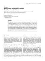
Two different cell types were isolated from the “distal”
vascular tissue in the patients with CTEPH. The cell
typesweredeterminedbymorphologytobeELCs
(rounded appearance and cell-cell contact in the mono-
layer) and MFLCs (spindle-shaped with cytoplasmic
extensions) (Figure 1). They were dissociated and pas-
saged free from surrounding cells using cloning cylin-
ders. MFLCs were prepared from each of the six
patients and ELCs could be isolated from 4 of the six
patients.
Figure 1 Cells from endarterectomized tissue. The MFCsL and
ELCs from endarterectomized tissue were microscopically assessed.
The magnification was 100×. Scale bar = 100 μm; MFLCs =
myofibroblast-like cells; ELCs = endothelial-like cells.
Sakao et al. Respiratory Research 2011, 12:109
/>Page 4 of 16
Moreover, another cell type was isolated from the ves-
sel wall tissues of control pulmonary arteries, defined
morphologically as fibroblast-like cells (pulmonary arter-
ial fibroblast-like cells) (data not shown). These cells
were used as control cells, and were prepared in the
same way as the CTEPH specimens.
The cells outgrown from the organized thrombotic tis-
sue and control pulmonary arteries were further charac-
terized by immunohistochemical staining for desmin,
vimentin, von Willebrand factor (Factor VIII) anda-
SMA. ELCs were positively stained for the endothelial
cell (EC)-specific marker (Factor VIII) and the mesenchy-
mal-specific marker (vimentin) and negative for the 2
smooth muscle cell (SMC)-specific markers (desmin and
a-SMA) [12]. MFLCs were Factor VIII and desmin nega-
tive and vimentin and a-SMA positive [12]. Pulmonary
arterial fibroblast-like cells were Factor VIII, desmin and
a-SMA negative, and vimentin positive (data not shown).
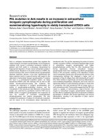
Phenotypic alteration of ELCs
After a few passages, morphological alterations were
detected in the ELCs. The cell-cell contact of the
endothelial monolayers became disrupted, and some
ELCs had lost their rounded appearance and acquired
an elongated, mesenchymal-like morphology. At the 2nd
passages, the morphological alterations could not to be
detected micr oscopically (Figure 2A), but some SM-like
cells (as defined by expression of a-SMA) (Figure 2B)
and a few transitional cells (co-expressing both endothe-
lial- and SM-cell markers) were consistently observed
(Figure 2C) by immunohistochemical staining. These
transitional cells could be observed in ELCs prepared
from 4 of the six samples.
Since this result suggested that ELCs were contami-
nated with SMCs, at the 3rd passage, they were sorted
for the EC marker CD31 in order to establish that the
ELCs were free of contamination with SMCs. After
magnetic cell sorting for the EC marker CD31, ELCs
were examined microscopically, and unusual “ pile”
growth and disrupted formation of the endothelial
monolayer were detected (Figure 2D). Moreover, SM-
like cells (Figure 2E) and transitional cells were consis-
tently observed (Figure 2F).
Transitional cells in endarterectomized CTEPH tissue
To detect transitional cells which co-express both
endothelial (CD31) and SM (a-SMA) markers in the
PEA tissues of patients with CTEPH, a double immu-
nostaining method for CD31/a-SMA was performed.
The HE staining of the neointimal layers of both the
“proximal” and the “distal” vascular tissues indicated the
presence of a fibrin network, and nuclei are seen within
this region (Figure 2G). These neointimal layers are
composed of some a-SMA positive cells (Figure 2H).
Although the neointimal layers of both the “ proximal”
and the “distal” vascular tissues were composed of a-
SMA positive cells, CD31 positive cells were found in
the “distal” vascular wall tissue but not in the “proximal”
vascular tissue (Figure 2I). As shown in Figure 2J, a few
CD31 and a-SMA double-positive cells were identified
in the “distal” vascular tissues, thus indicating the pre-
sence of “ intermediate” cells, which were intermediate
between ECs and muscle cells in structure, in the neoin-
timal lesions of CTEPH patients.
Decreased expression of Autophagic marker LC3
(microtubule-associated protein1 light chain 3; MAP1LC3),
abnormal mitochondria, and decreased expression of
superoxide dismutase (SOD)-2 in ELCs
To assess ELC alterations, an immunofluorescence
staining method for LC3, mitochondrial mark er mito-
tracker red, and SOD-2 was performed.
LC3 is a major constituent of the autophagosome, a
double-membrane structure that sequesters the target
organelle/protein and then fuses with endo/lysosomes
where the contents and LC3 are degraded. Confocal
microscopy showe d that th e ELCs did not express LC3.
The formation of autophagosomes (green punctate
structures) was not detected in these cells (Figure 2K).
SOD-2 is an enzyme that catalyzes the dissociation of
superoxide into oxygen and hydrogen peroxide. As such,
this is an important antioxidant defense in nearly all
cell s exposed to oxygen and i s located in the mitochon-
dria. Immunofluorescence staining for mitochondrial
marker mitotracker red revealed that the normal fila-
mentous mitochondrial reticulum was disrupted and
rarefied in ELCs (Figure 2L). Moreover, SOD-2 was
decreased in ELCs (Figure 2M).
Phenotypic alteration of human pulmonary microvascular
ECs is induced by MFLCs-conditioned medium
As mentioned above, ELCs isolated from the PEA tis-
sues could easily change their phenotype during passa-
ging. We postulated that the interactions of ELCs and
MFLCs, which exist in close contact in the PEA tissues,
are involved in a process of o rganized thrombus forma-
tion that occurs during the development of CTEPH.
One basic component of this interaction may be the
MFLC-induced transition of ELCs. To test this hypoth-
esis, the commercially available human pulmonary
microvascular ECs were incubated in serum-free med-
ium conditioned by MFLCs to determine whether
MFLCs release m ediators which cause phenotypic
alteration of human pulmonary microvascular ECs.
We first established that the human pulmonary micro-
vascular ECs were free of contamination with vascular
smooth muscle cells (VSMCs) by morphology (rounded
appearance and cell-cell contact of the monolayer)
Sakao et al. Respiratory Research 2011, 12:109
/>Page 5 of 16
(Figure3A)andbyimmunofluorescencestainingusing
anti-von Willebrand factor (Figure 3D), anti-a-SMA
(Figure 3D), anti- vimentin (data not shown), and anti-
human desmin (data not shown) antibodies. The
endothelial cell-specific marker and the mesenchymal-
specific marker were positive, and the 2 smooth muscle-
specific markers were negative, providing evidence that
the human pulm onary microvascular ECs were not con-
taminated with VSMCs.
At the 2nd passage after incubation in serum-free
medium conditioned by pulmonary arterial fibroblast-
like cells and MFLCs, the phenotypic alteration of
human pulmonary microvascular ECs was assessed
microscopically and by immunofluorescence staining.
The cell-ce ll contact of the endothelial mo nolayers
became disrupted, and many ECs had lost their rounded
appearance and acquired an elon gated, mesenchymal-
like morphology in the medium conditioned by MFLCs
(Figure 3C) in comparison to the medium conditioned
by pulmonary a rterial fibroblast-like cells (Figure 3B).
The number of ECs (as defined by ex pression of von
Willebrand factor) decreased, and SM-like cells (as
defined by expression of a-SMA) were consistently
obs erved in the medium conditioned by MFLCs (Figure
3F, G), but not in the medium conditioned by pulmon-
ary arterial fibroblast-like cells (Figure 3E, G).
Expression of TGF-b1 protein in the conditioned medium
Because TGF-b1 is known to be involved in inducing the
endothelial-mesenchymal transition [22] and is known to
promote a-SMA expression in non-muscle cells (ECs
and fibroblasts derived from various t issues) [23,24], the
protein levels in the conditioned medium were measured
by ELISA. Serum-free medium conditioned by MFLCs
contained higher TGF-b1 levels than medium condi-
tioned by pulmonary arterial fibroblast- like cells, but the
difference was not statistically significant (Figure 3H).
Phenotypic alteration of human pulmonary microvascular
ECs co-cultured with MFLCs
After a 14 day incubation period, morphological altera-
tions were detected in human pulmonary microvascular
Figure 2 ELCs from endarterectomized tissue. A-F), ELCs were assessed by immunofluorescence staining for anti-Factor VIII (green) and anti-
a-SMA (red) to confirm the phenotypes of the cells. A), B) and C), ELCs before sorting; D), E) and F), ELCs after sorting; A) and D), the
magnification was 100×. Scale bar = 100 μm; B) and E), the magnification was 200×. Scale bar = 50 μm; C) and F), the magnification was 400×.
Scale bar = 25 μm. The blue staining was DAPI. G-J), Immunohistochemical staining of endarterectomized tissue. The neointimal layer of distal
vascular wall tissues was assessed by immunohistochemical staining. G), Hematoxylin and Eosin (HE) staining; H), Single staining for a-SMA; I),
Single staining for CD31; J), Double staining for CD31 and a-SMA; the magnification was 200×. Scale bar = 50 μm. K, L, M), Immunofluorescence
staining of ELCs for the autophagic marker, LC3 (K), mitochondrial marker mitotracker red (L), and SOD-2 (M). K), The formation of
autophagosomes (green punctate structures) was not detected. L), The normal filamentous mitochondrial reticulum (red punctate structures) was
not detected. M), SOD-2 expression (green punctate structures) was not detected. The blue staining was DAPI. The magnification was 400×.
Scale bar = 25 μm. ELCs = endothelial-like cells.
Sakao et al. Respiratory Research 2011, 12:109
/>Page 6 of 16
Figure 3 Human pulmonary microvascular ECs (HPMVECs) in serum-free medium conditioned by pulmonary arterial fibroblast-like
cells (PAFLCs) or myofibroblast-like cells (MFLCs). The phenotypic alteration of HPMVECs was assessed microscopically and by
immunofluorescence staining. A) and D), Before incubation in serum-free medium conditioned by PAFLCs and MFLCs; B) and E), At the 2nd
passage after incubation in serum-free medium conditioned by PAFLCs; C) and F), At the 2nd passage after incubation in serum-free medium
conditioned by MFLCs; A), B) and C), microscopic findings; the magnification was 100×. Scale bar = 100 μm; D), E) and F), Immunofluorescence
staining for anti-Factor VIII (green) and anti-a-SMA (red). The blue staining was DAPI. The magnification was 200×. Scale bar = 50 μm. F), Some
cells were positive for smooth muscle actin fibers (see inset); HPMVECs = human pulmonary microvascular endothelial cells; MFLCs =
myofibroblast like cells; PAFLCs = fibroblast-like cells from control pulmonary arteries. G) Positive cells for anti-von Willebrand factor and anti-a-
SM-actin were counted in 3 different fields at a magnification of × 200 in a fluorescence microscope. *P < 0.05
VS.
PAFLCs, n ≥ 3. H) The TGF-b1
protein levels in the conditioned medium were measured by ELISA. There were no significant differences between the serum-free medium
conditioned by PAFLCs and MFLCs.
Sakao et al. Respiratory Research 2011, 12:109
/>Page 7 of 16
ECs co-cultured with MFLCs (Figure 4B, D), but not
those cultured with pulmonary arterial fibroblast-like
cells (Figure 4A, C). The cell-cell contact of the
endothelial monolayers (Figure 4A) became disrupted,
and hill and valley formation appeared. Moreover, some
ECs had lost their rounded appearance and acquired an
elongated, mesenchymal-like morphology (Figure 4B).
Some SM-like cells (as defined by their expression of a-
SMA) and a few transitional cells (co-expressing both
endothelial- and SM- cell markers) were consistently
observed (Figure 4D, E) by immunohistochemical
staining.
Autophagy PCR array analysis of human pulmonary
microvascular ECs co-cultured with MFLCs
There were decreases in the expression of 17 autop-
hagy-related genes in ECs co-cultured with MFLCs in
comparison to the expression in ECs co-cultured with
pulmonary arterial fibroblast-like cells (Figure 5A)
(Table 1). Four of these genes; AMBRA1, ATG4D,
MAP1LC3B, and RGS19, are involved in autophagic
vacuole formation. In particular, ATG4D is responsi-
ble for protein targeting to the membrane/vacuole,
and is responsible for protein transport and protease
activity. Ten of the 17 genes; BCL2, BID, CDKN2A,
CTSB, HSP90AA1, HTT, IFNG, IGF1, INS, and
PRKAA1 are co-regulators of autophagy and apopto-
sis. Three genes; RPS6KB1, TMEM77, and UVRAG
are related to autophagy in response to other intracel-
lular signals.
Autophagic marker LC3 expression in human pulmonary
microvascular ECs co-cultured with MFLCs
Confocal microscopy showed that the ECs co-cultured
with pulmonary a rterial fibroblast-like cells expressed
LC3. The formation of aut ophagosomes (green punctate
structures) was detected in these cells (Figure 6A), but
not in E Cs co-cultured with MFLCs (Figure 6B) no r in
ELCs (Figure 2K).
Abnormal mitochondria and decreased expression of
superoxide dismutase (SOD)-2 in human pulmonary
microvascular ECs co-cultured with MFLCs
Immunofluorescence staining for mitochondrial marker
mitotracker red revealed that the normal filamentous
mitochondrial reticulum observed in ECs co-cultured
with pulmonary arterial fibroblast-like cells (Figure 6D)
was disrupted and rarefied in both ECs co-cultured with
MFLCs (Figure 6E) and ELCs (Figure 2L). Moreover,
SOD-2 was decreased in ECs co-cultured with MFLCs
(Figure 6H) and ELCs (Figure 2M) compared to those
co-cultured with pulmonary arterial fibroblast-like cells
(Figure 6G). T he decrease in SOD-2 expression in ECs
co-cultured with MFLCs and ELCs might be associated
with a reduction in SOD-2 activity.
Endothelial cell biology PCR array of human pulmonary
microvascular ECs co-cultured with MFLCs
These results, including the phenotypic alterations, inac-
tivation of autophagy, and mitochondrial dysfunction,
suggested that the endothelialcellbiologyisalteredin
patients with CTEPH. Therefore, an endothelial cell
biology PCR array was done to fur ther explore the
effects of MFLCs on endothelial cell biology.
Figure 4 Human pulmonary microvascular ECs (HPMVECs) co-
cultured with pulmonary arterial fibroblast-like cells (PAFLCs)
or myofibroblast-like cells (MFLCs). The phenotypical alteration of
HPMVECs was assessed microscopically and by immunofluorescence
staining after a 14 day incubation period. A) and C), HPMVECs co-
cultured with PAFLCs; B) and D), HPMVECs co-cultured with MFLCs;
A) and B), Microscopic findings; the magnification was 100×. Scale
bar = 100 μm; C) and D), Immunofluorescence staining for anti-
Factor VIII (green) and anti-a-SMA (red). The blue staining was DAPI.
The magnification was 400×. Scale bar = 25 μm. D), Some cells
coexpressed both anti-Factor VIII and anti-a-SMA (see inset);
HPMVECs = human pulmonary microvascular endothelial cells;
MFLCs = myofibroblast-like cells; PAFLCs = fibroblast-like cells from
control pulmonary arteries. E) Positive cells for anti-von Willebrand
factor and anti-a-SM-actin were counted in 3 different fields at a
magnification of × 200 in a fluorescence microscope. *P < 0.05
VS.
PAFLCs, n ≥ 3.
Sakao et al. Respiratory Research 2011, 12:109
/>Page 8 of 16
There were decreases in the expression of 15 and
increases in the expression of 3 genes in ECs co-cul-
tured with MFLCs in comparison to the expression in
those co-cultured with pulmonary arterial fibroblast-like
cell s (Figure 5B). The 15 decreased genes were ANXA5,
BCL2, CDH5, COL18A1, CX3CL1, ITGA5, ITGAV,
ITGB1, MMP1, NPPB, PGF, PLA2G4C, PLAU, RHOB,
and SOD1 (Table 2). C DH5, COL18A1, CX3CL1,
ITGA5, ITGAV, ITGB1 and RHOB are related to
endothelial cell activation as adhesion molecules.
MMP1, NPPB, PLAU and RHOB are related to endothe-
lial cell activation, and are part of the extracellular
matrix (ECM) molecules. ANXA5 and PLAU are related
to endothelial cell activation with regard to thrombin
activity. PGF is related to angiogenesis. PLA2G4C and
SOD-1 are both related to the endothelial cell response
to stress.
The 3 genes with increased expression were AGTR1,
CASP1, and TIMP1 (Table 3). AGTR1 is related to the
permissibility and vessel tone of the angiotensin system.
CASP1 is related to endothelial cell injury and resulting
apoptosis. TIMP1 is related to endothelial cell activation
and cell growth.
SMAD reporter signal in human pulmonary microvascular
ECs co-cultured with MFLCs
The SMAD2 and SMAD3 proteins are phosphorylated
and activated by TGF-b signaling. These activ ated
SMAD 2 a nd SMAD 3 t hen form complexes with the
SMAD4. These SMAD complexes then migrate to the
nucleus, where they activate the expression of TGF-b-
responsive genes.
Besides simple concentration measurements of TGF-
b1 in the conditioned medium (Figure 3H), the activa-
tion of the TGF-b signaling in human pulmonary micro-
vascular ECs co-cultured with MFLCs were measured by
the SMAD reporter assay. There was no statistical dif-
ference in the expression of SMAD reporter signal in
ECs co-cultured with MFLCs in comparison to the
expression in those co-cultured with pulmonary arterial
fibroblast-like cells (Figure 5C).
Accumulation of ROS in human pulmonary microvascular
ECs co-cultured with MFLCs
Accumulation of ROS coupled with an increase in oxi-
dative stress has been implicated in the pathogenesis of
numerous disease states. As SOD1 and SOD2 downre-
gulation have been shown by the PCR-Arrays (Figure
5B) and immunofluorescence (Figure 6H), the missing
production of ROS might be involved in ECs co-cul-
turedwithMFLCs[25].Thedecreasedproductionof
ROS has been detected in ECs co-cultured with MFLCs
in comparison to the expression in those co-cultured
with pulmonary arterial fibroblast-like cells (Figure 5D).
Rapamycin treatment
Prolonged rapamycin treatme nt of ECs co-cultu red with
MFLCs reversed the decrease in the 17 autophagy-
Figure 5 Human pulmonary microvascular ECs (HPMVECs) co-
cultured with pulmonary arterial fibroblast-like cells (PAFLCs)
or myofibroblast-like cells (MFLCs). Autophagy and Endothelial
cell biology. A) Autophagy PCR array analysis of HPMVECs co-
cultured with PAFLCs, MFLCs or MFLCs+Rapamycin. There were
decreases in the expression of 17 autophagy-related genes in the
ECs co-cultured with MFLCs in comparison those co-cultured with
PAFLCs (P < 0.05; n = 3). This result is related to 3 different patients
out of six of co-culture or conditioned medium. See table 1 for
definitions of the abbreviations. B) Endothelial cell biology PCR array
analysis of HPMVECs co-cultured with PAFLCs, MFLCs or MFLCs
+Rapamycin. There were decreases in 15 and increases of 3 genes
in ECs co-cultured with MFLCs in comparison to the expression in
ECs co-cultured with PAFLCs (P < 0.05; n = 3). This result is related
to 3 different patients out of six of co-culture or conditioned
medium. See table 2 and 3 for the definitions. C) SMAD reporter
signal in HPMVECs co-cultured with MFLCs. There was no statistical
difference in the expression of SMAD reporter signal in ECs co-
cultured with MFLCs in comparison to the expression in those co-
cultured with PAFLCs treated with or without rapamycin. D)
Accumulation of ROS in HPMVECs co-cultured with MFLCs. The
decreased production of ROS has been detected in ECs co-cultured
with MFLCs in comparison to the expression in those co-cultured
with PAFLCs (P < 0.05; n = 3). Although there was a tendency that
rapamycin treatment of ECs co-cultured with MFLCs reversed the
decreased production of ROS, there was no statistical difference
between them.
Sakao et al. Respiratory Research 2011, 12:109
/>Page 9 of 16
related genes (Figure 5A) (Table 1) and prevented the
changes in expression in 11 of the 15 decreased and all
three of the increased genes related to endothelial cell
biology (Figure 5B) (Table 2, 3). There was no statistical
diff erence in the expression of SMAD reporter signal in
ECs co-cultured with MFL Cs with rapamycin (Figure
5C). Although rapamycin treatment of ECs co-cultured
with MFLCs seemed to reverse the decreased produc-
tion of ROS (Figure 5D), there was no statistical differ-
ence between them.
Confocal microscopy showed that the ECs co-cultured
with MFLCs that were treated with rapamycin expressed
LC3. Although the formation of autophagosomes (green
punctate structures) was not detected in ECs co-
cultured with MFLCs (Figure 6B), it was detected in
these cells when they were treated with rapamycin (Fig-
ure 6C). In the ECs co-cultured with MFLCs, the co-
localization of Mitotracker red and SOD-2 was lost,
indicating that the mitochondrial reticulum is disru pted
(Figure 6E, 2M). However, the mitochondria in the ECs
co-cultured with MFLCs that were treated with rapamy-
cin form an intricate, filamentous network, in which
SOD-2 and Mitotracker red are tightly co-localized (Fig-
ure 6F, I).
Discussion
EnMT is a term which has been used to describe the
process through which ECs lose their endothelial
Table 1 Autophagy PCR array
Biological process description Gene name Gene
symbol
Public ID P-value
Autophagy Machinary Components: Genes Involved in
Autophagic Vacuole Formation
Autophagy/beclin-1 regulator 1 AMBRA1 NM_017749 0.00308
Autophagy Machinary Components: Genes Involved in
Autophagic Vacuole Formation
Genes Responsible for Protein Targeting to Membrane/
Vacuole
Genes Responsible for Protein Transport
Genes with Protease Activity
ATG4 autophagy related 4 homolog D (S.
cerevisiae)
ATG4D NM_032885
NM_017749
0.01167
Regulation of Autophagy:
Co-Regulators of Autophagy and Apoptosis
B-cell CLL/lymphoma 2 BCL2 NM_000633 0.000727
Regulation of Autophagy:
Co-Regulators of Autophagy and Apoptosis
BH3 interacting domain death agonist BID NM_001196 0.047933
Regulation of Autophagy:
Co-Regulators of Autophagy and Apoptosis
Cyclin-dependent kinase inhibitor 2A
(melanoma, p16, inhibits CDK4)
CDKN2A NM_000077 0.044888
Regulation of Autophagy:
Co-Regulators of Autophagy and Apoptosis
Cathepsin B CTSB NM_001908 0.010802
Regulation of Autophagy:
Chaperone-Mediated Autophagy
Heat shock protein 90 kDa alpha (cytosolic),
class A member 1
HSP90AA1 NM_001017963 0.037151
Regulation of Autophagy:
Co-Regulators of Autophagy and Apoptosis
Huntingtin HTT NM_002111 0.033212
Regulation of Autophagy:
Co-Regulators of Autophagy and Apoptosis
Co-Regulators of Autophagy and the Cell Cycle
Interferon, gamma IFNG NM_000619 0.017749
Regulation of Autophagy:
Co-Regulators of Autophagy and Apoptosis
Insulin-like growth factor 1 (somatomedin C) IGF1 NM_000618 0.017282
Regulation of Autophagy:
Co-Regulators of Autophagy and Apoptosis
Insulin INS NM_000207 0.045037
Autophagy Machinary Components: Genes Involved in
Autophagic Vacuole Formation
Microtubule-associated protein 1 light chain 3
beta
MAP1LC3B NM_022818 0.011251
Regulation of Autophagy:
Co-Regulators of Autophagy and Apoptosis
Autophagy in Response to Other Intracellular Signals
Protein kinase, AMP-activated, alpha 1 catalytic
subunit
PRKAA1 NM_006251 0.005633
Autophagy Machinary Components: Genes Involved in
Autophagic Vacuole Formation
Regulator of G-protein signaling 19 RGS19 NM_005873 0.021592
Regulation of Autophagy:
Autophagy in Response to Other Intracellular Signals
Ribosomal protein S6 kinase, 70 kDa,
polypeptide 1
RPS6KB1 NM_003161 0.024072
Regulation of Autophagy:
Autophagy in Response to Other Intracellular Signals
Transmembrane protein 77 TMEM77 NM_178454 0.019285
Regulation of Autophagy:
Autophagy in Response to Other Intracellular Signals
UV radiation resistance associated gene UVRAG NM_003369 0.016479
Functional classification of low expressed genes in co-cultured HPMVECs with MFLCs in comparison to PAFLCs
Sakao et al. Respiratory Research 2011, 12:109
/>Page 10 of 16
characteristics and gain expression of mesenchymal,
myofibroblast-like characteristics [26]. In the present
study, a few transitional cells (co-expressing both
endothelial- and SM- cell markers) were shown in the
primary culture of endarterectomized tissue specimen
(Figure 2C, F). The microenvironment created by the
stabilized clot is suggested to induce EnMT (Figure 3F,
G, 4D, E). Moreover, CD31 and a-SMA double-positive
cells were identified in the neointimal layer of vascular
wall tissue, thus indicating the presence of transitional
cells in the neointim al lesions of CTEPH (Figure 2J). In
support of our finding, Yao et al showed the presence of
CD34 (endothelial marker) positive cells co-expressing
a-SMA (SM-cell marker) in endarterectomized tissues
from patients with CTEPH [27]. Moreover, they sug-
gested that the microenvironment provided by throm-
boemboli might promote the putative progenitor cells to
differentiate and enhance intimal remode ling [27]. In
this study, our data suggest that MFLC-related EnMT
may enhance intimal remodeling. However, we fully rea-
lize the limitations of our data interpretation, which was
based on in vitro studies of cultured cells, and acknowl-
edge that data provided in this study were not strong to
support EnMT hypothesis because this study failed to
show mechanisms responsible for this process. More-
over, it may be possible that transitional cells are more
likely progenitor cells rather than they are transdifferen-
tiated by EnMT.
There was no significant difference in TGF-b1 levels
between serum-free medium conditioned by MFLCs and
by pulmonary arterial fibroblast-like cells (Figure 3H).
Moreover, there was no statistical difference in the
expression of SMAD reporter signal in ECs co-cultured
with MFLCs in comparison to the expression in those
co-cultured with pulmonary arterial fibroblast-like cells
(Figure 5C). A recent study provides evidence that Ras/
MAPK, via T GF-b1 signaling, mediates completion of
EnMT in a bleomycin model of pulmonary fibrosis [28].
However, an endothelial cell biology PCR array in this
study demonstrated the decreased expression of RHOB
(Ras homolog gene family, member B) in co-cultured
human pulmonary microvascular ECs with MFLCs in
Figure 6 Immunofluorescence staining of human pulmonary microvascular ECs (HPMVECs) and endothelial-like cells (ELCs) for the
autophagic marker, LC3 (A-C), mitochondrial marker mitotracker red (D-F), and SOD-2 (G-I). A), HPMVECs co-cultured with PAFLCs; B),
with MFLCs; C), with MFLCs + Rapamycin; A) and C), The formation of autophagosomes (green punctate structures) was detected (see inset). D),
HPMVECs co-cultured with PAFLCs; E), MFLCs; F), with MFLCs + Rapamycin; D) and F), The normal filamentous mitochondrial reticulum (red
punctate structures) was detected (see inset). G), HPMVECs co-cultured with PAFLCs; H), with MFLCs; I), with MFLCs + Rapamycin; G) and I), SOD-
2 expression (green punctate structures) was detected (see inset). The blue staining was DAPI. The magnification was 400×. Scale bar = 25 μm.
ELCs = endothelial-like cells.
Sakao et al. Respiratory Research 2011, 12:109
/>Page 11 of 16
comparison to pulmonary arterial fibroblast-like cells.
These results suggest that not only TGF-b1 nor Ras, but
also additional factors, may be essential for this transi-
tional pathway. Indeed, TGF-b1 is currently thought t o
be insufficient to induce the late stage of SM differentia-
tion in non-SMC lineage cells [24]. Moreover, neither
TGF-b1 nor activated Ras alone were capable of indu-
cing a-SMA expression [28].
The effects of conditioned media may be particularly
remarkable if chemically defined culture me dia without
serum additions is employed. Therefore, serum free
media was adapted for the conditioned media
Table 2 Endothelial Cell Biology PCR Array
Biological process
description
Gene name Gene
symbol
Public ID P-value
Endothelial Cell Activation:
Thrombin Activity
Annexin A5 ANXA5 NM_001154 0.023464
Endothelial Cell Injury:
Response to Stress
Anti-Apoptosis
B-cell CLL/lymphoma 2 BCL2 NM_000633 0.000247
Endothelial Cell Activation:
Adhesion Molecules
Cadherin 5, type 2 (vascular endothelium) CDH5 NM_001795 0.003968
Endothelial Cell Activation:
Adhesion Molecules
Collagen, type XVIII, alpha 1 COL18A1 NM_030582 0.004024
Endothelial Cell Activation:
Adhesion Molecules
Chemokine (C-X3-C motif) ligand 1 CX3CL1 NM_002996 0.000779
Endothelial Cell Activation:
Adhesion Molecules
Integrin, alpha 5 (fibronectin receptor, alpha polypeptide) ITGA5 NM_002205 0.000147
Endothelial Cell Activation:
Adhesion Molecules
Integrin, alpha V (vitronectin receptor, alpha polypeptide, antigen CD51) ITGAV NM_002210 0.02125
Endothelial Cell Activation:
Adhesion Molecules
Integrin, beta 1 (fibronectin receptor, beta polypeptide, antigen CD29
includes MDF2, MSK12)
ITGB1 NM_002211 0.018399
Endothelial Cell Activation:
Extracellular Matrix (ECM)
Molecules
Matrix metallopeptidase 1 (interstitial collagenase) MMP1 NM_002421 0.021783
Permissibility and Vessel Tone:
Regulation of Blood Pressure
Regulation of Vascular
Permeability
Angiogenesis:
Negative Regulation of
Angiogenesis
Endothelial Cell Activation:
Extracellular Matrix (ECM)
Molecules
Natriuretic peptide precursor B NPPB NM_002521 0.041345
Angiogenesis:
Other Genes Involved in
Angiogenesis
Placental growth factor PGF NM_002632 0.029734
Endothelial Cell Injury:
Response to Stress
Phospholipase A2, group IVC (cytosolic, calcium-independent) PLA2G4C NM_003706 0.014626
Endothelial Cell Activation:
Extracellular Matrix (ECM)
Molecules
Thrombin Activity
Plasminogen activator, urokinase PLAU NM_002658 0.039877
Endothelial Cell Activation
Adhesion Molecules
Angiogenesis:
Positive Regulation of
Angiogenesis
Endothelial Cell Activation:
Adhesion Molecules
Endothelial Cell Injury:
Other Genes Related to
Apoptosis
Ras homolog gene family, member B RHOB NM_004040 0.035874
Endothelial Cell Injury:
Response to Stress
Superoxide dismutase 1, soluble SOD1 NM_000454 0.001855
Functional classification of low expressed genes in co-cultured HPMVECs with MFLCs in comparison to PAFLCs
Sakao et al. Respiratory Research 2011, 12:109
/>Page 12 of 16
experiments. However, this leads to serum starvation on
the cells, which commonly leads to cell cycle arrest and
induces changes in protein synthesis. Accordingly, co-
culture experiments were conducted in media with
serum, which allows different cell types to grow on
eithersideofthemembraneandmaybeabletodetect
the mutual effects of cell types on one another.
An inactivation of autophagy was found in both ELCs
(Figure 2K) and human pulmonary microvascular ECs
co-cultured with MFLCs (Figure 6B) compared to the
expression in human pulmonary microvascular ECs co-
cultured with pulmonary arterial fibroblast-like cells
(Figure 6A), t hus suggesting that in these cells, protei n
synthesis predominates over protein degradation. More-
over, the decreased expression of cell death-related
genes indicated that cell growth may be stimulated (Fig-
ure 5A). This in activation could ben efit cancer cells.
Recently several genetic links between autophagy defects
and cancers have been shown, providing increasing su p-
port for the concept that autophagy is a genuine tumor
suppressor pathway [29]. Signaling pathways that regu-
late autophagy overlaps with those that regulate tumori-
genesis [16].
This study has shown that human pulmonary micro-
vascular ECs co-cultured with MFLC s and ELCs have
fewer mitochondria with an organized r eticulum (Figure
6E,2H)andSOD-2,whichisanenzymefoundonlyin
the mitochondria, is decreased in these cells (Figure 6H,
2L). Endothelial cell biology PCR array demonstrated
the decreased expression of SOD1 (Table 2), which is
located in the cytoplasm. Both SOD1 and 2 are an
important antioxidant defense in almost all cells exposed
to oxygen . Moreover, the decreased production of ROS
has been detected in ECs co-cultured with MFLCs in
comparison to the e xpression in those co-cultured with
pulmonary arterial fibroblast-like cell s (Figure 5D).
These results including fewer mitochondria, the
decreased expression of SOD, and normoxic decreases
in ROS are compatible with the characteristics of mito-
chondrial abnormalities in PAH, demonstrated by
Archer et al [25]. The metabolic shift from oxidative
mitochondrial metabolism to the glycolytic metabolism
inhibits acetyl-CoA to enter the Krebs’ Cycle, resulting
in reduced production of R OS. However, gene and pro-
tein expression of SOD are not directly translated into
activity and the decreased production of ROS is not suf-
ficient to determine SOD activity. It has been shown
that pulmonary artery SMCs in PAH are associated with
mitochond rial disorders [30-32]. Xu and colleagues used
an in vitro experiment with pulmonary artery ECs from
idiopathic PAH (IPAH ECs) and control l ungs (control
ECs) to show th at glucose metabolism plays the primary
role in the energy requirements of IPAH ECs, based on
the 3-fold greater glycolytic rate of IPAH ECs compared
with control ECs. This indicates that there is mitochon-
drial dysfunction in ECs in patients with idiopathic
PAH, similar to the SMCs in PAH [33]. The existence
of mitochondrial diso rder/dysfunct ion in com mercially
available pulmonary microvascular ECs co-cultured with
MFLCs in CTEPH and ECs in PAH, may support the
similarities in the microvascular remodeling in the two
disease.
Although several protein kinases regulate a utophagy,
the mammalian target of rapamycin (mTOR), which
negatively regulates the pathway in organisms from
yeast to man, is the best characterized [15]. Rapamycin
is an inhibitor of mTOR and an anti-proliferative immu-
nosuppressor that arrests cells in the G1 phase of the
cell cycle [34]. Rapamycin is used clinically in cardiovas-
cular medicine as an anti-proliferative agent applied to
coronary stents to reduce local restenosis [35]. Rapamy-
cin inhibits hypoxia-induced activation of S6 kinase in
pulmonary arterial adventitial fibroblasts [36], suggesting
the possibility that there may be a therapeutic benefit in
PAH. Moreover, rapamycin has an anti-proliferative
effect on pulmonary arterial SMCs derived from endar-
terectomized tissues of CTEPH patients [37]. In this
study, we demonstrated that rapamycin reversed the
decrease in autophagy in the ECs co-cultured with
MFLCs(Figure5A,6C).Moreover,rapamycinalso
reversed the disruption of the mitochondrial reticulum
and restored the localization of SOD-2 (Figure 6F, I). It
is acknowledged that mTOR activity antagonize induc-
tion of the general stress response genes including
Table 3 Endothelial Cell Biology PCR Array
Biological process description Gene name Gene
symbol
Public ID P-value
Permissibility and Vessel Tone:
Angiotensin System
Angiotensin II receptor, type 1 AGTR1 NM_031850 0.030612
Endothelial Cell Injury:
Caspase Activation
Caspase 1, apoptosis-related cysteine peptidase (interleukin 1, beta,
convertase)
CASP1 NM_033292 0.005519
Endothelial Cell Activation:
Other Genes Involved in Cell
Growth
TIMP metallopeptidase inhibitor 1 TIMP1 NM_003254 0.049858
Functional classification of highly expressed genes in co-cultured HPMVECs with MFLCs in comparison to PAFLCs
Sakao et al. Respiratory Research 2011, 12:109
/>Page 13 of 16
SOD-2 gene [38-40]. This may explain the mechanisms
by which rapamy cin exerts its beneficial changes on cel-
lular mitochondria and SOD2 expression. Indeed, SOD2
is located within the mitochondrial matrix and was
strongly induced in response to rapamycin in normal
and neoplastic mammalian cells [41]. It also reversed
thechangeinexpressionof11ofthe15genes
decreased by co-culture and the 3 genes increased by
co-culture that were related to endothelial cell biology
(Figure 5B) (Table 2, 3), thus suggesting that rapamycin
(as an anti-proliferativ e agent) has beneficial therapeutic
effects, not o nly on pulmonary arterial SMCs, but also
on pulmonary arterial ECs which exist in the close con-
tact with MFLCs, in the patients with CTEPH. However,
because rapamycin may act on the proliferation rate of
MFLCs more than pulmonary arterial fibroblast-like
cells [37], it is possible that this action may be an alter-
native explanation for the observed differences. More-
over, a few transitional cells were observed in the ECs
co-culture with MFLCs that were treated with rapamy-
cin (data not shown), indicating that rapamycin might
exert no beneficial effect on EnMT.
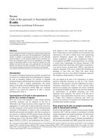
Conclusions
Our experiments with ECs and MFLCs demonstrated
that factors associated with MFLCs in the microenviron-
ment created by the unresolved clot might induce EC
dysfunction through EnMT (3F, 3G, 4D, 4E),
inactivation o f autophagy (Figure 5A, 6B), disruption of
the mitochondrial reticulum, and improper localization
of SOD-2 (Figure 6E, H). Indeed, ELCs, which were iso-
lated from the PEA tissues of CTEPH patients, included
a few transitional cells (coexpressing both endothelial-
and SM- cel l markers) (Figure 2J), lost their ability to
form autophagosomes (Figure 2K) and had defective
mitochondrial structure/function (Figure 2L). Although
it is uncertain whether MFLCs induce EC dysfunction
in vivo and whether EC dysfunctio n contribute to the
vascular lesions in th e patients with CTEPH, it is possi-
ble that there exist dysfunctional ECs in the microenvir-
onment created by the u nresolved clot (Figure 7).
However, non-resolving pulmonary thromboemboli in
CTEPH mainly consist of fibrotic tissue representing the
end-stage of a thrombus organization process. There-
fore, it remains uncertain whether any of the cellular or
molecular findings at this s tage of disease are causally
involved in disease pathogenesis. We should acknowl-
edge the purely descriptive nature of this study that
does not confer any pathophysiological evidence in
CTEPH.
Acknowledgements
All authors read and approved the final manuscript. We thank Dr. J. T.
Reeves, who enriched our research for many years.
All sources of support
This study was supported by Research Grants for the Respiratory Failure
Research Group, the Cardiovascular Diseases (19-9), and Research on
Intractable Diseases (22-33) from the Ministry of Health, Labor and Welfare,
Japan, and a Grant-in-Aid for Scientific Research (Category C 22590851) from
the Japanese Ministry of Education and Science.
Author details
1
Department of Respirology (B2), Graduate School of Medicine, Chiba
University, 1-8-1 Inohana, Chuo-ku, Chiba 260-8670, Japan.
2
Department of
Surgical Pathology, Hyogo College of Medicine, 1-1 Mukogawa-cho,
Nishinomiya, Hyogo, 663-8501, Japan.
Authors’ contributions
SS conceived of the report, contributed to its design and conception,
drafted the manuscript and carried out the all studies. HH carried out the
pathological studies. NT drafted the manuscript and contributed to its
design and conception. YK contributed to its design. KK carried out the
pathological studies. KT contributed to its design and drafted the
manuscript. All authors read and approved the final manuscript.
Competing interests
Dr. Tatsumi has received honoraria for lectures from Glaxo Smith Kline,
Actelion Pharmaceutical Ltd. Dr. Tanabe has received honoraria for lectures
from Actelion, Glaxo Smith Kline, Astellas and Pfizer and research grant
support from Actelion Pharmaceutical Ltd. The other authors report no
conflicts.
Received: 23 May 2011 Accepted: 22 August 2011
Published: 22 August 2011
References
1. Klepetko W, Mayer E, Sandoval J, Trulock EP, Vachiery JL, Dartevelle P,
Pepke-Zaba J, Jamieson SW, Lang I, Corris P: Interventional and surgical
modalities of treatment for pulmonary arterial hypertension. J Am Coll
Cardiol 2004, 43(suppl S):73S-80S.
Figure 7 EC dysfunction in CTEPH (a proposed mechanism).
Our experiments with ECs and MFLCs demonstrated that the
microenvironment provided by the thrombus cells in CTEPH
patients might induce EC dysfunction through EnMT, inactivation of
autophagy, disruption of mitochondrial reticulum, and the improper
SOD-2 localization, although it remains unknown whether both
EnMT and other cell function alterations are taking place
simultaneously in the same ECs. Although it is uncertain whether
MFLCs induce EC dysfunction in vivo and whether EC dysfunction
contribute to the vascular lesions in the patients with CTEPH, it is
possible that there exist dysfunctional ECs in the microenvironment
created by the unresolved clot.
Sakao et al. Respiratory Research 2011, 12:109
/>Page 14 of 16
2. Jamieson SW, Kapelanski DP, Sakakibara N, Manecke GR, Thistlethwaite PA,
Kerr KM, Channick RN, Fedullo PF, Auger WR: Pulmonary endarterectomy:
experience and lessons learned in 1,500 cases. Ann Thorac Surg 2003,
76:1457-1462; discussion 1462-1454.
3. Fanikos J, Piazza G, Zayaruzny M, Goldhaber SZ: Long-term complications
of medical patients with hospital-acquired venous thromboembolism.
Thromb Haemost 2009, 102:688-693.
4. Cohen AT, Agnelli G, Anderson FA, Arcelus JI, Bergqvist D, Brecht JG,
Greer IA, Heit JA, Hutchinson JL, Kakkar AK, Mottier D, Oger E, Samama MM,
Spannagl M: VTE Impact Assessment Group in Europe (VITAE). Venous
thromboembolism (VTE) in Europe. The number of VTE events and
associated morbidity and mortality. Thromb Haemost 2007, 98:756-764.
5. Hoeper MM, Mayer E, Simonneau G, Rubin L: Chronic thromboembolic
pulmonary hypertension. Circulation 2006, 113:2011-2020.
6. Moser KM, Bloor CM: Pulmonary vascular lesions occurring in patients
with chronic major vessel thromboembolic pulmonary hypertension.
Chest 1993, 103:685-692.
7. Azarian R, Wartski M, Collignon MA, Parent F, Herve P, Sors H, Simonneau G:
Lung perfusion scans and hemodynamics in acute and chronic
pulmonary embolism. J Nucl Med 1997, 38:980-983.
8. Yi ES, Kim H, Ahn H, Strother J, Morris T, Masliah E, Hansen LA, Park K,
Friedman PJ: Distribution of obstructive intimal lesions and their cellular
phenotypes in chronic pulmonary hypertension: a morphometric and
immunohistochemical study. Am J Respir Crit Care Med 2000,
162:1577-1586.
9. Sakao S, Taraseviciene-Stewart L, Lee JD, Wood K, Cool CD, Voelkel NF:
Initial apoptosis is followed by increased proliferation of apoptosis-
resistant endothelial cells. FASEB J 2005, 19:1178-1180.
10. Masri FA, Xu W, Comhair SA, Asosingh K, Koo M, Vasanji A, Drazba J,
Anand-Apte B, Erzurum SC: Hyperproliferative apoptosis-resistant
endothelial cells in idiopathic pulmonary arterial hypertension. Am J
Physiol Lung Cell Mol Physiol 2007, 293:L548-L554.
11. Firth AL, Yao W, Ogawa A, Madani MM, Lin GY, Yuan JX: Multipotent
mesenchymal progenitor cells are present in endarterectomized tissues
from patients with chronic thromboembolic pulmonary hypertension.
Am J Physiol Cell Physiol 2010, 298:C1217-C1225.
12. Maruoka M, Sakao S, Kantake M, Tanabe N, Kasahara Y, Kurosu K,
Takiguchi Y, Masuda M, Yoshino I, Voelkel NF, Tatsumi K: Characterization
of myofibroblasts in chronic thromboembolic pulmonary hypertension.
Int J Cardiol 2011.
13. Sakao S, Taraseviciene-Stewart L, Cool CD, Tada Y, Kasahara Y, Kurosu K,
Tanabe N, Takiguchi Y, Tatsumi K, Kuriyama T, Voelkel NF: VEGF-R blockade
causes endothelial cell apoptosis, expansion of surviving CD34+
precursor cells and transdifferentiation to smooth muscle-like and
neuronal-like cells. FASEB J 2007, 21:3640-3652.
14. Sahara M, Sata M, Morita T, Nakamura K, Hirata Y, Nagai R: Diverse
contribution of bone marrow-derived cells to vascular remodeling
associated with pulmonary arterial hypertension and arterial neointimal
formation. Circulation 2007, 115:509-517.
15. Klionsky DJ, Emr SD: Autophagy as a regulated pathway of cellular
degradation. Science 2000, 290:1717-1721.
16. Levine B, Kroemer G: Autophagy
in the pathogenesis of disease. Cell
2008, 132:27-42.
17. Lindahl P, Johansson BR, Levéen P, Betsholtz C: Pericyte loss and
microaneurysm formation in PDGF-B-deficient mice. Science 1997,
277:242-245.
18. Hirschi K, Rohovsky SA, D’Amore PA: PDGF, TGF-β and heterotypic cell-cell
interactions mediate the recruitment and differentiation of 10T1/2 cells
to a smooth muscle cell fate. J Cell Biol 1998, 141:805-814.
19. Hellström M, Kalén M, Lindahl P, Abramsson A, Betsholtz C: Role of PDGF-B
and PDGFR-β in recruitment of vascular smooth muscle cells and
pericytes during embryonic blood vessel formation in the mouse.
Development 1999, 126:3047-3055.
20. Allanach K, Mengel M, Einecke G, Sis B, Hidalgo LG, Mueller T, Halloran PF:
Comparing microarray versus RT-PCR assessment of renal allograft
biopsies: similar performance despite different dynamic ranges. Am J
Transplant 2008, 8:1006-1015.
21. Wang Y, Barbacioru C, Hyland F, Xiao W, Hunkapiller KL, Blake J, Chan F,
Gonzalez C, Zhang L, Samaha RR: Large scale real-time PCR validation on
gene expression measurements from two commercial long-
oligonucleotide microarrays. BMC Genomics 2006, 7:59.
22. Frid MG, Kale VA, Stenmark KR: Mature vascular endothelium can give rise
to smooth muscle cells via endothelial-mesenchymal
transdifferentiation: in vitro analysis. Circ Res 2002, 14:1189-1196.
23. Arciniegas E, Sutton AB, Allen TD, Schor AM: Transforming growth factor
beta 1 promotes the differentiation of endothelial cells into smooth
muscle-like cells in vitro. J Cell Sci 1992, 103:521-529.
24. Hautmann MB, Adam PJ, Owens GK: Similarities and differences in
smooth muscle α-actin induction by TGF-s in smooth muscle versus
non-smooth muscle cells. Arterioscler Thromb Vasc Biol 1999, 19:2049-2058.
25. Archer SL, Gomberg-Maitland M, Maitland ML, Rich S, Garcia JG, Weir EK:
Mitochondrial metabolism, redox signaling, and fusion: a mitochondria-
ROS-HIF-1alpha-Kv1.5 O2-sensing pathway at the intersection of
pulmonary hypertension and cancer. Am J Physiol Heart Circ Physiol 2008,
294:570-578.
26. Arciniegas E, Frid MG, Douglas IS, Stenmark KR: Perspectives on
endothelial-to-mesenchymal transition: potential contribution to
vascular remodeling in chronic pulmonary hypertension. Am J Physiol
Lung Cell Mol Physiol 2007, 293:L1-8.
27. Yao W, Firth AL, Sacks RS, Ogawa A, Auger WR, Fedullo PF, Madani MM,
Lin GY, Sakakibara N, Thistlethwaite PA, Jamieson SW, Rubin LJ, Yuan JX:
Identification of putative endothelial progenitor cells (CD34+CD133+Flk-
1+) in endarterectomized tissue of patients with chronic
thromboembolic pulmonary hypertension. Am J Physiol Lung Cell Mol
Physiol 2009, 296:L870-878.
28. Hashimoto N, Phan SH, Imaizumi K, Matsuo M, Nakashima H, Kawabe T,
Shimokata K, Hasegawa Y: Endothelial-mesenchymal transition in
bleomycin-induced pulmonary fibrosis.
Am J Respir Cell Mol Biol 2010,
43:161-172.
29.
Levine B: Cell biology: autophagy and cancer. Nature 2007, 446:745-747.
30. Bonnet S, Archer SL, Allaluis-Turner J, Haromy A, Beaulieu C, Thompson R,
Lee CT, Lopaschuk GD, Puttagunta L, Bonnet S, Harry G, Hashimoto K,
Porter CJ, Andrade MA, Thebaud B, Michelakis ED: A mitochondria-K
channel axis is suppressed in cancer and its normalization promotes
apoptosis and inhibits cancer growth. Cancer Cell 2003, 11:37-51.
31. Bonnet S, Michelakis ED, Porter CJ, Andrade-Navarro MA, Thébaud B,
Bonnet S, Haromy A, Harry G, Moudgil R, McMurtry MS, Weir EK, Archer SL:
An abnormal mitochondrial-HIF-1-Kv channel pathway disrupts oxygen-
sensing and triggers pulmonary arterial hypertension (PAH) in fawn-
hooded rats: similarities to human PAH. Circulation 2006, 113:2630-2641.
32. Bonnet S, Rochefort G, Sutendra G, Archer SL, Haromy A, Webster L,
Hashimoto K, Bonnet SN, Michelakis ED: The nuclear factor of activated T
cells in pulmonary arterial hypertension can be therapeutically targeted.
Proc Natl Acad Sci USA 2007, 104:11418-11423.
33. Xu W, Koeck T, Lara AR, Neumann D, DiFilippo FP, Koo M, Janocha AJ,
Masri FA, Arroliga AC, Jennings C, Dweik RA, Tuder RM, Stuehr DJ,
Erzurum SC: Alterations of cellular bioenergetics in pulmonary artery
endothelial cells. Proc Natl Acad Sci USA 2007, 104:1342-1347.
34. Brown EJ, Albers MW, Shin TB, Ichikawa K, Keith CT, Lane WS, Schreiber SL:
A mammalian protein targeted by G1-arresting rapamycinreceptor
complex. Nature 1994, 369:756-758.
35. Morice MC, Serruys PW, Sousa JE, Fajadet J, Ban Hayashi E, Perin M,
Colombo A, Schuler G, Barragan P, Guagliumi G, Molnàr F, Falotico R, RAVEL
Study Group. RAVEL Study Group: A randomized comparison of a
sirolimus-eluting stent with a standard stent for coronary
revascularization. N Engl J Med 2002, 346:1773-1780.
36. Gerasimovskaya EV, Tucker DA, Stenmark KR: Activation of
phosphatidylinositol 3-kinase, Akt, and mammalian target of rapamycin
is necessary for hypoxia-induced pulmonary artery adventitial fibroblast
proliferation. J Appl Physiol 2005, 98:722-731.
37. Ogawa A, Firth AL, Yao W, Madani MM, Kerr KM, Auger WR, Jamieson SW,
Thistlethwaite PA, Yuan JX: Inhibition of mTOR attenuates store-operated
Ca2+ entry in cells from endarterectomized tissues of patients with
chronic thromboembolic pulmonary hypertension. Am J Physiol Lung Cell
Mol Physiol 2009, 297:L666-676.
38. Beck T, Hall MN: The TOR signalling pathway controls nuclear localization
of nutrient-regulated transcription factors. Nature 1999, 402:689-692.
39. Görner W, Durchschlag E, Martinez-Pastor MT, Estruch F, Ammerer G,
Hamilton B, Ruis H, Schüller C: Nuclear localization of the C2H2 zinc
finger protein Msn2p is regulated by stress and protein kinase A
activity. Genes Dev 1998, 12:586-597.
Sakao et al. Respiratory Research 2011, 12:109
/>Page 15 of 16
40. Görner W, Durchschlag E, Wolf J, Brown EL, Ammerer G, Ruis H, Schüller C:
Acute glucose starvation activates the nuclear localization signal of a
stress-specific yeast transcription factor. EMBO J 2002, 21:135-144.
41. Zurita-Martinez SA, Cardenas ME: Tor and cyclic AMP-protein kinase A:
two parallel pathways regulating expression of genes required for cell
growth. Eukaryot Cell 2005, 4:63-71.
doi:10.1186/1465-9921-12-109
Cite this article as: Sakao et al.: Endothelial-like cells in chronic
thromboembolic pulmonary hypertension: crosstalk with myofibroblast-
like cells. Respiratory Research 2011 12:109.
Submit your next manuscript to BioMed Central
and take full advantage of:
• Convenient online submission
• Thorough peer review
• No space constraints or color figure charges
• Immediate publication on acceptance
• Inclusion in PubMed, CAS, Scopus and Google Scholar
• Research which is freely available for redistribution
Submit your manuscript at
www.biomedcentral.com/submit
Sakao et al. Respiratory Research 2011, 12:109
/>Page 16 of 16