Báo cáo y học: "Attenuation of acute lung inflammation induced by cigarette smoke in CXCR3 knockout mice" pps
Bạn đang xem bản rút gọn của tài liệu. Xem và tải ngay bản đầy đủ của tài liệu tại đây (7.75 MB, 10 trang )
BioMed Central
Page 1 of 10
(page number not for citation purposes)
Respiratory Research
Open Access
Research
Attenuation of acute lung inflammation induced by cigarette smoke
in CXCR3 knockout mice
Li Nie
†1,2
, Ruolan Xiang
†3
, Weixun Zhou
†4
, Bao Lu
5
, Deyun Cheng
2
and
Jinming Gao*
1
Address:
1
Department of Respiratory Disease, Peking Union Medical College Hospital, Chinese Academy of Medical Sciences & Peking Union
Medical College, Beijing 100730, PR China,
2
Department of Respiratory Disease, West China Hospital, Sichuan University, Chengdu 610041,
Sichuan Province, PR China,
3
Department of Pathophysiology, Peking University Health Sciences Center, Beijing 100088, PR China,
4
Department
of Pathology, Peking Union Medical College Hospital, Chinese Academy of Medical Sciences & Peking Union Medical College, Beijing 100730,
PR China and
5
Ina Sue Perlmutter Laboratory, Division of Pulmonary, Children's Hospital, Harvard Medical School, Boston, MA 02115, USA
Email: Li Nie - ; Ruolan Xiang - ; Weixun Zhou - ;
Bao Lu - ; Deyun Cheng - ; Jinming Gao* -
* Corresponding author †Equal contributors
Abstract
Background: CD8+ T cells may participate in cigarette smoke (CS) induced-lung inflammation in
mice. CXCL10/IP-10 (IFNγ-inducible protein 10) and CXCL9/Mig (monokine induced by IFN-γ) are
up-regulated in CS-induced lung injury and may attract T-cell recruitment to the lung. These
chemokines together with CXCL11/ITAC (IFN-inducible T-cell alpha chemoattractant) are ligands
for the chemokine receptor CXCR3 which is preferentially expressed chiefly in activated CD8+ T
cells. The purpose of this investigation was to study the contribution of CXCR3 to acute lung
inflammation induced by CS using CXCR3 knockout (KO) mice.
Methods: Mice (n = 8 per group) were placed in a closed plastic box connected to a smoke
generator and were exposed whole body to the tobacco smoke of five cigarettes four times a day
for three days. Lung pathological changes, expression of inflammatory mediators in
bronchoalveolar lavage (BAL) fluid and lungs at mRNA and protein levels, and lung infiltration of
CD8+ T cells were compared between CXCR3-/- mice and wild type (WT) mice.
Results: Compared with the WT littermates, CXCR3 KO mice showed less CS-induced lung
inflammation as evidenced by less infiltration of inflammatory cells in airways and lung tissue,
particularly fewer CD8+ T cells, lower levels of IFNγ and CXCR3 ligands (particularly CXCL10).
Conclusion: Our findings show that CXCR3 is important in promoting CD8+ T cell recruitment
and in initiating IFNγ and CXCL10 release following CS exposure. CXCR3 may represent a
promising therapeutic target for acute lung inflammation induced by CS.
Background
Exposure to cigarette smoke (CS) is a major risk factor for
the pathogenesis of chronic obstructive pulmonary dis-
ease (COPD) [1]. CS initiates the infiltration of innate and
adaptive inflammatory cells into the airways and the lung
parenchyma and further destroys the alveolar structure [2-
6]. The role of inflammation in the development of
COPD is supported by the finding of excess numbers of
Published: 16 December 2008
Respiratory Research 2008, 9:82 doi:10.1186/1465-9921-9-82
Received: 22 June 2008
Accepted: 16 December 2008
This article is available from: />© 2008 Nie et al; licensee BioMed Central Ltd.
This is an Open Access article distributed under the terms of the Creative Commons Attribution License ( />),
which permits unrestricted use, distribution, and reproduction in any medium, provided the original work is properly cited.
Respiratory Research 2008, 9:82 />Page 2 of 10
(page number not for citation purposes)
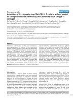
Effect of CXCR3 gene deficiency on infiltration of inflammatory cells in mice after 3 days of CS exposureFigure 1
Effect of CXCR3 gene deficiency on infiltration of inflammatory cells in mice after 3 days of CS exposure. Panels A and B, Total
inflammatory cells and differential populations recovered from BAL fluid and lung homogenates. Results are expressed as
means ± SEM, n = 5–8 animals per group, *, p < 0.05.
Respiratory Research 2008, 9:82 />Page 3 of 10
(page number not for citation purposes)
CD8+ T cells in lung tissues from patients with COPD and
an inverse relationship to the lung function [7,8]. CD8+ T
cells in epithelium and submucosa expressing CXCR3
were increased in numbers in smokers with COPD as
compared with nonsmokers. Excessive CD8+ T cells in the
lungs of COPD also produce large amounts of IFNγ and
IFNγ-induced CXC chemokines, such as CXCL10/inter-
feron-inducible protein-10 (IP-10). CXCL10, a CXCR3
ligand, was abundantly expressed in bronchiolar epithe-
lial cells and airway smooth muscle cells [9,10]. CXCL9/
Mig, CXCL10/IP-10, CXCL11/ITAC are chemokines that
attract activated T cells through binding to their receptor,
CXCR3 [11]. Collectively, these findings suggest that
CXCR3/CXCL10 interaction may play a pivotal role in the
pathogenesis and progression of COPD through T cell
recruitment in airways and lung parenchyma.
A key initiating event in COPD is the recruitment of
inflammatory cells into the lung in response to CS expo-
sure [1], which is regulated by a variety of chemokines [9-
11]. CXCL9/Mig, CXCL10/IP-10 and CXCL11/ITAC, lig-
ands for CXCR3, and CCL5/RANTES, ligand for CCR5,
were shown to be elevated in sputum from COPD patients
compared with nonsmokers [12]. Chemokine receptors
have been implicated in the pathogenesis of lung inflam-
mation in rodent models exposed to CS. For example,
CXCR2 has been demonstrated to be involved in acute
pulmonary inflammation induced by CS [13]. In CCR5
gene ablated mice, lung tissue inflammation and apopto-
sis induced by IFNγ and CS were also significantly
decreased [14,15]. Recent reports demonstrated that
CCR6, together with its ligand CXCL20/MIP-3α, was
involved in CS-induced lung inflammation and that the
interaction between CCR6 and CCL20/MIP-3α could also
mediate accumulation of dendritic cells (DCs) in the
lungs of COPD patients [16,17].
In terms of CXCR3, particularly expressed on activated
Th1/Tc1 cells [9,10,16-18], we hypothesized that CXCR3
gene deficiency would terminate or at least attenuate CS-
induced pulmonary inflammation and tissue damage. To
address this hypothesis, we used CXCR3 KO mice and
their WT littermates to investigate the contribution of
CXCR3 in CS-induced lung injury process.
Methods
Mice and cigarette smoke exposure
CXCR3 gene deficient mouse line has been established by
gene targeting as described elsewhere [19]. CXCR3 KO
mice and WT littermate mice (Experimental Animal
Research Center, Beijing, China) with C57BL/6 back-
ground (backcross > 14 generations), were maintained in
a pathogen-free mouse facility at Peking Union Medical
College. Clean food and water ad libitum were given. Ten
to 12 week old mice (~20–22 grams of weight) were used
in the experiments.
A commercially-available filter cigarette was used (White
Shark brand, Tobacco Company, China), and according
to manufacturer's specification, each cigarette contained 1
mg of nicotine and 13 mg of tar. CS exposure was per-
formed according to previously-described methods
[13,15,16]. Briefly, mice were placed in a closed plastic
box connected to smoke generator. The mice (n = 8 per
group) were exposed whole body to the tobacco smoke of
five cigarettes four times a day with 30-minute smoke free
interval for three consecutive days. Control mice were
received filtered air according to the same procedure. Ani-
Table 1: RT-PCR primers, conditions and products
RT-PCR Genes S/AS Primer sequence (5' to 3') Tm(°C) Product (bp)
CXCL9 S CTTGGGCATCATCTTCCT G 55 352
AS TGAACGACGACGACTTTGG
CXCL10 S GTCATTTTCTGCCTCATCC 55 273
AS GAGCCCTTTTAGACCTTTT
CXCL11 S CTGCTCAAGGCTTCCTTATGTT 55 166
AS CCTTTGTCGTTTATGAGCCTTC
IFNγ S CATCTTGGCTTTGCAGCTCTT 55 363
AS CTGGACCTGTGGGTTGTTGA
Granzyme-A S GAAACCAGGAACCAGATGC 55 390
AS GTGACAGGGATGGAGTGAA
Granzyme-B S CCCTCTGCCTTCTTCCTC 55 344
AS CTGGGTCTTCTCCTGTTCTT
Perforin S ATGGCACGCACTTTATCAC 55 413
AS CTTCGGGTTCTGTTCTTCC
β-actin S CTTCCTTAATGTCACGCACGATTTC 55 541
AS GTGGGGCGGCCCAGGCACCA
S, sense; AS, antisense
Respiratory Research 2008, 9:82 />Page 4 of 10
(page number not for citation purposes)
mals were killed on the fourth day after CS exposure by
pentobarbital overdose.
All experiments were performed according to interna-
tional and institutional guidelines for animal care, and
approved by Peking Union Medical College Hospital
Committee on Animal Care and Use.
Histological analysis of lung tissue
The mice were sacrificed and the lungs were removed,
inflated to 25 cmH
2
O with 10% formalin and fixed over-
night, embedded in paraffin, and sectioned at 5 μM.
Hematoxylin & eosin staining was performed at the
Department of Pathology, Peking University Health Sci-
ences Center. The pathological analysis was independ-
ently performed in each mouse in a blind manner by two
pathologists.
Bronchoalveolar lavage (BAL)
Mice were sacrificed, and the trachea was cannulated by
using a 20-gauge catheter. BAL was performed twice with
0.8 ml of ice-cold PBS (pH 7.4) each. 1.5 ml of total
injected volume was recovered in >95% of mice. The BAL
fluid was spun at 1500 rpm for 5 min at 4°C, and super-
natant was collected for the measurement of cytokines
and total protein. The pelleted cells were harvested, and
red cells were lysed, then the pelleted cells were washed
and resuspended in cold PBS. Total cells were enumerated
by counting on a hemocytometer. For differential cell
counting, cells were spun onto glass slides, air-dried, fixed
in ethanol, and stained with Diff-Quick reagents (Baxter
Scientific, Miami, FL). The number of macrophages, neu-
trophils and lymphocytes in 400 cells was counted based
on morphology.
Lung homogenates
Animals were euthanized and perfused with 3 ml of cold
saline via the heart. The left lobes were removed and
homogenized in 1 ml of PBS containing complete pro-
tease inhibitor cocktail (Sigma, St. Loius, MO). Then, the
samples were centrifuged for 10 min at 3000 rpm. Super-
natants were filtered through a 0.45 μm filter and kept in
-70°C until used.
Preparation of lung single-cell suspensions
The lungs were excised, minced and enzymatically
digested for 30 min in 15 ml of digestion buffer (RPMI,
10% FBS, 1% penicillin/streptomycin, 1 mg/ml colla-
genase (Sigma) and 30 μg/ml DNase (Sigma, St Louis,
MO). The undigested fragment was further dispersed by
repeated passage through a Nytex filter. The total cells
were pelleted, and any contaminating red cells were lysed
by ice-cold hypotonic RBC solution. After spinning, the
pellet was resuspended in 10 ml of completed medium
(RPMI 1640, 10% FBS, 1% penicillin/streptomycin). An
equal volume of 40% Percoll (Sigma, St Louis, MO) was
added, and the cells were spinned at 3000 rpm for 30 min
at room temperature. The cell pellets were resuspended in
complete medium, and leukocytes were counted on a
hemacytometer in the presence of 0.4% trypan blue. Cells
were >90% viable by trypan blue exclusion. Cytospins of
recovered cells were prepared for differential staining as
described above.
Labeling cells from BAL fluid and single lung cell
suspensions from lung tissue
50 ul of 2 × 10
7
/ml of cells from BALF and collagenase
digested lung cells was used. 10 μl of blocking buffer (1 μl
blocking antibody Fc in 9 ml PBS/2%BSA) was added to
the cells for 15 min on ice to block nonspecific binding.
After washing once, cells were incubated with 50 μl of
FITC-conjugated anti-CD4 Ab and PE-conjugated anti-
CD8 Ab or control mouse IgG2b (BD PharMingen, San
Diego, CA) for 1 hr on ice. Cells were washed twice by PBS
and fixed in PBS containing 2% formalin. Cells were sub-
jected to flow cytometer on a FACScan (Coulter).
Determination of protein content in BAL fluid
Total protein content in BAL fluid was measured using the
BCA Protein Assay Kit (Pierce, Rockford, IL) according to
manufacturer's instructions.
ELISA analysis of IFN
γ
and CXCL10
The concentrations of IFNγ, and CXCL10 (the limit of
detection were 12.5 pg/ml and 2.2 pg/ml, respectively) in
BAL fluid and lung homogenates were determined by
Effect of of CXCR3 gene deficiency on protein leakage from circulation to airways after 3 days of CS exposureFigure 2
Effect of of CXCR3 gene deficiency on protein leakage from
circulation to airways after 3 days of CS exposure. Results
are expressed as means ± SEM, n = 5–8 animals per group, *,
p < 0.05.
Respiratory Research 2008, 9:82 />Page 5 of 10
(page number not for citation purposes)
ELISA kits (R& D systems) according to manufacturer's
recommendations.
RNA extraction and semi-quantitative RT-PCR analysis
Total RNA was extracted from the lung using TRIzol rea-
gent (Invitrogen) according to manufacturer's instruc-
tions, and treated with RNase-free DNase. RNA was
reverse-transcribed and cDNA was subjected to PCR for
analyzing the expression of IFNγ, CXCL9, CXCL10,
CXCL11, granzyme A, granzyme B, perforin, and β-actin.
The primers and conditions for PCR are detailed in Table
1.
Statistical analysis
Data are expressed as means ± SEM. As appropriate, com-
parisons between two groups were carried out using
ANOVA and Student's t test (two-tailed) using GraphPad
PRISM software (Version 4.0 for windows; GraphGrad,
San Diego, CA). A value of P < 0.05 was considered signif-
icant.
Results
Inflammatory cells are reduced in CXCR3 KO mice
exposed to CS
To determine whether CXCR3 deficiency affects the CS-
induced infiltration of inflammatory cells into airways
and parenchyma, we estimated the cell subpopulations in
BAL fluid and lung tissue following CS exposure. There
was significantly less infiltration of inflammatory cells
into airways in CXCR3 KO mice compared with WT mice,
except for macrophages (figure 1A). Consistently, the
numbers of total inflammatory cells and differential sub-
populations harvested from lung parenchyma were signif-
Morphometry of the lungs in CXCR3 KO and WT mice after 3 days of air or CS exposureFigure 3
Morphometry of the lungs in CXCR3 KO and WT mice after 3 days of air or CS exposure. Representative photomicrographs
of hematoxylin- & eosin-stained lung tissues.
Respiratory Research 2008, 9:82 />Page 6 of 10
(page number not for citation purposes)
icantly decreased in CXCR3 KO mice group than in WT
mice (figure 1B).
Protein leakage was greater in WT mice than in CXCR3 KO
mice, indicating that there was more fluid accumulation
in alveolar spaces through the damaged alveolar and
endothelial cells in WT mice (figure 2).
Compared with CXCR3 KO mice, there was a greater
aggregation of leukocytes, and distortion of alveolar archi-
tecture in WT mice (Fig 3).
CD8+T cells in airways and lungs in CXCR3 KO mice
exposed to CS
The percentage of CD8+ T cells in both BAL fluid and lung
tissue from CXCR3 KO mice was decreased compared to
that from WT mice after CS exposure (BALF: 1.4 ± 0.1% vs
5.4 ± 0.4, p < 0.0001; Lung tissue: 7.9 ± 0.9% vs 18.9 ±
0.5%, p < 0.0001) (figure 4A and 4B). The percentage of
CD4+ T cells was similar in both BALF and lungs from WT
and CXCR3 KO mice (figure 4A and 4B). In the mice
exposed to air, CD4+T and CD8+ T cells were undetecta-
ble by FACS analysis (data not shown). These data dem-
onstrate that CXCR3 may be responsible for the initiation
of CS-induced inflammation through recruitment of
CD8+ T cells, as well as CD4+ T cells, into the airways and
lung parenchyma.
Expression of IFN
γ
and chemokines
The expression of IFNγ mRNA was increased in response
to CS in WT mice, but not in CXCR3 KO mice (figure 5A).
mRNA expression of CXCL9, CXCL10, and CXCL11 was
significantly up-regulated after CS exposure in the lungs
from WT mice relative to CXCR3 KO mice (figure 5B–D).
Effect of CXCR3 deficiency on CD8+ T cells and CD4+ T cell infiltration into airways and lungs in CXCR3 KO and WT mice after CS exposureFigure 4
Effect of CXCR3 deficiency on CD8+ T cells and CD4+ T cell infiltration into airways and lungs in CXCR3 KO and WT mice
after CS exposure. Panels A and B, representative histograms showing expression of CD4+ T cells and CD8+ T cells in BALF
and Lung. Panels C and D, pooled data showing the percentage of CD4+ T cells and CD8+ T cells in BALF and lung. Results are
expressed as means ± SEM, n = 4 separate experiments, ***, p < 0.001. The data presented are from one representative of
four independent experiments.
Respiratory Research 2008, 9:82 />Page 7 of 10
(page number not for citation purposes)
The level of IFNγ in BAL fluid and lung homogenates was
significantly lower in CXCR3 KO mice than in WT mice
(Fig 6A &6B). In addition, CXCL10 concentrations in BAL
fluid and lung homogenates were significantly decreased
in CXCR3 KO mice compared with WT mice after CS
exposure (figure 6C and 6D).
Expression of granzymes A and B, perforin
Upon activation, CD8+ T cells caused cytolysis and apop-
tosis of alveolar epithelial cells through the release of its
effector molecules, including granzymes and perforin
[20]. Although mRNA expression for granzymes A, B and
perforin was induced in both groups after CS exposure,
they were significantly reduced in CXCR3 KO mice as
compared with WT mice (figure 7A–C).
Effect of CXCR3 deficiency on mRNA expression of IFNγ and CXCR3 ligands in lung tissue of CXCR3 KO and WT miceFigure 5
Effect of CXCR3 deficiency on mRNA expression of IFNγ and CXCR3 ligands in lung tissue of CXCR3 KO and WT mice.
Panel A, mRNA expression of IFNγ. Panels B-D, mRNA expression of CXCR3 ligands. Results are expressed as means ± SEM,
n = 5–8 mice per group, *, p < 0.05, **p < 0.01.
Respiratory Research 2008, 9:82 />Page 8 of 10
(page number not for citation purposes)
Discussion
In this study, we have demonstrated that deletion of
CXCR3 gene in mice significantly prevented the lung
inflammation induced by exposure to CS. CXCR3 may be
a key factor in CS-induced pulmonary injury by regulating
the recruitment of CD8+ T cells as well as other inflamma-
tory cells such as neutrophils and macrophages and by
initiating the production of IFNγ, IFNγ-target CXCL10,
and the expression of effector molecules from CD8+ T
cells.
The local inflammatory response in CS-induced lung
injury is associated with infiltration of leukocytes, which
is regulated by the members of CXC family [11]. Consist-
ent with previous reports of murine model induced by
acute CS exposure [6,13,21], neutrophils represented the
majority of cells (~50% of the leukocytes in BAL fluid and
lung tissue) in this study. Neutrophilic inflammation is a
key factor in the pathogenesis of COPD, and neutrophil
infiltration has been shown to be essential for the subse-
quent recruitment of CD8+ T cells to sites of inflamma-
tion [22]. In CS-exposed CXCR3 KO mice, we observed
significant reduction in the severity of lung inflammation
as evidenced by fewer inflammatory cells in airways and
lung tissue and lesser protein leakage into the airway.
These observations point to an important role for CXCR3
in the pathogenesis of CS-induced pulmonary inflamma-
tion. However, it should be pointed out that CXCR3 KO
mice showed partial protection from CS-induced pulmo-
nary inflammation in this study. We have come to realize
that CS-induced pulmonary inflammation is not caused
by a single chemokine receptor but that multiple chemok-
ine receptors expressed on inflammatory and immune
cells are involved [13-16]. Further studies should be done
Effect of CXCR3 deficiency on IFNγ and CXCL10 concentration in BAL fluid and homogenates from mice exposed to CS or air for three consecutive daysFigure 6
Effect of CXCR3 deficiency on IFNγ and CXCL10 concentration in BAL fluid and homogenates from mice exposed to CS or air
for three consecutive days. Panels A-B, IFNγ concentration. Panels C-D, CXCL10 concentration. Results are expressed as
means ± SEM, n = 5–8 mice per group, *, p < 0.05, **p < 0.01.
Respiratory Research 2008, 9:82 />Page 9 of 10
(page number not for citation purposes)
to determine how they interact in a complex network to
contribute the pulmonary inflammation caused by CS.
Th1 type cells preferentially express CXCR3 and CCR5,
and the infiltrating T cells in COPD express high levels of
CXCR3 and CCR5, but not of CCR4 and CCR8 that are
preferentially expressed by Th2 cells [2,11,12]. Numerous
investigations performed both in vivo and in vitro have
consistently found CXCR3 to be associated with Th1/Tc1
responses [2,11,18,23]. In accordance with these findings,
we demonstrated that less CD8+ T cells infiltrated into air-
ways and lungs of CXCR3 KO mice. In mice where CD8+
T cells have been deleted, there is resistance to the devel-
opment of COPD [20]. The explanation for the relative
difference in CD8+ T cells between CXCR3 KO and WT
mice in this model may in part be due to the downstream
effect of CXCR3 activation. Moreover, CD8+ T cells dam-
age the lung interstitium through the release of lytic sub-
stances such granzyme A, granzyme B, and perforin
[20,24-26]. In support of this notion, we demonstrated
that the expression of these enzymes, to some extent, was
upregulated in WT mice upon CS exposure. However, the
upregulation was inhibited in CS-exposed CXCR3 KO
mice. This phenomenon can be attributed to the abroga-
tion of CD8+ T cells in the inflamed lungs from CXCR3
KO mice, which led to the decreased expression of these
effector molecules.
The inflammatory response in CS-induced pulmonary
damage is characterized by an increased number of Th1
cells, that secrete the Th1-type cytokine, IFNγ [27]. IFNγ
expression in BALF and lung homogenates at both mRNA
and protein levels was increased after CS exposure either
in CXCR3 KO or WT mice, but in CXCR3 KO mice, such
increase was blunted. We also demonstrated that CXCR3
ligands were significantly elevated at the transcriptional
level required for IFNγ in CS-exposed WT mice. Notably,
there was less CXCL10 in both BALF and lung homoge-
nates in CXCR3 KO mice. This can be explained by the
negative feedback effect of CXCR3 deletion, in which the
reduced accumulation of inflammatory cells in airways
and pulmonary parenchyma leads to a diminished release
of inflammatory mediators such as IFNγ; more impor-
tantly, this leads to inhibition of the activation of airway
epithelial cells to produce CXCL10 and decrease in the
recruitment of CXCR3 bearing CD8+ T cells [18].
Conclusion
To our knowledge, this study is the first to specifically
focus on the importance of CXCR3 in CS-induced lung
inflammation by using CXCR3 KO mice. In conclusion,
our study shows that CXCR3 regulates CS-induced lung
inflammation via recruitment of CD8+ T cells into the
lung to trigger the inflammatory response cascade with
over-expression of IFNγ and chemokines that activate
CXCR3 ligands, particularly CXCL10. Our findings may
provide a therapeutic target for treating CS-induced pul-
monary injury.
Effect of CXCR3 deficiency on effector molecules of CD8+ T cells in CXCR3 KO and WT miceFigure 7
Effect of CXCR3 deficiency on effector molecules of CD8+ T
cells in CXCR3 KO and WT mice. Panels A-C, mRNA
expression of granzyme A, granzyme B, and perforin in
CXCR3 KO and WT mice. Results are expressed as means ±
SEM, n = 5–8 mice per group, *, p < 0.05.
Respiratory Research 2008, 9:82 />Page 10 of 10
(page number not for citation purposes)
Editors' note
Following publication of this article, we have been
informed that results from this experiment looking at the
effects of cigarette smoke two hours after last exposure,
rather than 24 hours as in this article, have been pub-
lished as: Li Nie, Ruo-lan Xiang, Yong Liu, Wei-xun Zhou,
Lei Jiang, Bao Lu, Bao-sen Pang, De-yun Cheng, Jin-ming
Gao: Acta Pharmacologica Sinica 2008 December; 29 (12):
1432-1439.
Competing interests
The authors declare that they have no competing interests.
Authors' contributions
LN and RX performed the whole procedure of the experi-
ments. WZ carried out the pathological analysis. BL and
DC helped with designing and drafting the manuscript. JG
designed and supervised the experiment, and drafted the
manuscript.
Acknowledgements
This work was in part supported by grants from Natural Sciences Founda-
tion of China, Beijing Natural Sciences Foundation, and Education Ministry
of China New Century Excellent Talent, Key Laboratory of Comparative
Medicine of Healthy Ministry (No 30470767, No. 7072063, NCET 06-0156,
ZDS 200805, to Jinming Gao)
We are grateful for Professor Craig Gerard for providing the CXCR3
knockout mice, staff of Animal Center-PUMC for caring for the animals,
particularly Ms Huimin Zhao's kind help. We thank Professor Baosen Pang
for providing the smoke generating apparatus. We gratefully acknowledge
Professors Richard E. Ruffin and Surendral K Bansal for critically reading
this manuscript, for their helpful comments, and for their English editing of
this manuscript. We thank Dr. Zhiyong Liang's help in evaluating the path-
ological analysis.
References
1. Pauwels RA, Rabe KF: Burden and clinical features of chronic
obstructive pulmonary disease (COPD). Lancet 2004,
364:613-620.
2. Barnes PJ, Shapiro SD, Pauwels RA: Chronic obstructive pulmo-
nary disease: molecular and cellular mechanisms. Eur Respir J
2003, 22:672-688.
3. Hogg JC, Chu F, Utokaparch S, Woods R, Elliott WM, Buzatu L, Cher-
niack RM, Rogers RM, Sciurba FC, Coxson HO, Paré PD: The
nature of small-airway obstruction in chronic obstructive
pulmonary disease. N Engl J Med 2004, 350:2645-2653.
4. Hogg JC, Macklem PT, Thurlbeck WM: Site and nature of airway
obstruction in chronic obstructive lung disease. N Engl J Med
1968, 278:1355-1360.
5. van der Strate BW, Postma DS, Brandsma CA, Melgert BN, Luinge
MA, Geerlings M, Hylkema MN, van den Berg A, Timens W, Kerstjens
HA: Cigarette smoke-induced emphysema: a role for B cells?
Am J Respir Crit Care Med 2006, 173:751-758.
6. Stevenson CS, Coote K, Webster R, Johnson H, Atherton HC,
Nicholls A, Giddings J, Sugar R, Jackson A, Press NJ, Brown Z, Butler
K, Danahay H: Characterization of cigarette smoke-induced
inflammatory and mucus hypersecretory changes in rat lung
and the role of CXCR2 ligands in mediating this effect. Am J
Physiol Lung Cell Mol Physiol 2005, 288:L514-L522.
7. Saetta M, Di Stefano A, Turato G, Facchini FM, Corbino L, Mapp CE,
Maestrelli P, Ciaccia A, Fabbri LM: CD8+ T-lymphocytes in
peripheral airways of smokers with chronic obstructive pul-
monary disease. Am J Respir Crit Care Med 1998, 157:822-826.
8. O'Shaughnessy TC, Ansari TW, Barnes NC, Jeffery PK: Inflamma-
tion in bronchial biopsies of subjects with chronic bronchitis:
inverse relationship of CD8 + T lymphocytes with FEV1. Am
J Respir Crit Care Med 1997, 155:852-857.
9. Saetta M, Mariani M, Panina-Bordignon P, Turato G, Buonsanti C, Bar-
aldo S, Bellettato CM, Papi A, Corbetta L, Zuin R, Sinigaglia F, Fabbri
LM: Increased expression of the chemokine receptor CXCR3
and its ligand CXCL10 in peripheral airways of smokers with
chronic obstructive pulmonary disease. Am J Respir Crit Care
Med
2002, 165:1404-9.
10. Grumelli S, Corry DB, Song LZ, Song L, Green L, Huh J, Hacken J,
Espada R, Bag R, Lewis DE, Kheradmand F: An immune basis for
lung parenchymal destruction in chronic obstructive pulmo-
nary disease and emphysema. PLoS Med 2004, 1:e8.
11. D'Ambrosio D, Mariani M, Panina-Bordignon P, Sinigaglia F: Chem-
okines and their receptors guiding T lymphocyte recruit-
ment in lung inflammation. Am J Respir Crit Care Med 2001,
164:1266-1275.
12. Costa C, Rufino R, Traves SL, Lapa E, Silva JR, Barnes PJ, Donnelly LE:
CXCR3 and CCR5 chemokines in the induced sputum from
patients with COPD. Chest 2008, 133:26-33.
13. Thatcher TH, McHugh NA, Egan RW, Chapman RW, Hey JA, Turner
CK, Redonnet MR, Seweryniak KE, Sime PJ, Phipps RP: Role CXCR2
in cigarette smoke-induced lung inflammation. Am J Physiol
Lung Cell Mol Physiol 2005, 289:L322-328.
14. Ma B, Kang MJ, Lee CG, Chapoval S, Liu W, Chen Q, Coyle AJ, Lora
JM, Picarella D, Homer RJ, Elias JA: Role of CCR5 in IFN-gamma-
induced and cigarette smoke-induced emphysema. J Clin
Invest 2005, 115:3460-3472.
15. Bracke KR, D'hulst AI, Maes T, Demedts IK, Moerloose KB, Kuziel
WA, Joos GF, Brusselle GG: Cigarette smoke-induced pulmo-
nary inflammation, but not airway modelling, is attenuated
in chemokine receptor 5-deficient mice. Clin Exp Allergy 2007,
37:1467-1479.
16. Bracke KR, D'hulst AI, Maes T, Moerloose KB, Demedts IK, Lebecque
S, Joos GF, Brusselle GG: Cigarette smoke-induced pulmonary
inflammation and emphysema are attenuated in CCR6-defi-
ceinct mice. J Immunol 2006, 177:4350-4359.
17. Demedts IK, Bracke KR, Van Pottelberge G, Testelmans D, Verleden
GM, Vermassen FE, Joos GF, Brusselle GG: Accumulation of den-
dritic cells and increased CCL20 levels in the airways of
patients with chronic obstructive pulmonary disease. Am J
Respir Crit Care Med 2007, 175:998-1005.
18. Barnes PJ, Cosio MG: Characterization of T lymphocytes in
chronic obstructive pulmonary disease. PLoS Med 2004, 1:e20.
19. Hancock WW, Lu B, Gao W, Csizmadia V, Faia K, King JA, Smiley ST,
Ling M, Gerard NP, Gerard C: Requirement of the chemokine
receptor CXCR3 for acute allograft rejection. J Exp Med 2000,
192:1515-1520.
20. Maeno T, Houghton AM, Quintero PA, Grumelli S, Owen CA, Sha-
piro SD: CD8+ T cells are required for inflammation and
destruction in cigarette smoke-induced emphysema in mice.
J Immunol 2007, 178:8090-8096.
21. D'hulst AI, Vermaelen KY, Brusselle GG, Joos GF, Pauwels RA: Time
course of cigarette smoke-induced pulmonary inflammation
in mice. Eur Respir J 2005, 26:204-213.
22. Engeman T, Gorbachev AV, Kish DD, Fairchild RL: The intensity of
neutrophil infiltration controls the number of antigen-
primed CD8 T cells recruited into cutaneous antigen chal-
lenge site. J Leukoc Biol 2004, 76:941-949.
23. Lieberman J, Fan Z: Nuclear war: the granzyme A-bomb. Curr
Opin Immunol 2003, 15:553-559.
24. Trapani JA, Sutton VR: Granzyme B: pro-apoptotic, antiviral
and antitumor functions. Curr Opin Immunol 2003, 15:533-543.
25. Vernooy JH, Möller GM, van Suylen RJ, van Spijk MP, Cloots RH, Hoet
PH, Pennings HJ, Wouters EF: Increased granzyme A expression
in type II pneumocytes of patients with severe chronic
obstructive pulmonary disease. Am J Respir Crit Care Med 2007,
175:464-472.
26. O'Donnell R, Breen D, Wilson S, Djukanovic R: Inflammatory cells
in the airways in COPD. Thorax 2006, 61:448-454.