Báo cáo y học: "Vascular Endothelial Growth Factor (VEGF) isoform expression and activity in human and murine lung injury" pps
Bạn đang xem bản rút gọn của tài liệu. Xem và tải ngay bản đầy đủ của tài liệu tại đây (487.86 KB, 12 trang )
BioMed Central
Page 1 of 12
(page number not for citation purposes)
Respiratory Research
Open Access
Research
Vascular Endothelial Growth Factor (VEGF) isoform expression
and activity in human and murine lung injury
Andrew RL Medford
1
, Samantha K Douglas
1
, Sofia IH Godinho
1
,
Kay M Uppington
1
, Lynne Armstrong
1
, Kathleen M Gillespie
1
, Berendine van
Zyl
1
, Terry D Tetley
2
, Nassif BN Ibrahim
3
and Ann B Millar*
1
Address:
1
Department of Clinical Science at North Bristol, University of Bristol Paul O'Gorman Lifeline Centre, Southmead Hospital, Westbury-
on-Trym, Bristol, BS10 5NB, UK,
2
Lung Cell Biology, National Heart & Lung Institute, Imperial College, Dovehouse Street, London, SW3 6LY, UK
and
3
Department of Pathology, North Bristol NHS Trust, Frenchay Hospital, Frenchay Park Road, Frenchay, Bristol, BS16 1LE, UK
Email: Andrew RL Medford - ; Samantha K Douglas - ;
Sofia IH Godinho - ; Kay M Uppington - ;
Lynne Armstrong - ; Kathleen M Gillespie - ; Berendine van
Zyl - ; Terry D Tetley - ; Nassif BN Ibrahim - ;
Ann B Millar* -
* Corresponding author
Abstract
Background: The properties of vascular endothelial growth factor (VEGF) as a potent vascular
permogen and mitogen have led to investigation of its potential role in lung injury. Alternate spliced
VEGF transcript generates several isoforms with potentially differing functions. The purpose of this
study was to determine VEGF isoform expression and source in normal and ARDS subjects and
investigate the expression and regulation of VEGF isoforms by human alveolar type 2 (ATII) cells.
Methods: VEGF protein expression was assessed immunohistochemically in archival normal and
ARDS human lung tissue. VEGF isoform mRNA expression was assessed in human and murine lung
tissue. Purified ATII cells were cultured with proinflammatory cytokines prior to RNA extraction/
cell supernatant sampling/proliferation assay.
Measurements and Main Results: VEGF was expressed on alveolar epithelium, vascular
endothelium and alveolar macrophages in normal and ARDS human lung tissue. Increases in VEGF
expression were detected in later ARDS in comparison to both normal subjects and early ARDS
(p < 0.001). VEGF
121
, VEGF
165
and VEGF
189
isoform mRNA expression increased in later ARDS (p
< 0.05). The ratio of soluble to cell-associated isoforms was lower in early ARDS than normal
subjects and later ARDS and also in murine lung injury. ATII cells constitutionally produced VEGF
165
and VEGF
121
protein which was increased by LPS (p < 0.05). VEGF
165
upregulated ATII cell
proliferation (p < 0.001) that was inhibited by soluble VEGF receptor 1 (sflt) (p < 0.05).
Conclusion: These data demonstrate that changes in VEGF isoform expression occur in ARDS
which may be related to their production by and mitogenic effect on ATII cells; with potentially
significant clinical consequences.
Published: 9 April 2009
Respiratory Research 2009, 10:27 doi:10.1186/1465-9921-10-27
Received: 23 September 2008
Accepted: 9 April 2009
This article is available from: />© 2009 Medford et al; licensee BioMed Central Ltd.
This is an Open Access article distributed under the terms of the Creative Commons Attribution License ( />),
which permits unrestricted use, distribution, and reproduction in any medium, provided the original work is properly cited.
Respiratory Research 2009, 10:27 />Page 2 of 12
(page number not for citation purposes)
Introduction
Functional and physical failure of the alveolar capillary
membrane is a pivotal event in the development of lung
injury, exemplified by the acute respiratory distress syn-
drome (ARDS)[1]. The characteristics of vascular endothe-
lial growth factor (VEGF), as both an angiogenic and
permogenic factor has led to interest in its potential role
in this condition[2,3]. It is known that VEGF protein is
compartmentalized within the lung[4] and alveolar type 2
epithelial (ATII) cells have been identified as a major
source of VEGF in both animal studies and human foetal
lung studies[5,6]. Observational data show plasma VEGF
levels rise and intrapulmonary (ie, measurable in the epi-
thelial lining fluid (ELF) obtained by broncho-alveolar
lavage) levels fall in the early stages of lung injury with
normalization of both during recovery[7,8]. These
changes in intrapulmonary VEGF have been confirmed in
ARDS but have also been observed in other conditions in
which alveolar injury may occur, such as high-altitude
pulmonary oedema [9-11]. To explore the significance of
these observations, it is necessary to understand the mech-
anisms that regulate VEGF bioactivity.
Alternative splicing of the VEGF transcript from exons 5 to
8 leads to the generation of several different isoforms with
variable diffusibilities depending on their length:
VEGF
121
, VEGF
165
, VEGF
189
being the main forms[3,12-
14]. Exon 6 (not present in VEGF
121
and VEGF
165
) and
exon 7 provide heparin-binding affinity, exon 8 (present
in all active isoforms) is necessary for the stimulation of
mitosis[15]. The longer isoforms are highly basic and
remain virtually completely cell-associated, whereas
VEGF
121
(lacking both exons 6 and 7) is freely diffusi-
ble[14]. VEGF
165
(lacking exon 6 but not 7) possesses
intermediary properties being largely soluble but a dis-
tinct fraction remains cell-associated[14]. It is the pre-
dominant isoform and most biologically active in the
physiological state[15].
Recovery from lung injury/ARDS requires functional/
physical repair of the alveolar epithelial surface to occur.
ATII cells proliferate and differentiate into alveolar epithe-
lial type 1 (ATI) cells to regenerate the alveolar epithelium
after injury[16]. Limited and conflicting data exists on the
effect of VEGF on alveolar epithelial proliferation; fetal
human lung cells[17,18] and pulmonary adenocarci-
noma derived cell lines proliferate[19], whereas adult rat
ATII cells do not[20]. There are no similar studies of adult
human lung epithelial cells.
We initially hypothesised that changes in VEGF intrapul-
monary levels observed by ourselves and others would be
reflected in whole lung tissue and associated with changes
in VEGF isoforms in ARDS lung which differ in early and
late stages of the disease. We further hypothesised that the
role of VEGF in the lung may be as an epithelial mitogen
integral to lung repair. We have explored this hypothesis
and additionally considered the effects of known inflam-
matory mediators, previously suggested to be involved in
the pathogenesis of ARDS, in these processes.
Methods
Specimens
Archival normal and ARDS lung tissue sections and paraf-
fin blocks for which consent for research had been
obtained, were utilized for immunohistochemistry (ARDS
had been histopathologically confirmed and groups
divided into either "early ARDS" within 48 hours or "later
ARDS" after day 7). ARDS had been diagnosed according
to the internationally used 1994 American-European
Consensus Conference criteria[21]. Isolation of ATII cells
was undertaken from macroscopically normal lung tissue
sections (15 × 5 × 5 cm approximately) were donated by
13 patients (5 females and 8 males) undergoing lobar
resection for malignancy. The median age was 71. Ethical
approval was obtained from the North Bristol and United
Bristol Healthcare Trusts.
Immunohistochemistry for VEGF
Normal, early ARDS and later ARDS lung tissue sections
were examined (n = 8 for each group). Normal lung tissue
implied that there was no lung involvement in the cause
of death. Paraffinised 4 μm sections were de-waxed in
serial xylene (BDH Laboratory Supplies, Poole, UK),
dehydrated in absolute ethanol (BDH Laboratory Sup-
plies, Poole, UK) and pressure cooked in 0.01 M tri-
sodium citrate (BDH Laboratory Supplies, Poole, UK)
buffer (pH 6) to facilitate antigen retrieval. Endogenous
peroxidase was blocked with 3% hydrogen peroxide
(BDH Laboratory Supplies, Poole, UK) in methanol
(BDH Laboratory Supplies, Poole, UK). Sections were
incubated in 2.5% horse blocking serum (Vectastain Uni-
versal Quick Kit, Vector Laboratories, Peterborough, UK)
prior to Avidin D and Biotin blocking sera (Vector Labo-
ratories, Peterborough, UK). A rabbit polyclonal antibody
to VEGF Autogen Bioclear, UK Ltd, Wiltshire, UK) and iso-
typic rabbit IgG controls (Vector Laboratories, Peterbor-
ough, UK) were used. Isotypic control antibodies were
used on normal, early ARDS and later ARDS tissues. The
samples were then stained with pan-specific biotinylated
antibody, streptavidin-peroxidase complex with diami-
nobenzidine substrate (Vectastain Universal Quick Kit,
Vector Laboratories, Peterborough, UK), counterstained
in haematoxylin (BDH Laboratory Supplies, Poole, UK).
Image capture and semi-quantitative densitometry were
achieved using Histometrix version 6 software version 1.4
(Kinetic Imaging) linked to a JVC TK-C1360B camera with
a resolution of 470 TV lines. Pixels representing immuno-
positivity were chosen and this threshold was memorised
by the software. Anything within the selected pixel range
was accounted for and expressed as a percentage of the
Respiratory Research 2009, 10:27 />Page 3 of 12
(page number not for citation purposes)
pixels in the selected area. This gave a composite intensity
score per unit area derived from the staining intensity
value divided by the staining cross-sectional area assessed.
Densitometry was performed on all slides from the same
procedure assigning the same random coloration as unit
of intensity on each slide. Five randomly chosen (compu-
ter generated) areas on each section for each patient were
assessed giving twenty values. Densities on negative con-
trol sections were subtracted from positively stained sec-
tion densities to control for differing background pixel
intensities detected.
Formalin-fixed paraffin embedded (FFPE) RNA extraction
and measurement
This method is a modified version of the Krafft tech-
nique[22]. Briefly, 8 × 6 μm sections were cut on a micro-
tome from formalin-fixed paraffin-embedded (FFPE)
blocks of archival normal, "early" (within 48 hours) and
"later" ARDS (after day 7) lung tissue. Sections were de-
waxed and washed in Histoclear II and absolute ethanol.
Sections were then dried at 55°C for 3 minutes before
being digested for 6 hours in digest buffer (1 mg/ml pro-
teinase K, 20 mM Tris, 20 mM EDTA buffer at pH 7.4, 1%
SDS) at the same temperature. After ice cooling, RNA was
extracted using phenol:chloroform:isoamyl ethanol mix-
ture before precipitation in sodium acetate, washing and
pelleting.
Animal model of lung injury
Male C57BL/6 mice (18–20 g) were used in a LPS induced
model of lung injury. The experiments were done in
accordance with Home Office guidelines. Briefly mice
were exposed to intranasal LPS (Sigma, Poole, UK; sero-
type 011:B4) dissolved in pyrogen-free saline. Animals
were lightly anaesthetised by placing in a vapour-filled
chamber with halothane (Merial Animal Health). While
anaesthetised, intranasal inoculation of 50 μl of 10 μg
LPS/mouse was performed. This procedure was repeated
daily for 4 days. Control animals were treated with the
same volume of pyrogen-free saline. Animals were sacri-
ficed by halothane hyperanaesthesia and exsanguinated
by cardiac puncture. The lungs were removed, inflated
and snap frozen at day 2 and day 5 post initial LPS insult,
24 hours after last LPS dose. A minimum of n = 4 individ-
ual specimens was utilized in each experiment. Lung
injury was confirmed by histological analysis and RNA
extracted as previously described[23]. Quantitative real-
time PCR was undertaken in the day 5 post LPS mice only.
Isolation and purification of ATII cells
ATII cells were purified according to the method of With-
erden et al[24] as previously described. Sections were per-
fused with 0.9% saline, digested with 0.25% trypsin
(Sigma, Poole, UK) and minced with newborn calf serum
(NCS) (Invitrogen, Paisley). DNAse I was added to the
suspension at 250 μg/ml in 7 ml Hanks Balanced Salt
Solution (HBSS). The suspension was shaken before filter-
ing through a ~500 μm filter and then 40 μm mesh (Fahr-
enheit, Milton Keynes). ATII cells were purified by
differential adhesion. The non-adherent cells were centri-
fuged at 300 g for 10 minutes at 4°C and resuspended at
1 × 10
6
ATII cells/ml in complete media and put into 60
mm dishes (Greiner Cell Star, Stonehouse) pre-coated
with Vitrogen-100 (Cohesion Technologies, Palo Alto,
USA). The ATII cells were subsequently adhered at 37°C
for 24 hours. The medium and any remaining contami-
nating cells were removed and fresh complete medium
added. The cells were incubated for 16 hours, the medium
removed and the cells washed with HBSS. Fresh complete
medium was added and the cells incubated for a further
24 hours to establish confluent monolayers with ATII cell
morphology confirmed as previously described[24]. ATII
cell phenotype was confirmed by positive staining for
alkaline phosphatase and mRNA transcripts for SP-C and
aquaporin-3. Morphological characteristics were con-
firmed by electron microscopy. ATI cell phenotype was
excluded by negative staining for aquaporin-5.
Purified cells were then either cultured with the pro-
inflammatory agents LPS (10 μg/ml), TNF-α (10 ng/ml),
IL-1b (1 ng/ml) for 4 hours prior to RNA extraction and/
or sequential cell culture supernatant sampling or used in
the proliferation assays described below. A minimum of
10 subject samples were used for each experiment. In vitro
these cells rapidly differentiate and this precludes a pro-
longed culture timepoint.
VEGF isoform-specific RT-PCR
Human and murine RNA was extracted using TRIzol rea-
gent. Reverse transcription (RT) was carried out using the
Superscript II system (Invitrogen). Beta
2
-microglobulin
(B
2
M) was used as a housekeeping gene. Amplifications
were carried out in a 20 μl reaction volume containing 13
μl RNase free water, 1.2 μl 25 mM MgCl
2
(final concentra-
tion 1.5 mM) (Abgene), 0.4 μl 25 μM dNTPs (Abgene), 1
μl of 20 μM forward and reverse primers, 2 μl 10× reaction
buffer (Abgene) and 0.4 μl 5 U/μl Taq DNA polymerase
(Abgene). The following PCR conditions were used: dena-
turation at 94°C for 3 minutes, annealing at 72°C for 30
seconds then denaturation at 94°C for 30 seconds. The
coding sequences for human VEGF (accession no.
NM_000493, NM_001844, and NM_000088, respec-
tively) and murine VEGF (MN_00441242 for mouse
VEGF) were used to design primers using the online soft-
ware, Primer3 (Whitehead Institute for Biomedical
Research, Cambridge, MA). The primers span intronic
junctions to avoid the amplification of genomic
sequences. Primer sequences used were as in Table 1.
Amplified products were visualized by gel electrophoresis
using ethidium bromide (Sigma, Poole, UK) on a transil-
luminator (BioRad, Hertfordshire, UK) using semi-quan-
titative densitometric analysis (Biorad Geldoc software).
Respiratory Research 2009, 10:27 />Page 4 of 12
(page number not for citation purposes)
Control reactions were run without the addition of reverse
transcriptase. Previous experiments had determined the
RT-PCR data would be in linear phase at a cycle number
of 35 indicating the data were truly semi-quantitative. The
inter-assay variability of densitometry measurements was
11.2%.
Quantitative real-time RT-PCR
Quantitative real-time RT-PCR for VEGF isoforms was per-
formed in a 25 μl reaction volume containing 12.5 μl of
the SYBR Green PCR master mix (Sigma, Poole, UK), 5 μl
of the RT reaction mixture, and 300 nM each primer using
the Smart Cycler II System (Cepheid, Sunnyvale, CA). A
standard curve was generated for each test mRNA using 8
serial dilutions from neat RNA to 1:200,000. The amplifi-
cation program consisted of initial denaturation at 95°C
for 2 minutes followed by 40 cycles of 95°C for 15 sec-
onds, annealing at 58°C for 30 seconds, and extension at
72°C for 15 seconds. Primer sequences were as in Table 2.
Commercially available GAPDH primers were used http:/
/www.primerdesign.co.uk and used as a reference for nor-
malization in all RT-polymerase chain reactions (RT-
PCRs).
VEGF ELISA
Cell culture supernatants were assayed for VEGF
(VEGF
121
, VEGF
165
only) using a commercial ELISA kit
(R&D Systems). In brief, a specific monoclonal antibody
was coated onto a microplate. Standards and samples
were added and a polyclonal detection antibody added.
The resultant colour developed in proportion to the
amount of growth factor present and was read spectro-
photometrically. The detection limit was 3 pg/ml.
Cell Proliferation Assay (
3
H-thymidine incorporation)
ATII cells were seeded in 24-well plates (Greiner bio-one
Ltd, Stonehouse, UK) in complete medium (10%NCS/
DCCM/1% penicillin/streptomycin/amphotericin B)
(Sigma, Poole, UK) at 100,000 cells per well. For 48 hours
the cells were incubated in 5 ng/ml VEGF
165
± 10 ng/ml
sflt, 10 ng/ml KGF (as a positive control) and 10 ng/ml sflt
(as a specific VEGF inhibitor) alone. The concentrations of
KGF and sflt correspond to previously published concen-
trations in primary cell studies. 5 ng/ml VEGF
165
approxi-
mates to epithelial lining fluid concentrations of VEGF in
normal subjects and also following recovery from
ARDS[4,8]. The cells were then washed with HBSS (Sigma,
Poole, UK) and incubated in complete medium. Recom-
binant proteins and 37 kBq methyl-[
3
H] thymidine
(Amersham Biosciences) were added to each well. At 48
hours incubation at 37°C, the cells were washed with
trichloracetic acid 5% in PBS and then solubilized by add-
ing 0.5 ml of 0.3 M NaOH (Sigma, Poole, UK). Cell lysates
were subsequently pipetted into scintillation vials (Fisher
Scientific UK Ltd, Loughborough, Leicestershire, UK) con-
taining 2 mls of scintillation liquid (Amersham Bio-
sciences) and counted by a β-counter; Beckmann
Instruments (Beckman Coulter Ltd, High Wycombe,
Buckinghamshire, UK).
Cell count
ATII were seeded in 24-well plates (Greiner Bio-one Ltd,
Stonehouse, UK) in complete medium, at 100,000 cells
per well. After 48 hours, cells were washed with HBSSS
and the protein of interest added. After 48 hours incuba-
tion, cells were washed with PBS and incubated with 100
μl of Trypsin-EDTA (Sigma, Poole, UK). When the cells
Table 1: Table of primer sequences for semi-quantitative RT-PCR for human VEGF and beta-microglobulin (B
2
M)
RT-PCR Product sense antisense
human VEGF
121,165,189
5'-GAGATGAGCT TCCTACAGCAC-3' 5'-TCACCGCCTCGGCTTGTCACAT-3'
B
2
M 5'-GCATCATGGAG GTTTGAAGATG-3 5'-TAAGTTGCCAG CCCTCCTAGAG-3'
Table 2: Table of primer sequences for quantitative real time PCR (QPCR) for human and murine VEGF
QPCR Product sense antisense
human VEGF
165
5'-ATCTTCAAGCCATCCTGTGTGC-3' 5'-CAAGGCCCACAGGGATTTTC-3'
human VEGF
189
5'-ATCTTCAAGCCATCCTGTGTGC-3' 5'-CACAGGGAACGCTCCAGGAC-3'
murine VEGF
120
5'-AACGATGAAGCCCTGGAGTG-3' 5'-TGAGAGGTCTGGTTCCCGA-3'
murine VEGF
164
5'-AACGATGAAGCCCTGGAGTG-3' 5'-GACAAACAAATGCTTTCTCCG-3'
murine VEGF
188
5'-AACGATGAAGCCCTGGAGTG-3' 5'-AACAAGGCTCACAGTGAACG-3'
Respiratory Research 2009, 10:27 />Page 5 of 12
(page number not for citation purposes)
were unattached, 100 μl of culture medium containing
10% FBS was added. Cells were then counted under the
microscope using a haemocytometer.
Statistics
Data were analyzed by ANOVA with Bonferroni post hoc
multiple comparison correction using GraphPad Prism
version 4.0 software. A p value of < 0.05 was considered
significant.
Results
Human VEGF lung expression and quantification in
normal, early and late ARDS
Human VEGF expression was noted on alveolar epithe-
lium and macrophages with weaker endothelial expres-
sion in all human tissue sections. VEGF expression was
significantly increased in late ARDS compared to both
normal subjects and early ARDS (p < 0.001) (Figure 1).
This represented all VEGF isoforms both soluble and
membrane-bound.
Human VEGF isoform RT-PCR in normal, early and late
ARDS
In order to determine the individual VEGF isoforms, RT-
PCR was initially used. A representative RT-PCR gel from
the human FFPE tissue is shown in Figure 2a. When indi-
vidual isoforms were compared between groups, a signif-
icant increase in all three isoforms was detected in later
versus early ARDS (p < 0.05) as seen in Figure 2b–d. How-
ever, when considering individual relative isoform pro-
duction in disease there was a significant decrease in the
relative ratio of soluble to cell-associated isoforms in early
ARDS (p < 0.05) as seen in Figure 2e (comparing com-
bined densitometry data).
Murine VEGF mRNA expression
The human samples used were of archival post-mortem
origin and hence potentially subject to post mortem deg-
radation although a recent study suggests that this is min-
imal[25]. In order to support these human findings, we
repeated the mRNA analysis in samples extracted from
snap frozen murine whole lung injury samples. We dem-
onstrated a significant fold increase in all isoforms (p <
0.05) (Figure 3). Analysis of absolute values showed a sig-
nificantly greater amount of VEGF
188
than VEGF
164
and
VEGF
120
(p < 0.05).
ATII VEGF mRNA isoform expression (RT-PCR)
The main source of VEGF on the basis of our immunocy-
tochemistry results were ATII cells so we isolated these for
further investigation. We found that ATII cells constitu-
tively express mRNA for all the three main VEGF isoforms:
VEGF
121
, VEGF
165
and VEGF
189
(Figure 4a). All isoforms
were significantly increased in comparison to control fol-
lowing treatment with 10 ng/ml VEGF
165
(p < 0.05 for
VEGF
165
, p < 0.01 for other isoforms) and 10 μg/ml LPS
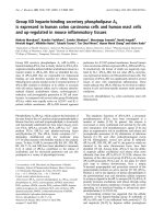
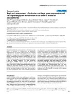
A) Immunocytochemistry of human whole lung tissue (× 40) showing (A) isotypic control (from later ARDS), (B) normal con-trols, (C) early ARDS, (D) later ARDS (magnification × 40)Figure 1
A) Immunocytochemistry of human whole lung tissue (× 40) showing (A) isotypic control (from later ARDS),
(B) normal controls, (C) early ARDS, (D) later ARDS (magnification × 40). Immunostaining shows positive VEGF
expression in alveolar epithelium (AE), alveolar macrophages (AM) and (to a lesser extent) vascular endothelium (VE). Signifi-
cant increase in staining noted in ARDS, especially later ARDS. Staining was assessed semiquantitatively using Histometrix soft-
ware analysis. B) Histometric analysis of human VEGF in healthy human lung, early and later ARDS. *P < 0.001 in later ARDS
versus early ARDS and normal, (ANOVA with post-hoc Bonferroni). Data are normal and plotted as mean and standard error.
D
B
C
A
VEGF
VEGF N VEGF E VEGF L
0
5000
10000
15000
20000
25000
*
Patient Groups
staining intensity (pixel s/unit area)
normal early ARDS late ARDS
A
B
VE
AM
VE
AE
AM
AE
AM
AE
AE
AM
VE
AM
VE
Respiratory Research 2009, 10:27 />Page 6 of 12
(page number not for citation purposes)
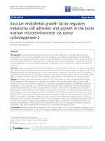
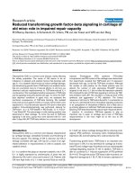
A): Representative RT-PCR gel for human VEGF isoforms in normal, early and later ARDS, lanes as follows: (1) early ARDS, (2) later ARDS, (3) negative control, (4,5,6) sequenced positive controls (VEGF
121
, VEGF
165
and VEGF
189
respectively), (7) 100 kb ladder (bottom marker denotes 100 bp, top marker 300 bp)Figure 2
A): Representative RT-PCR gel for human VEGF isoforms in normal, early and later ARDS, lanes as follows:
(1) early ARDS, (2) later ARDS, (3) negative control, (4,5,6) sequenced positive controls (VEGF
121
, VEGF
165
and VEGF
189
respectively), (7) 100 kb ladder (bottom marker denotes 100 bp, top marker 300 bp). B-E): Semi-
quantitative densitometry of (B) VEGF
189
, (C) VEGF
165
, (D) VEGF
121
relative to β-microglobulin (B
2
M) in normal, early and later
ARDS (*p < 0.05 late versus early ARDS), (E) ratio of soluble (VEGF
121
, VEGF
165
) to cell-associated (VEGF
189
) isoforms (*p <
0.05 early versus normal and later ARDS). Data are plotted as means with bars denoting standard errors (ANOVA with post-
hoc Bonferroni) for B-E).
A
VEGF
189
VEGF
165
VEGF
121
1 2 3 4 5 6 7
B
2
M
D
B
normal early ARDS late ARDS
0
30
60
90
Patient Groups
% intensity VEGF
121
/B
2
M
*
normal early ARDS late ARDS
0
30
60
90
Patient Groups
% intensity VEGF
189
/B
2
M
*
E
C
normal early ARDS late ARDS
0
30
60
90
Patient Groups
% intensity VEGF
165
/B
2
M
*
2.5
2.0
1.5
normal early ARDS late ARDS
3.0
*
Patient Groups
sol vs cell associated isoform
Respiratory Research 2009, 10:27 />Page 7 of 12
(page number not for citation purposes)
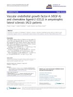
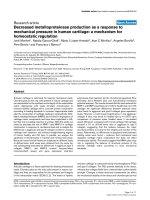
4 samples were examined for each treatment groupFigure 3
4 samples were examined for each treatment group. A) Representative RT-PCR gel of VEGF isoforms in control and
injured murine lung. Lanes are as follows: (1–8) injured murine lung at day 5 (24 hours post last LPS dose) (9–12) injured
murine lung at day 2 (24 hours post last LPS dose), (13–14) control murine lung. B-D): Further real-time PCR analysis was
undertaken in the samples at day 5. This confirmed the increased levels of VEGF
120
, VEGF
164
and VEGF
188
in injured murine
lung (*p < 0.05 fold change compared to control).
1 2 3 4 5 6 7 8 9 10 11 12 13 14
GAPDH
VEGF
188
VEGF
164
VEGF
120
A
0
1
2
3
Control
*
B
LPS 10ug/mouse
LPS 10u
g
/mouse
0
1
2
3
Control
*
C
Fold
change in
VEGF
164
mRNA
Fold
change in
VEGF
120
mRNA
3
D
*
Control
LPS 10ug/mouse
Fold
change in
VEGF
188
mRNA
2
1
0
Respiratory Research 2009, 10:27 />Page 8 of 12
(page number not for citation purposes)
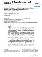
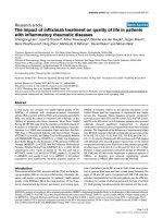
A) Representative RT-PCR gel of VEGF isoforms in ATII cellsFigure 4
A) Representative RT-PCR gel of VEGF isoforms in ATII cells. Lanes are as follows: (1–4) human ATII cell samples, (5)
blank, (6) negative control, (7–9) sequenced positive controls for VEGF
121
, VEGF
165
and VEGF
189
respectively, (10) 100 kb lad-
der (bottom marker denotes 100 bp, top marker denotes 300 bp). B-D): Semiquantitative densitometry data (n = 8, in tripli-
cate) showing VEGF
121
, VEGF
165
and VEGF
189
in response to LPS (10 μg/ml), TNF-α (10 ng/ml), IL-1β, VEGF
165
(0.1,1,10 ng/ml)
at 4 hours. Data are plotted as mean with bars denoting standard error. *P < 0.05 (ANOVA post hoc Bonferroni) 10 μg/ml LPS
versus control (all isoforms) and 10 ng/ml VEGF
165
versus control (VEGF
165
only).
#
P < 0.01 (ANOVA, post hoc Bonferroni) 10
ng/ml VEGF versus control (VEGF
121
and VEGF
189
). E-F): Further real-time PCR analysis confirmed the effect of LPS and VEGF
on VEGF
165
and VEGF
189
(*p < 0.05 fold change compared to control).
VEGF 121
VEGF
165
VEGF 189
A
1 2 3 4 5 6 7 8 9 10
B
CTRL LPS TNF IL-1 VEGF 0.1 VEGF 1 VEGF 10
0
25
50
75
100
125
*
#
VEGF
121
/GDH intensity (%)
C
E
CTRL LPS TNF IL-1 VEGF 0.1 VEGF 1 VEGF 10
0
10
20
30
40
50
60
70
80
90
100
110
120
130
140
*
*
VEGF
165
/GDH intensity (%)
Fold change
in VEGF
165
mRNA
1
*
*
Control
VEGF
10 ng
3
2
0
LPS
10 ng
D
CTRL LPS TNF IL-1 VEGF 0.1 VEGF 1 VEGF 10
0
25
50
75
100
125
150
#
*
VEGF
189
/GDH intensity (%)
Fold change
in VEG
F
189
mRNA
VEGF
10 ng
0
1
2
3
Control
*
*
F
LPS
10 ng
Respiratory Research 2009, 10:27 />Page 9 of 12
(page number not for citation purposes)
(p < 0.05) at 4 hours (Fig 4b–d). Other pro-inflammatory
stimuli (TNF, IL-1) and lower concentrations of VEGF
165
did not alter relative VEGF isoform expression. These
results were confirmed by Q-PCR showing a doubling of
VEGF
189
and VEGF
165
compared to control (p < 0.05) in
response to LPS 10 μg/ml and VEGF 10 ng/ml (Figure 4e–
f).
ATII VEGF protein levels and response to LPS
Having determined the production of VEGF isoforms at
the mRNA level we went on to assess the release of the sol-
uble isoforms (VEGF
121
, VEGF
165
) by the cultured cells.
ATII cells express significant amounts of VEGF
121
and
VEGF
165
constitutively (Figure 5). These levels signifi-
cantly increased with time in human ATII supernatant (p
< 0.01 vs control at 24 hours). At 24 hours, LPS (100 ng/
ml) further stimulated VEGF production (p < 0.05).
ATII proliferation
Having determined the production of VEGF by the ATII
cell population, we explored the potential effect of
VEGF
165
on these cells. An increase in human ATII cell
proliferation as assessed by
3
H-thymidine was detected
with 5 ng/ml VEGF
165
(p < 0.001), comparable to levels
detected previously in bronchoalveolar lavage fluid[8]).
Furthermore, with the addition of a natural VEGF inhibi-
tor, soluble VEGFR1 (sflt), there was a significant reduc-
tion in proliferation compared to serum control (p <
0.05) suggesting an autocrine effect (Figure 6). The addi-
tion of sflt alone and in combination with VEGF
165
showed no significant difference in cell number compared
to serum control (p > 0.05) but did significantly inhibit
VEGF
165
-induced increase in cell number (p < 0.001 com-
pared to 5 ng/ml VEGF
165
). This suggested that VEGF
165
was inducing proliferation rather than survival alone.
Discussion
Previous observational data by ourselves and others have
demonstrated plasma VEGF levels rise and intrapulmo-
nary levels fall in the early stages of lung injury with nor-
malization of both during recovery[7,8]. Similarly,
reduced levels of VEGF have been described in normal
smokers and patients with idiopathic pulmonary fibrosis
(IPF); other conditions in which damage to the alveolar
epithelium may be present[26].
Potential explanations for the apparent reduction in
intrapulmonary VEGF levels in early ARDS are manifold
and not mutually exclusive. They include increased mem-
brane-bound rather than soluble isoforms, changes in iso-
form expression and damage to the alveolar-capillary
membrane with consequent leakage of intrapulmonary
VEGF into the vascular bed [27-30].
We therefore assessed expression of VEGF and its specific
isoforms by immunohistochemistry and isoform-specific
RT-PCR (as isoform-specific antibodies are not available)
in archival normal and ARDS lung tissue. We demon-
strated a significant up-regulation of VEGF in later ARDS
tissue compared to normal subjects. However in early
ARDS, in contrast to our epithelial lining fluid (ELF) find-
ings we did not detect a reduction in total VEGF expres-
sion[8]. One other group has also explored this area, but
using different timepoint characteristics, whole tissue
homogenates including inflammatory cells and did not
assess differential isoform expression[11]. In both these
studies ELISA methodology was used which detects only
the soluble isoforms, VEGF
121
and VEGF
165
[8].
In order to consider changes in isoform expression as a
possible explanation for these discrepancies, we assessed
mRNA as there are no isoform-specific antibodies cur-
rently. No significant reduction in VEGF isoform expres-
sion occurred in early ARDS in comparison to normal
subjects although a trend was suggested. However, signif-
icant changes were detected between early and later ARDS
in all isoforms and there was a significant decrease in the
relative ratio of soluble to cell-associated isoforms in early
ARDS compared to later ARDS and normal subjects. In the
context of our previous ELF findings, this data is support-
ive of the suggestion that isoform switching is a critical
regulatory mechanism for VEGF bioactivity in that iso-
form-specificity may be important in ligand-VEGF recep-
tor interactions[31]. Furthermore, there is more potential
for soluble VEGF to have extrapulmonary effects which is
extremely relevant since the most common cause of death
in ARDS is multi-organ system failure[32,33].
Obtaining ARDS lung tissue is limited by the lack of sur-
gical biopsies of this disease in our clinical practice and
theoretically necropsy lung tissue might introduce selec-
ATII cell supernatant levels of VEGF protein significantly increases with time in unstimulated cells, unstimulated cell supernatants control v 24 hrs, filled bars, (#p < 0.01) with a significant increase in response to LPS 100 ng/ml at 24 hours, unfilled bars, (*p < 0.05), ANOVA with post hoc BonferroniFigure 5
ATII cell supernatant levels of VEGF protein signifi-
cantly increases with time in unstimulated cells,
unstimulated cell supernatants control v 24 hrs, filled
bars, (#p < 0.01) with a significant increase in
response to LPS 100 ng/ml at 24 hours, unfilled bars,
(*p < 0.05), ANOVA with post hoc Bonferroni.
850
*
Control
750
LPS
650
#
550
VEGF
pg/ml
450
350
3 hrs
6 hrs
12
hrs
250
150
50
24
hrs
Control
Time
Respiratory Research 2009, 10:27 />Page 10 of 12
(page number not for citation purposes)
tion bias for a more severe spectrum of ARDS, as intrapul-
monary VEGF levels are known to be lower in non-
survivors with ARDS[8]. There was no evidence of any sig-
nificant lung disease in the normal necropsy lung tissue,
but it is conceivable that the extra-pulmonary disease
process contributing to death might have affected VEGF
levels although recent data suggests this is not the
case[25]. In order to investigate this possibility we
repeated this analysis in snap frozen lung tissue from our
multiple dose LPS-induced lung injury model at day 5
post initial injury, reflecting early ARDS [23]. These data
supported the human post mortem findings of an increase
in cell-associated VEGF in an ARDS situation.
Previous in vitro studies had confirmed VEGF is abundant
in lung and suggested the alveolar epithelium as a key
source [5,6,34]. Several lines of in vitro evidence have
pointed to a possible role for VEGF in lung repair and
recovery following injury[19,29,35,36]. In one LPS-
induced murine model of lung injury, intrapulmonary
levels of VEGF increased following injury for 96 hours,
mirroring the increase in bronchoalveolar lavage fluid
protein and neutrophils with significant VEGF localiza-
tion to lung epithelium but increases mainly in inflamma-
tory cells [37]. However, previous experiments performed
in our laboratory have confirmed pg/ml levels of expres-
sion of VEGF in cultured alveolar macrophage superna-
tants from patients with ARDS and "at risk" of ARDS
suggesting that they are unlikely to be the main cellular
source of VEGF, although they may contribute [8]. In both
newborn and adult rabbit, hyperoxic lung injury resulted
in a relative reduction in VEGF
189
and parallel increase in
VEGF
121
and VEGF
165
mRNA expression with normaliza-
tion to control values during recovery[38]. Therefore, we
went on to investigate the role of human ATII cells as both
a source and potential target for VEGF bioactivity.
We initially established at the mRNA level that ATII cells
express the major VEGF isoforms (VEGF
121,165
) (both sol-
uble) and VEGF
189
(membrane-associated). The specific
functions of these isoforms have not been clearly identi-
fied in humans although genetically modified mouse
models suggest they may be significant. Ideally, we would
have isolated ATII cells from ARDS lung biopsies and
undertaken mRNA analysis but this was not possible as
described above. We have also shown that VEGF
165
is dif-
ferentially upregulated by various exogenous compounds,
in particular LPS.
Bar graphs depicting
3
H-thymidine incorporation into ATII cells (A) and cell number (B) following treatment for 48 hours with VEGF 5 ng/ml, sflt 10 ng/ml and combination confirming a significant proliferation with VEGF 5 ng/ml (#p < 0.001 vs serum con-trol)Figure 6
Bar graphs depicting
3
H-thymidine incorporation into ATII cells (A) and cell number (B) following treatment
for 48 hours with VEGF 5 ng/ml, sflt 10 ng/ml and combination confirming a significant proliferation with VEGF
5 ng/ml (#p < 0.001 vs serum control). The presence of sflt not only inhibited the proliferative effect of VEGF but also
reduced proliferation below that of serum controls (*p < 0.05), suggesting an autocrine effect. Cell number also increased sig-
nificantly with 5 ng/ml VEGF
165
(**p < 0.01) compared to serum control. The addition of sflt alone and in combination with
VEGF
165
showed no significant difference compared to serum control (p > 0.05) but did significantly inhibit VEGF
165
-induced
proliferation (p < 0.001 compared to 5 ng/ml VEGF
165
).
Serum control
10ng/ml
KG
F
5n
g
/ml V
E
GF1
6
5
10
ng
/ml
s
Fl
t
1
0
n
g
/ml sFl
t
+ 5
n
g/
m
l
V
EGF16
5
-50
0
50
100
150
200
250
#
**
[
3
H]thymidine incorporation:
% change of serum control
-ve co
n
trol
S
e
ru
m
c
on
trol
1
0n
g/
ml
KGF
5
ng/m
l
10
ng
/
m
l
s
Flt
1
0n
g
/ml
s
Flt + 5ng/m
l
VEGF165
0
100000
200000
300000
**
Cell number
Respiratory Research 2009, 10:27 />Page 11 of 12
(page number not for citation purposes)
We demonstrated for the first time that primary human
ATII cells constitutively produce soluble VEGF
(VEGF
121,165
) isoforms in a dose-dependent manner that
increased in response to LPS (p < 0.05). This is in agree-
ment with other studies showing high ELF levels in nor-
mal human subjects[4]. The increase in constitutional
production in time would suggest active secretion. The
relationship of these findings at the protein level to those
at the mRNA level is not clear-cut. The ELISA used does
not differentiate between VEGF
121
and VEGF
165
and only
detects the unbound free protein. Cell-associated VEGF is
not detected and may be considerable.
The high intrapulmonary levels of VEGF and its changes
in ARDS led us to hypothesise that VEGF may be an epi-
thelial mitogen or survival factor. This has particular rele-
vance in repair following injury as occurs in ARDS when
the alveolar epithelial surface must be regenerated to clear
fluid and restore the normal ATI cells and gas
exchange[39]. The evidence for VEGF as an epithelial
mitogen conflicts. Proliferation in human fetal
explants[18] and acid-injured A549 cells[19,40] and sur-
factant production by murine ATII cells[36] have been
described, but these data were not supported when rat
ATII cells were used[20]. In the current study, we have
shown for the first time that VEGF at 5 ng/ml (akin to nor-
mal human ELF VEGF levels), induces significant prolifer-
ation of these cells which is inhibited by the specific
inhibitor sflt. Furthermore, the reduction in proliferation
by the addition of sflt suggests a potential autocrine effect
of this protein.
We have only explored the effects of VEGF
165
in this study
and other isoforms may also be relevant. In addition,
sample numbers are limited and subject to biological het-
erogeneity inherent in primary cell studies and our pre-
liminary findings need to be expanded upon and analysed
individually in greater depth using techniques such as
laser capture microdissection for single cell PCR analysis.
VEGF
189
can be cleaved in into smaller units. Therefore,
repeating these experiments with a cleavage inhibitor
should be considered in the future. The relevance of this is
that the concept of soluble versus cell-associated isoforms
has not yet been fully resolved and proteolytic cleavage
may alter isoform ratios in ways not detected in this study.
In conclusion, we present evidence that changes in VEGF
isoforms occur between early and late ARDS. These data
are supported by both murine model and isolated human
ATII cell data. We have demonstrated ATII cells to be a
source of VEGF isoforms upregulated by lipopolysaccha-
ride, often implicated in the ARDS process. Finally we
show evidence that VEGF
165
is an ATII cell mitogen, induc-
ing proliferation which was inhibited by soluble VEGFR1.
These data suggest a key role for VEGF bioactivity in lung
injury and ARDS.
Ethics approval
The protocol was approved by the North Bristol NHS
Trust Local Research Ethics Committee.
Competing interests
The authors declare that they have no competing interests.
Authors' contributions
ARLM carried out the immunohistochemistry, FFPE RNA
extraction, ATII cell isolation and culture, ELISA, semi-
quantitative RT-PCR, statistical analysis, drafted the man-
uscript and contributed to its design and conception. SKD
carried out the proliferation studies and drafting of part of
the manuscript. SIHG designed and generated the murine
model and performed the murine RT-PCR. KMU and LA
contributed to the ATII cell isolation and culture. KMG
and BZ performed the real time PCR and contributed to
drafting of part of the manuscript. TDT contributed to the
ATII cell culture and drafting of part of the manuscript.
NBN contributed to the immunohistochemistry and
drafting of part of the manuscript. ABM conceived of the
study, contributed to its design and drafted the manu-
script. All authors read and approved the final manu-
script.
Acknowledgements
We would like to thank the following for technical advice: Mr Haydn Ken-
dall (immunohistochemistry), Ms Katy Chalmers (Histometrix software),
Ms Rachel Perrin (VEGF isoform RT-PCR). We would also like to thank Dr
Dave Bates for donation of positive control VEGF
121
and VEGF
165
isoform
cDNA.
References
1. Ware LB, Matthay MA: The acute respiratory distress syn-
drome. N Engl J Med 2000, 342(18):1334-1349.
2. Carmeliet P, Ferreira V, Breier G, Pollefeyt S, Kieckens L, Gertsen-
stein M, Fahrig M, Vandenhoeck A, Harpal K, Eberhardt C, Declercq
C, Pawling J, Moons L, Collen D, Risau W, Nagy A: Abnormal blood
vessel development and lethality in embryos lacking a single
VEGF allele. Nature 1996, 380(6573):435-439.
3. Ferrara N, Gerber HP, LeCouter J: The biology of VEGF and its
receptors. Nat Med 2003, 9:669-676.
4. Kaner RJ, Crystal RG: Compartmentalization of vascular
endothelial growth factor to the epithelial surface of the
human lung. Mol Med 2001, 7(4):240-246.
5. Koyama S, Sato E, Tsukadaira A, Haniuda M, Numanami H, Kurai M,
Nagai S, Izumi T: Vascular endothelial growth factor mRNA
and protein expression in airway epithelial cell lines in vitro.
Eur Respir J 2002, 20(6):1449-1456.
6. Boussat S, Eddahibi S, Coste A, Fataccioli V, Gouge M, Housset B,
Adnot S, Maitre B: Expression and regulation of vascular
endothelial growth factor in human pulmonary epithelial
cells. Am J Physiol Lung Cell Mol Physiol 2000, 279(2):L371-L378.
7. Thickett DR, Armstrong L, Christie SJ, Millar AB: Vascular
endothelial growth factor may contribute to increased vas-
cular permeability in acute respiratory distress syndrome.
Am J Respir Crit Care Med 2001, 164(9):1601-1605.
8. Thickett DR, Armstrong L, Millar AB: A role for vascular endothe-
lial growth factor in acute and resolving lung injury. Am J
Respir Crit Care Med 2002, 166(10):1332-1337.
9. Maitre B, Boussat S, Jean D, Gouge M, Brochard L, Housset B, Adnot
S, Delclaux C: Vascular endothelial growth factor synthesis in
the acute phase of experimental and clinical lung injury. Eur
Respir J 2001, 18(1):100-106.
Publish with BioMed Central and every
scientist can read your work free of charge
"BioMed Central will be the most significant development for
disseminating the results of biomedical research in our lifetime."
Sir Paul Nurse, Cancer Research UK
Your research papers will be:
available free of charge to the entire biomedical community
peer reviewed and published immediately upon acceptance
cited in PubMed and archived on PubMed Central
yours — you keep the copyright
Submit your manuscript here:
/>BioMedcentral
Respiratory Research 2009, 10:27 />Page 12 of 12
(page number not for citation purposes)
10. Hanaoka M, Droma Y, Naramoto A, Honda T, Kobayashi T, Kubo K:
Vascular endothelial growth factor in patients with high-alti-
tude pulmonary edema. J Appl Physiol 2003, 94:1836-1840.
11. Abadie Y, Bregeon F, Papazian L, Lange F, Chailley-Heu B, Thomas P,
Duvaldestin P, Adnot S, Maitre B, Delclaux C: Decreased VEGF
concentration in lung tissue and vascular injury during
ARDS. Eur Respir J 2005, 25(1):139-146.
12. Tischer E, Mitchell R, Hartman T, Silva M, Gospodarowicz D, Fiddes
JC, Abraham JA: The human gene for vascular endothelial
growth factor. Multiple protein forms are encoded through
alternative exon splicing. J Biol Chem 1991,
266(18):11947-11954.
13. Robinson CJ, Stringer SE: The splice variants of vascular
endothelial growth factor (VEGF) and their receptors. J Cell
Sci 2001, 114(Pt 5):853-865.
14. Houck KA, Ferrara N, Winer J, Cachianes G, Li B, Leung DW: The
vascular endothelial growth factor family: identification of a
fourth molecular species and characterization of alternative
splicing of RNA. Mol Endocrinol 1991, 5(12):1806-1814.
15. Ferrara N: Vascular endothelial growth factor: basic science
and clinical progress. Endocr Rev 2004, 25(4):581-611.
16. Fehrenbach H: Alveolar epithelial type II cell: defender of the
alveolus revisited. Respir Res 2001, 2(1):33-46.
17. Maniscalco WM, Watkins RH, Finkelstein JN, Campbell MH: Vascu-
lar endothelial growth factor mRNA increases in alveolar
epithelial cells during recovery from oxygen injury. Am J
Respir Cell Mol Biol 1995, 13(4):377-386.
18. Brown KR, England KM, Goss KL, Snyder JM, Acarregui MJ: VEGF
induces airway epithelial cell proliferation in human fetal
lung in vitro. Am J Physiol Lung Cell Mol Physiol 2001,
281(4):L1001-L1010.
19. Roberts JR, Perkins GD, Fujisawa T, Pettigrew KA, Gao F, Ahmed A,
Thickett DR: Vascular endothelial growth factor promotes
physical wound repair and is anti-apoptotic in primary distal
lung epithelial and A549 cells. Crit Care Med 2007,
35(9):2164-2170.
20. Raoul W, Chailley-Heu B, Barlier-Mur AM, Delacourt C, Maitre B,
Bourbon JR: Effects of vascular endothelial growth factor
(VEGF) on isolated fetal alveolar type II cells. Am J Physiol Lung
Cell Mol Physiol 2004, 286(6):L1293-L1301.
21. Bernard GR, Artigas A, Brigham KL, Carlet J, Falke K, Hudson L, Lamy
M, LeGall JR, Morris A, Spragg R: Report of the American-Euro-
pean consensus conference on ARDS: definitions, mecha-
nisms, relevant outcomes and clinical trial coordination. The
Consensus Committee. Intensive Care Med 1994, 20(3):225-232.
22. Krafft AE, Duncan BW, Bijwaard KE, Taubenberger JK, Lichy JH:
Optimization of the Isolation and Amplification of RNA
From Formalin-fixed, Paraffin-embedded Tissue: The
Armed Forces Institute of Pathology Experience and Litera-
ture Review. Mol Diagn 1997, 2(3):217-230.
23. Godinho SIMA: An animal model of the acute respiratory dis-
tress syndrome (ARDS). European Respiratory Journal 2002,
20:486s.
24. Witherden IR, Tetley TD, Rogers DF, Donnelly LE: Isolation and
culture of human alveolar type II pneumocytes. In Methods in
Molecular Medicine, Volume 56 (book) Volume Chapter 11. Human air-
way inflammation; sampling techniques and analytical protocols:
Humana Press Inc., Totawa, New Jersey; 2001:137-146.
25. Zhao D, Zhu BL, Ishikawa T, Quan L, Li DR, Maeda H: Real-time
RT-PCR quantitative assays and postmortem degradation
profiles of erythropoietin, vascular endothelial growth factor
and hypoxia-inducible factor 1 alpha mRNA transcripts in
forensic autopsy materials. Legal medicine (Tokyo, Japan) 2006,
8(2):132-136.
26. Koyama S, Sato E, Haniuda M, Numanami H, Nagai S, Izumi T:
Decreased level of vascular endothelial growth factor in
bronchoalveolar lavage fluid of normal smokers and patients
with pulmonary fibrosis. Am J Respir Crit Care Med 2002,
166(3):382-385.
27. Lieto E, Ferraraccio F, Orditura M, Castellano P, Mura AL, Pinto M,
Zamboli A, De Vita F, Galizia G: Expression of vascular endothe-
lial growth factor (VEGF) and epidermal growth factor
receptor (EGFR) is an independent prognostic indicator of
worse outcome in gastric cancer patients.
Annals of Surgical
Oncology 2008, 15(1):69-79.
28. Mura M, Dos Santos CC, Stewart D, Liu M: Vascular endothelial
growth factor and related molecules in acute lung injury. J
Appl Physiol 2004, 97(5):1605-1617.
29. Mura M, Han B, Andrade CF, Seth R, Hwang D, Waddell TK, Kes-
havjee S, Liu M: The early responses of VEGF and its receptors
during acute lung injury: implication of VEGF in alveolar epi-
thelial cell survival. Critical care (London, England) 2006,
10(5):R130.
30. Perkins GD, Roberts J, McAuley DF, Armstrong L, Millar A, Gao F,
Thickett DR: Regulation of vascular endothelial growth factor
bioactivity in patients with acute lung injury. Thorax 2005,
60(2):153-158.
31. Shraga-Heled N, Kessler O, Prahst C, Kroll J, Augustin H, Neufeld G:
Neuropilin-1 and neuropilin-2 enhance VEGF121 stimulated
signal transduction by the VEGFR-2 receptor. Faseb J 2007,
21(3):915-926.
32. Montgomery AB, Stager MA, Carrico CJ, Hudson LD: Causes of
mortality in patients with the adult respiratory distress syn-
drome. Am Rev Respir Dis 1985, 132(3):485-489.
33. Medford AR, Millar AB: Vascular endothelial growth factor
(VEGF) in acute lung injury (ALI) and acute respiratory dis-
tress syndrome (ARDS): paradox or paradigm? Thorax 2006,
61(7):621-626.
34. Berse B, Brown LF, Van de WL, Dvorak HF, Senger DR: Vascular
permeability factor (vascular endothelial growth factor)
gene is expressed differentially in normal tissues, macro-
phages, and tumors. Mol Biol Cell 1992, 3(2):211-220.
35. Corne J, Chupp G, Lee CG, Homer RJ, Zhu Z, Chen Q, Ma B, Du Y,
Roux F, McArdle J, Waxman AB, Elias JA: IL-13 stimulates vascu-
lar endothelial cell growth factor and protects against hyper-
oxic acute lung injury. J Clin Invest 2000, 106(6):783-791.
36. Compernolle V, Brusselmans K, Acker T, Hoet P, Tjwa M, Beck H,
Plaisance S, Dor Y, Keshet E, Lupu F, Nemery B, Dewerchin M, Van
Veldhoven P, Plate K, Moons L, Collen D, Carmeliet P: Loss of HIF-
2alpha and inhibition of VEGF impair fetal lung maturation,
whereas treatment with VEGF prevents fatal respiratory
distress in premature mice. Nat Med 2002, 8(7):702-710.
37. Karmpaliotis D, Kosmidou I, Ingenito EP, Hong K, Malhotra A, Sunday
ME, Haley KJ: Angiogenic growth factors in the pathophysiol-
ogy of a murine model of acute lung injury. Am J Physiol Lung
Cell Mol Physiol 2002, 283(3):L585-L595.
38. Watkins RH, D'Angio CT, Ryan RM, Patel A, Maniscalco WM: Differ-
ential expression of VEGF mRNA splice variants in newborn
and adult hyperoxic lung injury. Am J Physiol 1999, 276(5 Pt
1):L858-L867.
39. Berthiaume Y, Lesur O, Dagenais A: Treatment of adult respira-
tory distress syndrome: plea for rescue therapy of the alveo-
lar epithelium. Thorax 1999, 54(2):150-160.
40. Ohwada A, Yoshioka Y, Iwabuchi K, Nagaoka I, Dambara T, Fukuchi
Y: VEGF regulates the proliferation of acid-exposed alveolar
lining epithelial cells. Thorax 2003, 58(4):328-332.