Báo cáo y học: " Macrophage derived chemokine (CCL22), thymus and activation-regulated chemokine (CCL17), and CCR4 in idiopathic pulmonary fibrosis" pdf
Bạn đang xem bản rút gọn của tài liệu. Xem và tải ngay bản đầy đủ của tài liệu tại đây (1.14 MB, 11 trang )
BioMed Central
Page 1 of 11
(page number not for citation purposes)
Respiratory Research
Open Access
Research
Macrophage derived chemokine (CCL22), thymus and
activation-regulated chemokine (CCL17), and CCR4 in idiopathic
pulmonary fibrosis
Yurika Yogo
1
, Seitaro Fujishima*
2
, Takashi Inoue
1
, Fumitake Saito
1
,
Takayuki Shiomi
3
, Kazuhiro Yamaguchi
1
and Akitoshi Ishizaka
1
Address:
1
Division of Pulmonary Medicine, Department of Medicine, School of Medicine, Keio University, Tokyo, Japan,
2
Department of
Emergency and Critical Care Medicine, School of Medicine, Keio University, Tokyo, Japan and
3
Department of Pathology, School of Medicine,
Keio University, Tokyo, JapanSadakazu Aiso, Department of Anatomy, School of Medicine, Keio University, Tokyo, Japan
Email: Yurika Yogo - ; Seitaro Fujishima* - ; Takashi Inoue - ;
Fumitake Saito - ; Takayuki Shiomi - ; Kazuhiro Yamaguchi - ;
Akitoshi Ishizaka -
* Corresponding author
Abstract
Background: Idiopathic pulmonary fibrosis (IPF) is a chronically progressive interstitial lung
disease of unknown etiology. Previously, we have demonstrated the selective upregulation of the
macrophage-derived chemokine CCL22 and the thymus activation-regulated chemokine CCL17
among chemokines, in a rat model of radiation pneumonitis/pulmonary fibrosis and preliminarily
observed an increase in bronchoalveolar (BAL) fluid CCL22 levels of IPF patients.
Methods: We examined the expression of CCR4, a specific receptor for CCL22 and CCL17, in
bronchoalveolar lavage (BAL) fluid cells, as well as the levels of CCL22 and CCL17, to elucidate
their pathophysiological roles in pulmonary fibrosis. We also studied their immunohistochemical
localization.
Results: BAL fluid CCL22 and CCL17 levels were significantly higher in patients with IPF than
those with collagen vascular diseases and healthy volunteers, and there was a significant correlation
between the levels of CCL22 and CCL17 in patients with IPF. CCL22 levels in the BAL fluid did not
correlate with the total cell numbers, alveolar lymphocytes, or macrophages in BAL fluid. However,
the CCL22 levels significantly correlated with the numbers of CCR4-expressing alveolar
macrophages. By immunohistochemical and immunofluorescence analysis, localization of CCL22
and CCR4 to CD68-positive alveolar macrophages as well as that of CCL17 to hyperplastic
epithelial cells were shown. Clinically, CCL22 BAL fluid levels inversely correlated with DLco/VA
values in IPF patients.
Conclusion: We speculated that locally overexpressed CCL22 may induce lung dysfunction
through recruitment and activation of CCR4-positive alveolar macrophages.
Published: 29 August 2009
Respiratory Research 2009, 10:80 doi:10.1186/1465-9921-10-80
Received: 21 March 2009
Accepted: 29 August 2009
This article is available from: />© 2009 Yogo et al; licensee BioMed Central Ltd.
This is an Open Access article distributed under the terms of the Creative Commons Attribution License ( />),
which permits unrestricted use, distribution, and reproduction in any medium, provided the original work is properly cited.
Respiratory Research 2009, 10:80 />Page 2 of 11
(page number not for citation purposes)
Background
Idiopathic pulmonary fibrosis (IPF), also called usual
interstitial pneumonia (UIP) on histological basis, is a
chronically progressive interstitial lung disease of
unknown etiology, characterized by diffuse interstitial
inflammation, fibroblast proliferation with accelerated
remodeling of extracellular matrix, and hyperplasia of
type II epithelial cells. The prognosis for IPF patients is
poor with a median survival of 3-5 years [1-3]. Although
several agents such as glucocorticoids, immunosuppres-
sants and pirfenidone, have been administered to IPF
patients, less than 30% patients show objective evidence
of improvement, and there is no established treatment
that certainly improves their outcomes [2-4]. The key
pathogenic mechanisms of pulmonary fibrosis are still ill
defined, but it is speculated that the disintegration of
inflammatory and structural cells, as well as disregulated
production of bioactive mediators including cytokines,
chemokines, and growth factors, contributes to its patho-
genesis [1-3]. Thus, novel therapies based on a novel
understanding of its pathophysiology are eagerly awaited.
The thymus and activation-regulated chemokine, CCL17,
and the macrophage-derived chemokine CCL22 are mem-
bers of the CC chemokine family, and CCR4 was identi-
fied as their specific receptor [5,6]. CCL17 and CCL22
have been recognized as Th2 chemokines, and their
involvement in allergic diseases, such as atopic dermatitis,
bronchial asthma and eosinophilic pneumonia has been
revealed [7,8]. However, there is increasing evidence that
these two chemokines are also involved in the pathophys-
iology of pulmonary fibrosis. Belperio et al. demonstrated
that CCL17, CCL22 and CCR4 were overexpressed in a
mice model of bleomycin-induced pulmonary fibrosis
[9], and Pignatti et al. showed that CCR4 expression on
bronchoalveolar lavage (BAL) fluid CD4 T cells were sig-
nificantly elevated in IPF patients [10]. We have previ-
ously demonstrated the selective upregulation of CCL22
and CCL17 in a rat model of radiation pneumonitis/pul-
monary fibrosis [11]. In this model, CCL22 and CCL17
were localized primarily to alveolar macrophages,
whereas CCR4 was expressed by alveolar macrophages as
well as lymphocytes. In addition, we observed elevated
levels of CCL22 in BAL fluid of IPF patients by prelimi-
nary experiments. Thus, the current study was aimed to
further elucidate the role of CCL22 and CCL17 in IPF. We
determined CCL22 and CCL17 levels in BAL fluid using
new sensitive ELISAs, and analyzed their correlation with
clinical parameters. Furthermore, we analyzed CCR4
expression on BAL fluid cells and obtained supportive
results that CCL22 and CCR4 contribute to the patho-
physiology of IPF.
Materials and methods
Study Population
We studied 19 patients with IPF (18 males and 1 female,
mean age 67.0 ± 1.9 years, SEM), 6 with sarcoidosis (3
males and 3 females, mean age 58.5 ± 23.2 years), and 9
with collagen vascular diseases associated with interstitial
pneumonia (CVD-IP; 3 males and 6 females, mean age
59.4 ± 14.8 years), along with 6 non-smoking healthy vol-
unteers without any medication in the previous six
months (6 males, aged between 20 and 24 years). After
obtaining informed consent from all patients and healthy
volunteers, BAL was performed by a standard procedure.
BAL total cell numbers were counted and differential cell
counts were analyzed. The study was approved by the Eth-
ical Committee of the School of Medicine, Keio Univer-
sity.
IPF was diagnosed, according to the diagnostic criteria by
American Thoracic Society (ATS)/European Respiratory
Society (ERS), for cases that satisfied all four major crite-
ria: (1) exclusion of other known causes of interstitial lung
disease; (2) abnormal pulmonary function; (3) bibasilar
reticular abnormalities with minimal ground-glass opaci-
ties on high resolution computed tomography (HRCT)
scans; (4) transbronchial lung biopsy specimen or BAL
fluid showing no features to support an alternative diag-
nosis [3]. In addition, at least three of the four minor cri-
teria had to be fulfilled: (1) age>50 years; (2) insidious
onset of otherwise unexplained dyspnea on exertion; (3)
duration of illness >3 months; (4) bibasilar, inspiratory
crackles. Open lung biopsy was performed in one IPF
patient, and transbronchial lung biopsy (TBLB) in 11
patients without any atypical findings. No patients
showed any atypical findings in BAL fluid cell analysis,
nor symptoms or signs of respiratory tract infection, and
none had been treated with corticosteroids or immuno-
suppressants. We excluded patients who showed massive
lung honeycombing on chest X-rays or chest CT scans, and
those with an acutely exacerbating clinical course.
Sarcoidosis was diagnosed from chest X-ray findings, BAL
fluid differential cell counts, and histological findings
from TBLB. Non-caseous granulomas were confirmed by
TBLB in all patients.
CVD-IP was diagnosed according to the criteria of the
American College of Rheumatology. Two patients with
rheumatoid arthritis (RA), 1 with polymyositis (PM)/der-
matomyositis (DM), 2 with mixed connective tissue dis-
ease (MCTD), 2 with systemic sclerosis (SSc), and 2 with
Sjogren's syndrome (SjS) were included in the study.
Respiratory Research 2009, 10:80 />Page 3 of 11
(page number not for citation purposes)
Lung Function Tests and Lung Fibrosis Scores on Chest X-
Rays
Spirometry was performed for all IPF and sarcoidosis
patients and 8 patients with CVD-IP. Single-breath carbon
monoxide diffusing capacity (DLco) was evaluated in 15
patients with IPF, 5 with sarcoidosis, and 5 with CVD-IP.
PaO
2
, PaCO
2
, and alveolar-arterial oxygen gradient
(AaDO
2
) were evaluated in 16 patients with IPF. In addi-
tion, scores for pulmonary fibrosis were assigned from
chest X-rays following a previously described method
[12].
BAL Fluid CCL22 and CCL17 Analysis
CCL22 and CCL17 concentrations in BAL fluids were
determined by sensitive sandwich ELISAs according to the
manufacturer's protocols (GT Development Co., Seattle
WA). The absorbance at 450 nm was determined on a
microplate reader (SPECTRAFluor Plus, Tecan Co., Min-
neapolis, MN), and the concentrations were determined
by interpolation of their absorbance from the standard
curve. Each sample was tested in triplicate and the mean
value was obtained. The detection limit for both CCL22
and CCL17 was 6.3 pg/ml.
Flow Cytometric Analysis of BAL Fluid Cell Subpopulations
For flow cytometric analysis, 5 × 10
5
BAL cells were sus-
pended in 100 μl phosphate-buffered saline (PBS) and
incubated with (FITC)-conjugated anti-human CD4 mon-
oclonal antibody (cat. #551120, Becton, Dickinson, Fran-
klin Lakes, NJ) and phycoerythrin-conjugated anti-
human CCR4 monoclonal antibody (Becton, Dickinson)
for 30 min. After incubation, the cells were washed twice
with PBS, and analyzed using a flow cytometer following
the previously established protocol (Epics XL•MC L, Beck-
man Coulter, Inc., Fullerton, CA) [13,14]. Alveolar macro-
phages were primarily identified on a forward and side
scattergram, and we additionally used CD4 as a marker of
alveolar macrophages as well as helper T lymphocytes to
better eliminate contaminated neutrophils and debris. A
weakly CD4-positive cell population was gated [15], and
the expression of CCR4 was analyzed.
Histological and Immunohistochemical Examination
For histological and immunohistochemical analysis, we
used lung tissue obtained through TBLB or open lung
biopsy. The lung tissue was fixed with 10% formalin,
embedded in paraffin, and the paraffin sections were
stained with hematoxylin and eosin (HE). For immuno-
histochemistry, the sections were stained with specific
goat polyclonal antibodies against human CCL22, CCL17
(Santa Cruz Biotechnology Inc, Santa Cruz, CA), CCR4
(Abcam, Cambridge, UK), or monoclonal antibody for
human CD68 (KP1, Santa Cruz Biotechnology Inc)
[16,17], using an indirect streptavidin-biotinylated com-
plex method. We additionally performed immunofluores-
cence staining using Alexa-488- and Cy3-labeld secondary
antibody to show the colocalization of CCL22, CCR4 and
CD68. In these analyses, DAPI was used for the staining of
nuclei.
Statistical Analysis
All data are presented as mean ± SEM. A one-way analysis
of variance (ANOVA) followed by Fisher's least significant
difference (LSD) test was applied to detect statistically sig-
nificant differences among groups. Significant differences
were accepted at p < 0.05.
Results
Patient Characteristics and BAL Fluid Analysis
Clinical characteristics as well as BAL fluid data of the
patients are summarized in Tables 1 and 2. DLco/VA was
significantly lower in patients with IPF than in those with
CVD-IP. The total BAL fluid cell number was significantly
higher in patients with CVD-IP than in the other groups.
The percentage of BAL fluid macrophages was signifi-
cantly lower in IPF, CVD-IP and sarcoidosis patients than
in healthy volunteers, and it was significantly lower in
CVD-IP patients than in IPF patients. Patients with sar-
coidosis and CVD-IP showed a significantly increased per-
centage of BAL fluid lymphocytes than those with IPF and
healthy volunteers. The percentage of BAL fluid neu-
trophils was significantly higher in patients with CVD-IP
than in the other groups. The percentage of BAL fluid eosi-
nophils was significantly higher in patients with IPF than
those with sarcoidosis and healthy volunteers.
Table 1: Patient Characteristics and Lung Functions
IPF Sar CVD-IP HV
Male/female 18/1 3/3 3/6 6/0
Age
(range)
67.0 ± 1.9
(48-83)
58.5 ± 23.2
(24-76)
59.4 ± 14.8
(33-76)
N.D.
(20-24)
Smoker 16*
†
6* 3 0
PaO
2
/FIO
2
372 ± 9.2
(307-453)
419 ± 29
(319-529)
358 ± 23
(278-448)
N.D.
%VC 62 ± 4.6
(33-110)
101 ± 6.1
#
(83-120)
67 ± 6.6
(43-98)
N.D.
DLCO/VA 4.0 ± 0.2
†
(2.6-5.4)
4.8 ± 0.4
(4.1-6.3)
7.1 ± 1.9
(4.4-14.0)
N.D.
IPF, idiopathic pulmonary fibrosis; Sar, sarcoidosis; CVD-IP, collagen
vascular disease with interstitial pneumonia; HV, healthy volunteers;
N.D, not determined; DLco, single-breath carbon monoxide diffusing
capacity; VA, alveolar ventilation per minute
Age data and lung function parameters are shown as mean ± SEM.
*p < 0.001 v. s. HV
§
p < 0.001 v. s. CVD-IP,
†
p < 0.005 v. s. CVD-IP
#
p < 0.0005 vs. IPF
Respiratory Research 2009, 10:80 />Page 4 of 11
(page number not for citation purposes)
BAL Fluid Chemokines, Cell Differentials and
Subpopulations
CCL22 and CCL17 BAL fluid levels were significantly
higher in patients with IPF than in those with CVD-IP and
healthy volunteers (Fig 1A, B). CCL22 BAL fluid levels
were significantly correlated with CCL17 levels in IPF
patients (Fig 1C). We found no correlation of CCL22 and
CCL17 with the total cell numbers and differential cell
counts in BAL fluid.
To further elucidate the roles of these chemokines in
recruiting cells to the lungs in fibrotic lung diseases, we
analyzed CCR4-positive BAL fluid cell subpopulations by
flow cytometry. CCL22 levels were significantly correlated
with the total number of CCR4-positive BAL fluid cells in
all patients examined. Furthermore, CCL22 levels were
significantly correlated with the number of CCR4-positive
alveolar macrophages (Fig 2A), but not with lymphocytes
(Fig 2B). These correlations were not observed between
these subpopulations and CCL17 BAL fluid levels. CCL22
levels in IPF patients were significantly correlated with the
number of CCR4-positive alveolar macrophages (R =
0.87, p < 0.001) and CCR4-positive lymphocytes (R =
0.75, p < 0.01). In contrast, BAL fluid CCL17 levels did
not correlate with CCR4-positive alveolar macrophages or
lymphocytes in IPF patients.
Immunohistochemical Localization of CCL22, CCL17, and
CCR4 in IPF
We also examined the localization of CCL22, CC17, and
CCR4 by immunohistochemistry. A fraction of alveolar
macrophages were positive for CCL22, whereas CCL17
was exclusively expressed by hyperplastic epithelial cells
(Fig 3A, B). CCR4 also seemed to be weakly positive for a
part of alveolar macrophages (Fig 3C). CD68, a specific
marker of macrophages, was localized in the cells identi-
cal or similar to CCL22- and CCR4-positive cells (Fig 3D).
There were very few lymphocytes, and CCR4-positive lym-
phocytes were barely found.
To further confirm the localization of CCL22 and CCR4 to
alveolar macrophages, we used dual immunofluorescence
staining technique. Localization of CCL22 and CCR4 to a
fraction of CD68-positive alveolar macrophages was
shown (Fig 4A, B). These observations suggested that alve-
olar macrophage-derived CCL22 as well as epithelial cell-
derived CCL17 contribute to the recruitment and activa-
tion of CCR4-positive cells, which are probably alveolar
macrophages in IPF patients.
Correlation between BAL Fluid Chemokines and Clinical
Parameters
We further examined the correlation between the BAL
fluid chemokines and various clinical parameters, includ-
ing serum lactate dehydrogenase, C-reactive protein, KL-6,
and semi-quantitative scores of chest X-ray abnormalities
Table 2: BAL Fluid Cell Characteristics
IPF (n = 19) Sar (n = 6) CVD-IP (n = 8) HV (n = 6)
Total cells
(10
5
/ml)
6.2 ± 0.8
(1.9-14.8)
4.9 ± 0.3
(4.0-6.0)
11.2 ± 3.1*
#£
(1.1-27.9)
2.7 ± 0.5
(0.6-4.0)
Macrophage
(%)
78.0 ± 2.6
∫
(60.2-97.0)
62.9 ± 10.8* (29.5-95.0) 44.0 ± 9.9
"§
(5.5-74.5) 95.6 ± 0.3
§$
(94.7-96.6)
Lymphocyte
(%)
11.3 ± 2.1
(0-27.4)
34.6 ± 10.5*
‡
(5.0-68.5)
33.8 ± 8.7*
‡
(12.0-87.5)
3.1 ± 0.2
(2.6-4.0)
Neutrophil
(%)
6.1 ± 1.4
(1.0-23.0)
1.7 ± 0.8
(0-4.0)
18.4 ± 8.6
|#£
(0-65.5)
1.1 ± 0.1
(0.7-1.6)
Eosinophil
(%)
4.4 ± 1.1
∫#
(0-14.5)
0.5 ± 0.3
(0-1.9)
1.7 ± 0.9
(0-7.5)
0.2 ± 0.2
(0-0.9)
CD4/CD8 3.1 ± 0.6
(0.2-9.6)
11.2 ± 4.0
&¥
(2.4-29.3)
1.8 ± 0.5
(0.4-3.9)
N.D.
IPF, idiopathic pulmonary fibrosis; Sar, sarcoidosis; CVD-IP, collagen vascular disease with interstitial pneumonia; HV, healthy volunteers; N.D., not
determined
All data were shown as mean ± SEM.
"p < 0.0001 v.s. HV, *p < 0.005 v.s. HV,
∫
p < 0.05 v.s. HV
†
p < 0.0005 v.s. CVD-IP,
&
p < 0.005 v.s. CVD-IP
$
p < 0.005 v.s. Sar,
#
p < 0.05 v.s. Sar
§
p < 0.0001 v.s. IPF,
¥
p < 0.001 v.s. IPF,
‡
p < 0.005 v.s. IPF,
£
p < 0.05 v.s. IPF
Respiratory Research 2009, 10:80 />Page 5 of 11
(page number not for citation purposes)
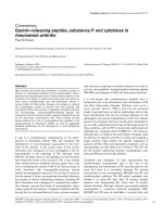
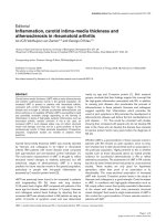
BAL fluid CCL22 and CCL17 in fibrotic lung diseasesFigure 1
BAL fluid CCL22 and CCL17 in fibrotic lung diseases. BAL fluid levels of CCL22 and CCL17 were determined by sensi-
tive ELISAs. CCL22 and CCL17 levels were significantly higher in patients with idiopathic pulmonary fibrosis (IPF) than in those
with CVD-IP and healthy volunteers (A, B). In IPF patients, BAL fluid CCL22 levels correlated significantly with CCL17 levels
(C). IPF, idiopathic pulmonary fibrosis; HV, healthy volunteers; CVD-IP, collagen vascular disease with interstitial pneumonia;
Sar, sarcoidosis.
Respiratory Research 2009, 10:80 />Page 6 of 11
(page number not for citation purposes)
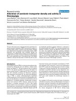
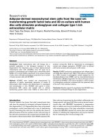
Correlations between BAL fluid CCL22 and CCR4-positive alveolar macrophages and lymphocytes in all patients examinedFigure 2
Correlations between BAL fluid CCL22 and CCR4-positive alveolar macrophages and lymphocytes in all
patients examined. To further elucidate the roles of the chemokines in recruiting cells to the lungs in fibrotic lung diseases,
we analyzed CCR4-positive BAL fluid cell subpopulations by flow cytometry in IPF. CCL22 levels significantly correlated with
the number of CCR4-positive alveolar macrophages (A). CCL22 levels in IPF patients were significantly correlated with the
number of CCR4-positive alveolar macrophages and lymphocytes. These correlations were not observed between these sub-
populations and BAL fluid CCL17 levels.
Respiratory Research 2009, 10:80 />Page 7 of 11
(page number not for citation purposes)
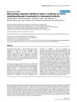
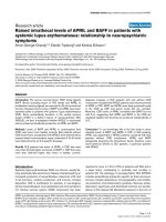
Lung immunohistochemical photomicrograph of CCL17, CCL22, CCR4, and CD68 in patients with idiopathic pulmonary fibro-sis (IPF)Figure 3
Lung immunohistochemical photomicrograph of CCL17, CCL22, CCR4, and CD68 in patients with idiopathic
pulmonary fibrosis (IPF). We examined the localization of CCL17, CCL22, CCR4, and CD68 by immunohistochemistry.
The sections were initially incubated with anti-CCL22 antibody (A), anti-CCL17 antibody (B), anti-CCR4 antibody (C), anti-
CD68 antibody (D), or their diluent buffer (E), and then stained using an indirect streptavidin-biotinylated complex method. A
fraction of the alveolar macrophages was positive for CCL22, whereas CCL17 was exclusively expressed by some hyperplastic
epithelial cells (A, B). There were few alveolar macrophages which were weakly positive for CCR4 (C). The tissue distribution
of alveolar macrophages was confirmed by their positivity for CD68 (D). In contrast, no lung cells were positively stained in
negative control (NC) sections (E).
Respiratory Research 2009, 10:80 />Page 8 of 11
(page number not for citation purposes)
in IPF patients. We assessed the degree of radiographic
abnormalities according to Watter's method [12]. Briefly,
areas of abnormal shadows, presence of honeycombing,
and the diameter of the main pulmonary artery were
assessed by expert pulmonologists, and a semi-quantita-
tive radiological score was calculated for each patient.
However, we did not find any significant correlations
between any of the clinical parameters examined and the
CCL22 and CCL17 levels in BAL fluid.
We next examined the correlation of the BAL fluid chem-
okines with indices of lung function tests in IPF patients.
An inverse correlation was observed between BAL fluid
CCL22 levels and DLco/VA values (Fig 5). Although BAL
fluid CCL17 also tended to correlate inversely with DLco/
VA, no statistical significance was present. There were no
significant correlations between the two BAL chemokines
levels and other parameters of lung function, including
%VC and PaO
2
/FIO
2
.
Discussion
In the present study, we examined the T-helper 2 (Th2)
chemokines, CCL22, CCL17, and BAL fluid cells express-
ing CCR4, a specific receptor for these chemokines, to elu-
cidate their pathophysiological roles in IPF patients. We
also studied the localization of CCL22, CCL17, and CCR4
by immunohistochemistry. The levels of CCL22 and
CCL17 in BAL fluid were significantly higher in patients
with IPF than in those with CVD-IP and healthy volun-
teers, and there was a significant correlation between the
levels of CCL22 and CCL17 in IPF. CCL22 levels in the
BAL fluid did not correlated with total cell numbers, alve-
olar lymphocytes, and macrophages in the BAL fluid.
However, the CCL22 levels were significantly correlated
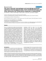
Lung immunofluorescence photomicrograph of CCL22 and CCR4 in patients with idiopathic pulmonary fibrosis (IPF)Figure 4
Lung immunofluorescence photomicrograph of
CCL22 and CCR4 in patients with idiopathic pulmo-
nary fibrosis (IPF). We examined the localization of
CCL22 and CCR4 in CD68-positive alveolar macrophages by
a dual immunofluorescence technique. A. Localization of
CCL22 (red) to a certain fraction of CD68 (green) -positive
alveolar macrophages was shown. B. Localization of CCR4
(red) to a small fraction of CD68 (green) -positive alveolar
macrophages was shown. Nuclei were counterstained with
DAPI (blue).
Correlation between BAL fluid CCL22 and lung diffusing capacity in idiopathic pulmonary fibrosis (IPF) patientsFigure 5
Correlation between BAL fluid CCL22 and lung dif-
fusing capacity in idiopathic pulmonary fibrosis (IPF)
patients. We examined the correlation of BAL fluid chem-
okines with indices of lung function tests in IPF patients. An
inverse correlation was observed between BAL fluid CCL22
levels and DLco/VA values. Although BAL fluid CCL17 also
tended to correlate inversely with DLco/VA, there was no
statistical significance. DLco, single-breath carbon monoxide
diffusing capacity; VA, alveolar ventilation per minute.
Respiratory Research 2009, 10:80 />Page 9 of 11
(page number not for citation purposes)
with the numbers of CCR4-expressing alveolar macro-
phages. By immunohistochemical analysis, localization
of CCL22 and CCR4 to alveolar macrophages as well as
that of CCL17 to hyperplastic epithelial cells were shown.
Clinically, CCL22 levels in BAL fluid inversely correlated
with DLco/VA values in IPF patients. Collectively, we
speculated that locally overexpressed CCL22 may contrib-
ute to the induction of lung dysfunction mainly through
recruitment of CCR4-positive alveolar macrophages.
Increased Production of CCL17and CCL22 in IPF
In our previous study, we showed that the production of
CCL22 and CCL17 in rat radiation pneumonitis increased
significantly, but CCL17 was undetectable in BAL fluid of
IPF patients [11]. Previous reports found no significant
increase in BAL fluid CCL17 [9,18]. Using a more sensi-
tive ELISA kit in the current experiment, we confirmed sig-
nificant increases in CCL17 and CCL22 BAL fluid levels in
IPF patients as compared with those in CVD-IP patients
and healthy volunteers. The levels of CCL17 were lower
than those of CCL22 in 14 out of 16 patients examined,
and there was a significant correlation between the two
levels, suggesting a common stimulus or stimuli for their
induction.
In our study, CCL17 was positive in hyperplastic epithe-
lial cells. Our results regarding CCL17 were consistent
with previous observations in IPF [9,19], and CCL17
detected in BAL fluid could be mainly derived from these
cells. Bronchial epithelial cells are the major source of
CCL17 under physiological and pathological conditions,
including bronchial asthma [20], and CCL17 is inducible
by various stimuli, such as TNF-alpha, interleukin (IL)-4,
interferon-gamma, and TGF-beta [21,22]. Because over-
production of these cytokine has been shown previously,
they also could be in vivo stimuli for CCL17 in IPF.
Our study revealed that immunoreactive CCL22 was pre-
dominantly localized to alveolar macrophages, whereas
Marchal-Sommé et al reported that CCL22 was positive in
hyperplastic epithelial cells, fibroblasts, and endothelial
cells, but not in alveolar macrophages [19]. However,
because our previous study showed the localization of
CCL22 to alveolar macrophages in a rat radiation pneu-
monitis model [11], and the augmented production of
CCL22 was shown in IPF [23], it is reasonable to speculate
that alveolar macrophages are at least partly responsible
for high levels of CCL22 in IPF. CCL22 is inducible in
alveolar macrophages by IL-4, PGE
2
, and TGF-beta [24].
Because overproduction of these mediators has been
shown previously [25], they may be in vivo inducers of
CCL22 in IPF.
Possible Contribution of Lung CCL22 to the Recruitment
of CCR4-Positive Alveolar Macrophages
In the present study, we found that BAL fluid levels of
CCL22 were significantly correlated with the number of
CCR4-positive alveolar macrophages among all patients
examined. CCL22 levels in IPF patients were significantly
correlated with the number of CCR4-positive alveolar
macrophages and lymphocytes. Thus, although the per-
centage of CCR4-positive cells was relatively small among
alveolar macrophages, the results may indicate that locally
overproduced CCL22, but not CCL17, contributes to the
recruitment of alveolar macrophages, and to a lesser
extent, alveolar lymphocytes to the lungs in IPF patients.
In animal models of pulmonary fibrosis, we have found
CCR4 expressed on alveolar macrophages in rat radiation
pneumonitis/pulmonary fibrosis, and Belperio et al. dem-
onstrated predominant CCR4 expression on alveolar mac-
rophages in mice bleomycin-induced pulmonary fibrosis
[9]. Furthermore, Trujillo et al. recently demonstrated that
bleomycin induced CCL17-dependent activation of CCR4
in alveolar macrophages using CCR4-deficient mice [26].
Thus, the CCL22-CCR4 axis may contribute to the activa-
tion of alveolar macrophages in pneumonitis and pulmo-
nary fibrosis.
Inverse Correlation of BAL Fluid CCL22 with Lung
Diffusing Capacity in IPF
Our current study demonstrated that CCL22 was inversely
correlated with DLco/VA. Because DLco/VA is affected by
both total surface area and thickness of alveolar walls, and
these regions are the major targets of alveolar macrophage
infiltration in IPF, the results may suggest that alveolar
macrophage recruitment by CCL22 induces a dose-
dependent decrease in DLco/VA. It is also possible that
CCL22 or CCR4-positive alveolar macrophages are
involved in the destruction of lung parenchyma in IPF.
Previously, Pignatti et al. demonstrated an increase in
CCR4-positive alveolar T-lymphocytes and their inverse
correlation with DLco in IPF [10]. In contrast, the increase
of CCR4 expression on T-lymphocytes was relatively small
and we did not find their significant correlation with the
parameters of lung functions, including DLco in our
study. The discrepancy between their and our results may
be derived from the difference in disease stages or charac-
teristics. All of our patients were in a stable stage, and we
excluded the patients who showed massive lung honey-
combing, or were treated with corticosteroids, whereas
they did not exclude such patients. In addition, the CCR4-
expressing alveolar macrophages, as well as BAL fluid
CCL22 levels, were not examined in their study. Since we
also found a significant correlation between BAL fluid
CCL22 levels and CCR4-positive lymphocytes in IPF
patients, it is possible to speculate that locally overpro-
Respiratory Research 2009, 10:80 />Page 10 of 11
(page number not for citation purposes)
duced CCL22 contributes to the recruitment of CCR4-pos-
itive alveolar macrophages, and to a lesser extent, to the
recruitment of CCR4-positive alveolar T-lymphocytes.
Conclusion
CCL22 and CCL17 were both increased in BAL fluid of IPF
patients and CCL22 levels in BAL fluid correlated propor-
tionally with the numbers of CCR4-positive alveolar mac-
rophages, and inversely with DLco/VA. CCL22 may
contribute to the recruitment and activation of alveolar
macrophages, and consequently to the destruction of
lungs in patients with IPF.
List of Abbreviations
AaDO
2
: alveolar-arterial oxygen gradient; BAL: bronchoal-
veolar lavage; CVD-IP: collagen vascular disease with
interstitial pneumonia; ELISAs: enzyme-linked immuno-
sorbent assay; DLco: single-breath carbon monoxide dif-
fusing capacity; HV: healthy volunteers; IPF: idiopathic
pulmonary fibrosis: N.D: not determined; Sar: sarcoido-
sis; TBLB: transbronchial lung biopsy; UIP: usual intersti-
tial pneumonia; VA: alveolar ventilation per minute
Competing interests
The authors declare that they have no competing interests.
Authors' contributions
YY primarily collected and analyzed the data, with the
help of TI and FS. This manuscript was prepared by YY
under SF's instruction. TS was involved in pathological
diagnosis and immunohistochemical analysis. SA con-
tributed to FACS analysis and interpretation of data. This
study was supported by the scientific fund for KY, AI, and
SF.
Acknowledgements
We thank Kazuko Sano for conducting immunohistochemical analysis. This
study was supported in part by Grants-in-Aid from the Japanese Ministry of
Education, Culture, Sports, Science and Technology, and the Keio Gijuku
Fukuzawa Memorial Fund for the Advancement of Education.
References
1. American Thoracic Society/European Respiratory Society
International Multidisciplinary Consensus Classification of
the Idiopathic Interstitial Pneumonias. This joint statement
of the American Thoracic Society (ATS), and the European
Respiratory Society (ERS) was adopted by the ATS board of
directors, June 2001 and by the ERS Executive Committee,
June 2001. Am J Respir Crit Care Med 2002, 165(2):277-304.
2. Gross TJ, Hunninghake GW: Idiopathic pulmonary fibrosis. N
Engl J Med 2001, 345(7):517-525.
3. American Thoracic Society. Idiopathic pulmonary fibrosis:
diagnosis and treatment. International consensus state-
ment. American Thoracic Society (ATS), and the European
Respiratory Society (ERS). Am J Respir Crit Care Med 2000, 161(2
Pt 1):646-664.
4. Azuma A, Nukiwa T, Tsuboi E, Suga M, Abe S, Nakata K, Taguchi Y,
Nagai S, Itoh H, Ohi M, et al.: Double-blind, placebo-controlled
trial of pirfenidone in patients with idiopathic pulmonary
fibrosis. Am J Respir Crit Care Med 2005, 171(9):1040-1047.
5. Imai T, Chantry D, Raport CJ, Wood CL, Nishimura M, Godiska R,
Yoshie O, Gray PW: Macrophage-derived chemokine is a func-
tional ligand for the CC chemokine receptor 4. J Biol Chem
1998, 273(3):1764-1768.
6. Imai T, Baba M, Nishimura M, Kakizaki M, Takagi S, Yoshie O: The T
cell-directed CC chemokine TARC is a highly specific biolog-
ical ligand for CC chemokine receptor 4. J Biol Chem 1997,
272(23):15036-15042.
7. Katoh S, Fukushima K, Matsumoto N, Matsumoto K, Abe K, Onai N,
Matsushima K, Matsukura S: Accumulation of CCR4-expressing
CD4+ T cells and high concentration of its ligands (TARC
and MDC) in bronchoalveolar lavage fluid of patients with
eosinophilic pneumonia. Allergy 2003, 58(6):518-523.
8. Romagnani S: Cytokines and chemoattractants in allergic
inflammation. Mol Immunol 2002, 38(12-13):881-885.
9. Belperio JA, Dy M, Murray L, Burdick MD, Xue YY, Strieter RM,
Keane MP: The role of the Th2 CC chemokine ligand CCL17
in pulmonary fibrosis.
J Immunol 2004, 173(7):4692-4698.
10. Pignatti P, Brunetti G, Moretto D, Yacoub MR, Fiori M, Balbi B, Bal-
estrino A, Cervio G, Nava S, Moscato G: Role of the chemokine
receptors CXCR3 and CCR4 in human pulmonary fibrosis.
Am J Respir Crit Care Med 2006, 173(3):310-317.
11. Inoue T, Fujishima S, Ikeda E, Yoshie O, Tsukamoto N, Aiso S, Aikawa
N, Kubo A, Matsushima K, Yamaguchi K: CCL22 and CCL17 in rat
radiation pneumonitis and in human idiopathic pulmonary
fibrosis. Eur Respir J 2004, 24(1):49-56.
12. Watters LC, King TE, Schwarz MI, Waldron JA, Stanford RE, Cherni-
ack RM: A clinical, radiographic, and physiologic scoring sys-
tem for the longitudinal assessment of patients with
idiopathic pulmonary fibrosis. Am Rev Respir Dis 1986,
133(1):97-103.
13. Nakamura H, Fujishima S, Soejima K, Waki Y, Nakamura M, Ishizaka
A, Kanazawa M: Flow cytometric detection of cell-associated
cytokines in alveolar macrophages. Eur Respir J 1996,
9(6):1181-1187.
14. Nakamura H, Fujishima S, Waki Y, Urano T, Sayama K, Sakamaki F,
Terashima T, Soejima K, Tasaka S, Ishizaka A, et al.: Priming of alve-
olar macrophages for interleukin-8 production in patients
with idiopathic pulmonary fibrosis. Am J Respir Crit Care Med
1995, 152(5 Pt 1):1579-1586.
15. Wood GS, Warner NL, Warnke RA: Anti-Leu-3/T4 antibodies
react with cells of monocyte/macrophage and Langerhans
lineage. J Immunol 1983, 131(1):212-216.
16. Marchal-Somme J, Uzunhan Y, Marchand-Adam S, Valeyre D, Soume-
lis V, Crestani B, Soler P: Cutting edge: nonproliferating mature
immune cells form a novel type of organized lymphoid struc-
ture in idiopathic pulmonary fibrosis. J Immunol 2006,
176(10):5735-5739.
17. Penna G, Vulcano M, Sozzani S, Adorini L: Differential migration
behavior and chemokine production by myeloid and plasma-
cytoid dendritic cells. Hum Immunol 2002, 63(12):1164-1171.
18. Miyazaki E, Nureki S, Fukami T, Shigenaga T, Ando M, Ito K, Ando H,
Sugisaki K, Kumamoto T, Tsuda T: Elevated levels of thymus- and
activation-regulated chemokine in bronchoalveolar lavage
fluid from patients with eosinophilic pneumonia. Am J Respir
Crit Care Med 2002,
165(8):1125-1131.
19. Marchal-Somme J, Uzunhan Y, Marchand-Adam S, Kambouchner M,
Valeyre D, Crestani B, Soler P: Dendritic cells accumulate in
human fibrotic interstitial lung disease. Am J Respir Crit Care
Med 2007, 176(10):1007-1014.
20. Sekiya T, Miyamasu M, Imanishi M, Yamada H, Nakajima T, Yamaguchi
M, Fujisawa T, Pawankar R, Sano Y, Ohta K, et al.: Inducible expres-
sion of a Th2-type CC chemokine thymus- and activation-
regulated chemokine by human bronchial epithelial cells. J
Immunol 2000, 165(4):2205-2213.
21. Heijink IH, Marcel Kies P, van Oosterhout AJ, Postma DS, Kauffman
HF, Vellenga E: Der p, IL-4, and TGF-beta cooperatively induce
EGFR-dependent TARC expression in airway epithelium.
Am J Respir Cell Mol Biol 2007, 36(3):351-359.
22. Berin MC, Eckmann L, Broide DH, Kagnoff MF: Regulated produc-
tion of the T helper 2-type T-cell chemoattractant TARC by
human bronchial epithelial cells in vitro and in human lung
xenografts. Am J Respir Cell Mol Biol 2001, 24(4):382-389.
23. Manabe K, Nishioka Y, Kishi J, Inayama M, Aono Y, Nakamura Y,
Ogushi F, Bando H, Tani K, Sone S: Elevation of macrophage-
Publish with Bio Med Central and every
scientist can read your work free of charge
"BioMed Central will be the most significant development for
disseminating the results of biomedical research in our lifetime."
Sir Paul Nurse, Cancer Research UK
Your research papers will be:
available free of charge to the entire biomedical community
peer reviewed and published immediately upon acceptance
cited in PubMed and archived on PubMed Central
yours — you keep the copyright
Submit your manuscript here:
/>BioMedcentral
Respiratory Research 2009, 10:80 />Page 11 of 11
(page number not for citation purposes)
derived chemokine in eosinophilic pneumonia: a role of alve-
olar macrophages. J Med Invest 2005, 52(1-2):85-92.
24. Kuroda E, Sugiura T, Okada K, Zeki K, Yamashita U: Prostaglandin
E2 up-regulates macrophage-derived chemokine production
but suppresses IFN-inducible protein-10 production by APC.
J Immunol 2001, 166(3):1650-1658.
25. Fireman E, Ben Efraim S, Greif J, Alguetti A, Ayalon D, Topilsky M:
Suppressive activity of alveolar macrophages and blood
monocytes from interstitial lung diseases: role of released
soluble factors. Int J Immunopharmacol 1989, 11(7):751-760.
26. Trujillo G, O'Connor EC, Kunkel SL, Hogaboam CM: A novel
mechanism for CCR4 in the regulation of macrophage acti-
vation in bleomycin-induced pulmonary fibrosis. Am J Pathol
2008, 172(5):1209-1221.