Báo cáo y học: " Gene promoter methylation assayed in exhaled breath, with differences in smokers and lung cancer patients" ppt
Bạn đang xem bản rút gọn của tài liệu. Xem và tải ngay bản đầy đủ của tài liệu tại đây (1.32 MB, 12 trang )
BioMed Central
Open Access
Page 1 of 12
(page number not for citation purposes)
Respiratory Research
Research
Gene promoter methylation assayed in exhaled breath, with
differences in smokers and lung cancer patients
Weiguo Han
1,5
, Tao Wang
1
, Andrew A Reilly
2
, Steven M Keller
4
and
Simon D Spivack*
1,3,5,6
Address:
1
Wadsworth Center, Human Toxicology & Molecular Epidemiology, Albany, NY, USA,
2
Biostatistics, NYS Dept of Health, Albany, NY,
USA,
3
Pulmonary & Critical Care Medicine, Albany Medical College, Bronx, NY, USA,
4
Thoracic Surgery, Albert Einstein College of Medicine,
Bronx, NY, USA,
5
Pulmonary Medicine, Albert Einstein College of Medicine, Bronx, NY, USA and
6
Depts. of Epidemiology and Genetics, Albert
Einstein College of Medicine, Bronx, NY, USA
Email: Weiguo Han - ; Tao Wang - ; Andrew A Reilly - ;
Steven M Keller - ; Simon D Spivack* -
* Corresponding author
Abstract
Background: There is a need for new, noninvasive risk assessment tools for use in lung cancer population
screening and prevention programs.
Methods: To investigate the technical feasibility of determining DNA methylation in exhaled breath condensate,
we applied our previously-developed method for tag-adapted bisulfite genomic DNA sequencing (tBGS) for
mapping of DNA methylation, and adapted it to exhaled breath condensate (EBC) from lung cancer cases and
non-cancer controls. Promoter methylation patterns were analyzed in DAPK, RASSF1A and PAX5
β
promoters in
EBC samples from 54 individuals, comprised of 37 controls [current- (n = 19), former- (n = 10), and never-
smokers (n = 8)] and 17 lung cancer cases [current- (n = 5), former- (n = 11), and never-smokers (n = 1)].
Results: We found: (1) Wide inter-individual variability in methylation density and spatial distribution for DAPK,
PAX5
β
and RASSF1A. (2) Methylation patterns from paired exhaled breath condensate and mouth rinse specimens
were completely divergent. (3) For smoking status, the methylation density of RASSF1A was statistically different
(p = 0.0285); pair-wise comparisons showed that the former smokers had higher methylation density versus never
smokers and current smokers (p = 0.019 and p = 0.031). For DAPK and PAX5
β
, there was no such significant
smoking-related difference. Underlying lung disease did not impact on methylation density for this geneset. (4) In
case-control comparisons, CpG at -63 of DAPK promoter and +52 of PAX5
β
promoter were significantly
associated with lung cancer status (p = 0.0042 and 0.0093, respectively). After adjusting for multiple testing, both
loci were of borderline significance (p
adj
= 0.054 and 0.031). (5) The DAPK gene had a regional methylation pattern
with two blocks (1)~-215~-113 and (2) -84 ~+26); while similar in block 1, there was a significant case-control
difference in methylation density in block 2 (p = 0.045); (6)Tumor stage and histology did not impact on the
methylation density among the cases. (7) The results of qMSP applied to EBC correlated with the corresponding
tBGS sequencing map loci.
Conclusion: Our results show that DNA methylation in exhaled breath condensate is detectable and is likely of
lung origin. Suggestive correlations with smoking and lung cancer case-control status depend on individual gene
and CpG site examined.
Published: 25 September 2009
Respiratory Research 2009, 10:86 doi:10.1186/1465-9921-10-86
Received: 12 June 2009
Accepted: 25 September 2009
This article is available from: />© 2009 Han et al; licensee BioMed Central Ltd.
This is an Open Access article distributed under the terms of the Creative Commons Attribution License ( />),
which permits unrestricted use, distribution, and reproduction in any medium, provided the original work is properly cited.
Respiratory Research 2009, 10:86 />Page 2 of 12
(page number not for citation purposes)
Background
Lung cancer is the leading cause of cancer mortality in the
U.S. [1]. Most patients will never undergo curative proce-
dures (surgery) because of the wide extent of disease at
diagnosis. For earlier diagnosis, screening programs in
asymptomatic, high-risk population groups have been
studied by several technologies, including cytology of the
sputum [2,3], circulating tumor biomarkers [4,5], blood
proteomic patterns [6,7], chest tomography [8,9], nuclear
magnetic resonance (NMR) [10], and other techniques.
Each approach has limited diagnostic specificity as cur-
rently applied [11,12], such that identifying particularly
high risk individuals for application of these candidate
early disease detection strategies may allow leveraging of
their performance.
Sampling the target visceral epithelia non-invasively for
risk assessment in asymptomatic subjects poses anatomic
challenges. Expectorated sputum has been intensively
studied for this reason, although up to 30% of current or
former smokers do not produce sputum, even after induc-
tion with nebulized saline [13-15]. Nonetheless, the suc-
cessful study of sputum, presumably derived solely from
lung epithelia, has been demonstrated in suggestive stud-
ies by the New Mexico/Colorado consortium where Belin-
sky, et al. have demonstrated the promise of a multiple
gene promoter hypermethylation panel for identifying
people at high risk for cancer incidence [14].
Exhaled breath contains aerosols and vapors that can be
collected for non-invasive analysis of physiologic and
pathologic processes in the lung. To capture the breath for
assay, exhaled air is passed through a cooled, condensing
apparatus, which is also available as a handheld, disposa-
ble device. The result is an accumulation of condensed
fluid that is referred to as exhaled breath condensate
(EBC). Predominantly derived from water vapor, EBC has
dissolved within it aqueous, soluble, nonvolatile com-
pounds. The technique has attracted broad research inter-
est, and there is a significant literature describing its utility
in procuring small metabolites for the investigation of
inflammatory lung diseases [16,17]. Several investigative
groups, including our own, have detected macromole-
cules in EBC, such as genomic DNA [18-21]. This suggests
the possibility of DNA-based analyses of lung processes,
including epigenetic alteration.
Promoter hypermethylation is known to cause stable
silencing of associated genes and plays an important role
in both normal development [22] and disease [23]. Gene
promoter hypermethylation is recognized as a crucial
component in lung cancer initiation and progression [24].
Most translational studies measuring CpG methylation
invoke methylation-specific PCR (MSP) assays that sam-
ple 1-4 CpG sites. We recently reported a method for the
facile annotation of larger expanses of gene sequence for
CpG methylation at single base resolution, using a tag-
modification of bisulfite genomic sequencing (tBGS) [21]
where all CpG sites could be sampled in a given fragment.
Because of consistent reports as a relevant biomarker class
in carcinogenesis, we pursued the appearance of promoter
hypermethylation of tumor suppressor genes in a non-
invasive exhaled (EBC) matrix putatively representing
lung-derived material. In the current study, we analyzed
comprehensive DNA methylation maps in EBC from non-
cancer control subjects who were never smokers, former
smokers, and current smokers, along with a pilot group of
incident lung cancer patients, to generate a new non-inva-
sive, epithelial-based method for ascertainment of lung
carcinogenesis in humans.
Methods
Subjects
A total of 54 subjects (37 non-cancer control subjects and
17 lung cancer case subjects) donated exhaled breath con-
densate. Thirty six of the first 37 consecutive subjects
donated sufficient mouth rinses for anatomic verification
for the purposes of this study, in an ongoing lung cancer
case-control study. Subjects were of predominantly
(>80%) Euro-Caucasian descent, equally women and
men, queried on lifetime and proximate smoking habits,
as well as medical history and other factors. Questionaire,
mouth rinses, and exhaled breath condensate were all
sampled prior to any other diagnostic (e.g., bronchos-
copy) or therapeutic (e.g., surgery, chemotherapy) inter-
vention. The procedures followed protocols approved by
both the Albany Medical Center, New York State Depart-
ment of Health Institutional Review Boards, and Albert
Einstein College of Medicine Committee on Clinical
Investigation (IRB).
Case status was confirmed by conventional positive clini-
cal and histopathologic criteria; for initially negative clin-
ical bronchoscopic biopsies, follow-up biopsy procedures
and clinical data were tracked for three months from time
of enrollment to affirm the case status. The 17 cases were
comprised of six with adenocarcinoma, three with squa-
mous cell carcinoma, five with undifferentiated non-
small cell carcinomas, and three subjects with small cell
carcinoma. The smoking status of these 17 cancer cases
included current smokers (n = 5), former smokers (n =
11), and never smoker (n = 1). The 37 non-cancer con-
trols, with no clinical evidence of cancer at time of enroll-
ment, included current-smokers (n = 19), former-smokers
(n = 10), and never-smokers (n = 8). Those control sub-
jects (n = 9) undergoing biopsy of what proved ultimately
to be benign nodule were histologically confirmed as con-
trols. The other 28 control subjects were designated as
controls by common clinical criteria (no recent suggestive
symptoms, or suggestive CXR).
Respiratory Research 2009, 10:86 />Page 3 of 12
(page number not for citation purposes)
Exhaled breath condensate (EBC) collection
Exhaled breath condensate (EBC) collection was per-
formed by standard methods. EBC is collected in a hand-
held, disposable RTube
®
exhaled breath condenser
(Respiratory Research, Charlottesville, VA) which entails a
airway valve, inner protective sleeve, outer (cooled to -
80°C) aluminum sleeveand insulates, during 10 to 15
minutes of quiet tidal volume breathing, with the excep-
tion that subjects were asked to swallow or expectorate all
saliva, and to sigh once each minute. Approximately 1.0
ml of EBC was collected from each subject. The collected
EBC was stored at -20°C.
DNA preparation from EBC
From each sample, 0.8 ml of EBC was used for DNA prep-
aration. DNA was prepared with DNA Blood Mini Kit per
manufacturer's instructions (Qiagen). We added 5 μg of
60-mer oligo-dT as a DNA carrier to enhance template
recovery. DNA was eluted in 55 μl buffer AE (Qiagen). The
presence of genomic DNA was confirmed by PCR using 5
μl of sample.
Bisulfite treatment
Of the EBC DNA extract, 45 μl was used for bisulfite treat-
ment. Bisulfite treatment was performed with DNA meth-
ylation kit (Zymo Research), with the reaction condition
optimized to 37°C for 3 hours. Finally, DNA was eluted
in 10 μl of elution buffer. Non-CpG cytosines were
checked for complete conversion to uracils/thymidine in
the sequence trace as a positive control, before CpG site
data analysis commenced. Samples with any incomplete
conversion of non-CpG C's in the sequence trace were to
be omitted from further CpG site data analysis; however,
there were no cases of incomplete conversion.
Multiplex PCR
Three sets of gene-specific primers (Table 1) were
designed to flank each promoter region of DAPK,
RASSF1A and PAX5
β
, The multiplex PCR contained
1×buffer (Qiagen, Valencia, CA) with 1.5 mM MgCl
2
, 1
μM of each promoter-specific sense and anti-sense primer,
5 units of HotStar
®
Taq polymerase (Qiagen) and 5 μl
bisulfite-modified EBC DNA. PCR conditions were: 95°C
for 15 min, then 5 cycles of 95°C for 10 sec, 52°C for 30
sec, 72°C for 1 min, and 35 cycles of 95°C for 10 sec,
49°C for 30 sec, 72°C for 1 min, and finally 7 min at
72°C. The PCR thermal profiles were programmed into a
Perkin-Elmer 9700 thermocycler. The presence of ampli-
cons was confirmed by electrophoresis on a 1.5% agarose
gel. In many samples, only one (27.8%) or two (35.2%)
of three bisulfite treated amplicons could be detected.
GC tag-modified bisulfite genomic DNA sequencing
(tBGS)[21]
The multiplex PCR products were used as template (1 μl)
and re-amplified by GC-tagged primers separately (Table
1). The PCR conditions were: 95°C for 15 min, and 5
cycles of 95°C for 10 sec, 50°C for 30 sec, 72°C for 1 min,
30 cycles of 95°C for 10 sec, 65°C for 30 sec, 72°C for 1
min, and finally 7 min at 72°C. PCR products were then
purified with a Gel Extraction Kit (Qiagen) and subjected
to direct-cycle sequencing on a Perkin-Elmer Biosystems
ABI model 3700 automated DNA sequencer, using tag-tar-
geted sequencing primers: 5'-ATTAACCCTCACTAAAG-3'
(Forward); 5'-AATACGACTCACTATAG-3' (reverse). Man-
ual review of sequence chromatograms containing two
peaks at any one CpG locus was performed by measuring
the peak height of the C (or anti-sense G) versus the com-
bined height of the C+T peaks, and generating a C/C+T (or
anti-sense A/A+G) peak height representing the methyl-
ated fraction of DNA molecules at that CpG site, as a per-
centage [25,26].
Quantitative methylation-specific PCR (MSP)
In order to (a) complement the sensitivity limits inherent
to sequencing-based technologies such as tBGS, (b) to
replicate CpG site sampling approaches used in the litera-
ture, and (c) to provide independent corroboration of
technical feasibility of exhaled DNA methylation analy-
ses, we analyzed a consecutive subset of 36 available EBC
specimens (16 current smokers, 9 former smokers, 7
never-smokers, and 4 lung cancer patients) from the ini-
tial 37 EBC samples, using quantitative MSP. Two sets of
MSP probes were used. Probe 1 (Table 1) was specific for
-82 to -99 (a low methylation region by tBGS), and probe-
2 specific for -144 to -158 (a high methylation region by
tBGS).
Quantitative MSP for DAPK promoter was performed on
an ABI Prism-7500 realtime thermocycler, using a 96-well
optical tray with caps at a final reaction volume of 20 μl.
Samples contained 10 μl of TaqMan
®
Universal PCR Mas-
ter Mix, No AmpErase
®
UNG (uracil-N-glycosylase), 1 μl
of 1:1000 diluted multiplex PCR product, an additional
2.5 U of AmpliTaq Gold (Perkin Elmer), 2.5 μM each of
the primers and 150 nM each of the fluorescently labeled
probes for methylated and unmethylated templates. The
specificity of each probe was confirmed by positive and
negative control templates, and water blanks. The cloned
DAPK promoter methylated with CpG methyltransferase
was used as positive control included in all experiments.
To generate a standard curve, we prepared different ratios
of methylated versus unmethylated target sequences by
mixing methylated and unmethylated DNA. The follow-
ing ratios were prepared (methylated/unmethylated): 0/
100, 10/90, 20/80, 30/70, 40/60, 50/50, 60/40, 70/30,
80/20, 90/10, 100/0. To verify whether MSP sampling
probes, targetting variable regions of methylation, would
indicate discordant patterns of MSP-designated methyla-
tion, we designed two spatially separated sets of probes
for the DAPK promoter, one in a 5' upstream, tBGS-
defined high methylation region (adjacent to CpG residue
Respiratory Research 2009, 10:86 />Page 4 of 12
(page number not for citation purposes)
-158), and one in a 3' downstream low methylation
region (adjacent to CpG residue -99) (Table 1). Results
were verified by gel electrophoresis of the PCR product.
Correlations were made between qMSP and tBGS results
at the relevant two target loci, by correlating the percent
methylation determined by the respective MSP probe,
with the fraction of sites found methylated by tBGS at that
same four-CpG MSP site locus (where individual CpG
sites were generally dichotomous as methylated or not).
Data analysis
The tBGS-generated CpG methylation sequence chroma-
togram tracings data were converted to dichotomous data
at each CpG site, where >20% C/C+T peak height ratio by
sequence trace was considered methylated, and <20%
ratio was considered unmethylated, as the limits of detec-
tion for the technology are 5-10% methylated/total DNA
molecules, at any given CpG site. Methylation density was
defined as the methylated CpGs divided by total CpGs
examined in a gene promoter in a given sample. The
methylation densities among smoking groups and case
group were evaluated by ANOVA and the position specific
CpG methylation state was tested for correlation substruc-
ture, and then tested by Fisher's exact test. Further tests on
each CpG locus within each promoter region were per-
formed by logistic regression [27,28]. Correlations
between the qMSP data and tBGS data at the two respec-
tive probe loci were tested by Pearson product moment
analysis.
Results
Reproducibility of DNA methylation mapping in EBC
To initially test the reproducibility of DNA methylation
mapping in EBC, we collected two consecutive EBC sam-
Table 1: PCR primers
Multiplex PCR primers Sequence Product
RASSF1A-F
RASSF1A-R
TTAGTAAAT(C/T)GGATTAGGAGGGTTAG
CCACAAAAC(A/G)AACCCC(A/G)ACTTCAAC
325 bp
(-254~+70)
DAPK-F
DAPK-R
AGGGTAGTTTAGTAATGTGTTATAG
ACCCTACC(A/G)CTAC(A/G)AATTACC(A/G)AATC
391 bp
(-312~+78)
PAX5β-F
PAX5β-R
GAGTTTGTGGGTTGTTTAGTTAATGG-3'
AACAAAAAATCCCAACCACCAAAACC-3'
322 bp
(-147~+174)
tBGS Primer
RASSF1A-TF
RASSF1A-TR
CGACTCCTGCACTCATTAACCCTCACTAAAGAGGGT(T/C)GGATGTGGGGATTT
GGCCAGTGAATTGTAATACGACTCACTATAG
GGAGGCGGCCCAAAATCCAAACTAAAC
337 bp
(-254~+39)
DAPK-TF
DAPK-TR
CGACTCCTGCACTCATTAACCCTCACTAAAGTGGGTGTGGGG(T/C)GAGTGGGTG
GGCCAGTGAATTGTAATACGACTCACTATAG
GGAGGCGGCTCC(A/G)C(A/
G)AAAAAAACAAAATC
358 bp
(-240~+50)
PAX5β-TF
PAX5β-TR
CGACTCCTGCACTCATTAACCCTCACTAAAGGTTATTTTGATTGGTTTGGTG
GGCCAGTGAATTGTAATACGACTCACTATAG
GGAGGCGGCTACC(A/G)AAACTAAAATAAAAC
301 bp
(-92~+141)
Quantitative MSP primers
DAPK-qF
DAPK-qR
AG(C/T)G(C/T)GGAGTTGGGAGGAGTA
CAAAC(A/G)ACCAATAAAAACCCTACAAAC
121 bp
(-179~-58)
Probe
DAPK-P1m VIC-AACGAACTAACGACGCGA-MGB -99 - -82
DAPK-P1u 6FAM-TACAAACAAACTAACAACACAA-MGB -99 - -78
DAPK-P2m VIC-CTACGCGACGCTCGC-MGB -158 - -144
DAPK-P2u 6FAM-AATTCTACACAACACTCACT-MGB -159~-140
All gene sequences are from Human Genome sequence using NCBI sequence viewer v2.0. Primer sequences displayed in 5' to 3'end. Italic letters
are tag sequence and the underlined is sequencing primer.
Respiratory Research 2009, 10:86 />Page 5 of 12
(page number not for citation purposes)
ples, separated in collection time by two hours, from each
of two individuals. Each EBC sample was split into two
technical replicates for DAPK promoter methylation map-
ping, and these technical and temporal replicates were
assayed. The results show that the methylation pattern is
completely consistent within samples as technical repli-
cates, and across this brief two hour time period as tempo-
ral/biological replicates, for each individual (Figures 1
and 2). There were no episodes of incomplete cytosine
conversion, using our protocol, within the 95% sensitiv-
ity/resolution limits inherent to sequencing-based chro-
matographic technologies.
Origin of exhaled DNA
To help verify that EBC-DNA is predominantly derived
from the lower airway, we reasoned that methylation pat-
terns themselves might differ between epithelia, confer-
ring the expression features unique to those epithelia. We
therefore compared the methylation pattern of DAPK in
paired EBC and mouthwash samples from the initial
recruitment set of 37 consecutive subjects with adequate
amounts still available from both specimens in 36 of the
37 donors. Results showed that DAPK methylation pat-
tern in mouthwash is largely unmethylated, except for the
first position CpG site, and therefore completely divergent
from that in exhaled breath (Figure 3).
Promoter methylation mapping across genes and subjects
Of the five initial genes selected for evaluation (DAPK,
RASSF1A, PAX5
β
, CDH1, p16) based on their literature
reported, methylation-specific PCR (MSP)-based preva-
lence in lung tumors (>25%), diversity of function, and
timing for inactivation during lung cancer development,
where known, we chose to pursue the three that showed
any promoter methylation at all. We mapped the pro-
moter methylation status of each gene by tBGS.
Overall, the methylation density and patterns for the three
promoters (DAPK, RASSF1A and PAX5
β
) differed quite
dramatically between individuals (Figure 4), otherwise
not readily explained by differences in pack-years, quit
years, and other factors (below). There were, for example,
high methylation outlier individuals apparent (e.g., the
methylation density of DAPK in subject 6113, male cur-
rent smoker, 27 pack-years, is 96%; Subject 6216, female
never smoker, is 91%).
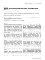
Tag-adapted sequencing chromatograms from exhaled breath condensateFigure 1
Tag-adapted sequencing chromatograms from exhaled breath condensate. For a portion (~250 bp) of the DAPK
promoter region just 5' to the transcription initiation site (TIS), displayed for two representative subjects A and B. Top two trac-
ings: Subject A (all CpG sites methylated, circled C's). The two top tracings are technical replicates from PCR to sequencing for
this subject. Bottom tracing: Subject B (several CpG sites unmethylated, circled Ts). Detection of partial methylation at a given
site is also feasible.
CGAG CCC GGA GCGC GGA GCT GGG A GG AG CA GCG AGC GC CG CGC AG A AC C CGC AG
Native, untreated genomic DNA sequence
Bisulfite-treated genomic DNA
Subject A (completely methylated, C), initial
Subject A (completely methylated, C), technical duplicate
Subject B (several sites unmethylated, T)
Respiratory Research 2009, 10:86 />Page 6 of 12
(page number not for citation purposes)
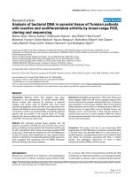
Reproducibility of DAPK promoter methylation mapping in EBCFigure 2
Reproducibility of DAPK promoter methylation mapping in EBC. Each of two subjects (W and S) had two consecu-
tive 10-minute EBC collections (1 and 2) separated in time by one hour. Displayed is the tBGS map readout from each of these
separate samples, additionally performed as technical replicates (A and B). Both temporal and technical replicates are identical,
for a given individual. Methylation density is the simple count of methylated CpG sites (W1A and W1B = 16) over total CpG
sites (=33), here yielding 48.5%.
26
23
15
10
-10
-23
-34
-45
-52
-55
-63
-84
-93
-148
-157
-175
-134
-139
-150
-131
-153
-229
-177
-113
-98
-96
-182
-188
-194
-204
-206
-196
-215
Sample
W1A
W1B
W2A
W2B
S1A
S1B
S2A
S2B
80~100% methylated
60~80% methylated
40~60% methylated
20~40% methylated
0~20% methylated
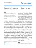
Comparison of Methylation mapping of DAPK promoter in exhaled breath and mouthwash-exfoliated DNAFigure 3
Comparison of Methylation mapping of DAPK promoter in exhaled breath and mouthwash-exfoliated DNA. (a):
Methylation mapping of exhaled breath DNA. (b) Methylation mapping of mouthwash-exfoliated DNA. Exhaled breath conden-
sate (EBC) from 37 of 38 initially recruited consecutive donors and available mouthwash from 36 of the 37 EBC donors, was
screened using the tBGS multiplex technique for simultaneous assay of three gene promoters' CpG islands within ~200-300 bp
surrounding the TIS. Only mapping results for the DAPK promoter are shown. Subject historical smoking features are listed on
the left. Mean percent of sites methylated is listed by smoking and case strata, in larger font, on the right. Wide inter-individual
methylation variability within any given smoking stratum is apparent. All samples are collected prior to any diagnostic or thera-
peutic procedure.
6102 38 n/a n/a
6251 n/a
6223 n/a
6238 n/a
317 n/a
9.1%
36.4%
94.0%
24.2%
9.1%
24.2%
30.3%
42.4%
21.2%
42.4%
15.2%
27.3%
27.2%
21.2%
36.4%
39.4%
12.1%
30.3%
48.5%
30.3%
12.1%
36.4%
15.2%
36.4%
54.5%
42.4%
30.3%
DAPK promoter methylation in EBC
324 n/a
33.3%
6216 n/a
6218 n/a
90.9%
21.2%
330 n/a
Never smoker
Former smoker
Current smoker
42.4%
30.0%
72.7%
Ƃ
30.4 %
39.6 %
35.6 %
Subject
Pack year
Quit year
Cancer type
6107 18 n/a n/a
6112 42 n/a n/a
6113 27 n/a n/a
6123 14 n/a n/a
6124 4 n/a n/a
6125 11 n/a n/a
6128 8 n/a n/a
6129 2 n/a n/a
6130 30 n/a n/a
6131 1 n/a n/a
6133 10 n/a n/a
6134 34 n/a n/a
6137 27 n/a n/a
326 60 n/a n/a
329 90 n/a n/a
6201 16 46 n/a
6202 30 9 n/a
6207 1 1 n/a
6211 n/a n/a n/a
6245 30 5 n/a
6255 23 7 n/a
315 20 3 n/a
319 1 40 n/a
328 2 10 n/a
Methylati on
density
48.5%
54.5%
84.8%
36.4%
295 57 0 NSCCa
325 10 30 NSCCa
331 50 0 SCCa
318 125 0 SCCa
50.0 %
Cancer
26
23
15
10
-10
-23
-34
-45
-52
-56
-84
-93
-148
-
157
-175
-134
-
139
-150
-131
-153
-229
-177
-113
-98
-96
-182
-188
-194
-204
-206
-196
-215
-63
6102 38 n/a n/a
6251 n/a
6223 n/a
6238 n/a
317 n/a
DAPK promoter methylation in mouthwash
324 n/a
6218 n/a
330 n/a
Never smoker
Former smoker
Current smoker
Sub ject
Pack year
Quit year
Cancer t ype
6107 18 n/a n/a
6112 42 n/a n/a
6113 27 n/a n/a
6123 14 n/a n/a
6124 4 n/a n/a
6125 11 n/a n/a
6128 8 n/a n/a
6129 2 n/a n/a
6130 30 n/a n/a
6131 1 n/a n/a
6133 10 n/a n/a
6134 34 n/a n/a
6137 27 n/a n/a
326 60 n/a n/a
329 90 n/a n/a
6201 16 46 n/a
6202 30 9 n/a
6207 1 1 n/a
6211 n/a n/a n/a
6245 30 5 n/a
6255 23 7 n/a
315 20 3 n/a
319 1 40 n/a
328 2 10 n/a
295 57 0 NSCCa
325 10 30 NSCCa
331 50 0 SCCa
318 125 0 SCCa
Cancer
26
23
15
10
-10
-23
-34
-45
-52
-56
-84
-93
-148
-
157
-175
-134
-
139
-150
-131
-153
-229
-177
-113
-98
-96
-182
-188
-194
-204
-206
-196
-215
-63
80~100% methylated
60~80% methylated
40~60% methylated
20~40% methylated
0~20% methylated
Respiratory Research 2009, 10:86 />Page 7 of 12
(page number not for citation purposes)
Promoter methylation density in non-cancer controls
EBC samples from 37 non-cancer controls were analysed
by tBGS, and included samples from 11 subjects with
asthma, 6 with COPD and 20 non-diseased subjects. In
initial univariate analyses of EBC methylation, inclusive
of all three methylated promoters, there was no signifi-
cant difference in the overall methylation densities. How-
ever, the methylation density of RASSF1A was statistically
different between smoker and nonsmoker group (p =
0.0285) and the differences between former versus never
smokers and former versus current smokers were also sig-
nificant (p = 0.019 and p = 0.031, resp.)(Table 2). We also
analyzed DAPK promoter methylation versus underlying
lung disease type in controls. There was no significant dif-
Methylation maps of DAPK, RASSF1A, PAX5
β
promoter from Exhaled Breath CondensateFigure 4
Methylation maps of DAPK, RASSF1A, PAX5
β
promoter from Exhaled Breath Condensate. The promoter methyla-
tion status of DAPK, RASSF1A, PAX5β, was mapped using tBGS. Overall, both the methylation density and patterns of DAPK,
RASSF1A or PAX5
β
promoters differed quite dramatically between individuals within any given smoking or clinical stratum.
Methylation density is given at right, for individuals and group means. [NSCCa: non-small cell lung cancer: SCCa: Small cell lung
cancer; SqCCa: squamous cell lung cancer; AdCa: Adenocarcinoma; AdBaCa: Adenocarcinoma with bronchalveolar features].
Data on smoking status (never, former and current), pack year, quit years and for tumors, histology and stages I, II, III, IV are
given at left.
6102 38 0 None
6251 None
6223 None
6238 None
317 None
26
23
15
10
-10
-23
-34
-45
-52
-56
-63
-84
-93
-148
-
157
-175
-134
-
139
-150
-131
-153
-229
-177
-113
-98
-96
-182
-188
-194
-204
-206
-196
-215
9.1%
36.4%
94.0%
24.2%
9.1%
24.2%
30.3%
42.4%
21.2%
42.4%
15.2%
27.3%
27.2%
21.2%
36.4%
39.4%
12.1%
30.3%
48.5%
30.3%
12.1%
36.4%
15.2%
36.4%
54.5%
42.4%
30.3%
DAPK promoter methylation
324 COPD
33.3%
6216 None
6218 COPD
90.9%
21.2%
330 Asthma
Never smoker
Former smoker
Current smoker
42.4%
30.0%
72.7%
Ƃ
9.1%
3.0%
29.2 %
39.6 %
35.6 %
Subject
Pack yea
Quit year
Disease
6107 18 0 None
6112 42 0 None
6113 27 0 None
6123 14 0 None
6124 4 0 COPD
6125 11 0 Asthma
6128 8 0 Asthma
6129 2 0 None
6130 30 0 None
6131 1 0 None
6133 10 0 Asthma
6134 34 0 None
6137 27 0 None
326 60 0 COPD
329 90 0 COPD
505 4 0 Asthma
6239 2 1 Asthma
6201 16 46 None
6202 30 9 Asthma
6207 1 1 None
6211 n/a n/a None
6245 30 5 Asthma
6255 23 7 None
315 20 3 COPD
319 1 40 None
328 2 10 Asthma
Methylation
r
density
6216 None
6223 None
6238 None
317 None
-4
-6
-41
-47
-57
-59
-65
-79
-103
-126
-130
-139
-142
-145
-149
-161
-163
-173
-225
15.8%
5.2%
36.8%
5.2%
15.8%
63.2%
26.3%
52.6%
57.9%
21.0%
52.6%
15.8%
52.6%
21.0%
21.0%
10.5%
84.2%
21.0%
21.0%
RASSF1A promoter methylation
Methylation
density
31.6%
10.5%
57.9%
21.0%
0.0%
21.0%
21.0%
10.5%
Sub ject
Pack year
Quit year
Disease
6107 18 0 None
6112 42 0 None
6123 14 0 None
6124 4 0 COPD
6125 11 0 Asthma
6128 8 0 Asthma
6129 2 0 None
6130 30 0 None
6133 10 0 Asthma
6134 34 0 None
326 60 0 COPD
329 90 0 COPD
511 15 0 Asthma
6201 16 46 None
6245 30 5 Asthma
6255 23 7 None
315 20 3 COPD
319 1 40 None
295 57 0 NSCCa
318 125 0 SCCa
531 80 4 SCCa
537 57 7 AdCa
539 75 0 AdCa
Current smoker
Former
smoker
Canc er
24.5 %
47.3 %
18.4 %
31.5 %
Never
smoker
80~100% methylated
60~80% methylated
40~60% methylated
20~40% methylated
0~20% methylated
48.5%
54.5%
84.8%
36.4%
Lung cancer cases
72.7%
3.0%
78.8%
66.7%
69.7%
3.0%
3.0%
63.6%
3.0%
3.0%
42.2 %
295 57 0 NSCCa
325 10 30 NSCCa
331 50 0 SCCa
318 125 0 SCCa
501 11 20 NSCCa
504 87 0 SqCCa
502 30 0 SqCCa
506 15 20 AdCa
509 0 0 NSCCa
510 35 1 SqCCa
542 9 20 AdBaCa
543 51 20 AdCa
545 47 1 AdBaCa
547 50 20 NSCCa
6216 None
6218 COPD
+97
+91
+88
+85
+82
+79
+76
+65
+35
+21
+8
+43
+37
+32
+27
-68
+3
+52
+54
+57
-8
-11
-15
-36
-42
-32
-47
0%
18.5%
59.2%
22.2%
61.5%
44.4%
0%
40.1%
14.8%
11.1%
59.2%
0%
29.6%
0.0%
0.0%
77.8%
63.0%
88.9%
PAX5
E
promoter methylation
6238 None
324 COPD
100%
7.4%
29.6%
0%
0%
0%
6223 None
0.0%
6251 None
0.0%
330 Asthma
0.0%
63.0%
74.1%
7.4%
26.0%
26.5%
13.2%
57.4%
Sub ject
Pack year
Quit year
Disease
6107 18 0 None
6112 42 0 None
6113 27 0 None
6123 14 0 None
6124 4 0 COPD
6125 11 0 Asthma
6128 8 0 Asthma
6129 2 0 None
6130 30 0 None
6131 1 0 None
6133 10 0 Asthma
6139 21 0 Asthma
326 60 0 COPD
511 15 0 Asthma
6211 n/a n/a None
6239 2 1 Asthma
6245 30 5 Asthma
6255 23 7 None
319 1 40 None
295 57 0 NSCCa
318 125 0 SCCa
325 10 30 NSCCa
501 11 20 NSCCa
Former
Methylation
density
Current smoker
smoker
Never
smoker
Cancer
Table 2: Methylation densities among smoking groups
Methylation density (SD)
Subjects (n) DAPK RASSF1A* PAX5
β
Pooled
Never smoker (8) 0.365(0.248) 0.184(0.0526) 0.132 (0.246) 0.240(0.168)
Current smoker (19) 0.294(0.198) 0.232(0.19675) 0.296 (0.328) 0.251 (0.153)
Former smoker (9) 0.377(0.211) 0.474(0.149) 0.244 (0.248) 0.374 (0.220)
*p = 0.02854 (Former vs Never: p = 0.019; Former vs Current: p = 0.031)
Respiratory Research 2009, 10:86 />Page 8 of 12
(page number not for citation purposes)
ference in methylation density between asthma, COPD
and the non-diseased group. (p = 0.806, Figure 5).
We further examined each CpG of the RASSF1A promoter
region using Fisher's exact test. There were five positions
with significant differences between former and never
smokers (-173, -103, -79, -65 and -57) and three positions
between former and current smokers (-173, -79 and -65).
After adjusting for multiple testing using a permutation
procedure, only two positions (-173 and -65) were signif-
icantly different between former smoker and never smok-
ers (p = 0.0079, p
adj
= 0.031)
Methylation density of DAPK, RASSF1A and PAX5
β
in
controls appeared to be increased with age, but this was
not statistically significant. Pack-years, diet, and occupa-
tional risk in controls also did not show association with
methylation densities in this small pilot analysis.
Promoter methylation density in lung cancer cases
While it appeared that methylation densities in cases
appeared higher than those in controls in promoters of
three candidate gene, global patterns were not statistically
significant (Table 3). In more localized tests on each CpG
locus within each promoter region, CpG at -63 of DAPK
promoter and CpG at +52 of PAX5β promoter were signif-
icantly associated with lung cancer versus non-cancer con-
trols (p = 0.0042 and 0.0093, respectively). After adjusting
for multiple testing, both loci were at the borderline of sig-
nificance (p
adj
= 0.054 and 0.031). We also analyzed the
DAPK promoter methylation for tumor histology and
clinical stage effects in cases (Figure 6, 7). There was no
significant difference in methylation density among
tumor histologies (p = 0.401, Figure 6) nor among stages
of non-small cell cancer (p = 0.728, Figure 7).
Regional methylation pattern analyses
We examined correlation substructure by position, to
reveal any clustering or spatial patterns using logistic
regression (Figure 8). The DAPK promoter uniquely
appeared to have a regional methylation pattern with two
blocks (block 1: -215~-113 and block 2: -84~+26), in
which different CpG positions tend to have similar meth-
ylation status. Applying logistic regression on methylation
density for each block, we found cases and controls had
similar methylation density in block 1, but were signifi-
cantly different in methylation density in block 2 which
lies near the transcription initiation site (p = 0.045)
(Table 4).
Quantitative MSP analysis of DAPK promoter
To analyze the EBC specimens with a second method, for
corroboration, quantitative MSP was performed, for the
33 EBC samples available after the primary tBGS mapping
assay was complete. We employed two sets of probes for
two different locations in the DAPK gene: Probe 1 was
specific for downstream positions -82 to -99 (a low meth-
ylation region as previously assayed by the tBGS assay);
and Probe 2 was specific for -144 to -158 (a high methyl-
ation region as previously assayed by the tBGS assay).
First, the results again indicated DNA methylation analy-
ses are feasible in exhaled breath, by this second assay
technique. Second, the qMSP results correlated with those
of tBGS at the same loci (Probe 1, r = 0.523, p = 0.00427;
Probe 2, r = 0.538, p = 0.00313). Third, the MSP results
from Probe 1 were divergent with those from Probe 2 (r =
0.329, p > 0.05), indicating that methylation status in any
Methylation density of DAPK promoter in non-cancer con-trols by underlying lung diseaseFigure 5
Methylation density of DAPK promoter in non-cancer
controls by underlying lung disease. The methylation
density of DAPK promoter in EBC samples from COPD,
asthma and non-lung disease donors was compared by
ANOVA multiple group comparison. There was no signifi-
cant difference in methylation density between asthma,
COPD and the non-diseased group (n = number of subjects).
(p = 0.806)
n=9
n=6
n=20
0
10
20
30
40
50
60
None COPD Asthma
Disease
Methylation %
Table 3: Methylation density in lung cancer cases versus controls.
Methylation density (SD)
Subjects (n) DAPK RASSF1A PAX5
β
Pooled*
Lung cancer (17) 0.422 (0.326) 0.316(0.298) 0.574 (0.397) 0.369(0.312)
Non-Cancer (37) 0.332(0.208) 0.285(0.196) 0.236 (0.288) 0.277 (0.176)
Total (54) 0.358(0.247) 0.292(0.213) 0.280 (0.318) 0.306 (0.229)
*p > 0.05.
Respiratory Research 2009, 10:86 />Page 9 of 12
(page number not for citation purposes)
one annealing site location, could not readily be inferred
from that of another site, even when closely spaced or
adjacent.
Discussion
The results of this study show that: (a) measurement of
DNA methylation in exhaled breath condensate is feasi-
ble; (b) the DNA appears to be of lower airway or lung ori-
gin; and (c) has some association with lung cancer and
smoker status, depending on gene and individual CpG
site examined.
It has long been clear that the gas phase of exhaled breath,
and the aqueous condensate phase, contains small mole-
cules that can be analyzed for pathologic processes in the
lung, such as for asthma. For larger molecules, such as
DNA-based studies, both Gessner et al. [18] and Carpag-
nano et al [19,20] have demonstrated the possibility of
detecting DNA-based sequence alterations in EBC from
patients with non-small cell lung cancer. We confirmed
that ability, and further optimized the collection and
DNA extraction procedures. We then adapted a bisulfite
conversion approach and developed two-step nested PCR
amplification, while limiting multiplexing, to allow for
consistent analyses of these trace specimens, in a recently-
devised and comprehensive methylation mapping assay
[21].
Our results showing the complete discordance between
the respective exhaled and mouthwash DNA methylation
map "fingerprints" implies that the predominant origin of
exhaled DNA was not contamination from the mouth.
Indeed, if mouth-derived DNA is present in EBC, it should
be less than 10% of total DNA in EBC. This conclusion is
based on the: (1) sensitivity limits of tBGS (>10%) that
preclude complete exclusion of mouth derived (unmeth-
ylated) DNA in EBC at CpG sites that show methylation;
and (2) the detection of a negative (unmethylated) signal
could potentially be subsumed in the positive signal at
methylated sites, although a review of the sequence trac-
ings did not bear this out. The precision limits of the semi-
quantitation afforded by sequence chromatograms for
partial methylation (intervals of ~20% intervals), were
previously published [21] and appear as shades of gray, in
the maps. This initial study therefore suggests that the
largest proportion of EBC derives from the lower airway,
as judged by the fact that exhaled specimens are discord-
ant from the mouthrinse specimens in methylation pat-
tern, when collected from the same individuals, for the
one gene promoter (DAPK) so tested. We have ongoing
studies more directly addressing the anatomic origin of
exhaled DNA, by direct bronchial brush and bronchoalve-
olar lavage methylation comparison to EBC methylation
from the same donors.
Critical to the development of a marker panel for early
detection of lung cancer is the selection of genes whose
methylation is common but occurs during different stages
of lung cancer development. In this study, three genes
(DAPK, RASSF1A and PAX5
β
) showed methylation
among the five candidate genes originally selected. While
the p16 gene methylation has been reported as one of the
earliest methylation events in lung cancer development,
occurring in the bronchial epithelium of some current
and former smokers [29], we did not find methylation in
pretested exhaled samples, nor in the lung cancer cell line
A549 cells (not shown). This may be because of the 5-
Methylation density of DAPK promoter by tumor histology in lung cancer casesFigure 6
Methylation density of DAPK promoter by tumor his-
tology in lung cancer cases. The methylation density of
DAPK promoter in EBC samples from adenocarcinoma, squa-
mous cell carcinoma, non-small cell carcinoma and small cell
carcinoma (n = number of subjects) was compared by
ANOVA multiple group comparison. There was no signifi-
cant difference in methylation density between adenocarci-
noma, squamous cell carcinoma, non-small cell carcinoma
and small cell carcinoma (p = 0.401)
n=2
n=5
n=3
n=4
0
20
40
60
80
100
Adca SqCCa NSCCa SCCa
Cancer type
Methylation %
Methylation density of DAPK promoter by stage in cancer casesFigure 7
Methylation density of DAPK promoter by stage in
cancer cases. The methylation density of DAPK promoter in
EBC samples from different stages of lung cancer was com-
pared by ANOVA multiple group comparison. There was no
significant difference in methylation density between lung
cancer stages(n = number of subjects) (p = 0.728).
n=3
n=3
n=5
n=3
0
10
20
30
40
50
60
70
80
90
100
ĉĊċČ
Stage
Methylation %
Respiratory Research 2009, 10:86 />Page 10 of 12
(page number not for citation purposes)
10% sensitivity limitations of tBGS and/or for A549 cells,
cell line differences that may not reflect tumor markers.
The vast majority of published data has employed some
form of methylation specific PCR, which is much more
sensitive than sequencing based tBGS for methylation at a
given CpG site, by perhaps 10-100-fold. It should be
noted that this relative insensitivity of tBGS for methyla-
tion at any given site, but broad coverage of multiple CpG
sites that may bear on expression, is suitable for many sit-
uations where minor degrees of methylation at isolated
sites may not be biologically relevant, as the ultimate pro-
moter readout is functional gene expression.
We chose commonly studied tumor suppressor genes
such as DAPK, and RASSF1A precisely because they had
been reported to be later events in lung cancer. Indeed,
methylation of the DAPK and RASSF1A genes is uncom-
mon (3% and 0%, respectively) in bronchial epithelium
from smokers without cancer, using MSP-based methods
[29]. Nonetheless, our bisulfite sequencing results
showed the methylation density of RASSF1A was statisti-
cally different between smoker and nonsmoker group (p
= 0.0285). Methylation of DAPK has been detected in
alveolar hyperplasias in a murine model of lung adeno-
carcinoma, supporting a role for this gene in the progres-
sion of carcinogenesis [30]. The PAX5
β
gene function
appears to entail nuclear transcription factors important
for cellular differentiation, migration, and proliferation
[31], and methylation is reportedly altered in lung
tumors. With work on technical limitations to multiplex-
ing underway in this laboratory, we envision an expanded
geneset for more comprehensive assessment of the utility
of exhaled DNA methylation biomarkers in classifying
phenotypes, and ultimately, assigning the risk status of
the epithelium.
Initial DNA methylation mapping projects illuminate
both the complex distribution of DNA methylation in the
human genome, and the importance of inter-individual
variation among DNA methylation profiles from different
individuals [32-34]. The complexity of methylation map
patterns in EBC suggests that comprehensive promoter
methylation mapping may be more reflective of the meth-
ylation state of a promoter than probe-based methods
Table 4: Regional methylation pattern of DAPK promoter
Regional methylation of DAPK promoter (SD)
Subjects (n) Block 1 (-215~-113) Block 2 (-84~+26)
Lung cancer (17) 0.546 (0.387) *0.304(0.389)
Non-cancer (37) 0.521(0.272) 0.110(0.220)
*:p < 0.05
Positional correlation substructure of EBC methylation in the three promotersFigure 8
Positional correlation substructure of EBC methylation in the three promoters. The non-independence of the posi-
tions (clustering of CpG sites that are methylated appears to be non-random, for both cases and controls) suggested a different
statistical analytic technique. The DAPK controls lower left, leftmost panel) shows mild grouping sufficient to define two regions
(about -215 -113 and -84 +26 near the transcription initiation site). For all cases (upper right of diagonal) and RASSF1A
and PAX5
β
controls (lower left) there is no apparent no clear grouping by region. The gradient goes from blue (no correlation,
r = 0) to green to yellow to red (complete correlation, r = 1.0)
Respiratory Research 2009, 10:86 />Page 11 of 12
(page number not for citation purposes)
that sample only 1-4 sites in aggregate, such as MSP. And
while chance is possible, the site-specific detail or cluster-
ing patterns of more comprehensive methylation map
patterns (e.g., DAPK) may have specific regulatory conse-
quences, particularly when considering broader regions of
a gene promoter. Functional studies approaching this
hypothesis are ongoing in the laboratory. Such functional
studies would be important for optimizing cancer
biomarker identification for robustness and precision;
and for targeting by genetic or small molecule interven-
tions.
The quantitive MSP analyses of DAPK using two spatially
separated probes did show the discordance between
methylation at the two designated sites that had originally
been mapped as discordant by tBGS. This reinforces the
idea that (a) tBGS data is generally concordant with MSP
data, based on CpG sites where both assays have been
applied; and (b) inference of methylation from one CpG
site or region to another is fraught with uncertainty. Addi-
tionally, the reasonable correlation between the quanti-
tive MSP and tBGS findings, at each of the two probe sites,
was reassuring to the validity of tBGS mapping in these
trace exhaled specimens.
For initial confirmation of control status, each control
subject who underwent biopsy for clinical indications did
also undergo imaging routinely, prior to consideration of
dominant lesion biopsy, per clinical routine. This would
exclude a significant "missed cancer", other than the one
biopsied. Additionally, any subject undergoing a biopsy
procedure that had initially negative clinical broncho-
scopic biopsies, follow-up surgical or other biopsy proce-
dures and clinical data were tracked for three months
from time of enrollment, to reconfirm control status. For
those controls not imaged/biopsied by clinical routine,
while control misclassification is always a potential prob-
lem in case-control studies where some controls are
drawn from an at-risk population, with little prospective
follow-up, we feel that the thorough vetting of all availa-
ble clinical and pathologic data in a three month time-
frame after enrollment minimized this potential problem.
Clearly, prospective follow-up is needed to definitively
ascertain outcome, a good design for future more ambi-
tious biomarker studies.
We do not envision exhaled DNA as a method for detec-
tion of a small, peripheral tumor. Rather, as field carcino-
genesis progresses over the lung epithelia, transforming
cells and their debris containing methylated tumor sup-
pressor genes will be shed, marking an increased probabil-
ity for a lung tumor to arise somewhere, but likely not
directly exfoliating from an existing lung tumor in a given
deep anatomic location. The exhaled DNA might better be
viewed as a whole lung epithelium sampling tool. There-
fore, the performance of this biomarker class in predicting
lung cancer (i.e. in risk assessment) could be viewed as
akin to other "risk factors" for any disease including lung
cancer - non-deterministic, but rather informing further
early diagnostic, disease detection, and preventive efforts.
These speculations, of course, require considerably more
extensive cross-sectional and prospective testing.
In summary, non-invasive access of lower airway tissues
for DNA methylation studies appears achievable. Our
work demonstrates that DNA methylation in EBC is
detectable, can be comprehensively mapped, and in pilot-
ing a small number of genes, shows some signal that cor-
relates with tobacco exposure, and perhaps with case-
control status. If further characterized and anatomically
validated, the approach could help facilitate the non-inva-
sive provision of components of human lung epithelia for
epigenetic studies of lung cancer and other lung disease
pathogenesis and risk assessment.
Conclusion
Our results suggest that DNA methylation in exhaled
breath condensate is detectable, and in pilot work shows
some correlation with smoking and lung cancer case-con-
trol status.
List of abbreviations
EBC: exhaled breath condensate; MSP: Methylation spe-
cific PCR; tBGS: tag-modified bisulfite genomic DNA
sequencing.
Competing interests
All four authors have no competing commercial interests.
A patent application at USPTO is pending on the tBGS
methylation assay.
Authors' contributions
WH carried out the EBC DNA methylation laboratory
studies, and drafted the manuscript. TW and AAR per-
formed the statistical analysis. SDS conceived of EBC
methylation, designed the study, aided technical trouble
shooting, helped perform the statistical analysis, and
drafted and edited the manuscript. All authors read and
approved the final manuscript.
Acknowledgements
Xiang-Lin Tan, MD, PhD, for help with subject characterization; Shengli
Xiong, for the mouthwash tBGS maps and general laboratory support; the
research nurses at Albany Medical Center, Kathy Mokhiber, Paula Malone,
and Angela Sheehan, and M Katherine Fernandez at Albert Einstein College
of Medicine/Montefiore, for exceptional efforts, along with medical and sur-
gical colleagues at Albany Medical Center and at Montefiore Medical Center
for allowing us to enroll their patients. And to the volunteer subjects and
patients themselves, for important acts of altruism in agreeing to participate
in the study. The P60 grant of Bronx CREED for Spanish translation serv-
ices, NIH National Center for Minority Health & Health Disparities, Grant
Publish with BioMed Central and every
scientist can read your work free of charge
"BioMed Central will be the most significant development for
disseminating the results of biomedical research in our lifetime."
Sir Paul Nurse, Cancer Research UK
Your research papers will be:
available free of charge to the entire biomedical community
peer reviewed and published immediately upon acceptance
cited in PubMed and archived on PubMed Central
yours — you keep the copyright
Submit your manuscript here:
/>BioMedcentral
Respiratory Research 2009, 10:86 />Page 12 of 12
(page number not for citation purposes)
No. P60 MD000514. NIH National Cancer Institute, Grant No.
1R03CA132145-01A1 and 1R21CA121068.
References
1. Alberg AJ, Ford JG, Samet JM: Epidemiology of lung cancer:
ACCP evidence-based clinical practice guidelines. Chest 2nd
edition. 2007, 132:29S-55S.
2. Johnson FL, Turic B, Kemp R, Palcic B, Sussman R, Voelker KG, Rob-
inette E: Improved diagnostic sensitivity for lung cancer using
an automated quantitative cytology system and uridine 5'-
triphosphate-induced sputum specimens. Chest 2004,
125:157S-158S.
3. Varella-Garcia M, Kittelson J, Schulte AP, Vu KO, Wolf HJ, Zeng C,
Hirsch FR, Byers T, Kennedy T, Miller YE, Keith RL, Franklin WA:
Multi-target interphase fluorescence in situ hybridization
assay increases sensitivity of sputum cytology as a predictor
of lung cancer. Cancer Detect Prev 2004, 28:244-251.
4. Helmig S, Schneider J: Oncogene and tumor-suppressor gene
products as serum biomarkers in occupational-derived lung
cancer. Expert Rev Mol Diagn 2007, 7:555-568.
5. Yildiz PB, Shyr Y, Rahman JS, Wardwell NR, Zimmerman LJ, Sha-
khtour B, Gray WH, Chen S, Li M, Roder H, Liebler DC, Bigbee WL,
Siegfried JM, Weissfeld JL, Gonzalez AL, Ninan M, Johnson DH, Car-
bone DP, Caprioli RM, Massion PP: Diagnostic accuracy of
MALDI mass spectrometric analysis of unfractionated
serum in lung cancer. J Thorac Oncol 2007, 2:893-901.
6. Maciel CM, Junqueira M, Paschoal ME, Kawamura MT, Duarte RL,
Carvalho MG, Domont GB: Differential proteomic serum pat-
tern of low molecular weight proteins expressed by adeno-
carcinoma lung cancer patients. J Exp Ther Oncol 2005, 5:31-38.
7. Kikuchi T, Carbone DP: Proteomics analysis in lung cancer:
challenges and opportunities. Respirology 2007, 12:22-28.
8. Diederich S: CT screening for lung cancer. Cancer Imaging 2008,
8(Suppl A):S24-S26.
9. Aisner J: CT screening for lung cancer: are we ready for wide-
scale application? Clin Cancer Res 2007, 13:4951-4953.
10. Lichy MP, Aschoff P, Plathow C, Stemmer A, Horger W, Mueller-Hor-
vat C, Steidle G, Horger M, Schafer J, Eschmann SM, Kiefer B, Claus-
sen CD, Pfannenberg C, Schlemmer HP: Tumor detection by
diffusion-weighted MRI and ADC-mapping initial clinical
experiences in comparison to PET-CT.
Invest Radiol 2007,
42:605-613.
11. Blanchon T, Brechot JM, Grenier PA, Ferretti GR, Lemarie E, Milleron
B, Chagué D, Laurent F, Martinet Y, Beigelman-Aubry C, Blanchon F,
Revel MP, Friard S, Rémy-Jardin M, Vasile M, Santelmo N, Lecalier A,
Lefébure P, Moro-Sibilot D, Breton JL, Carette MF, Brambilla C, Four-
nel F, Kieffer A, Frija G, Flahault A: Baseline results of the Depis-
can study: a French randomized pilot trial of lung cancer
screening comparing low dose CT scan (LDCT) and chest X-
ray (CXR). Lung Cancer 2007, 58:50-58.
12. Bach PB, Silvestri GA, Hanger M, Jett JR: Screening for lung can-
cer: ACCP evidence-based clinical practice guidelines. Chest
2nd edition. 2007, 132:69S-77S.
13. Belinsky SA, Klinge DM, Dekker JD, Smith MW, Bocklage TJ, Gilliland
FD, Crowell RE, Karp DD, Stidley CA, Picchi MA: Gene promoter
methylation in plasma and sputum increases with lung can-
cer risk. Clin Cancer Res 2005, 11:6505-6511.
14. Belinsky SA, Liechty KC, Gentry FD, Wolf HJ, Rogers J, Vu K, Haney
J, Kennedy TC, Hirsch FR, Miller Y, Franklin WA, Herman JG, Baylin
SB, Bunn PA, Byers T: Promoter hypermethylation of multiple
genes in sputum precedes lung cancer incidence in a high-
risk cohort. Cancer Res 2006, 66:3338-3344.
15. Hartung TK, Maulu A, Nash J, Fredlund VG: Suspected pulmonary
tuberculosis in rural South Africa sputum induction as a
simple diagnostic tool? S Afr Med J 2002, 92:455-458.
16. Kharitonov SA, Barnes PJ: Exhaled biomarkers. Chest 2006,
130:1541-1546.
17. Barnes PJ, Chowdhury B, Kharitonov SA, Magnussen H, Page CP,
Postma D, Saetta M: Pulmonary biomarkers in chronic obstruc-
tive pulmonary disease. Am J Respir Crit Care Med 2006, 174:6-14.
18. Gessner C, Kuhn H, Toepfer K, Hammerschmidt S, Schauer J, Wirtz
H: Detection of p53 gene mutations in exhaled breath con-
densate of non-small cell lung cancer patients. Lung Cancer
2004, 43:215-222.
19. Carpagnano GE, Foschino-Barbaro MP, Mule G, Resta O, Tommasi S,
Mangia A, Carpagnano F, Stea G, Susca A, Di Gioia G, De Lena M, Par-
adiso A: 3p microsatellite alterations in exhaled breath con-
densate from patients with non-small cell lung cancer. Am J
Respir Crit Care Med 2005, 172:738-744.
20. Carpagnano GE, Foschino-Barbaro MP, Spanevello A, Resta O, Carp-
agnano F, Mule G, Pinto R, Tommasi S, Paradiso A: 3p microsatel-
lite signature in exhaled breath condensate and tumor tissue
of patients with lung cancer. Am J Respir Crit Care Med 2008,
177:337-341.
21. Han W, Cauchi S, Herman JG, Spivack SD: DNA methylation map-
ping by tag-modified bisulfite genomic sequencing. Anal Bio-
chem 2006, 355:50-61.
22. Reik W: Stability and flexibility of epigenetic gene regulation
in mammalian development. Nature 2007, 447:425-432.
23. Feinberg AP: Phenotypic plasticity and the epigenetics of
human disease. Nature 2007, 447:433-440.
24. Herman JG, Baylin SB: Gene silencing in cancer in association
with promoter hypermethylation. N Engl J Med 2003,
349:2042-2054.
25. Rakyan VK, Hildmann T, Novik KL, Lewin J, Tost J, Cox AV, Andrews
TD, Howe KL, Otto T, Olek A, Fischer J, Gut IG, Berlin K, Beck S:
DNA methylation profiling of the human major histocom-
patibility complex: a pilot study for the human epigenome
project. PLoS Biol 2004, 2:e405.
26. Lewin J, Schmitt AO, Adorjan P, Hildmann T, Piepenbrock C: Quan-
titative DNA methylation analysis based on four-dye trace
data from direct sequencing of PCR amplificates. Bioinformat-
ics 2004, 20:3005-3012.
27. Cox DR, Snell EJ: Analysis of Binary Data. Second edition. Chap-
man and Hall/CRC. Boca Raton; 1989.
28. Paulson AS, Delehanty TA: Sensitivity Analysis in Experimental
Design. In Computer Science and Statistics: Proceedings of
the 14th Symposium on the Interface. Edited by: Heiner KW,
Sacher RS, Wilkinson JW. Springer-Verlag, New York; 1982:52-57.
29. Belinsky SA, Palmisano WA, Gilliland FD, Crooks LA, Divine KK,
Winters SA, Grimes MJ, Harms HJ, Tellez CS, Smith TM, Moots PP,
Lechner JF, Stidley CA, Crowell RE: Aberrant promoter methyl-
ation in bronchial epithelium and sputum from current and
former smokers. Cancer Res 2002, 62:2370-2377.
30. Pulling LC, Vuillemenot BR, Hutt JA, Devereux TR, Belinsky SA:
Aberrant promoter hypermethylation of the death-associ-
ated protein kinase gene is early and frequent in murine lung
tumors induced by cigarette smoke and tobacco carcino-
gens. Cancer Res 2004, 64:3844-3848.
31. Schafer BW: Emerging roles for PAX transcription factors in
cancer biology. Gen Physiol Biophys 1998, 17:211-224.
32. Bock C, Walter J, Paulsen M, Lengauer T: Inter-individual varia-
tion of DNA methylation and its implications for large-scale
epigenome mapping. Nucleic Acids Res 2008, 36:e55.
33. Bernstein BE, Meissner A, Lander ES: The mammalian epige-
nome. Cell 2007, 128:669-681.
34. Bock C, Lengauer T: Computational epigenetics. Bioinformatics
2008, 24:1-10.