Báo cáo y học: " Differential expression of Toll-like receptors on human alveolar macrophages and autologous peripheral monocytes" pdf
Bạn đang xem bản rút gọn của tài liệu. Xem và tải ngay bản đầy đủ của tài liệu tại đây (2.34 MB, 13 trang )
RESEARC H Open Access
Differential expression of Toll-like receptors on
human alveolar macrophages and autologous
peripheral monocytes
Esmeralda Juarez
1
, Carlos Nuñez
2
, Eduardo Sada
1
, Jerrold J Ellner
3,4
, Stephan K Schwander
3,5,6*†
, Martha Torres
1†
Abstract
Background: Toll-like receptors (TLRs) are critical components in the regulation of pulmonary immune responses
and the recognition of respiratory pathogens such as Mycobacterium Tuberculosis (M.tb). Through examination of
human alveolar macrophages this study attempts to better define the expression profiles of TLR2, TLR4 and TLR9 in
the human lung compartment which are as yet still poorly defined.
Methods: Sixteen healthy subjects underwent venipuncture, and eleven subjects underwent additional
bronchoalveolar lavage to obtain peripheral blood mononuclear and bronchoalveolar cells, respectively. Surface
and intracellular expression of TLRs was assessed by fluorescence-activated cell sorting and qRT-PCR. Cells were
stimulated with TLR-specific ligands and cytokine production assessed by ELISA and cytokine bead array.
Results: Surface expression of TLR2 was significantly lower on alveolar macrophages than on blood monocytes (1.2
± 0.4% vs. 57 ± 11.1%, relative me an fluorescence intensity [rMFI]: 0.9 ± 0.1 vs. 3.2 ± 0.1, p < 0.05). The proportion
of TLR4 and TLR9-expressing cells and the rMFIs of TLR4 were comparable between alveolar macrophages and
monocytes. The surface expression of TLR9 however, was higher on alveolar macro phages than on monocytes
(rMFI, 218.4 ± 187.3 vs. 4.4 ± 1.4, p < 0.05) while the intracellular expression of the receptor and the proportion of
TLR9 positive cells were similar in both cell types. TLR2, TLR4 and TLR9 mRNA expression was lower in
bronchoalveolar cells than in monocytes.
Pam3Cys, LPS, and M.tb DNA upregulated TLR2, TLR4 and TLR9 mRNA in both, bronchoalveolar cells and mono-
cytes. Corresponding with the reduced surface and mRNA expression of TLR2, Pam3Cys induced lower production
of TNF- a , IL-1b and IL-6 in bronchoalveolar cells than in monocytes. Despite comparable expression of TLR4 on
both cell types, LPS induced higher levels of IL-10 in monocytes than in alveolar macrophages. M.tb DNA, the
ligand for TLR9, induced similar levels of cytokines in both cell types.
Conclusion: The TLR expression profile of autologous human alveolar macrophages and monocytes is not
identical, therefore perhaps contributing to compartmentalized immune responses in the lungs and systemically.
These dissimilarities may have important implications for the design and efficacy evaluation of vaccines with TLR-
stimulating adjuvants that target the respiratory tract.
Introduction
As a consequence of the physiological breathing process,
lungs are the major portal of entry for airborne infec-
tious microorganisms and environmental p articulate
matter. Pulmonary host defense mechanisms against
these potential noxious insults rely in large part on
coordinated local immune responses in the bronchoal-
veolar spaces of alveolar macrophages, lymphocytes,
neutrophils, NK, NKT, gδ T cells and epithelial cells [1].
Alveolar macrophages are sentinel cells in the immune
response against infectious pathogens in the lungs and
involved in phagocytosis, ant igen presentation, produc-
tion of antimicrobial e ffector molecules, and release of
cytokines and chemokines that in turn contribute to
immune cell recruitment and activation [2-5]. The
rec ognition of micro organisms by alveolar macro phages
* Correspondence:
† Contributed equally
Juarez et al. Respiratory Research 2010, 11:2
/>© 2010 Juarez et al; licensee BioMed Central Ltd. This is an Open Access article distributed under the terms of the Creative Commons
Attribution License ( which permits unrestricted use, distribution, and re prod uctio n in
any medium, provided the original work is properly cited.
occurs through the sensory functions of pattern recogni-
tion receptors such as complement receptor 3 (CR3), c-
type lectin Dectin-1, receptors for the Fc portion of IgG,
scavenger receptors, chemokine receptors, mannose
receptors, DC-SIGN, adenosine receptor and toll-like
receptors (TLRs) [6-8].
Although TLRs are not implicated in the uptake of
microorganisms, binding of their ligands activates
monocytes, macrophages and dendritic cells, and trig-
gers a host of innate and adaptive antimicrobial immune
responses [4,9]. There are currently 11 known human
TLRs [10,11], which are differentially expressed in dis-
tinct cell subsets and tissues. These TLRs recognize
multiple components of microorganisms ranging from
nucleic acids to complex proteins. Ligation of the TLR s
triggers signaling pathways that involve the adaptor pro-
tein MyD88, activate the transcription factor NF-B,
and induce the release of proinflammatory cytokines or
of secondary signals, which can be MyD88-independent
[12-14]. TLR2, TLR4 and TLR9 are relevant in the
recognition of mycobacterial antigens. For example in
the mouse model of tuberculosis, 38 kDa glycolipi d and
PIM6 are sensed through TLR4 and have been found to
trigger a protective type Th1 cytokine response in the
lungs during Mycobacterium tuberculosis (M.tb) infec-
tion [15,16], whereas TLR2 ligation by mycobacterial
liparabinomannan modulates inflammatory responses in
mouse macrophages [17]. Moreover, potent immune
response induced by mycobacterial DNA (M.tb DNA)
through TLR9 has recently been described in mouse
macrophages[18].TLRstherefore play a critical role in
the immune response against M.tb.
Tissue-specific TLR expression patterns are believed
to reflect unique ad aptations to the requirements within
tissues for efficient innate immune responses under the
special local exposure conditions to the external envir-
onment. Indeed, the expression of TLRs differs consid-
erably between cell types and tissues in humans and
mice [19,20]. For example, human peripheral blood
monocytes and macrophages from lung tissue o r colon
express TLR1, TLR2, TLR3, TLR4 and TLR5 [20],
whereas gut epithelial cells express TLR3 and TLR5
only [21].
TLR2 mRNA and surface expression has been
described in human alveolar macrophages and lung
epithelial cells from tumor-free lobectomy material of
lung cancer pa tients [22]. TLR1, TLR2, a nd TLR4
expression was found on lymphocytes , myeloid cells and
type II pneumocytes from granulomas of TB pat ients by
immunocytochemistry, whereas TLR9 expression was
restricted to ma crophages and lymphocy tes [23]. The
same study found that TLR3 and TLR5 were expressed
exclusively on al veolar macrophages and that TLR2 and
IL-4 expression were inversely correlated. The latter
suggests that TLR expression patterns may affect the
profile of local host immune responses and Th immu-
nity [23].
However, the expression of TLRs on human alveolar
macrophages has remained ill-defined despite their pre-
sumed importance in protective immune responses
against airborne pathogens such as M.tb.Thepresent
work therefore aimed at characterizing the expression of
TLR2, TLR4 and TLR9 on human alveolar macrophages.
Alveolar macrophages from healthy volunteers were
compared with their autologous blood monocytes and
monocyte-derived macrophages. A differential expres-
sion profile of the TLRs on the alveolar macrophages
and monocyt es emerged. Alveol ar macrophages
expressed lower levels of TLR2, comparable levels of
TLR4, and higher levels of TLR9 than monocytes. These
findings suggest that the capability of immune cells to
recognize infectious pathogens or noxious particulate
matter may be tissue and thus compartment-specific.
Materials and methods
Study subjects
Sixteen healthy pe rsons (HIV-1 seronegative, with nor-
mal chest radiographs), three female, thirteen male, with
ameanageof29±7years,residents of Mexico City,
were recruited by advertisement at the National Institute
for Respiratory Diseases (INER) in Mexico City. Five of
the study subjects were tuberculin skin test positive and
11 were tuberculin skin test negative. All study subjects
underwent a venipuncture, and 11 of the 16 subjects
underwent an additional fiberoptic bronchoscopy with
bronchoalveolar lavage. Approval to perform these stu-
dies was given by the Institutional Review Boards of
INER and the University of Medicine and Dentistry
New Jersey ( UMDNJ). Written informed consent was
obtained prior to any procedures from all study subjects
according to the guidelines of the U.S. Department of
Health and Human Services.
Culture medium
Unless otherwise specified, cells were cultured in RPMI
1640 (Cambrex, Walkersville, MD) supplemented with
50 μg/mL gentamycin sulfate, 200 m M L-glutamine and
10% heat-inactivate d pooled human AB serum (Gemini
Bioproducts, Sacramento, CA) at 37°C in 5% CO
2
.
Preparation of bronchoalveolar cells
Bronchoalveolar cells were obtained by bronchoalveolar
lavage as described previously [24]. Briefly, after local
anesthesia of the upper airways with 2% lidocaine a flex-
ible bronchoscope (P30, Olympus BF, New Hyde Park,
NY) was introduced into the nose, throat and trachea
with further instillation of 1% lidocaine to prevent
coughing. The bronchoscope was wedged into the right
middle lobe or the lingula and 150 mL of 0.9% sterile
saline fluid instilled in 20-30 mL aliquots into each of
Juarez et al. Respiratory Research 2010, 11:2
/>Page 2 of 13
two adjacent lung subsegments. Bronchoalveolar lavage
fluid was centrifuged at 400 × g for 15 minutes at 4°C.
Pellets of bronchoalveolar cells were resuspended in cul-
ture medium and viability of the bronchoalveolar cells
assessed by Trypan blue exclusion (>98% in all cases).
Bronchoalveolar cells were 95 ± 2.6% alveolar macro-
phages by flow cytometry using a gate based on size,
granularity and HLA-DR expression. Basal TLR expres-
sion levels on alveolar macrophages were determined on
freshly isolated bronchoalveolar cells within 2-4 hours of
the bronchoalveolar lavage procedure.
Preparation of peripheral blood mononuclear cells and
purification of monocytes
Peripheral blood mononuclear cells were obtained from
heparinized venous whole blood by gradient centrifuga-
tion over Ficoll (Axis-Shield PoC As, Oslo, Norway)
using standard procedures [25]. Monocytes were
obtained by positive selection from peripheral blood
mononuclear cells using magnetic CD14
+
microbeads
(Miltenyi Biotec, Auburn, CA) according to the manu-
facturer’ s instructions. Monocytes were washed twice
and resuspended in culture medium. Viability of the
monocytes was assessed by Trypan blue exclusion and
was >98% in all cases. CD14 expression was greater
than 90% (91.4% ± 1.9). Basal TLR expression was
assessed by flow cytometry on these freshly isolated
monocytes.
Preparation of monocyte-derived macrophages
Monocytes were adjusted at 10
6
cells/mL in three mL
culture medium and incubated in six-well plates for
one, four and seven days. Cells were harvested using cell
lifters (Corning Inc., Acton, MA), resuspended in cul-
ture medium, and used for flow cytometry and produc-
tion of cell lysates for qRT-PCR.
Culture and TLR staining of HEK293 cells
To assure specificity of binding of the TLR mABs, stably
TLR-transfected human embryonic kidney cells
(HEK293, kindly provided by Dr . Golenbock, University
of Massachusetts) were used as positive controls.
HEK293 cells were transfected with two types of fluores-
cent fusion proteins (YFP and CFP) fused to TLRs at the
C-terminus: TLR 2-YFP, TLR4-YFP and TLR9-CFP
[26,27]. HEK293 cells were cultured in DMEM medium
(Cambrex, Walkersville, MD) containing 4.5 g/L Glu-
cose, 200 mM L-glutamine, 10% fetal bovine serum
(Hyclone, Logan, Utah), 0.5 mg/mL G418-sulfate (MP
Biomedicals, Solon, Ohio), 3.7 g/l sodium bicarbonate
and 10 μg/mL Ciprofloxacin (Senosiain, Celaya, Mexico).
HEK293 cells were harvested and stained for membrane
and intracellular TLR detection with phycoerythrine
(PE)-coupled anti-TLR2, TLR4 and TLR9 monoclonal
and matched isotype control antibodies (all from
eBioscience, San Diego, CA). Cells were subsequently
fixed with 1% paraformaldehyde and kept at 4°C until
acquisition of 20,000 cells with a FACSCalibur flow cyt-
ometer (Becton Dickinson, BD, San José, CA) within 24
hours. Flow cytometry was performed using a morpho-
logic gate set on large granular cells (high FSC and SSC)
with fluorescence detection in the PE (FL2) channel.
This allowed discriminating fluorescence emitted from
YFP and CFP-expressing TLR-transfected HEK cells.
TLR2 and TLR4-transfected HEK293 cell s expressed
TLR2 and TLR4 on their surfaces only. TLR9 trans-
fected HEK293 cells expressed intracellular TLR9 only
(as previously reported [27]). TLR2, TLR4 and TLR9-
transfected HEK293 cells were antibody positive in 90%,
80% and 99.9%, respectively. None of the antibodies
showed nonspecific crossreactive binding.
Preparation of M.tb DNA
M.tb DNA was prepared as described previously by our
group[28,29].Briefly,10
9
M.tb H37 Rv bacteria w ere
digested with 2 mg/mL proteinase K in lysis buffer (50
mM TRIS-1 mM EDTA-0.5% Tween 20) at 56°C in a
water bath overnight. Genomic bacterial DNA was
extracted using a chloroform: isoamyl alcohol (49:1)
mixture, precipitated with sodium acetate-ethanol (1:30)
and then dissolved in pyrogen-free sterile water and
stored at -20°C in aliquots. Human DNA was prepared
inthesamewayfrom5×10
6
peripheral blood mono-
nuclear cells and used as a negative stimulation control.
Concentration and purity of mycobacterial and human
DNA were determined by spectrophotometry. Both
DNA preparations were lipopolysaccharide (LPS) free as
determined by Limulus Amebocyte Lysate Assay (Pyro-
gentPlus, Cambrex, Walkersville, MD).
Stimulation of monocytes and bronchoalveolar cells with
TLR ligands
To assess ligand-induced TLR expression of monocytes
and bronchoalveolar cells, 10
6
cells were cultured in a
finalvolumeof1mLinduplicate wells in ultra-low
attachment polystyrene 24-well plates (Corning Inc.).
Cells were stimulated with 1 ng/mL synthetic lipoprotein
Pal mitylated N-acyl-S-diacylglyceryl Cystein e (Pam3Cys)
(EMC Microcollections, Tübingen, Germany), 100 ng/
mL LPS from Escherichia coli (Sigma, St Louis, Missouri),
M.tb DNA (5 μg/mL), and human DNA (5 μg/mL) as
control DNA. In a pilot study, cells were stimulated for
periodsof10min,30min,1h,4h,6h,18h,20hand
24 h to define the optimal incubation periods for each
TLR ligand. Optimal incubation periods were defined by
the time points at which ligand-i nduced TLR express ion
either increased or decreased relative to basal values and
remained constant thereafter. Following stimulation, one
set of cultures from monocytes and bronchoal veolar cells
was harvested and prepared for flow cytometry, and one
set for mRNA extraction.
To assess TLR ligand-induced cytokine production, 10
6
purified monocytes or bronchoalveolar cells were
Juarez et al. Respiratory Research 2010, 11:2
/>Page 3 of 13
stimulated for 24 h in 24-well plates (Corning Inc) at the
following final concentrations per mL: 1 ng Pam3Cys, 100
ng of LPS, 5 μg of mycobacterial DNA (M.tb DNA), and 5
μg of human DNA (control DNA). Culture medium alone
was used as a negative control. TNF-a and IL-6 concen-
trations were determined in culture supernatants using in-
house ELISAs [30]. Mouse anti-human TNF-a [1 μg/mL,
Pharmingen, San Diego, CA], and anti-human IL-6 [2 μg/
mL, R&D, Minneapolis, MN] were used as capture antibo-
dies, mouse anti-human biotinylated anti-TNF-a0.5 μg/
mL,Pharmingen],andanti-IL-6[0.3mg/mL,R&D])as
secondary detection antibodies. Standard curves (0-2000
pg/mL) were prepared with recombinant human cytokines
(TNF-a, Endogen, Woburn, MA; IL6, R&D). IL-1b, IL-10
and IL-12 were assessed in 24-hour culture supernatants
using the human inflammation cytokine bead array kit
(BD Biosciences, San Jose, CA).
Surface and Intracellular TLR Expression by Fluorescence
Activated Cell Sorting
Surface expression levels of TLR2, TLR4 and TLR9 on
human alveolar macrophages, autologous monocytes
and monocyte-derived macrophages were determined by
FACS analysis. Prior to speci fic antibody staining and in
order to block nonspecific Fc receptor binding, 10
6
bronchoalveolar cells and monocy te-derived macro-
phages were incubated in 1 × phosphate buffered saline
(Cambrex, Walkersville, MD) with 50% rabbit serum for
10 min at room temperature in agitation (30 rpm).
Satu rating amounts of phycoerythrin (PE)-labeled mAbs
against TLR2, TLR4, TLR9 (eBioscience, San Diego,
CA), HLA-DR and matching isotype control antibodies
(BD PharMingen, San Diego, CA), were then added and
incubated for 30 minutes at room temperature in the
dark. Cells were then washed once with 1 × phosphate
buffered saline by centrifugation at 600 × g for 5 min-
utes. Cells were subsequently fixed with 1% paraformal-
dehyde and kept at 4°C until acquisition of 20,000 cells
with a FACSCalibur flow cytometer (Becton Dickinson,
BD, San José, CA) within 24 hours. Flow cytometric
analysis was performe d using a morphologic gate set on
large granular cells (high FSC and SSC). To assess the
intracellular and cell surface expression of TLR9, cells
were permeabilized (permeabili zing buffer, Becton Dick-
inson) or remained unpermeabilized, respectively.
Macrophage autofluore scence was compensated by set-
ting the PE detector voltage to a minimum level that
discriminates between autofluorescence and specific
staining in both negative and positive controls. Isotype
control antibodies were used to define settings in histo-
gram plot analyses. TLR expression of the cells is pre-
sented in two ways: as proportions of positive cells and
as relative mean fluorescence intensity (rMFI) of the
specific monoclonal antibody/mean fluorescence inten-
sity of the corresponding isotype control.
Reverse transcription and real-time PCR for TLR2, TLR4
and TLR9 gene expression
Total RNA was isolated from cell lysates of 10
6
unsti-
mulated or of 10
6
ligand-stimulated monocytes and
bronchoalveolar cells using RNAeasy Kit (Qiagen, Ger-
mantown, MD) according to manufacturer’sprotocol.
DNAse-treated RNA was reverse transcribed using 2 μg
of RNA and random hexamer s following a protocol of
the Superscript First-Strand Synthesis kit (Invitrogen,
Carlsbad, CA) and subjected to quantitative PCR.
Quantitative real-time PCR (qRT-PCR, TaqMan) was
performed to determine the r elative TLR2, TLR4 and
TLR9 mRNA expression levels using the comparative
threshold cycle (ΔΔCt) method o f relative quantitation
(PerkinElmer User Bulletin no. 2). All real time PCR
reagents were purchased from Applied Biosystems
(Carlsbad, CA). Real time PCR reactions were performed
in duplicate wells using 12.5 μl PCR master mix, 5 μlof
cDNA and 1.25 μl of Taqman pre-designed gene assa y
for TLR2 ( Hs00610101_m1), TLR4 (Hs00152939_m1)
and TLR9 (Hs00152973_m1). Volumes were adjusted to
25 μl per well with RNAse free water. PCR cycles were
as follows: 50°C for 2 min, 95°C for 10 min, followed by
40 cycles of 95°C for 15 s and 60°C for 1 min, on an
ABI Prism 7500 Sequence Detection S ystem (Applied
Biosystems). Threshold values were set on the amplifica-
tion plots, and the calculated Ct values were exported to
Microsoft Excel for analysis. The Ct values for each
gene were normalized to the endogenous control gene
18 S rRNA (431941 3 E). The effect of DNA concentra-
tion on PCR efficiency was validated (PerkinElmer User
Bulletin no. 2). To analyze the constitutive expression of
each of the TLR genes in bronchoalveolar cells and
monocytes, TLR gene expression in autologous mono-
cytes was set as 1, and the TLR gene e xpression of the
autologous bronchoalveolar cells reported relative to
that of the monocytes. To analyze the ligand-induced
TLR mRNA expression at 1 h and 24 h post-stimulation
TLR mRNA expression of unstimulated bronchoalveolar
cells and monocytes was set as 1, and the TLR mRNA
expression of the ligand-stimulated cells reported rela-
tive to that of the unstimulated cells.
Statistical analysis
Data were analyzed using the non- parametric two-tailed
Wilcoxon signed-rank test. Means and standard errors
(SEs) a re presented. Statistical significance was set at p
< 0.05. Analyses were done using SPSS 13.0 for Win-
dows (SPSS, Chicago, IL, 2005).
Results
Alveolar macrophages express lower cell surface TLR2
and higher TLR9 levels than autologous monocytes
The proportion of TLR2-expressing cells and the rMFI
levels of TLR2 by flow cytometry were significantly
Juarez et al. Respiratory Research 2010, 11:2
/>Page 4 of 13
lower in alveolar macrophages than in monocytes (1.2 ±
0.4% vs. 57 ± 11.1% and 0.9 ± 0.1 vs. 3.2 ± 0.1, respec-
tively, p < 0.05). The proportion of TLR4-expressing
cells and rMFIs of TLR4 were comparable between
alveolar macrophages and monocytes (1.3 ± 0.2% and 3
± 0.8% and 1.1 ± 0.1 vs. 1.5 ± 0.2, respectively). To
deter mine cell surface expression of TLR9, unpermeabi-
lized alveolar macrophages and monocytes were assessed
by flow cytometry. Interestingly, the proportion of alveo-
lar macrophages that expressed TLR9 on their surface
was similar to that of monocytes (54.6 ± 15.5% vs. 39.8
±14.7%), however, the TLR9 rMFI, was significantly
higher in alveolar macrophages than in monocytes
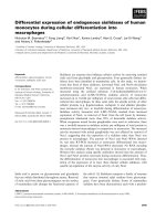
(rMFI, 218.4 ± 187.3 vs. 4.4 ± 1.4, p < 0.05) (Figure 1
and Table 1). The expression of intracellular TLR9 was
comparable in both monocytes and alveolar macro-
phages (data not shown).
TLR2 expression is modified during the monocyte
differentiation process
The observed differences in TLR expression levels
between alveolar macrophages and monocytes may have
resulted from differences in the source tissue
Figure 1 Differential constitutive surface expression of TLR2, TLR4 and TLR9 on human alveolar macrophages and monocytes. Alveolar
macrophages and monocytes from healthy donors were analyzed by flow cytometry using phycoerythrin (PE)-coupled mouse anti-TLR2, TLR4
and TLR9 antibodies and their corresponding isotype controls (gray thin lines). Histograms are representative of eight independent experiments.
Juarez et al. Respiratory Research 2010, 11:2
/>Page 5 of 13
microenvironment or the maturation stages of the cells.
To test the latter possibility, we modeled the impact of
the differentiation process from monocytes to macro-
phages on the expression of TLRs by in vitro monocyte
maturation. Expression levels of TLR2, TLR4 and TLR9
were monitored by flow cytometry in the transition pro-
cess from monocytes to monocyte-derived macrophages.
Interestingly, TLR2 surface expression (rMFI) and the
proportion of TLR2 positive cells decreased after 24
hours of culture in Petri dishes and through day 7 (D7)
when cells portrayed a macrophage phenotype as deter-
mined by light microscopy (Day 0, basal rMFI 3.9 ± 0.9,
54 ± 10.4%; Day 4 rMFI 1.4 ± 0.36, 8.5 ± 7.8%, Day 7
rMFI 1.4 ± 0.5, 1.5 ± 1.2%, p < 0.05). TLR4 expression
remained unchanged during the differentiation of mono-
cytes into macrophages (D0, rMFI 1.3 ± 0.2, D4 rMFI
1.25 ± 0.22, D7 rMFI 1.3 ± 0.3) while the expression of
TLR9 varied although not statistically significant (D0,
basal rMFI 6.3 ± 1.2, rMFI at D1, 3 ± 0.6, rMFI at D4
4.85 ± 1.43, rMFI at D7 3.6 ± 0.9) (Figure 2 and Table 1).
TLR2, TLR4 and TLR9 mRNA expression in monocyte-
derived and alveolar macrophages
The mRNA expression levels of TLRs were assessed by
qRT-PCR (TaqMan) in alveolar macrophages and mono-
cytes using the ΔΔCt method allowing a comparison of
the TLR mRNA expression of alveolar ma crophages
Table 1 Constitutive surface expression of TLR2, TLR4 and TLR9
Receptor % Cells expressing TLRs Cell Surface Expression (rMFI)
MN AM MDM MN AM MDM
TLR2 57 ± 11.1 1.2 ± 0.4* 1.5 ± 1.2* 3.2 ± 0.1 0.9 ± 0.1* 1.4 ± 0.5*
TLR4 3.0 ± 0.8 1.3 ± 0.2 3.8 ± 1.4 1.5 ± 0.2 1.1 ± 0.1 1.3 ± 0.3
TLR9 39.8 ± 14.7 54.6 ± 15.5 38 ± 20 4.4 ± 1.4 218.4 ± 187.3* 3.6 ± 0.9
Constitutive surface expression of TLR2, TLR4 and TLR9 on human monocytes and alveolar macrophages and monocyte-derived macrophages.TLR
levels were determined on monocytes (MN, n = 8), alveolar macrophages (AM, n = 7) and monocyte-derived macrophages (MDM, n = 8) by flow cytom etry.
Results present mean percentages ± SE of cells expressing TLR and relative mean fluorescence index (rMFI) ± SE as a measure of the TLR expression density. (*)
statistically significant differences compared to monocytes (p < 0.05).
Figure 2 Modulation of TLR2, TLR4, and TLR9 expression during macrophage maturation. Monocyte-derived macro phages (MDM) were
obtained from monocytes during a 7-day culture period in plastic dishes. Surface TLR expression was assessed by flow cytometry on freshly
isolated monocytes (D0) and on cultured monocytes after 1 day (D1), 4 days (D4) and 7 days (D7) of differentiation. Histograms are
representative of five independent experiments.
Juarez et al. Respiratory Research 2010, 11:2
/>Page 6 of 13
relative to that of monocytes. The expression of TLR2,
TLR4 and TLR9 mRNA of alveolar macrophages was
lower than that of autologous monocytes (Figure 3A).
To determine the TLR mR NA expression during
monocyte differentiation into macrophages, mRNA from
monocyte cultures during seven-day plastic adherence
was extracted and TLR mRNA expression of mono cyte-
derived macrophages was assessed relative to that of
autologous monocytes on day 0. Monocyte-derived
macrophages expressed lower TLR2, TLR4 and TLR9
mRNA levels than monocytes thus resembling alveolar
macrophages (Figure 3B).
Regulation of TLR surface expression in response to TLR
ligands in monocytes and alveolar macrophages
To assess the expression of TLR2, TLR4 and TLR9 by flow
cytometry following ligand exposure, alveolar ma cro-
phages and monocytes were stimulated for the optimal
incubation periods (described in the Methods section)
with Pam3Cys (30 minutes), LPS (10 minutes) and M.tb
DNA (24 hours), respectively. Following the 30-minute-
exposure to Pam3Cys, TLR2 expression levels on alveolar
macrophages remained unchanged, whereas on monocytes
it was decreased below constitutive (culture medium)
levels in all the individuals tested (Figure 4A).
Stimulation of alveolar macrophages and monocytes
with LPS, however, augmented the expression of TLR4
on both alveolar macrophages and monocytes already
after 10 minutes (Figure 4B). No further changes of
TLR2 and TLR4 surface expression had been observed
within a 24-hour observation period in our pilot study
(data not shown). TLR9 expression after M.tb DNA sti-
mulation was redu ced in monocytes from six of nine and
in alveolar macrophages from seven of nine subjects after
24 hours, however, statistical significance was not
reached (data not shown). Cell exposure to human DNA
did not alter the expression of TLR9 (data not shown).
Regulation of TLR mRNA expression by TLR specific
ligands
To determine whether cellular activation may regulate
TLR mRNA levels, cells were stimulated with LPS,
Figure 3 Bronchoalveol ar cell mRNA expression of TLR2, TLR4 and TLR9 is lower than that of monocytes. TLR gene expression in
unstimulated cells was assessed by qRT-PCR and relative quantification determined using the ΔΔCT method. Gene expression was normalized to
18 S rRNA. TLR expression of bronchoalveolar cells (BAC, panel A) and monocyte-derived macrophages (MDM, panel B) is reported relative to
monocytes (MN). TLR expression on monocytes was set at 1. Depicted are mean ± SE of five individuals.
Juarez et al. Respiratory Research 2010, 11:2
/>Page 7 of 13
Pam3Cys and M.tb DNA, for 1 h and 24 h, respectively.
Total RNA was extracted from the cells and analyzed by
qRT-PCR.
Pam3Cys upregulated the expression of TLR2 mRNA
in monocytes within a 1-hour incubation period only,
whereas in alveolar macrophages TLR2 mRNA upregu-
lation was detected after 1 h and then maintained u ntil
24 h (Figure 5A).
LPS upregulated TLR4 mRNA in both monocy tes and
alveolar macrophages after 1 h only, and was d ecreased
below basal levels in both cells types after 24 h (Figure 5B).
M.tb DNA, in contrast, upregulated TLR9 mRNA in
monocytes and alveolar macrophages after 24 h only
(Figure 5C). These observations suggest that the expres-
sion of TLR2, TLR4 and TLR9 may b e regulated differ-
entially in vivo at sites o f infection or inflammation by
bacterial components or TLR specific ligands. There
were no differences noted in the cell surface expression
or the mRNA levels of TLR2, TLR4, and TLR9 or the
responsiveness of the TLRs to their ligands between
cells from TST positive ( n = 4) and TST negative (n =
7) subjects.
TLR ligands induce production of pro-inflammatory
cytokines by bronchoalveolar cells and monocytes
To assess the cytokine-inducing functional capability of
TLR2, TLR4 and TLR9, we assessed the release of TNF-
a, IL-1b, IL-6, IL-10 and IL-12 following ligand-stimula-
tion of bronchoalveolar cells (95 ± 2.6% alveolar macro-
phages) and monocytes in response to Pam3Cys, LPS,
M.tb-DNA, and human DNA and culture medium (con-
trol). Stimulation with Pam3Cys showed a trend towards
lower TNF-a production levels (mean ± SD [pg/mL],
376 ± 152 versus 1080 ± 495, Figure 6A) and signifi-
cantly lower levels of IL-6 (887 ± 150 versus 8485 ±
4548 , p < 0.05, Figure 6C) in bronchoalveolar cells than
in monocytes. Levels of IL-1b (Figure 6B) were compar-
ably low (me an ± SD [pg/mL], IL-1b: 27.8 ± 18.1 versus
333.8 ± 179.0) and levels of IL-10 and IL-12 undetect-
able (Figure 6D and 6E). These findings coincided with
the lower surface expression levels of TLR2 on bronch-
oalveolar cells compared with monocytes and sugges ted
that Pam3Cys may preferentially induce the production
of TNF-a and IL-6.
LPS induced similar levels of TNF-a,IL-1b IL-6 and
IL-12 in bronchoalveolar cells and monocytes, (mean ±
SD [pg/mL], TNF-a 6915 ± 1675 versus 5436 ± 2008,
IL-1b 3653.8 ± 1695.6 versus 2459.1 ±1211, IL-6: 11931
± 2983 versus 9985 ± 3770, IL-12: 1.7 ± 0.7 versus 2.8 ±
1.2, Figure 6A, B and 6E, respectively) while the induc-
tion of IL-10 was significantly lower in bronchoalveolar
cells than in monocytes (mean ± SD [pg/mL], IL-10:
65.4 ± 14.6 versus 1471.6 ± 250.8, p < 0.05).
M.tb-DNA induced comparable levels of TNF-a,IL-
1b and IL-6 b ut did not induce IL-10 or IL-12 in
bronchoalveolar cells and monocytes (mean ± SD [pg/
mL], TNF-a: 2049 ± 421 and 1779 ± 560; IL-1b: 910.3
± 1138.9 and 700.3 ± 899; IL-6: 5142 ± 2153 and 4485
± 1922, respectively, Figure 6A, B, C). Culture medium
Figure 4 TLR expression upon ligand recogni tion. Alveolar macrophages and monocytes were cultured for 30 minutes in presence of 1 ng/
mL Pam3Cys (TLR2, panel A). Alveolar macrophages and monocytes were cultured for 10 minutes in presence of 100 ng/mL LPS (TLR4, panel B).
TLR expression after ligand stimulation was determined by flow cytometry. Histograms are representative of six independent experiments.
Juarez et al. Respiratory Research 2010, 11:2
/>Page 8 of 13
alone or human DNA (control stimuli) induced compar-
ably low levels o f all the cytokines (<30 pg/mL) studied
in both cell types.
Discussion
The expression profile of TLR s and its potential contri-
bution to human innate pulmonary immune responses
in the alveolar spaces in response to bacterial compo-
nents are poorly understood. We therefore compared
the cons tituti ve and ligand-induced expression of TLR2,
TLR4 and TLR9 that are involved in the recognition of
M.tb on alveolar macrophages, with that on autologous
blood monocytes and monocyte-derived macrophages
from healthy persons.
Resting human alveolar macrophages were character-
ized by significantly lower TLR2 and comparably low
TLR4 surface expression levels than autologous mono-
cytes. The flow cytometry findings for TLR2 were c on-
sistent with the mRNA expression levels in the c urrent
work. The se findings also coincide with reports of five-
fold decreased TLR2 mRNA levels in healthy lung tis-
sues compared to that i n human peri pheral leukocytes
1 h
24 h
LPS - Induced
TLR4 Gene Expression
B
0
5
10
15
20
25
30
MN BAC
Pam3cys – Induced
TLR2 Gene Expression
0
2
4
6
8
10
12
MN BAC
A
M.tb DNA -Induced
TLR9 Gene Expression
0
2
4
6
8
10
MN BAC
C
Figure 5 Regulation of TLR mRNA expression after ligand exposure. Bronc hoalveolar cells (BAC) and monocytes (MN) were incubated in
presence of 1 ng/ml of Pam3Cys and 100 ng/ml of LPS during 1 h or 24 h. Total RNA from cell lysates was reverse transcribed and qRT-PCR
performed to quantify mRNA expression. Ligand-induced TLR2, TLR4 and TLR9 expression is reported relative to that of the unstimulated
autologous cells. Mean ± SE of five independent experiments are depicted.
Juarez et al. Respiratory Research 2010, 11:2
/>Page 9 of 13
[20,31], and with lower TLR2 mRN A levels in human
alveolar macrophages than in autologous monocytes
[32]. Our observation of reduced TLR2 surface expres-
sion on alveolar macrophages coincides functionally
with the lower production of TNF-a and IL-6 following
Pam3Cys stimulation of bronchoalveolar cells compared
with autologous monocytes.
TLR4 cell surface expression was low and comparable
in alveolar macrophages and monocytes, and TLR4
mRNA lower in alveolar macrophages than monocytes.
These discrepancies may be explained by dif ferences in
the time kinetics of TLR4 trafficking and surface expres-
sion and mRNA expression. Nevertheless, despite the
low e xpression levels of TLR4 in alveolar macrophages,
these cells produced significantly higher (p < 0.05)
amounts of IL-1b,IL-6andTNF-a in response to LPS
than to culture medium. This suggests that small
expression levels of TLR4 may suffice to induce cytokine
production and TLR4 mRNA expression. Intriguingly,
TLR9 surface expression detected by flow cytometry was
50-fo ld greater on resting primary alveolar macrophages
than on primary autologous monocytes, although the
proportion of cells expressing the recep tor and the
intracellular e xpression levels were similar. This obser-
vation contrasts the notion that TLR9 is expressed pri-
marily intracellular, as was previously suggested by some
authors i n macrophages and dendritic cells [33-35]. The
findings in the current study and that of other authors
[36-38], however, provide evidence that the expression
of TLR9 may in fact be both, intracellular and on the
cell surface. The higher expression density of TLR9 on
the cell surface of the alve olar macrophages (compared
with that on the monocytes) was inconsistent with the
lower TLR9 mRNA expression of these cells. This may
for example be due to the half life of the receptors, or
dissociation between TLR9 trafficking and de novo pro-
tein synthesis in the two cell types.
Because the distinct expression levels of TLR2 found
on alveolar macrophages and monocytes may have been
due to differences in the maturation stages of these cells
we assessed monoc yte-derived macrophages in parallel.
We had previously reported that monocyte-derived
macrophages obtained under plastic adherence culture
conditions resemble alveolar macrophag es in their capa-
city to phagocytose M.tb and to express LL-37 [29]. In
the current study, we found by flow cytometry and
qRT-PCR that TLR2 was downregulated within 24
hours of monocyte culture and remained low through-
out the seven-day differentiation period into macro-
phages. Interestingly, the low TLR2 expression levels on
monocyte-derived macrophages on day seven coincided
with the low constitutive TLR2 expressio n found o n
alveolar macrophages (Figures 1 and 2). These results
are also compatible with t hose from Henning et al who
reported a significant reduction of TLR2 protein and
mRNA, and unaltered TLR4 expression during the in
vitro maturation of human monocytes to macrophages
in Teflon wells [39]. Thus, t he low-level expression o f
TLR2 appears to be a feature of primary alveolar macro-
phages as well as of in vitro generated monocyte-derived
macrophages. In contrast, induction of macrophage
maturation by M-CSF, has been shown to result i n
unchanged TLR2, increased TLR4 and very low TLR9
mRNA expression levels [40]. Macrophage TLR expres-
sion assessed in experimental culture microenviron-
ments thus depends on the presence or absence of a
variety o f factors, including type and concentrations of
cytokines and of additional proteins such as surfactant
protein A [39].
We also assessed the effects of TLR-specific ligands on
the expression of TLR2, TLR4 and TLR9, as both, TLR
ligands and cytokines have been reported to regulate
TLR expression [31,41].
TLR2 cell surface expression by flow cytometry was
decreased on monocytes after stimulation with
Pam3Cys, whereas the expression of TLR2 on alveolar
macrophages in response to Pam3Cys remained
unchanged. Pam3Cys induced TLR2 mRNA expression
was increased as early as after 1 h in both cells types,
but was maintained for a longer time in bronchoalveolar
cells. These findings su ggest that TLR2 may be differen-
tially regulated in monocytes and alveolar macrophages.
TLR4, in contrast was shown to be upregulated in
response to LPS on monocytes and alveolar macro-
phages in a kinetic similar to that of TLR2 using both
flow cytometry and qRT-PCR. Taken together these
results indicate a differential, cell-type-specific ligand-
mediated regulation of the expression of TLR2 and
TLR4.
It was beyond the scope of this study to assess in
detail whether ligand-binding alone, and/or cytokine
release in the cellular microenvironment affected the
regulation of the TLRs. While TLR2 regulation may be
due to Pam3Cys ligation and/or cytokine production
from macrophages or other cellular subsets within the
bronchoalveolar cells (5-8% are lymphocytes), regulation
of TLR4 expression may result from a direct effect of
LPS on the cell membrane as it was noted already
within 10 minutes of LPS stimulation.
TLR9 cell surface expression detected by flow cytome-
try in respo nse to M. tb DNA did not show a uniform
pattern, however, was diminished on alveolar macro-
phages and on monocytes in 65% to 75% of all study
subjects. We speculate that this phenomenon may be
due to the internalization of cell surface TLR9 after
binding to its ligand. Alternatively, TLR9 may become
undetectable to the antibodies used during the flow
cytometry after binding to its ligand. TLR9 mRNA was
Juarez et al. Respiratory Research 2010, 11:2
/>Page 10 of 13
upregulated after 24 hours of incubation with M.tb
DNA in both monocytes and alveolar macrophages in
five of five study subjects indicating a slower kinetic of
TLR9 mRNA generation than in the case of TLR2 and
TLR4.
We also assessed the induction of cytokines foll owing
sti mulation of the cells with TLR-specific ligand s. TNF-
a and IL-6-production levels after Pam3Cys (TLR2) sti-
mulation were significantly lower in bronchoalveolar
cells than in autologous monocytes. This data is consis-
tent with the lo wer TLR2 expression levels found on
alveolar macrophages in the current study and with data
from a recent study in which lipoteichoic acid, a TLR2
ligand, was instilled experimentally into lung segments
of human volunteers and resulted in a poor transcrip-
tion of IL-1b, IL-6, and IL-8 genes [42].
In the current study, TNF-a,IL-1b and IL-6 produc-
tion in response to LPS (TLR4) was not signifi cantly
Figure 6 TLR-ligands induce di fferential cytokine production. Bronchoalveolar cells (BAC) and monocytes (MN) were stimulated with 5 mg/
mL DNA from M.tb H37 Rv, 100 ng/mL LPS and 1 ng/mL Pam3Cys for 20 hours. Cytokines were determined in culture supernatants by ELISA for
TNF-a and IL-6 (A, C) and Cytokine Bead Array for IL-1b IL-10 and IL-12 (B, D and E). Culture medium and human DNA (5 mg/mL) were used as
negative stimulation controls. Mean cytokine pg/mL ± SE of seven independent experiments are depicted. Statistically significant differences (p <
0.05): (*) comparing stimulated vs. controls (medium) within respective cell groups, (++) comparing bronchoalveolar cells with monocytes.
Juarez et al. Respiratory Research 2010, 11:2
/>Page 11 of 13
different between bronchoalveolar cells and monocytes.
However, LPS induced higher production of IL-10 in
monocytes than in bronchoalveolar cells suggesting
that activation via TLR4 may result in differential cyto-
kine production from alveolar macrophages and mono-
cytes. This observation may find a mechanistic
explanation i n a study of human alv eolar macrophages
in which LPS-induced IL-10 production was associated
with a reduced capacity to activate STAT3. This sug-
gested that TLR ligand activation may modulate the
anti-inflammatory activity of IL-10 either by altering its
production or by inhibiting the cellular responsiveness
to the c ytokine [43].
M.tb DNA (TLR9) induced similar levels of TNF-a,
IL-1b and IL-6 in bronchoalveolar cells and monocytes
although the density of TLR9 expression was higher on
alveolar ma crophages than mon ocytes. This finding may
suggest expression of non-functional cell surface TLR9,
while the comparable induction of cytokine production
in both cell types suggests that intracellular TLR9 may
be the functional c omponent of the receptor. One may
speculate that the cell surface form of TLR9 binds bac-
terial DNA (released from infected and dying cells) and
that the ligand is then transferred from the cell surface
into the intracellular compartment. This may provide an
extra safety step prior to inducing the inflammatory cas-
cade. Indeed, there is evidence that for TLR9 to be func-
tional, an ecto domain cleavage in the endolysosome is
required to recruit MyD88 upon activation, and that a
truncated rather than the full-length form of the recep-
tor is functional. Both, full-length and cleaved forms of
TLR9, however, are capable of binding ligand [44].
Tissue compartment-specific immune responses thus
may be characterized by differential expression levels of
TLRs, such as those shown here for TLR2, that result in
distinct production levels of cytokines. These immunor-
egulatory mechanisms may shape the inflammatory
cytokine response and thus the potential o f damage to
the tissue that, as in the case of the lungs, is exposed
continuously to inhaled pathogens and noninfectious
particulate matter. A suppressive immunoregulatory
mechanism involving TLRs has been described recently
as alveolar surfactant protein A (SP-A) was shown to
downregulate TLR2 expression and TNF- a production
in human macrophages [39].
Limitations of the current study are related to the dif-
ficulty to recruit healthy volunteers for lung immunity
studies and the resulting small study subject numbers.
In vitro studies also may not reflect exactly processes in
vivo. Further, the bronchoalveolar cells contained a
small proportion (5-8%) o f alveolar lymphocytes of
which a small subset may express TLRs [45-47]. Simi-
larly, monocyte populations contained up to 10% of
contaminating lymphocytes, despite plastic adherence
and magnetic bead enrichment. It is thus possible that a
small component of TLR ligand-induced cytokines
derived from lymphocytes and not from alveolar macro-
phages or monocytes.
Conclusions
The observations in this study have clinical implications.
Differences in the expression profile of TLRs between
the blood and lung compartments may have to be con-
sidered for the design and efficacy evaluation of new
vaccine-adjuvant combinations. New vaccines against
respiratory pathogens, such as M.tb, may target the
respiratory system to provide optimal protection locally
in the near future.
Acknowledgements
This work was supported by grant 2R01HL51630 from NHLBI and by grant
R21ES016928-02 from NIEHS.
Author details
1
Departamento de Microbiología, Instituto Nacional de Enfermedades
Respiratorias, (Calzada de Tlalpan) México City, (14080), México.
2
Servicio de
Broncoscopia, Instituto Nacional de Enfermedades Respiratorias, (Calzada de
Tlalpan) México City, (14080), México.
3
Center for Emerging Reemerging
Pathogens, University of Medicine and Dentistry New Jersey, (S Orange Ave),
Newark, (07103), USA.
4
Department of Medicine, Section of Infectious
Diseases, Boston Medical Center (Albany Street), Boston, (02118), USA.
5
Department of Environmental and Occupational Health, University of
Medicine and Dentistry New Jersey - School of Public Health (Hoes Lane)
Piscataway, (08854), USA.
6
Center for Global Public Health, University of
Medicine and Dentistry New Jersey - School of Public Health (Hoes Lane)
Piscataway, (08854), USA.
Authors’ contributions
EJ carried out the cell culture flow cytometry assays and prepared the first
draft of the manuscript.
CN carried out the bronchoalveolar lavages.
ES participated in the preparation of the manuscript
JJE participated in the preparation of the manuscript and was the PI of the
NIH grant that supported much of this project.
SKS co-developed the study idea, participated in the design of the study
and the experimental work, and spearheaded the final preparation of the
manuscript.
MT co-developed the study idea, participated in the design of the study and
the preparation of the manuscript and coordinated the experimental work.
All authors have read and approved the final manuscript.
Competing interests
The authors declare that they have no competing interests.
Received: 14 March 2009
Accepted: 5 January 2010 Published: 5 January 2010
References
1. Suzuki T, Chow CW, Downey GP: Role of innate immune cells and their
products in lung immunopathology. Int J Biochem Cell Biol 2008, 40:1348-
61.
2. Segal BH: Role of macrophages in host defense against aspergillosis and
strategies for immune augmentation. Oncologist 2007, 12(Suppl 2):7-13.
3. Gordon S: Pattern recognition receptors: doubling up for the innate
immune response. Cell 2002, 111:927-930.
4. Sibille Y, Reynolds HY: Macrophages and polymorphonuclear neutrophils
in lung defense and injury. Am Rev Respir Dis 1990, 141:471-501.
5. Gordon SB, Read RC: Macrophage defences against respiratory tract
infections. Br Med Bull 2002, 61:45-61.
Juarez et al. Respiratory Research 2010, 11:2
/>Page 12 of 13
6. Politis AD, Vogel SN: Measurement of Fc gamma receptor-mediated binding
and phagocytosis. Curr Protoc Immunol 2005, Chapter 14(Unit 14):18.
7. Groves E, Dart AE, Covarelli V, Caron E: Molecular mechanisms of
phagocytic uptake in mammalian cells. Cell Mol Life Sci 2008, 65:1957-76.
8. Brekke OL, Christiansen D, Fure H, Fung M, Mollnes TE: The role of
complement C3 opsonization, C5a receptor, and CD14 in E. coli-induced
up-regulation of granulocyte and monocyte CD11b/CD18 (CR3),
phagocytosis, and oxidative burst in human whole blood. J Leukoc Biol
2007, 81:1404-1413.
9. Taylor PR, Martinez-Pomares L, Stacey M, Lin HH, Brown GD, Gordon S:
Macrophage receptors and immune recognition. Annu Rev Immunol 2005,
23:901-944.
10. Leulier F, Lemaitre B: Toll-like receptors–taking an evolutionary approach.
Nat Rev Genet 2008, 9:165-178.
11. Mukhopadhyay S, Herre J, Brown GD, Gordon S: The potential for Toll-like
receptors to collaborate with other innate immune receptors.
Immunology 2004, 112:521-530.
12. Takeda K, Akira S: Regulation of innate immune responses by Toll-like
receptors. Jpn J Infect Dis 2001, 54:209-219.
13. Takeda K, Kaisho T, Akira S: Toll-like receptors. Annu Rev Immunol 2003,
21:335-376.
14. Akira S, Takeda K, Kaisho T: Toll-like receptors: critical proteins linking
innate and acquired immunity. Nat Immunol 2001, 2:675-680.
15. Abel B, Thieblemont N, Quesniaux VJ, Brown N, Mpagi J, Miyake K, Bihl F,
Ryffel B: Toll-like receptor 4 expression is required to control chronic
Mycobacterium tuberculosis infection in mice. J Immunol 2002, 169:3155-
3162.
16. Jung SB, Yang CS, Lee JS, Shin AR, Jung SS, Son JW, Harding CV, Kim HJ,
Park JK, Paik TH, Song CH, Jo EK: The mycobacterial 38-kilodalton
glycolipoprotein antigen activates the mitogen-activated protein kinase
pathway and release of proinflammatory cytokines through Toll-like
receptors 2 and 4 in human monocytes. Infect Immun 2006, 74:2686-2696.
17. Underhill DM, Ozinsky A, Smith KD, Aderem A: Toll-like receptor-2
mediates mycobacteria-induced proinflammatory signaling in
macrophages. Proc Natl Acad Sci USA 1999, 96:14459-14463.
18. Bafica A, Scanga CA, Feng CG, Leifer C, Cheever A, Sher A: TLR9 regulates
Th1 responses and cooperates with TLR2 in mediating optimal
resistance to Mycobacterium tuberculosis. J Exp Med 2005, 202:1715-1724.
19. Harju K, Glumoff V, Hallman M: Ontogeny of Toll-like receptors Tlr2 and
Tlr4 in mice. Pediatr Res 2001,
49:81-83.
20. Zarember KA, Godowski PJ: Tissue expression of human Toll-like
receptors and differential regulation of Toll-like receptor mRNAs in
leukocytes in response to microbes, their products, and cytokines. J
Immunol 2002, 168:554-561.
21. Cario E, Podolsky DK: Differential alteration in intestinal epithelial cell
expression of toll-like receptor 3 (TLR3) and TLR4 in inflammatory bowel
disease. Infect Immun 2000, 68:7010-7017.
22. Droemann D, Goldmann T, Branscheid D, Clark R, Dalhoff K, Zabel P,
Vollmer E: Toll-like receptor 2 is expressed by alveolar epithelial cells
type II and macrophages in the human lung. Histochem Cell Biol 2003,
119:103-108.
23. Fenhalls G, Squires GR, Stevens-Muller L, Bezuidenhout J, Amphlett G,
Duncan K, Lukey PT: Associations between toll-like receptors and
interleukin-4 in the lungs of patients with tuberculosis. Am J Respir Cell
Mol Biol 2003, 29:28-38.
24. Schwander SK, Torres M, Carranza CC, Escobedo D, Tary-Lehmann M,
Anderson P, Toossi Z, Ellner JJ, Rich EA, Sada E: Pulmonary mononuclear
cell responses to antigens of Mycobacterium tuberculosis in healthy
household contacts of patients with active tuberculosis and healthy
controls from the community. J Immunol 2000, 165:1479-1485.
25. Boyum A: Isolation of lymphocytes, granulocytes and macrophages.
Scand J Immunol 1976, , Suppl 5: 9-15.
26. Latz E, Visintin A, Lien E, Fitzgerald KA, Monks BG, Kurt-Jones EA,
Golenbock DT, Espevik T: Lipopolysaccharide rapidly traffics to and from
the Golgi apparatus with the toll-like receptor 4-MD-2-CD14 complex in
a process that is distinct from the initiation of signal transduction. J Biol
Chem 2002, 277:47834-47843.
27. Latz E, Visintin A, Espevik T, Golenbock DT: Mechanisms of TLR9 activation.
J Endotoxin Res 2004, 10:406-412.
28. Diaz ML, Herrera T, Lopez-Vidal Y, Calva JJ, Hernandez R, Palacios GR,
Sada E: Polymerase chain reaction for the detection of Mycobacterium
tuberculosis DNA in tissue and assessment of its utility in the diagnosis
of hepatic granulomas. J Lab Clin Med 1996, 127:359-363.
29. Rivas-Santiago B, Hernandez-Pando R, Carranza C, Juarez E, Contreras JL,
Aguilar-Leon D, Torres M, Sada E: Expression of cathelicidin LL-37 during
Mycobacterium tuberculosis infection in human alveolar macrophages,
monocytes, neutrophils, and epithelial cells. Infect Immun 2008, 76:935-
941.
30. Carranza C, Juarez E, Torres M, Ellner JJ, Sada E, Schwander SK:
Mycobacterium tuberculosis growth control by lung macrophages and
CD8 cells from patient contacts. Am J Respir Crit Care Med 2006, 173:238-
245.
31. Flo TH, Halaas O, Torp S, Ryan L, Lien E, Dybdahl B, Sundan A, Espevik T:
Differential expression of Toll-like receptor 2 in human cells. J Leukoc Biol
2001, 69:474-481.
32. Droemann D, Goldmann T, Tiedje T, Zabel P, Dalhoff K, Schaaf B: Toll-like
receptor 2 expression is decreased on alveolar macrophages in cigarette
smokers and COPD patients. Respir Res 2005, 6
:68.
33. Wagner H: The immunobiology of the TLR9 subfamily. Trends Immunol
2004, 25:381-386.
34. Ahmad-Nejad P, Hacker H, Rutz M, Bauer S, Vabulas RM, Wagner H:
Bacterial CpG-DNA and lipopolysaccharides activate Toll-like receptors at
distinct cellular compartments. Eur J Immunol 2002, 32:1958-1968.
35. Latz E, Schoenemeyer A, Visintin A, Fitzgerald KA, Monks BG, Knetter CF,
Lien E, Nilsen NJ, Espevik T, Golenbock DT: TLR9 signals after translocating
from the ER to CpG DNA in the lysosome. Nat Immunol 2004, 5:190-198.
36. Cognasse F, Hamzeh-Cognasse H, Lafarge S, Chavarin P, Pozzetto B,
Richard Y, Garraud O: Identification of two subpopulations of purified
human blood B cells, CD27(-) CD23(+) and CD27(high) CD80(+), that
strongly express cell surface Toll-like receptor 9 and secrete high levels
of interleukin-6. Immunology 2008, 125(3):430-7, Epub 2008 Apr 28.
37. Ewaschuk JB, Backer JL, Churchill TA, Obermeier F, Krause DO, Madsen KL:
Surface expression of Toll-like receptor 9 is upregulated on intestinal
epithelial cells in response to pathogenic bacterial DNA. Infect Immun
2007, 75:2572-2579.
38. Eaton-Bassiri A, Dillon SB, Cunningham M, Rycyzyn MA, Mills J, Sarisky RT,
Mbow ML: Toll-like receptor 9 can be expressed at the cell surface of
distinct populations of tonsils and human peripheral blood
mononuclear cells. Infect Immun 2004, 72:7202-7211.
39. Henning LN, Azad AK, Parsa KV, Crowther JE, Tridandapani S, Schlesinger LS:
Pulmonary surfactant protein A regulates TLR expression and activity in
human macrophages. J Immunol 2008, 180:7847-7858.
40. O’Mahony DS, Pham U, Iyer R, Hawn TR, Liles WC: Differential constitutive
and cytokine-modulated expression of human Toll-like receptors in
primary neutrophils, monocytes, and macrophages. Int J Med Sci 2008,
5:1-8.
41. Marshall JD, Heeke DS, Gesner ML, Livingston B, Van Nest G: Negative
regulation of TLR9-mediated IFN-alpha induction by a small-molecule,
synthetic TLR7 ligand. J Leukoc Biol 2007, 82:497-508.
42. Hoogerwerf JJ, de Vos AF, Bresser P, Zee van der JS, Pater JM, de Boer A,
Tanck M, Lundell DL, Her-Jenh C, Draing C, von Aulock S, Poll van der T:
Lung Inflammation Induced by Lipoteichoic Acid or Lipopolysaccharide
in Humans. Am J Respir Crit Care Med 2008, 178:34-41.
43. Fernandez S, Jose P, Avdiushko MG, Kaplan AM, Cohen DA: Inhibition of IL-
10 receptor function in alveolar macrophages by Toll-like receptor
agonists. J Immunol 2004, 172:2613-2620.
44. Ewald SE, Lee BL, Lau L, Wickliffe KE, Shi GP, Chapman HA, Barton GM: The
ectodomain of Toll-like receptor 9 is cleaved to generate a functional
receptor. Nature 2008, 456:658-662.
45. Prabha C, Rajashree P, Sulochana DD: TLR2 and TLR4 expression on the
immune cells of tuberculous pleural fluid. Immunol Lett 2008, 117:26-34.
46. Girart MV, Fuertes MB, Domaica CI, Rossi LE, Zwirner NW: Engagement of
TLR3, TLR7, and NKG2D regulate IFN-gamma secretion but not NKG2D-
mediated cytotoxicity by human NK cells stimulated with suboptimal
doses of IL-12.
J Immunol 2007, 179:3472-3479.
47. Xu D, Komai-Koma M, Liew FY: Expression and function of Toll-like
receptor on T cells. Cell Immunol 2005, 233:85-89.
doi:10.1186/1465-9921-11-2
Cite this article as: Juarez et al.: Differential expression of Toll-like
receptors on human alveolar macrophages and autologous peripheral
monocytes. Respiratory Research 2010 11:2.
Juarez et al. Respiratory Research 2010, 11:2
/>Page 13 of 13