Báo cáo y học: "Cytokine profile of autologous conditioned serum for treatment of osteoarthritis, in vitro effects on cartilage metabolism and intra-articular levels after injection" potx
Bạn đang xem bản rút gọn của tài liệu. Xem và tải ngay bản đầy đủ của tài liệu tại đây (929.37 KB, 11 trang )
Rutgers et al. Arthritis Research & Therapy 2010, 12:R114
/>Open Access
RESEARCH ARTICLE
© 2010 Rutgers et al.; licensee BioMed Central Ltd. This is an open access article distributed under the terms of the Creative Commons
Attribution License ( which permits unrestricted use, distribution, and reproduction in
any medium, provided the original work is properly cited.
Research article
Cytokine profile of autologous conditioned serum
for treatment of osteoarthritis,
in vitro
effects on
cartilage metabolism and intra-articular levels after
injection
Marijn Rutgers
1
, Daniël BF Saris
1
, Wouter JA Dhert
1,2
and Laura B Creemers*
1
Abstract
Introduction: Intraarticular administration of autologous conditioned serum (ACS) recently demonstrated some
clinical effectiveness in treatment of osteoarthritis (OA). The current study aims to evaluate the in vitro effects of ACS on
cartilage proteoglycan (PG) metabolism, its composition and the effects on synovial fluid (SF) cytokine levels following
intraarticular ACS administration.
Methods: The effect of conditioned serum on PG metabolism of cultured OA cartilage explants was compared to
unconditioned serum. The effect of serum conditioning on levels of interleukin-1beta (IL-1β), IL-4, IL-6, IL-10, IL-13,
interferon gamma (IFN-γ), tumor necrosis factor alpha (TNF-α), osteoprotegerin (OPG), oncostatin M (OSM), interleukin-
1 receptor (IL-1ra) and transforming growth factor beta (TGF-β) were measured by multiplex ELISA. As TNF-α levels
were found to be increased in conditioned serum, the effect of TNF-α inhibition by etanercept on PG metabolism was
studied in cartilage explants cultured in the presence of conditioned serum. Furthermore, cytokine levels in SF were
measured three days after intraarticular ACS injection in OA patients to verify their retention time in the joint space.
Results: PG metabolism was not different in the presence of conditioned serum compared to unconditioned serum.
Levels of the anti-inflammatory cytokines IL-1ra, TGF-β, IL-10 as well as of pro-inflammatory cytokines IL-1β, IL-6, TNF-α
and OSM were increased. IL-4, IL-13 and IFN-γ levels remained similar, while OPG levels decreased. TNF-α inhibition did
not influence PG metabolism in cartilage explant culture in the presence of condtioned serum. Although OPG levels
were higher and TGF-β levels were clearly lower in ACS than in SF, intraarticular ACS injection in OA patients did not
result in significant changes in these cytokine levels.
Conclusions: ACS for treatment of osteoarthritis contains increased levels of anti-inflammatory as well as pro-
inflammatory cytokines, in particular TNF-α, but conditioned serum does not seem to have a net direct effect on
cartilage metabolism, even upon inhibition of TNF-α. The fast intraarticular clearance of cytokines in the injected ACS
may explain the limited effects found previously in vivo.
Introduction
Osteoarthritis (OA)-associated cartilage degradation is
mediated in part by cytokines and growth factors,
excreted into the intraarticular environment by synovio-
cytes, activated immune cells, or by the articular cartilage
itself [1,2]. Therapies interfering with these cytokines
may influence disease progression and improve the
patient's quality of life.
A pivotal role in the progression of OA has been
assigned to the pro-inflammatory cytokine interleukin-
1β (IL-1β), which induces a cascade of inflammatory and
catabolic events including the expression of cartilage
degrading matrix metalloproteinases (MMP) [3], nitric
oxygen (NO) production and prostaglandin E
2
(PGE
2
)
release [4], while inhibiting proteoglycan and collagen
synthesis [5,6]. The number of type-1 IL-1 receptors is
* Correspondence:
1
Department of Orthopaedics, University Medical Center Utrecht,
Heidelberglaan 100, 3584 CX Utrecht, The Netherlands
Full list of author information is available at the end of the article
Rutgers et al. Arthritis Research & Therapy 2010, 12:R114
/>Page 2 of 11
significantly increased in OA chondrocytes [7] and syn-
ovial fibroblasts [8], increasing the susceptibility for IL-1α
and IL-1β mediated effects. In addition, it was suggested
that in OA synovium, a relative deficit in IL-1ra-produc-
tion exists [1]. As intraarticular administration of recom-
binant human interleukin-1 receptor antagonist has been
shown to alleviate symptoms in several animal models of
OA and rheumatoid arthritis [9-11], intraarticular treat-
ment with IL-1ra was also suggested as a feasible treat-
ment for patients with OA.
One example of a disease-modifying osteoarthritis-
drug (DMOAD) based on blocking the intraarticular
effects of IL-1 associated with OA, is autologous condi-
tioned serum (ACS or Orthokine
®
; Orthogen, Düsseldorf,
Germany). Autologous conditioned serum (ACS) treat-
ment consists of six repetitive injections of ACS over a
period of 21 days. ACS is prepared from whole blood that
is incubated in the presence of glass beads to initiate
monocyte activation [12,13]. The resulting conditioned
serum (ACS), has been shown to contain increased levels
of IL-1ra as well as IL-4 and IL-10 [12]. In horses with
arthroscopically induced osteochondral defects, ACS
treatment demonstrated a reduction in lameness and a
decrease in synovial membrane hyperplasia [14]. ACS
treatment of human OA patients, however, demonstrated
limited to moderate clinical effects [15,16]. Despite the
fact that this approach has already been introduced in the
clinic, the mechanisms by which administration of this
product may result in reduction of OA symptoms is not
yet fully understood [14,16,17]. Although the primary
goal of ACS treatment is alleviation of OA symptoms,
one of the mechanisms may be enhancement of cartilage
integrity through the inhibition of inflammatory activity,
in particular with respect to Il-1 signalling. In fact, the
direct effect of the entire blend of known and unknown
factors present in ACS on cartilage metabolism in human
OA cartilage has not been described. Moreover, only lim-
ited data are available on the actual composition of the
conditioned serum. Besides IL-1ra, growth factors, such
as transforming growth factor beta 1 (TGF-β1), which
stimulates chondrocyte proliferation [18,19], are upregu-
lated during incubation [17]. Of several pro-inflamma-
tory cytokines like IL-1β, tumour necrosis factor-alpha
(TNF-α) [20,21] and IL-6 [22], of the last of which also
anti-inflammatory effects have been described [23], it is
not entirely clear if their levels remain equal or are upreg-
ulated during incubation [12,17]. As a consequence of
monocyte activation during incubation of blood, anti-
inflammatory cytokines such as IL-13, which was shown
to inhibit the production of IL-1β and enhance produc-
tion of IL-1ra [24], and osteoprotegerin (OPG) [25],
which protected cartilage in a murine model of surgically
induced osteoarthritis from further degeneration [26],
may be upregulated. Also pro-inflammatory cytokines
oncostatin-M (OSM) [27] and interferon-gamma (IFN-γ)
[28] which act synergistically with IL-1β to stimulate pro-
duction of MMPs and aggrecanases, may be upregulated
together with the anti-inflammatory cytokines. Even if
the composition of ACS would be favourable to cartilage
regeneration, it is still unknown to what extent the
intraarticularly injected cytokines are present long
enough in the knee joint to exert their actions. The
intraarticular availability of adequate levels of IL-1ra is
important, as IL-1β is considered to be active at low con-
centrations and relatively high levels of IL-1ra are
required to inhibit the effects of IL-1β [29]. In vivo,
increased IL-1ra levels were found in equine synovial
fluid 35 days after the last (of four) injections with ACS
[14].
The current study aims to evaluate the direct in vitro
effect of conditioned serum on cartilage proteoglycan
metabolism, to further evaluate the composition of ACS
and to examine to what extent intraarticular injection of
ACS is reflected in cytokine level changes in human
osteoarthritic synovial fluid.
Materials and methods
Preparation of conditioned serum
To prepare conditioned serum, 35 ml of whole blood was
acquired through venapunction and aspirated in six poly-
propylene syringes (5 ml) containing glass beads (Ortho-
gen, Düsseldorf, Germany). The syringes were incubated
at 37°C for six hours. After incubation, the blood was
centrifuged at 1,000 × g for 10 minutes, and serum was
aspirated and stored at -80°C until further use. Control
syringes containing whole blood (5 ml without glass
beads) were centrifuged and serum was stored at -80°C.
Effect of conditioned serum on proteoglycan metabolism
To measure the effects of conditioned serum on proteo-
glycan metabolism, 48 full thickness osteoarthritic carti-
lage explants were taken of the femoral condyles of OA
patients undergoing a total knee arthroplasty (Kellgren-
Lawrence grade III) and satisfying the OA criteria of the
American College of Rheumatology [30]. The explants
were cultured in the presence of conditioned serum (n =
24) or non-conditioned control serum (n = 24) of healthy
serum donors. The cartilage was washed, cut into cubes
of approximately 3 × 3 × 3 mm, weighed and cultured for
16 days in Dulbecco's Modified Eagles Medium contain-
ing 1% penicillin/streptomycin, 1% ascorbic acid (ASAP)
and either 25% conditioned serum or 25% control (non-
stimulated) serum. The experiment was repeated with
two other OA cartilage and serum donor combinations
(Table 1).
Rutgers et al. Arthritis Research & Therapy 2010, 12:R114
/>Page 3 of 11
Effect of TNF-α inhibition by etanercept on proteoglycan
metabolism in the presence of conditioned serum
In another series of three experiments comparing the
effect of conditioned serum and unconditioned serum on
in vitro cartilage metabolism, Etanercept (Enbrel
®
, Wyeth
Pharmaceuticals Inc., Collegeville, PA, USA) was added
to full-thickness cartilage explants of femoral condyles of
OA patients undergoing a knee replacement surgery and
cultured in vitro. The explants were cultured with 25%
control serum (n = 8), 25% conditioned serum (n = 8) and
25% conditioned serum with etanercept (1 μg/ml etaner-
cept, n = 8). This concentration was based on a previous
publication showing that this concentration was capable
of inhibiting the activity of 40 ng/ml of TNFα [31].
35
S incorporation was measured by means of a four-
hour incubation with
35
SO
4
2-
, on Day 4 for all conditions
(see below). The medium released on Days 4, 8, 12 and on
Day 16 was analysed for proteoglycan release, including
release of newly synthesised PGs. The experiment was
repeated with two other OA cartilage and serum donor
combinations (Table 1).
35
S incorporation
At Days 4, 8 and 12 of the culture,
35
SO
4
2-
incorporation
(Na
2
35
SO
4
, carrier-free; Perkin Elmer, Boston, MA, USA)
was measured in order to quantify proteoglycan incorpo-
ration by means of a four-hour incubation in culture
medium containing 20 μCi of
35
SO
4
2-
. For the control as
well as for the conditioned serum cultured cartilage
explants, eight separate cartilage explants were used for
each incorporation time point. Explants were then rinsed
in plain culture medium during three 45-minute changes,
and the culture of these explants was continued in iso-
tope-free medium until the end of culture on Day 16. At
Days 4, 8, 12 and 16, conditioned media were collected to
quantify the release of newly synthesised PG.
35
SO
4
2-
incorporation was quantified using a scintillation counter
(Tri-carb 1900CA, Packard, Ramsey, MN, USA), and
results were normalized to DNA content and weight of
the sample.
Alcian blue immunoprecipitation and DNA assay
On Day 16, all cartilage explants were washed three times
in phosphate-buffered saline (PBS) at 4°C. Explants were
then digested in 2% papain (Sigma, St. Louis, MO, USA)
in 50 mM phosphate buffer, 2 mM N-acetylcysteine, and
2 mM Na
2
-EDTA (pH 6.5) at 65°C for two hours. Part of
the digest was used to measure DNA content and part
was used for the quantification of the glycosaminoglycan
content as a measure of proteoglycan content using an
Alcian Blue precipitation assay (described below).
Another part was used to measure
35
SO
4
2-
activity.
Glycosaminoglycans (GAGs) were precipitated from
the explant digests as well as from the culture medium
and stained with an Alcian blue dye solution (Alcian blue
8GX, Sigma-Aldrich, Zwijndrecht, The Netherlands), sat-
urated in 0.1 M sodium acetate buffer, containing 0.3 M
MgCl
2
(pH 6.2) for 30 minutes at 37°C [32]. The blue
staining of the medium was quantified photospectromet-
rically from the change in absorbance at 620 nm, using
chondroitin sulphate (Sigma) as a reference. DNA was
stained with the fluorescent dye Hoechst 33258 (Sigma)
and fluorescence was measured on the Cytofluor (MTX
Lab Systems, Vienna, VA, USA) [33], using calf thymus
DNA (Sigma) as a reference.
Composition of autologous conditioned serum (ACS)
Whole blood was obtained from 22 OA patients meeting
the American College of Rheumatology criteria for OA
(mean age 52 years, range 35 to 72). ACS for intraarticu-
lar treatment was prepared by whole blood incubation in
the presence of ACS-specific glass beads. Unconditioned
serum was taken as control.
Multiplex ELISA
Multiplex ELISA was used for measurement of cytokine
levels in conditioned and unconditioned serum and in SF.
Table 1: Patient characteristics of cartilage explant experiments
Experiment series Cartilage donor OA grade
(Kellgren-Lawrence)
Cartilage explants/
condition
Serum donor
control vs CS 56 year old male III 24 (48) 26 year old male
65 year old female IV 24 (48) 33 year old male
76 year old male III 24 (48) 30 year old male
control vs CS vs
etanercept
54 year old male III 8 (24) 27 year old male
55 year old male III 8 (24) 27 year old male
44 year old male IV 8 (24) 26 year old female
CS, conditioned serum; OA, osteoarthritis.
Rutgers et al. Arthritis Research & Therapy 2010, 12:R114
/>Page 4 of 11
Earlier validation studies showed high correlation of mul-
tiplex ELISA readings with conventional ELISA [34] and
demonstrated that multiplex ELISA is suitable for SF
analysis [35]. The cytokines measured were IL-1β, IL-4,
IL-6, IL-10, IL-13, IFN-γ, OSM and OPG. Measurements
and data analysis were performed using the Bio-Plex sys-
tem in combination with the Bio-Plex Manager software
version 3.0 using five parametric curve fitting (Bio-Rad
Laboratories, Hercules, CA, USA). Coating antibodies for
IL-1β, IL-6, IL-10 and TNF-α were provided by Strath-
man Biotec (Hannover, Germany); coating antibodies for
IL-4, OPG and OSM by R&D Systems (Abingdon, UK),
coating antibody for IL-13 by National Institute for Bio-
logical Standards and control (Potters Bar, UK) and coat-
ing antibody for IFN-γ by BD Biosciences (San Diego,
CA, USA). The recombinant proteins for IL-1β, IL-6, IL-
10 and IL-13 were provided by Sanquin (Amsterdam, The
Netherlands), for IL-4 and IFN-γ by eBioscience (San
Diego, CA, USA), for TNF-α by BD Biosciences, for OPG
by R&D Systems (Abingdon, UK) and for OSM by Bio-
carta (Hamburg, Germany).
Preparation of recombinant cytokine mixes, covalent
coupling of the captured antibodies to the microspheres
and preparation of detection antibodies were performed
as described previously [34,35]. For determination of
cytokine profiles in SF, aliquots of 200 μl were first pre-
treated with 20 μl of hyaluronidase (0.5 mg/ml, type IV-S,
Sigma-Aldrich, Zwijndrecht, The Netherlands) for 30
minutes at 37°C, spun over 0.22 μm nylon membrane
(Spin-X column; Corning, The Netherlands) and diluted
with High-Performance ELISA-buffer (Sanquin Blood
Supply Foundation, Utrecht, Netherlands) at a 1:2 dilu-
tion. Recombinant protein standards and calibration
curves were prepared in serum diluents (R&D Systems).
A mix containing 1,000 coupled microspheres per
cytokine (total volume of 10 μl/well) was added to the
standard, sample or blank, and incubated for 60 minutes.
Next, a 10 μl mix of biotinylated antibodies (final concen-
tration 16.6 μg/ml for each antibody) was added to each
well and incubated for an additional 60 minutes. Beads
were washed in PBS containing 1% BSA and 0.5% Tween
20 (pH 7.4) in order to remove residual sample and
unbound antibodies. After incubation for 10 minutes
with 0.5 μg/ml streptavidin R-phycoerythrin (BD Biosci-
ences) and washing twice with 1% BSA and 0.5% Tween
20 (pH 7.4), the fluorescence intensity of the beads was
measured in 100 μl High Performance ELISA buffer (San-
quin). Measurements and data analysis were performed
using the Bio-Plex system in combination with the Bio-
Plex Manager software version 3.0 using five parametric
curve fitting (Bio-Rad Laboratories). To eliminate the
possibility of inter-assay variability, control and condi-
tioned serum samples were measured in duplo in the
same assay.
ELISA
IL-1ra and TGF-β1 levels were measured using commer-
cially available ELISA kits (Quantikine
®
, DRA00 and
DB100B, R&D Systems), following the manufacturer's
protocol.
Analysis of cytokines in synovial fluid after ACS injection
Twenty-two OA patients were treated with six consecu-
tive injections of ACS at Days 0, 3, 7, 10, 14 and 21,
according to the ACS treatment schedule. To this end, a
21 gauge needle was inserted into the knee joint through
a lateral supra-patellar approach. After aspiration of the
SF, 2 ml of ACS was injected into the joint through a 0.22
mm sterile nitrocellulose filter (Millex
®
, Millipore
Express, Carrigtwohill, Co. Cork, Ireland). The knee was
flexed and extended manually to ensure thorough distri-
bution of the serum throughout the joint. Within 30 min-
utes after aspiration, the aspirated SF was centrifuged for
10 minutes at 1,000 × g and the aspirate and the residual
serum samples were stored at -80°C until further analysis.
Treatment of patients with ACS was performed in com-
pliance with the Helsinki Declaration. Written informed
consent was given by all participants, and approval by the
Medical Ethics Committee (University Medical Center
Utrecht, The Netherlands; trial registration ID 03-232/G-
O) was obtained before initiation of the trial.
Statistical analysis
SPSS version 15.0 for Windows (SPSS Inc., Chicago, IL,
USA) was used for data analysis. Paired t-tests were used
to compare cytokine levels in ACS and control serum of
OA patients (n = 22 patients), and independent t-tests
were used to compare PG content, DNA content and PG/
DNA for each of the in vitro experiments. Analysis of
variance (ANOVA) was performed on the pooled data of
the experiments, with randomised block design to cor-
rect for inter-donor variability. Repeated measurement
analysis was used to identify changes in SF cytokine levels
during treatment. Comparisons between different treat-
ments (control, ACS, Etanercept) were followed by a Bon-
ferroni correction. P-values less than 0.05 were
considered statistically significant. Graphs show mean
values with standard deviation (SD).
Results
Proteoglycan metabolism of cartilage explant culture
An average of 40% of explant proteoglycan (PG) was
released into the culture medium in 25% conditioned
serum or in 25% control serum (Figure 1). PG release, PG
content at the end of the culture, nor
35
S incorporation on
Days 4, 8 or 12 differed between OA cartilage explants
cultured in either condition, measured with independent
t-tests and ANOVA (Figures 1 and 2).
Rutgers et al. Arthritis Research & Therapy 2010, 12:R114
/>Page 5 of 11
Proteoglycan metabolism upon TNF-α inhibition by
etanercept
Addition of etanercept to conditioned serum or control
serum did not alter PG release, PG incorporation and
final PG or DNA content after culture measured with
independent t-tests and ANOVA (Figures 3 and 4).
Cytokines in unconditioned serum and ACS
Serum levels of IL-10 and IL-1ra increased after condi-
tioning (3.0-fold and 7.9-fold, respectively) (P < 0.01). Of
the other anti-inflammatory cytokines, TGF-β1 was
upregulated (14.9-fold) and OPG was downregulated
(2.8-fold) (both P < 0.001). Pro-inflammatory cytokines
IL-1β, OSM and TNF-α were upregulated (20.9-fold, 2.9
fold and 10.2-fold, respectively) (all P < 0.01) while IFN-γ
levels did not change. IL-6 levels were upregulated 19.3
fold (P < 0.001). IL-4 levels and IL-13 levels were below
detection limits in non-stimulated serum as well as in
ACS (Figure 5).
Cytokines in synovial fluid before and after treatment
Sufficient amounts of SF for all treatment time points
were available for analysis in 14 patients. To verify
whether this implied a bias in the ensuing experiments,
clinical grade of OA and baseline serum cytokine levels
were compared between this group of patients and the
eight patients from whom no SF could be aspirated. No
statistically significant differences between these patients
and the other group of eight patients were noted (Table
2).
Levels of IL-1β, IL-4, IL-13, TNF-α, and IFN-γ were low
or undetectable in SF before and during treatment with
ACS. IL-6, OSM, OPG, IL-10, TGF-β and IL-1ra were
detectable in synovial fluid, but only OPG and TGF-β lev-
els differed significantly from ACS levels. The levels of
OPG in SF at baseline were higher than in ACS (14,476
pg/ml vs. 134 pg/ml, P < 0.001), but had not changed sig-
nificantly three days after injection of the serum. Baseline
synovial fluid TGF-β levels were lower than in ACS (580.7
vs. 21,670.9 pg/ml, P < 0.001), but did not change signifi-
cantly after ACS injection either (Figure 6).
Discussion
Disease-modifying drugs for conservative treatment of
osteoarthritis have proven effective in a variety of ran-
domized controlled clinical trials [36,37]. Although autol-
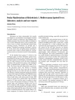
Figure 1 Proteoglycan incorporation and release during culture of cartilage explants (mean +/- SD). (a) Proteoglycan release during culture
of cartilage explants. A similar amount of proteoglycans were released into the culture medium by explants cultured with unstimulated (n = 24) or
conditioned serum (CS; n = 24). (b) PG incorporation rate on Days 4, 8 and 12 of the culture, measured using
35
SO
4
2-
incorporation (n = 8 per timepoint).
Results are representative of three separate experiments with different OA cartilage donor - serum donor combinations.
Ϭ
ϭ
Ϯ
ϯ
ϰ
ϱ
ϲ
ϳ
ϴ
ϵ
ĚĂLJϰ ĚĂLJϴ ĚĂLJϭϮ ĚĂLJϭϲ
ƵŐ''ƌĞůĞĂƐĞͬŵŐĐĂƌƚŝůĂŐĞ
ŽŶƚƌŽů
^
Ϭ
ϭ
Ϯ
ϯ
ϰ
ϱ
ϲ
ϳ
ĚĂLJϰ ĚĂLJϴ ĚĂLJϭϮ
ϯϱͲ^ŝŶĐŽƌƉŽƌĂƚŝŽŶ
;ŶŵŽůͬŚƌΎŵŐͿ
ŽŶƚƌŽů
^
A
B
^
^
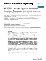
Figure 2 Proteoglycan and DNA content during culture of carti-
lage explants (mean +/- SD). Proteoglycan and DNA content of car-
tilage explants after culturing with unconditioned serum (n = 24) or
conditioned serum (CS, n = 24 cartilage explants). (a) Proteoglycan
content. (b) DNA content, (c) PG/DNA ratio.
Ϭ͘ϬϬ
Ϭ͘Ϭϱ
Ϭ͘ϭϬ
Ϭ͘ϭϱ
Ϭ͘ϮϬ
ŽŶƚƌŽů ^
ƵŐEͬŵŐĐĂƌƚŝůĂŐĞ
0
50
100
150
200
ŽŶƚƌŽů ^
ƵŐ''ͬƵŐE
Ϭ
ϱ
ϭϬ
ϭϱ
ϮϬ
Ϯϱ
ŽŶƚƌŽů ^
ƵŐ''ͬŵŐĐĂƌƚŝůĂŐĞ
A
C
B
^
^
^
Rutgers et al. Arthritis Research & Therapy 2010, 12:R114
/>Page 6 of 11
Figure 3 Proteoglycan incorporation and release during culture in unconditioned, conditioned or conditioned serum with Etanercept
(mean +/- SD). Proteoglycan release and incorporation (mean +/- SD) in the presence of unconditioned serum (control, n = 8), conditioned serum
(CS, n = 8) or conditioned serum with etanercept (n = 8). (a) Proteoglycan release, (b) Proteogycan incorporation on Day 4, measured by
35
SO
4
2-
in-
corporation (n = 8). Results are representative for three separate experiments with different OA cartilage donor - serum donor combinations.
Ϭ͘Ϭ
ϭ͘Ϭ
Ϯ͘Ϭ
ϯ͘Ϭ
ϰ͘Ϭ
ĚĂLJϰ ĚĂLJϴ ĚĂLJϭϮ ĚĂLJϭϲ
''ƌĞůĞĂƐĞ;ƵŐͿͬŵŐĐĂƌƚŝůĂŐĞ
ŽŶƚƌŽů
^
ƚĂŶĞƌĐĞƉƚ
Ϭ͘Ϭ
Ϭ͘ϱ
ϭ͘Ϭ
ϭ͘ϱ
Ϯ͘Ϭ
Ϯ͘ϱ
ϯ͘Ϭ
ϯ͘ϱ
ϰ͘Ϭ
ϰ͘ϱ
ŽŶƚƌŽů ^ ƚĂŶĞƌĐĞƉƚ
ϯϱͲ^ŝŶĐŽƌƉŽƌĂƚŝŽŶĚĂLJϰ
;ŶŵŽůͬŚƌΎŵŐĐĂƌƚŝůĂŐĞͿ
AB
^
^
Figure 4 Proteoglycan and DNA content during culture in unconditioned, conditioned serum or conditioned serum with Etanercept (mean
+/- SD). Proteoglycan (PG) metabolism in the presence of unconditioned serum (control, n = 8), conditioned serum (CS, n = 8) or conditioned serum
with etanercept (n = 8). (a) PG content, (b) DNA content, (c) PG/DNA ratio.
Ϭ͘ϬϬ
Ϭ͘Ϭϱ
Ϭ͘ϭϬ
Ϭ͘ϭϱ
Ϭ͘ϮϬ
Ϭ͘Ϯϱ
Ϭ͘ϯϬ
ŽŶƚƌŽů ^ ƚĂŶĞƌĐĞƉƚ
ƵŐEͬŵŐĐĂƌƚŝůĂŐĞ
Ϭ
ϱϬ
ϭϬϬ
ϭϱϬ
ϮϬϬ
ϮϱϬ
ŽŶƚƌŽů ^ ƚĂŶĞƌĐĞƉƚ
ƵŐ''ͬƵŐE
Ϭ
ϱ
ϭϬ
ϭϱ
ϮϬ
Ϯϱ
ŽŶƚƌŽů ^ ƚĂŶĞƌĐĞƉƚ
ƵŐ''ͬŵŐĐĂƌƚŝůĂŐĞ
A
C
B
^
^
^
Rutgers et al. Arthritis Research & Therapy 2010, 12:R114
/>Page 7 of 11
ogous conditioned serum (ACS, Orthokine
®
) proved
slightly to moderately effective for alleviation of OA
symptoms up to two years after treatment in human OA
patients [15,16], many aspects of this therapy have
remained unclear so far.
In vitro, conditioned serum does not seem to have a
direct effect on cartilage metabolism compared to
unstimulated serum. In line with earlier studies, IL-1ra
levels of ACS in the current study were upregulated,
although the reported relative increases in conditoned
serum differed an order of a magnitude with those from
the current study [12,17]. Also IL-10 levels were upregu-
lated two-fold as found earlier [4], but IL-4 was hardly
detectable. It is not known to what extent this is related to
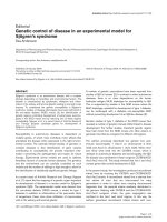
Figure 5 Effects of whole blood conditioning on serum cytokine levels of 22 OA patients. Cytokine levels in serum of 22 OA patients, before
incubation (control) and after six hours of incubation in the presence of glass beads (ACS). Note the increase in anti-inflammatory cytokines (IL-1ra,
TGF-β1, IL-10) and pro-inflammatory cytokines (IL-1β, IL-6, IFN-γ, OSM, TNF-α) after incubation. OPG levels were decreased. All values are displayed as
mean ± SD in pg/ml. * P < 0.01; ** P < 0.001.
Table 2: Clinical scores and serum cytokine levels in patients with sufficient and with non-sufficient SF for analysis
KOOS score KSCRS
score
Baseline serum cytokine levels
IL-1 IL-4 IL-6 IL-10 IL-13 TNFα IFNγ OSM OPG IL-1ra
SF available and analysed
(14 patients)
46
(10)
79
(19)
0.2
(0.7)
0.0
(0.0)
8.5
(28)
1.5
(2.7)
0.0
(0.1)
0.0
(0.2)
2.2
(2.2)
13
(27)
376
(128
)
180
(137)
No SF available
(8 patients)
51
(15)
75
(15)
0.2
(0.6)
0.0
(0.0)
0.1
(0.2)
0.1
(0.2)
0.1
(0.2)
14
(29)
16
(44)
14
(29)
371
(229
)
276
(288)
Pre-treatment clinical OA and baseline serum characteristics (mean +/- SD) of patients whose synovial fluid were analysed, were similar to those
of patients whose synovial fluid were not analysed due to insufficient amounts of SF in the knee at time of aspiration.
KOOS, Knee and Osteoarthritis Outcome Score; KSCRS, Knee Society Clinical Rating Scale; SF, synovial fluid; OA, osteoarthritis; IL-4, interleukin-4;
IL-6, interleukin-6; IL-10, interleukin-10; IL-13, interleukin-13; IFN-γ, interferon gamma; OSM, oncostatin M; TNF-α, tumor necrosis factor alpha;
OPG, osteoprotegerin; IL-1ra, interleukin-1 receptor antagonist.
Rutgers et al. Arthritis Research & Therapy 2010, 12:R114
/>Page 8 of 11
the change in the manufacturer's protocol, in which the
conditioning period is reduced to six hours [17], as
opposed to the 24 hours initially included in the prepara-
tion protocol [14,15]. Allegedly, most of the cytokine pro-
duction occurs after six hours. Moreover, although this
has not been argued as such by the manufacturer, long
incubation periods at body temperature are known to
reduce the bioactivity of most cytokines, while their
immunoreactivity as determined by ELISA is still
retained [38]. In particular Il-10, one of the anti-inflam-
matory cytokines upregulated in ACS, has been shown to
have a half-life of several hours under these conditions
[39]. This may also represent another explanation for the
limited effects found in vivo thus far. More important,
however, pro-inflammatory cytokines, in particular IL-1β
and TNF-α, were found to be significantly upregulated in
the current ACS study, in contrast to previous results
[17]. As, unlike for IL-1, the increased TNF-α levels were
not counterbalanced by an increase in levels of natural
inhibitors, and TNF-α has been postulated to have
degenerative effects on cartilage [21,40], this may have
explained the limited effects found in OA patients treated
with conditioned serum. However, blocking the action of
TNF-α [31] did not result in a net positive effect of condi-
tioned serum on matrix metabolism in vitro, suggesting
that, if any, in vivo effects of the TNFα in the injected
ACS would have been indirect. It is not clear to what
extent the increased levels of IL-6, OSM and lower levels
of OPG in conditioned serum may have had a pro-inflam-
matory effect, but as conditioned serum addition did not
result in decreased sulphate incorporation after four days
of cartilage explant culture, or a lower PG content after
16 days of culture, conditioning of serum is not likely to
have any effect on OA cartilage. These findings were
strengthened by the large number of explants used per
experiment, and by repeating both experiments in a total
of six different OA donors. Nevertheless, it cannot be
excluded that factors present in conditioned serum,
either known or as yet undiscovered, play a role in vivo by
inducing other mediators, not determined in the current
study, in the joint space. With respect to the role of Il-1
signalling in OA, in the one human clinical study using
recombinant IL-1ra as a treatment for OA, a single injec-
tion into the knee joint did not result in an improvement
of OA symptoms [41]. This may have been due to fast
clearance from the joint space. Injection of ACS led to an
increase of IL-1ra SF levels in osteoarthritic equine knee
joints during ACS treatment in vivo [14]. However, in the
Figure 6 Cytokine levels in control serum and ACS, and in synovial fluid during treatment. Control serum (control), autologous conditioned
serum (ACS) and synovial fluid cytokine levels (mean +/- SD) of IL-1RA, TGF-β1, IL-10, IL-6, OSM and OPG during treatment with ACS in 14 OA patients.
The large symbols next to the y-axis correspond to the levels of these cytokines in control serum and the injected ACS. TGF-β1 levels in SF were lower
than in the injected ACS, and OPG levels in SF were higher than in ACS (P < 0.01). During the course of treatment, no significant changes in cytokine
levels occurred despite repeated ACS injection (SF was aspirated before each of the six injections with ACS, at t = 0, Day 3, Day 7, Day 10, Day 14 and
Day 21).
1
10
100
1000
10000
100000
037101421
Day of aspiration
synovial
fluid
cytokine
level
log (pg/ml)
IL-1ra TGF-ȕ IL-10 IL-6 OSM OPG
*
ACS
SF
0 3 7 10 14 21
Day of aspiration
IL-1ra TGF-ȕ IL-10 IL-6 OSM OPG
*
ACS
SF
SF
ĺ
*
0 3 7 10 14 21
Day of aspiration
IL-1ra TGF-ȕ IL-10 IL-6 OSM OPG
S
SF
IL-1ra TGF-ȕ IL-10 IL-6 OSM OPG
Control
serum
,
ACS and SF
cytokine levels (pg/ml)
**
**
Control
*
0 3 7 10 14 21
ACS
Synovial Fluid (day of aspiration)
*
ĺ
**
**
Rutgers et al. Arthritis Research & Therapy 2010, 12:R114
/>Page 9 of 11
current study, IL-1ra levels did not increase during the
course of the treatment, even though the interval
between injection and measurement was shorter than in
the former study (3 days vs 7 and 35 days [14]). The fast
clearance of injected cytokines from the joint found in
the current study suggests that any in vitro net effect
would still have been difficult to reproduce in vivo. Con-
tinuous intraarticular availability of IL-1ra may be more
effective. In vivo injection of synoviocytes transduced
with the IL-1ra gene into a canine knee joint after sec-
tioning of the anterior cruciate ligament [11] and intraar-
ticular injection of IL-1ra plasmid into a rabbit knee joint
after meniscectomy resulted in reduction of OA clinical
symptoms (histological parameters, preservation of artic-
ular cartilage quality) [42]. Eventually these long term
approaches may be more effective than the limited num-
ber of injections of this treatment, but currently they are
not practically feasible in a clinical setting. Nevertheless,
even if IL-1ra levels are increased in the synovial fluid in
vivo, it is uncertain if these IL-1ra levels correlate with
OA symptoms or disease progression, as the ratio of IL-
1ra to IL-1β in the SF of human OA subjects were shown
not to correlate with pain or with the Lequesne OA index
[43].
With respect to the upregulation of IL-1β, its role in
progression of OA may actually be disputed [44]. In our
study, IL-1β levels in OA SF were extremely low, which is
in line with previous reports [34,45,46]. Although there
are studies in which IL-1β inhibited proteoglycan synthe-
sis in vitro at concentrations as low as 10 pg/ml [47],
commonly IL-1β concentrations of at least 1,000 pg/ml
are used to induce detectable cartilage damage [48,49].
Studies demonstrating synergistic effects of IL-1β with
IFN-γ, TNF-α, IL-17 or Oncostatin-M [50] also departed
from IL-1β concentrations much higher than detected in
OA synovial fluid and hence synergistic effects of low IL-
1β levels with other cytokines in the current study do not
seem likely. Moreover, the baseline IL-1ra levels in the SF
of the currently studied OA patients were already in the
effective range to block IL-1β [29]. Although it may be
argued that the patients with sufficient amounts of SF
may have differed from the group as a whole, this is con-
tradicted by the observation that serum cytokine levels as
well as the clinical response of the patients with sufficient
amounts of SF were similar to the patients with insuffi-
cient amounts of SF.
A high standard deviation in synovial fluid cytokine
levels was encountered, as is common in OA. Increasing
the number of subjects may decrease this standard devia-
tion and possibly enable subgroup analysis (for example,
by progression of OA according to radiological parame-
ters (dGEMRIC) or by clinical parameters (KOOS
scores). However, as the group of patients was already
small, this would have even further reduced the likeli-
hood of finding statistically significant changes.
Future evaluation of intraarticular cytokine changes
following ACS injection might include SF analysis earlier
after injection, which would give further insight on
intraarticular half-life. Also, effects of ACS on synovium,
either alone or in coculture with cartilage explants may
be studied. Even though multiplex ELISA showed good to
excellent correlation with ELISA [34], separate ELISAs
may give slightly more accurate information about abso-
lute cytokine levels.
Although the present in vitro data show no effect of
ACS and a short intraarticular half life, a recent clinical
study demonstrated a two-year lasting improvement of
ACS treatment compared to hyaluronic acid and placebo
treatment [51]. However, it must be noted that the treat-
ment regimens differed, with six injections of ACS being
compared to three injections of hyaluronic acid or pla-
cebo. Moreover, as none of the clinical trials carried out
so far included unconditioned serum as a placebo, it can
actually not be excluded that injection of serum without
prior conditioning per se has a beneficial effect on pro-
teoglycan metabolism in vivo.
Conclusions
In conclusion, ACS is a mix of counteracting growth fac-
tors and cytokines that does not have a direct effect on
cartilage metabolism and probably has a minimal influ-
ence in the joint space, given the fast disappearance of
cytokines from the synovial fluid after injection. Develop-
ment of new intraarticular therapies may focus on their
prolonged presence in the joint space.
Abbreviations
ACS: autologous conditioned serum; ASAP: ascorbic acid; DMOAD: disease-
modifying osteoarthritis-drug; GAGS: glycosaminoglycans; IFN-γ: interferon
gamma; IL-1ra: interleukin-1 receptor antagonist; IL-1β: interleukin-1 beta; IL-4:
interleukin-4; IL-6: interleukin-6; IL-10: interleukin-10; IL-13: interleukin-13; MMP:
matrix metalloproteinases; NO: nitric oxygen; OA: osteoarthritis; OPG: osteo-
protegerin; OSM: oncostatin M; PG: proteoglycan; PGE
2
: prostaglandin E
2
; SD:
standard deviation; SF: synovial fluid; TNF-α: tumor necrosis factor alpha; TGF-β:
transforming growth factor beta.
Competing interests
The authors declare that they have no competing interests.
Authors' contributions
MR carried out the in vitro and in vivo experiments, analysed and interpreted
the data and drafted the manuscript. DS participated in design and coordina-
tion of the study and revised the manuscript. WD was involved in design of the
study and revised the manuscript. LC conceived of the study and participated
in its design and coordination, aided with statistics and revised the manuscript.
All authors read and approved the final manuscript.
Acknowledgements
The authors wish to thank the Anna Foundation for Musculoskeletal Research
in The Netherlands, the Netherlands Organisation for Health Research and
Development (NWO) and the Dutch Arthritis Association (Reumafonds) for
their continuous support. The authors wish to thank W. de Jager, PhD for his
assistance with the multiplex ELISA and P. Westers, PhD for his statistical advice.
Rutgers et al. Arthritis Research & Therapy 2010, 12:R114
/>Page 10 of 11
Author Details
1
Department of Orthopaedics, University Medical Center Utrecht,
Heidelberglaan 100, 3584 CX Utrecht, The Netherlands and
2
Faculty of
Veterinary Sciences, Utrecht University, Yalelaan 1, De Uithof, 3584 CL Utrecht,
The Netherlands
References
1. Smith MD, Triantafillou S, Parker A, Youssef PP, Coleman M: Synovial
membrane inflammation and cytokine production in patients with
early osteoarthritis. J Rheumatol 1997, 24:365-371.
2. Goldring SR, Goldring MB: The role of cytokines in cartilage matrix
degeneration in osteoarthritis. Clin Orthop Relat Res 2004:S27-36.
3. Stove J, Huch K, Gunther KP, Scharf HP: Interleukin-1beta induces
different gene expression of stromelysin, aggrecan and tumor-
necrosis-factor-stimulated gene 6 in human osteoarthritic
chondrocytes in vitro. Pathobiology 2000, 68:144-149.
4. Goldring MB, Berenbaum F: The regulation of chondrocyte function by
proinflammatory mediators: prostaglandins and nitric oxide. Clin
Orthop Relat Res 2004, 427(suppl):S37-46.
5. Smith RL, Allison AC, Schurman DJ: Induction of articular cartilage
degradation by recombinant interleukin 1 alpha and 1 beta. Connect
Tissue Res 1989, 18:307-316.
6. Fernandes JC, Martel-Pelletier J, Pelletier JP: The role of cytokines in
osteoarthritis pathophysiology. Biorheology 2002, 39:237-246.
7. Martel-Pelletier J, McCollum R, DiBattista J, Faure MP, Chin JA, Fournier S,
Sarfati M, Pelletier JP: The interleukin-1 receptor in normal and
osteoarthritic human articular chondrocytes. Identification as the type
I receptor and analysis of binding kinetics and biologic function.
Arthritis Rheum 1992, 35:530-540.
8. Sadouk MB, Pelletier JP, Tardif G, Kiansa K, Cloutier JM, Martel-Pelletier J:
Human synovial fibroblasts coexpress IL-1 receptor type I and type II
mRNA. The increased level of the IL-1 receptor in osteoarthritic cells is
related to an increased level of the type I receptor. Lab Invest 1995,
73:347-355.
9. Caron JP, Fernandes JC, Martel-Pelletier J, Tardif G, Mineau F, Geng C,
Pelletier JP: Chondroprotective effect of intraarticular injections of
interleukin-1 receptor antagonist in experimental osteoarthritis.
Suppression of collagenase-1 expression. Arthritis Rheum 1996,
39:1535-1544.
10. Frisbie DD, Ghivizzani SC, Robbins PD, Evans CH, McIlwraith CW:
Treatment of experimental equine osteoarthritis by in vivo delivery of
the equine interleukin-1 receptor antagonist gene. Gene Ther 2002,
9:12-20.
11. Pelletier JP, Caron JP, Evans C, Robbins PD, Georgescu HI, Jovanovic D,
Fernandes JC, Martel-Pelletier J: In vivo suppression of early
experimental osteoarthritis by interleukin-1 receptor antagonist using
gene therapy. Arthritis Rheum 1997, 40:1012-1019.
12. Meijer H, Reinecke J, Becker C, Tholen G, Wehling P: The production of
anti-inflammatory cytokines in whole blood by physico-chemical
induction. Inflamm Res 2003, 52:404-407.
13. Arend WP, Leung DY: IgG induction of IL-1 receptor antagonist
production by human monocytes. Immunol Rev 1994, 139:71-78.
14. Frisbie DD, Kawcak CE, Werpy NM, Park RD, McIlwraith CW: Clinical,
biochemical, and histologic effects of intra-articular administration of
autologous conditioned serum in horses with experimentally induced
osteoarthritis. Am J Vet Res 2007, 68:290-296.
15. Yang KG, Raijmakers NJ, van Arkel ER, Caron JJ, Rijk PC, Willems WJ, Zijl JA,
Verbout AJ, Dhert WJ, Saris DB: Autologous interleukin-1 receptor
antagonist improves function and symptoms in osteoarthritis when
compared to placebo in a prospective randomized controlled trial.
Osteoarthritis Cartilage 2008, 16:498-505.
16. Baltzer AW, Moser C, Jansen SA, Krauspe R: Autologous conditioned
serum (Orthokine) is an effective treatment for knee osteoarthritis.
Osteoarthritis Cartilage 2009, 17:152-60.
17. Wehling P, Moser C, Frisbie D, McIlwraith CW, Kawcak CE, Krauspe R,
Reinecke JA: Autologous conditioned serum in the treatment of
orthopedic diseases: the orthokine therapy. BioDrugs 2007, 21:323-332.
18. Bos PK, Verhaar JA, van Osch GJ: Age-related differences in articular
cartilage wound healing: a potential role for transforming growth
factor beta1 in adult cartilage repair. Adv Exp Med Biol 2006,
585:297-309.
19. Mello MA, Tuan RS: Effects of TGF-beta1 and triiodothyronine on
cartilage maturation: in vitro analysis using long-term high-density
micromass cultures of chick embryonic limb mesenchymal cells. J
Orthop Res 2006, 24:2095-2105.
20. Malfait AM, Verbruggen G, Veys EM, Lambert J, De Ridder L, Cornelissen M:
Comparative and combined effects of interleukin 6, interleukin 1 beta,
and tumor necrosis factor alpha on proteoglycan metabolism of
human articular chondrocytes cultured in agarose. J Rheumatol 1994,
21:
314-320.
21. Westacott CI, Barakat AF, Wood L, Perry MJ, Neison P, Bisbinas I, Armstrong
L, Millar AB, Elson CJ: Tumor necrosis factor alpha can contribute to focal
loss of cartilage in osteoarthritis. Osteoarthritis Cartilage 2000,
8:213-221.
22. Flannery CR, Little CB, Hughes CE, Curtis CL, Caterson B, Jones SA: IL-6 and
its soluble receptor augment aggrecanase-mediated proteoglycan
catabolism in articular cartilage. Matrix Biol 2000, 19:549-553.
23. Henrotin YE, De Groote DD, Labasse AH, Gaspar SE, Zheng SX, Geenen VG,
Reginster JY: Effects of exogenous IL-1 beta, TNF alpha, IL-6, IL-8 and LIF
on cytokine production by human articular chondrocytes.
Osteoarthritis Cartilage 1996, 4:163-173.
24. de Waal Malefyt R, Figdor CG, Huijbens R, Mohan-Peterson S, Bennett B,
Culpepper J, Dang W, Zurawski G, de Vries JE: Effects of IL-13 on
phenotype, cytokine production, and cytotoxic function of human
monocytes. Comparison with IL-4 and modulation by IFN-gamma or
IL-10. J Immunol 1993, 151:6370-6381.
25. Kong YY, Feige U, Sarosi I, Bolon B, Tafuri A, Morony S, Capparelli C, Li J,
Elliott R, McCabe S, Wong T, Campagnuolo G, Moran E, Bogoch ER, Van G,
Nguyen LT, Ohashi PS, Lacey DL, Fish E, Boyle WJ, Penninger JM: Activated
T cells regulate bone loss and joint destruction in adjuvant arthritis
through osteoprotegerin ligand. Nature 1999, 402:304-309.
26. Shimizu S, Asou Y, Itoh S, Chung UI, Kawaguchi H, Shinomiya K, Muneta T:
Prevention of cartilage destruction with intraarticular
osteoclastogenesis inhibitory factor/osteoprotegerin in a murine
model of osteoarthritis. Arthritis Rheum 2007, 56:3358-3365.
27. Koshy PJ, Lundy CJ, Rowan AD, Porter S, Edwards DR, Hogan A, Clark IM,
Cawston TE: The modulation of matrix metalloproteinase and ADAM
gene expression in human chondrocytes by interleukin-1 and
oncostatin M: a time-course study using real-time quantitative reverse
transcription-polymerase chain reaction. Arthritis Rheum 2002,
46:961-967.
28. Henrotin YE, Zheng SX, Labasse AH, Deby GP, Crielaard JM, Reginster JY:
Modulation of human chondrocyte metabolism by recombinant
human interferon. Osteoarthritis Cartilage 2000, 8:474-482.
29. Arend WP, Welgus HG, Thompson RC, Eisenberg SP: Biological properties
of recombinant human monocyte-derived interleukin 1 receptor
antagonist. J Clin Invest 1990, 85:1694-1697.
30. Altman RD: Criteria for the classification of osteoarthritis of the knee
and hip. Scand J Rheumatol Suppl 1987, 65:31-39.
31. Vuolteenaho K, Moilanen T, Hamalainen M, Moilanen E: Effects of
TNFalpha-antagonists on nitric oxide production in human cartilage.
Osteoarthritis Cartilage 2002, 10:327-332.
32. Whiteman P: The quantitative measurement of Alcian Blue-
glycosaminoglycan complexes. Biochem J 1973, 131:343-350.
33. Kim YJ, Sah RL, Doong JY, Grodzinsky AJ: Fluorometric assay of DNA in
cartilage explants using Hoechst 33258. Anal Biochem 1988,
174:168-176.
34. de Jager W, te Velthuis H, Prakken BJ, Kuis W, Rijkers GT: Simultaneous
detection of 15 human cytokines in a single sample of stimulated
peripheral blood mononuclear cells. Clin Diagn Lab Immunol 2003,
10:133-139.
35. de Jager W, Prakken BJ, Bijlsma JW, Kuis W, Rijkers GT: Improved multiplex
immunoassay performance in human plasma and synovial fluid
following removal of interfering heterophilic antibodies. J Immunol
Methods 2005, 300:124-135.
36. Clegg DO, Reda DJ, Harris CL, Klein MA, O'Dell JR, Hooper MM, Bradley JD,
Bingham CO, Weisman MH, Jackson CG, Lane NE, Cush JJ, Moreland LW,
Schumacher HR Jr, Oddis CV, Wolfe F, Molitor JA, Yocum DE, Schnitzer TJ,
Furst DE, Sawitzke AD, Shi H, Brandt KD, Moskowitz RW, Williams HJ:
Glucosamine, chondroitin sulfate, and the two in combination for
painful knee osteoarthritis. N Engl J Med 2006, 354:795-808.
Received: 24 October 2009 Revised: 3 May 2010
Accepted: 10 June 2010 Published: 10 June 2010
This article is available from: 2010 Rutgers et al.; licensee BioMed Central Ltd. This is an open access article distributed under the terms of the Creative Commons Attribution License ( which permits unrestricted use, distribution, and reproduction in any medium, provided the original work is properly cited.Arthritis R esearch & Therapy 2010, 12:R114
Rutgers et al. Arthritis Research & Therapy 2010, 12:R114
/>Page 11 of 11
37. Juni P, Reichenbach S, Trelle S, Tschannen B, Wandel S, Jordi B, Zullig M,
Guetg R, Jorg Hauselmann H, Schwarz H, Theiler R, Ziswiler HR, Dieppe PA,
Villiger PM, Egger M: Efficacy and safety of intraarticular hylan or
hyaluronic acids for osteoarthritis of the knee: a randomized controlled
trial. Arthritis Rheum 2007, 56:3610-3619.
38. de Jager W, Bourcier K, Rijkers GT, Prakken BJ, Seyfert-Margolis V:
Prerequisites for cytokine measurements in clinical trials with
multiplex immunoassays. BMC Immunol 2009, 10:52.
39. Suga M, Keshavjee S, Liu M: Instability of cytokines at body
temperature. J Heart Lung Transplant 2005, 24:504-505.
40. Henderson B, Pettipher ER: Arthritogenic actions of recombinant IL-1
and tumour necrosis factor alpha in the rabbit: evidence for synergistic
interactions between cytokines in vivo. Clin Exp Immunol 1989,
75:306-310.
41. Joosten LA, Helsen MM, van de Loo FA, van den Berg WB: Anticytokine
treatment of established type II collagen-induced arthritis in DBA/1
mice. A comparative study using anti-TNF alpha, anti-IL-1 alpha/beta,
and IL-1Ra. Arthritis Rheum 1996, 39:797-809.
42. Fernandes J, Tardif G, Martel-Pelletier J, Lascau-Coman V, Dupuis M,
Moldovan F, Sheppard M, Krishnan BR, Pelletier JP: In vivo transfer of
interleukin-1 receptor antagonist gene in osteoarthritic rabbit knee
joints: prevention of osteoarthritis progression. Am J Pathol 1999,
154:1159-1169.
43. Richette P, Francois M, Vicaut E, Fitting C, Bardin T, Corvol M, Savouret JF,
Rannou F: A high interleukin 1 receptor antagonist/IL-1beta ratio
occurs naturally in knee osteoarthritis. J Rheumatol 2008, 35:1650-1654.
44. Chevalier X, Goupille P, Beaulieu AD, Burch FX, Conrozier T, Loeuille D,
Kivitz AJ, Silver D, Kiefer P, Zhou L, Bevirt T, Appleton B: Results from a
double blind, placebo-controlled, multicenter trial of a single intra-
articular injection of anakinra (kineret (R)) in patients with
osteoarthritis of the knee. Arthritis and Rheumatism 2005, 52:S507-S507.
45. Schlaak JF, Pfers I, Meyer Zum Buschenfelde KH, Marker-Hermann E:
Different cytokine profiles in the synovial fluid of patients with
osteoarthritis, rheumatoid arthritis and seronegative
spondylarthropathies. Clin Exp Rheumatol 1996, 14:155-162.
46. Vignon E, Balblanc JC, Mathieu P, Louisot P, Richard M: Metalloprotease
activity, phospholipase A2 activity and cytokine concentration in
osteoarthritis synovial fluids. Osteoarthritis Cartilage 1993, 1:115-120.
47. Huch K, Wilbrink B, Flechtenmacher J, Koepp HE, Aydelotte MB, Sampath
TK, Kuettner KE, Mollenhauer J, Thonar EJ: Effects of recombinant human
osteogenic protein 1 on the production of proteoglycan,
prostaglandin E2, and interleukin-1 receptor antagonist by human
articular chondrocytes cultured in the presence of interleukin-1beta.
Arthritis Rheum 1997, 40:2157-2161.
48. Shakibaei M, John T, Seifarth C, Mobasheri A: Resveratrol inhibits IL-1
beta-induced stimulation of caspase-3 and cleavage of PARP in human
articular chondrocytes in vitro. Ann N Y Acad Sci 2007, 1095:554-563.
49. Lippiello L: Glucosamine and chondroitin sulfate: biological response
modifiers of chondrocytes under simulated conditions of joint stress.
Osteoarthritis Cartilage 2003, 11:335-342.
50. Durigova M, Roughley PJ, Mort JS: Mechanism of proteoglycan
aggregate degradation in cartilage stimulated with oncostatin M.
Osteoarthritis Cartilage 2008, 16:98-104.
51. Baltzer AW, Moser C, Jansen SA, Krauspe R: Autologous conditioned
serum (Orthokine) is an effective treatment for knee osteoarthritis.
Osteoarthritis Cartilage 2009, 17:152-160.
doi: 10.1186/ar3050
Cite this article as: Rutgers et al., Cytokine profile of autologous conditioned
serum for treatment of osteoarthritis, in vitro effects on cartilage metabolism
and intra-articular levels after injection Arthritis Research & Therapy 2010,
12:R114