Báo cáo khoa học: "Changes in some Blood Micronutrients, Leukocytes and Neutrophil Expression of Adhesion Molecules in Periparturient Dairy Cows" ppt
Bạn đang xem bản rút gọn của tài liệu. Xem và tải ngay bản đầy đủ của tài liệu tại đây (129.3 KB, 12 trang )
Meglia GE, Johannisson A, Petersson L, Persson Waller K: Changes in some blood
micronutrients, leukocytes and neutrophil expression of adhesion molecules in
periparturient dairy cows. Acta vet. scand. 2001, 42, 139-150. – Dairy cows are
highly susceptible to infectious diseases, like mastitis, during the period around calving.
Although factors contributing to increased susceptibility to infection have not been fully
elucidated, impaired neutrophil recruitment to the site of infection and changes in the
concentrations of some micronutrients related with the function of the immune defence
has been implicated. Most of the current information is based on studies outside the
Nordic countries where the conditions for dairy cows are different. Therefore, the aim
of the study was to evaluate changes in blood concentrations of the vitamins A and E,
the minerals calcium (Ca), phosphorous (P), and magnesium (Mg), the electrolytes po-
tassium (K) and sodium (Na) and the trace elements selenium (Se), copper (Cu) and zinc
(Zn), as well as changes in total and differential white blood cell counts (WBC) and ex-
pression of the adhesion molecules CD62L and CD18 on blood neutrophils in Swedish
dairy cows during the period around calving. Blood samples were taken from 10 cows
one month before expected calving, at calving and one month after calving. The results
were mainly in line with reports from other countries. The concentrations of vitamins A
and E, and of Zn, Ca and P decreased significantly at calving, while Se, Cu, and Na in-
creased. Leukocytosis was detected at calving, mainly explained by neutrophilia, but
also by monocytosis. The numbers of lymphocytes tended to decrease at the same time.
The mean fluorescent intensity (MFI) of CD62L and CD18 molecules on blood neu-
trophils remained constant over time. The proportion of CD62L
+
neutrophils decreased
significantly at calving. The animals were fed according to, or above, their requirements.
Therefore, changes in blood levels of vitamins, minerals and trace elements were mainly
in response to colostrum formation, changes in dry matter intake, and ruminal metab-
olism around calving. Decreased levels of vitamins A and E, and of Zn at calving might
have negative implications for the functions of the immune defence. The lower propor-
tion of CD62L+ neutrophils at calving may result in less migration of blood neutroph-
ils into the tissues, and might contribute to the increased susceptibility to infections at
this time.
dairy cows; periparturient period; leukocytes; neutrophils; CD18; CD62L; vitamin A;
vitamin E; calcium; phosphorous; potassium; sodium; magnesium; selenium; cop-
per; zinc.
Acta vet. scand. 2001, 42, 139-150.
Acta vet. scand. vol. 42 no. 1, 2001
Changes in some Blood Micronutrients, Leukocytes
and Neutrophil Expression of Adhesion Molecules in
Periparturient Dairy Cows
By G.E. Meglia
1
, A. Johannisson
2
, L. Petersson
3
, and K. Persson Waller
1
1
Department of Obstetrics and Gynaecology, and
2
Department of Pathology, Faculty of Veterinary Medicine,
Swedish University of Agricultural Sciences,
3
Department of Chemistry, National Veterinary Institute (SVA),
Uppsala, Sweden.
Introduction
The susceptibility of dairy cows to infectious
diseases, like mastitis, is higher during the pe-
riod around calving than any other time. Host
resistance mechanisms are usually depressed
from approximately 3 weeks before calving un-
til 3 weeks after calving (Mallard et al. 1998).
Underlying mechanisms and factors have not
been fully explained. However, many metabolic
and hormonal changes take place during this
period, which may contribute to the impaired
immune defence (Smith et al. 1973, Va n
Kampen & Mallard 1997, Kehrli et al. 1998).
Changes in white blood cell counts are ob-
served around parturition, for example an in-
crease in the numbers of circulating neutrophils
(e.g. Guidry et al. 1976, Kehrli et al. 1989).
Neutrophils are considered the first line of cel-
lular defence against pathogens. However, at
calving, important neutrophil functions, like
migration and phagocytosis, are impaired (Hill
1981, Kehrli et al. 1989, Saad et al. 1989). Re-
duced migration of blood neutrophils can be ex-
plained by a lower expression of the adhesion
molecules CD62L (L-selectin) and CD11/
CD18, which are of vital importance for their
migration to the site of inflammation (Naga-
hata et al. 1995, Lee and Kehrli 1998).
The nutritional status of the animals has been
associated with the ability to resist infections.
Reports have shown a depression in the blood
levels of calcium (Ca), zinc (Zn), magnesium
(Mg), phosphorous (P), potassium (K), sele-
nium (Se), vitamins A and E during the peripar-
turient period (Johnston and Chew 1984, Goff
and Stabel 1990, Weiss et al. 1990, Dukes 1993,
Xin et al. 1993). Several of these nutrients are
important for the immune system. Increased in-
cidence of mastitis was reported at calving
when the concentrations of vitamins A and E
were decreased (Chew et al. 1982, Michal et al.
1994, Politis et al. 1995, Smith et al. 1997). Se-
lenium plays an important role in preventing
impaired function of the immune response
(Smith et al. 1997). Neutrophils from Se-defi-
cient animals were less capable of intracellular
killing of mastitis pathogens (Gyang et al.
1984, Smith et al. 1997). Cu deficiencies have
been shown to result in lowered bactericidal ac-
tivities of blood leukocytes in cattle and sheep
(Jones and Suttle 1981, Xin et al. 1991). More-
over, Harmon et al. (1998) reported a higher
proportion of uninfected quarters during the
peripartum period in Holstein heifers after ad-
ditional Cu supplementation. Zinc sufficiency
has also been linked to proper immune func-
tions, whereas deficiencies were related with ir-
regular immunological profiles (Hutcheson
1989, Reddy and Frey 1990).
Most of the available information in this field is
based on studies outside the Nordic countries
where the conditions for dairy cows are differ-
ent, for example in housing systems, feeding,
climate and management. Therefore, the aim of
this study was to evaluate leukocyte numbers
and the expression of the adhesion molecules
CD62L and CD18 on blood neutrophils, as well
as blood vitamins A and E, the minerals Ca, P
and Mg, the electrolytes K and Na, and the trace
elements Se, Cu and Zn, during the periparturi-
ent period in Swedish dairy cows. This would
also give baseline data for future studies in
which different management routines could be
compared.
Materials and methods
Animals
Ten healthy dairy cows of the Swedish Red and
White breed at the university farm were moni-
tored from one month before expected calving
to one month after calving. The animals were in
their second to sixth lactation and calved during
March and April. They were fed with grass si-
lage, concentrates and hay depending on their
stage of lactation (Table 1). The animals were
supplemented with 150 g/d of a commercial
mineral and vitamin mix. Samples of hay, con-
centrate and silage were frozen at –20ºC and
analysed for contents of vitamins, minerals and
trace elements. The total daily requirements
and allotments of nutrients are given in Table 2.
140 G. E. Meglia et al.
Acta vet. scand. vol. 42 no. 1, 2001
Experimental design
From each cow, jugular blood samples were
collected in the morning, using Vacutainer
®
tubes (Becton Dickinson Vacutainer Systems,
Meylan, France), one month before estimated
calving, at calving (within 24 hours after calv-
ing) and one month after calving. Before sam-
pling, the skin was cleaned with Milli-Q-water
(Milli-Q, Millipore Corp., Bedford, MA, USA).
Blood collected in a Zn-free vacutainer tube
without additives was used for serum analyses
of Zn, Cu, Ca, P, K, Na and Mg. Heparinized
blood was used for separation of plasma and
erythrocytes which was analysed for Se. Blood
without additives was taken for serum vitamin
E and vitamin A analysis. The tubes were cen-
trifuged at 1500 g for 35 min to get plasma or
serum, which was frozen at –20ºC until analy-
sis of the nutrients. Blood samples with EDTA
added were taken for neutrophil immunostain-
ing of CD18 and CD62L adhesion molecules,
and for total and differential white blood cell
counts.
Leukocyte counts
Total and differential leukocyte counts were de-
termined within 2 h using a Cell-Dyn
R
3500
(Abbott diagnostics, Abbott Laboratories, Ab-
bott Park, IL, USA) according to standard pro-
cedures at the Department of Clinical Chemis-
Changes in blood in periparturient cows 141
Acta vet. scand. vol. 42 no. 1, 2001
Table 1. Diet composition and estimated dry matter
intake (DMI) of 10 dairy cows one month before ex-
pected calving, at calving, and one month after calv-
ing, expressed in kilograms of dry matter and in per-
centage (%) of the total diet.
Before calving At calving After calving
Kg % Kg % kg %
Concentrate 1 12.5 4 36.4 14 58.3
Grass hay 0
1
00
1
0 1 4.2
Grass silage 7 87.5 7 63.6 9 37.5
DMI 8 100 11 100 24 100
1
The cows had access to straw
Table 2. Nutrient requirements according to NRC (National Research Council, 1989) and approximate daily
allotments to ten dairy cows one month before expected calving (-1), at calving (0), and one month after calv-
ing (+1), calculated on a body weight of 600 kg, and an average milk production of 30 l/day one month after
calving.
Daily requirements (NRC) Daily allotments
-1 0 +1 -1 0 +1
Energy ME
1
Mcal 16.3 30.8 65.0 21.5 31.4 71.9
Protein g 960 2090 3840 1237 1798 4068
Ca g 31.2 84.7 139.2 83.9 94.7 151.1
K g 52 110 216 217 243 409
Mg g 12.8 27.5 48 21.2 26.6 49.0
Na g 8 19.8 43.2 12.8 13.4 15.9
P g 19.2 52.8 88.8 45.4 61.9 124.2
Cu mg 80 110 240 246 291 473
Zn mg 320 440 960 1140 1236 1650
Se mg 2.4 3.3 7.2 7.1 7.2 7.9
Vit. A
2
mg 32000 44000 76800 -
2
Vit. E mg 120 165 360 865 917 1127
1
Metabolizable energy.
2
The carotenoid content in the feedstuffs was not determined in this study. However, the estimated allotment was above
NRC requirements.
try, Swedish University of Agricultural Scien-
ces, Uppsala, Sweden.
Polymorphonuclear leukocyte immunostaining
and flow cytometry analysis
For immunostaining with monoclonal antibod-
ies (mAb), erythrocytes were lysed with ammo-
nium chloride (NH
4
Cl) before the staining pro-
cedure, and washed three times with phosphate
buffered saline (PBS) without Ca and Mg. A
double staining procedure was used to identify
CD45
+
leukocytes bearing the other markers of
interest as described by Colditz et al. (1996).
The cell suspensions were labeled for flow cy-
tometry with CD45 (clone CACTB51A, Veteri-
nary Medical Research and Development
(VMRD), Pullman, WA, USA), and either
CD18 (clone BAQ30A, VMRD) or CD62L
(clone BAQ92A, VMRD). Two secondary anti-
bodies, goat anti-mouse IgG
1
FITC (Caltag La-
boratories, Burlingame, CA, USA), and goat
anti-mouse IgG
2a
PE (Caltag), were used. The
following controls were performed, blood with-
out antibodies and blood with primary mono-
clonal antibodies CD45 (clone CACTB51A,
VMRD) and a negative IgG
1
isotype control
(clone DAK-G01, DAKO, Glostrup, Denmark).
Finally, the cell pellet was fixed in 200 µl of 1%
paraformaldehyde in PBS and was stored in
darkness at 4ºC and analysed within a week.
Before analysis, cells were washed twice and
resuspended in PBS.
Stained cells were analysed on a FACStar Plus
flow cytometer (Becton Dickinson Immunocy-
tometry systems, Mountain View, CA, USA)
with standard optical equipment using an argon
ion laser at 200 mW tuned to 488 nm. The data
were acquired with a FACstation, with the soft-
ware Cellquest, version 1.2.2 (Becton Dickin-
son Immunocytometry Systems). Thirty thou-
sand events were collected. The following
parameters were obtained: forward light scatter
(FSC), orthogonal light scatter (SSC), FITC
fluorescence (FL1), and PE fluorescence (FL2).
Leukocytes were identified by their expression
of CD45, while their size (FSC) and granularity
(SSC) identified polymorphonuclear leuko-
cytes (PMNL). PMNL were gated to identify
the proportions of CD18
+
and CD62L
+
cells.
The discrimination between positive and nega-
tive cells was set using the isotype control. The
mean fluorescent intensity (MFI) of each cell in
FL1 was determined using quantum beads
(Flow Cytometry Standards Corporation, San
Juan, Puerto Rico).
Analysis of vitamins, minerals and trace
elements
Vitamin A and E were extracted from the serum
samples with hexan. The separation was done
by High Performance Liquid Chromatography
(HPLC) on a C18 colonn. Vitamin A and E
were determined by using ultraviolet and fluo-
rescence detection, respectively according to
standard procedures at the Department of
Chemistry, National Veterinary Institute, Upp-
sala, Sweden.
Serum samples were diluted (1:10) with ultra-
pure water (Milli-Q). The determination of Ca,
Cu, K, Mg, Na and Zn was performed using in-
ductively coupled plasma emission spectrome-
try (ICP-AES, Jobin Yvon 238 emission-spec-
trometer, Instruments S.A., Division Jobin
Yvon, Longjuemeau, France) with set-up and
conditions according to the method accredited
by SWEDAC (Swedish Board for Accreditation
and Conformity Assessment). Serum inorganic
phosphate (P) was determined according to
standard procedures at the Department of Clin-
ical Chemistry, Swedish University of Agricul-
tural Sciences, Uppsala, Sweden.
The Se concentrations in the plasma and eryth-
rocyte fractions were determined with flow in-
jection hydrid generation atomic absorption
spectrometry (FI-HG-AAS) after wet digestion
of the biological material with a mixture of ox-
142 G. E. Meglia et al.
Acta vet. scand. vol. 42 no. 1, 2001
idizing acids (Galgan and Frank 1993). Sele-
nium content in whole blood was calculated
from plasma Se and erythrocyte Se assuming
an average hematocrit content of 35% (Schalm
1986).
Statistical analysis
Analyses of variance for the concentrations of
nutrients and leukocytes, and the proportions
and MFI for the neutrophil adhesion molecules
were done using the General Linear Model
(SAS Institute Inc., Cary, NC, USA). The ef-
fects of cow and period were included in the
model. Mean fluorescent intensity for CD18
and CD62L were log-transformed. The results
are presented as least square means ± standard
Changes in blood in periparturient cows 143
Acta vet. scand. vol. 42 no. 1, 2001
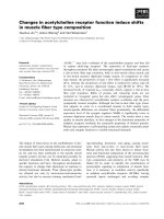
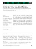
Figure 2. Proportions (%, LSM±SEM) of CD62L
+
and CD18
+
blood neutrophils in blood samples taken one
month before expected calving (-1), at calving (0), and one month after calving (+1) from ten dairy cows. Val-
ues with different letters within each parameter differ significantly (p<0.05).
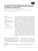
Figure 1. Numbers (×10
9
/l, LSM±SEM) of white blood cells (WBC), neutrophils (N), lymphocytes (L), and
monocytes (M) in blood samples taken one month before expected calving (-1), at calving (0), and one month
after calving (+1) from ten dairy cows. Values with different letters within each parameter differ significantly
(p<0.05).
x10
9
/l
error of the mean (LSM ± SEM). Probabilities
less than 0.05 were considered significant.
Results
Total and differential blood leukocytes
The total white blood cell counts (WBC) were
significantly (p<0.05) higher at parturition than
before and after calving (Figure 1). This was
mainly due to a significant increase in the num-
bers of neutrophils reaching values over the
normal range (0.6–4.0 × 10
9
/l) in 6 cows, and to
a lesser extent, to a significant increase in the
numbers of monocytes at this time point (Fig-
ure 1). The numbers of lymphocytes did not dif-
fer significantly between sampling occasions,
but was lower than the normal range (2.5–7.5 ×
10
9
/l) in 8 cows at calving (Figure 1).
Neutrophil adhesion molecules
Most neutrophils were positive for both CD18
and CD62L (Figure 2). The proportion of
CD18+ neutrophils remained fairly constant,
but was significantly (p<0.05) higher after calv-
ing than before calving. In contrast, the propor-
144 G. E. Meglia et al.
Acta vet. scand. vol. 42 no. 1, 2001
Figure 4. Serum concentrations of calcium (Ca), phosphorous (P), and potassium (K) (mmol/l, LSM±SEM)
in blood samples taken one month before expected calving (-1), at calving (0), and one month after calving (+1)
from ten dairy cows. Values with different letters within each parameter differ significantly (p<0.05).
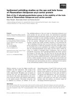
Figure 3. Serum concentrations of vitamins A and E (mg/l, LSM±SEM) in blood samples taken one month be-
fore expected calving (-1), at calving (0), and one month after calving (+1) from ten dairy cows. Samples with
different letters within each parameter differ significantly (p<0.05).
tion of CD62L
+
neutrophils decreased signifi-
cantly (p<0.05) at calving. A fairly large varia-
tion in proportion positive cells at calving ex-
plained the large standard error of means before
and after calving. The log MFI for CD62L and
CD18 on blood neutrophils was, on average,
11.63 ± 0.09 and 11.90 ± 0.11 at calving, re-
spectively, and did not change significantly dur-
ing the sampling period (data not shown).
Vitamins A and E
The serum concentrations of vitamins A and E
are shown in Figure 3. The level of vitamin A
changed significantly (p<0.001) over time. It
was significantly lower at parturition than be-
fore or after calving, reaching values (0.23 ±
0.02 mg/l) considered marginal (Puls 1995).
The levels of vitamin E did also tend (p=0.065)
to decrease at calving and was significantly
(p<0.05) higher one month after calving than
before and at calving.
Changes in blood in periparturient cows 145
Acta vet. scand. vol. 42 no. 1, 2001
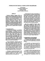
Figure 6. Whole blood Se (TSe), erythrocyte (ESe), and plasma selenium (PSe) concentrations (mg/kg,
LSM±SEM) in blood samples taken one month before expected calving (-1), at calving (0), and one month af-
ter calving (+1) from ten dairy cows. Values with different letters within each parameter differ significantly
(p<0.05).
Figure 5. Serum concentrations of zinc (Zn), and copper (Cu) (µmol/l, LSM±SEM) in blood samples taken
one month before expected calving (-1), at calving (0), and one month after calving (+1) from ten dairy cows.
Values with different letters within each parameter differ significantly (p<0.05).
Minerals, electrolytes and trace elements
The serum concentrations of Ca, P, K, Cu, Zn
and Se are shown in Figures 4-6. The levels of
Ca and Zn were significantly (p<0.05) lowered
at calving, reaching levels just under the refer-
ence values 2.1-2.7 mmol/l and 11-23 µmol/l,
respectively. In contrast, the serum concentra-
tion of Cu was significantly (p<0.05) higher at
calving and one month after calving (p<0.001)
compared with before calving. The P levels de-
creased significantly (p<0.05) at calving and re-
mained depressed after calving compared with
before calving. The K levels did not decrease
significantly (p<0.05) until after calving, reach-
ing values under the normal reference range of
4.0-5.6 mmol/l. The concentration of plasma,
erythrocytic and whole blood Se changed
slightly, but significantly, over time (Figure 6).
Plasma Se was significantly (p<0.05) higher at
calving compared with after calving, whereas
erythrocytic Se was significantly (p<0.05)
higher at calving than before calving. Whole
blood Se was significantly (p<0.001) higher at
parturition than before and after calving.
The Na concentrations were 138.3 ± 1.2, 142 ±
1.2 and 138 ± 1.2 mmol/l before, at and after
calving, respectively. The value at calving was
significantly (p<0.05) higher than at the other
time points. The Mg concentrations remained
fairly constant over time, at approximately 1.05
± 0.05 mmol/l.
Discussion
In agreement with earlier studies (e.g. Guidry et
al. 1976, Kehrli et al. 1989), we detected a sig-
nificant increase in the numbers of WBC at
calving. This was mainly due to an increase in
the numbers of circulating neutrophils, and to a
less extent, an increase in monocytes. At calv-
ing, the levels of corticosteroids are elevated
(Smith et al. 1973, Guidry et al. 1976). Corti-
costeroids induce neutrophilia by an increased
output of neutrophils from the bone marrow, by
neutrophil demargination from the blood vessel
wall, or by a combination of the two (Roth et al.
1982, Lee and Kehrli 1998). According to Lee
& Kehrli (1998), the neutrophil expression of
CD18 increases, while the expression of
CD62L decreases at calving. Such changes
were not observed in this study as the expres-
sion of CD62L and CD18 remained constant
over time. However, we observed a depression
in the proportion of CD62L
+
neutrophils at
calving, in accordance with Lee & Kehrli
(1998). Fewer cells expressing this molecule
means that the marginating pool of neutrophils,
rolling along the vessel wall, will shift to the
main blood flow stream contributing to the leu-
kocytosis. As a result, fewer neutrophils are
able to migrate into the tissues. In agreement
with Shafer-Weaver et al. (1996), we found that
the numbers of blood lymphocytes were re-
duced at calving. Alon et al. (1995) presented
the hypothesis that lymphocytes migrate in a
different manner than neutrophils, suggesting
that the high levels of cortisol detected at calv-
ing do not affect the adhesion molecules of
lymphocytes and therefore they can migrate
into the tissues.
Our results have shown a marked decline at
calving in serum concentrations of vitamins A
and E, and in Zn in agreement with earlier re-
ports (Johnston & Chew 1984, Goff & Stabel
1990, Weiss et al. 1990, Xin et al. 1993). A drop
in the serum concentrations of these nutrients is
associated with impaired immune functions
and a higher incidence of diseases, like mastitis
(Johnston & Chew 1984, Reddy & Frey 1990,
Michal et al. 1994, Smith et al. 1997).
The drop in serum concentrations of vitamins A
and E is largely due to colostrum formation
(Goff & Stabel 1990), but can also be due to
changes in dry matter intake and ruminal me-
tabolism (Weiss et al. 1994). Moreover, storage
and season can have negative effects on the
amount of vitamins A and E in the feedstuffs
146 G. E. Meglia et al.
Acta vet. scand. vol. 42 no. 1, 2001
(Goff & Stabel 1990, Miller et al. 1995, Puls
1994). Dry matter intake (DMI) can drop re-
markably during the week before calving (Ber-
tics et al. 1992, Grummer et al. 1995). As a re-
sult, reduced blood concentrations of nutrients
can be expected, especially as the nutrient de-
mands to initiate milk synthesis is increasing.
However, Olsson (1996) reported less reduction
in DMI before calving in Swedish dairy cows
fed high quality feedstuffs. Ruminal metab-
olism has been implicated in the destruction of
vitamin E (Shin & Owens 1990), but others
have suggested that ruminal vitamin E metab-
olism is essentially nil (Leedle et al. 1993,
Weiss et al. 1995). Vitamin E in blood is present
mainly as a component of lipoproteins. As par-
turition approaches, the liver secretion of lipo-
proteins decreases. As a consequence, its trans-
port capacity of vitamin E is lowered (Herdt &
Stowe 1991). However, the ruminal destruction
of vitamin A can be substantial and increases as
the level of concentrates in the diet is elevated
(Rode et al. 1990, Weiss et al. 1995).
The significant drop in serum Zn concentration
reported at calving, is most likely a conse-
quence of colostrum formation (Goff & Stabel
1990) and increased stress e.g. in association
with an acute phase response due to inflamma-
tory reactions in the uterus. Stress induces syn-
thesis of metallothionein, a protein associated
with Zn distribution. As a consequence, Zn is
redistributed from blood to other tissues, such
as the liver (Spears et al. 1991, Xin et al. 1993).
Physiological fluctuations occur immediately
before and after calving in the blood levels of
Ca, P, K and Na (Dukes 1993). Blood levels of
Ca and P is expected to decrease at calving due
to the large demand of colostrum and milk pro-
duction. In agreement with Forar et al. (1982),
we detected a reduced blood P concentration
one month after calving. There is an inverse re-
lationship between milk production and plasma
P concentration (Forar et al. 1982). The K val-
ues were also depressed one month after calv-
ing, which might be related with K being the
major cation secreted into the milk of cattle
(Underwood & Suttle 1999).
The blood Cu status undergoes several changes
during the periparturient period. The lower
value before calving could be due to the drain-
age by the fetal liver (Xin et al. 1993). In con-
trast to other reports (Hidiroglou & Knipfel
1981, Xin et al. 1993), an increased blood level
of Cu was detected at calving in this study. Wa r d
& Spears (1999) suggest that cattle undergoing
stressful periods have increased blood levels of
Cu and ceruloplasmin, as Cu transport protein.
Ceruloplasmin is considered an acute phase pro-
tein and its concentration increase in response to
injury, infections and inflammation (Conner et
al. 1986). This might be one reason for the in-
creased blood level of this nutrient, as calving is
considered a stressful period with tissue dam-
ages for example in the uterus.
There is a relationship between the Se status of
the animals around parturition and the func-
tions of the immune system and disease resis-
tance (Gyang et al. 1984, Smith et al. 1997).
From these studies it can be concluded that ben-
eficial effects of Se supplementation occur only
when the animals are Se deficient. Whole blood
Se levels in the range of 0.1-0.2 mg/l could be
considered optimal from immunological stand-
point (Koller et al. 1983, Jukola et al. 1996). In
this study, whole blood Se concentrations were
in the range of 0.167-0.180 mg/kg, i.e. accord-
ing to recommendations. Weiss et al. (1990) hy-
pothesised that the increase in the level of Se at
calving may be related to the high fragility of
the red blood cells detected at calving.
In conclusion, the results obtained under Swed-
ish conditions were mainly in line with earlier
reports. At calving, leukocytosis due to neutro-
philia and monocytosis was detected. A lower
proportion of CD62L
+
neutrophils at calving
suggests that fewer of these cells can migrate
Changes in blood in periparturient cows 147
Acta vet. scand. vol. 42 no. 1, 2001
into the tissues with negative consequences for
the defence against infections. Moreover, re-
duced concentrations of vitamins A and E, and
the trace element Zn, were observed at this
time. This can also have negative effects on the
functions of the immune system resulting in in-
creased susceptibility to diseases, such as mas-
titis. Despite the fact that vitamin A was fed ac-
cording to recommendations, and vitamin E
above recommendations, the levels of both nu-
trients decreased at calving. Vitamin A levels
dropped below the normal reference value,
while vitamin E levels remained within the nor-
mal range. Several authors (Smith et al. 1984,
Michal et al. 1994, Weiss 1998) reported im-
provements in milk production, immune func-
tions and mammary gland health when addi-
tional vitamins A and E were given compared
with NRC (National Research Council 1989)
recommendations. Results from the present
study, in combination with aforementioned
data, suggest that the NRC vitamin A and E rec-
ommendations may not be adequate, at least not
around calving. However, further studies are
needed to evaluate whether the low blood val-
ues reflect a true body deficiency and the im-
portance of decreased absorption of vitamins
during this period.
Acknowledgements
The authors thank Inga-Lena Örde-Öström, Gabor
Sellei, and Anna Stepinska, National Veterinary In-
stitute (SVA), Uppsala, Sweden for skilful technical
assistance.
References
Alon R, Kassner PD, Woldemar Carr M, Finger EB,
Hemler ME, Springer TA: The integrin VLA-4
supports tethering and rolling in flow on VCAM-
1. J. Cell Biol. 1995, 128, 1243-1253.
Bertics SJ, Grummer RR, Cadorniga-Valino C, Stod-
dard EE: Effect of prepartum dry matter intake
on liver triglyceride concentration and early lac-
tation. J. Dairy Sci., 1992, 75, 1914-1922.
Chew BP, Hollen LL, Hillers JK, Herlugson ML: Re-
lationship between vitamin A and ß-carotene in
blood plasma and milk and mastitis in Holsteins.
J. Dairy Sci., 1982, 65, 2111-2118.
Colditz IG, Eisemann CH, Tellam RL, McClure SJ,
Mortimer SI, Husband AJ: Growth of Lucilia cu-
prina larvae following treatment of sheep diver-
gently selected for fleece rot and fly strike with
monoclonal antibodies to T lymphocyte subsets
and interferon ␥ Int. J. Parasitol., 1996, 26, 775-
782.
Conner JG, Eckersall PD, Doherty M, Douglas TA:
Acute phase response and mastitis in the cow.
Res. Vet. Sci., 1986, 41, 126-128.
Dukes HH: Physiology of domestic animals. 11
th
ed.
M. J. Swenson, W. O. Reece Editors. Cornell Uni-
versity Press. Ithaca and London, 962 pp, 1993.
Forar FL, Kincaid RL, Preston RL, Hillers JK: Vari-
ation of inorganic phosphorous in blood plasma
and milk of lactating cows. J. Dairy Sci., 1982,
65‚ 760-763.
Galgan V, Frank A: Notes and comments of the deter-
mination of selenium in biological materials.
Now. J. Anim. Sci., 1993, 11, 57-74.
Goff JP, Stabel JR: Decreased plasma retinol, α-to-
copherol, and zinc concentration during the peri-
partum period: effect of milk fever. J. Dairy Sci.,
1990, 73, 3195-3199.
Grummer RR, Hoffman PC, Luck ML, Bertics SJ: Ef-
fect of prepartum and postpartum dietary energy
on growth and lactation of primiparous cows. J.
Dairy Sci., 1995, 78, 172-180.
Guidry AJ, Paape MJ, Pearson RE: Effects of partu-
rition and lactation on blood and milk cell con-
centrations, corticosteroids, and neutrophil phag-
ocytosis in the cow. Am. J. Vet. Res., 1976, 37,
1195-1200.
Gyang EA, Stevens JB, Olson WG, Tsitsamis SD,
Usenik EA: Effects of selenium-vitamin E injec-
tion on bovine polymorphonucleated leukocytes
phagocytosis and killing Staphylococcus aureus.
Am. J. Vet. Res., 1984, 45, 175-177.
Harmon RJ, Trammell DS, Smith BA, Scaletti RW: Ef-
fects of dietary copper insufficiency and sources
of dietary copper on copper status and intramam-
mary infections at calving. J. Dairy Sci., 1998, 81
(Suppl. 1): 43.
Herdt TH, Stowe HD: Fat-soluble vitamin nutrition
for dairy cattle. Vet. Clin. North Am. Food Anim.
Prac., 1991, 7, 391-415.
Hidiroglou M, Knipfel JE: Maternal-fetal relation-
148 G. E. Meglia et al.
Acta vet. scand. vol. 42 no. 1, 2001
ships of copper, manganese, and sulfur in rumi-
nants. A review. J. Dairy Sci., 1981, 64, 1637-
1647.
Hill AW: Factors influencing the outcome of Escheri-
chia coli mastitis in the dairy cows. Res. Vet. Sci.,
1981, 31, 107-112.
Hutcheson DP: Nutritional factors affect immune re-
sponse in cattle. Feedstuffs, 1989, 61, 16-24.
Johnston LA, Chew BP: Peripartum changes of
plasma and milk vitamin A and ß-carotene
among dairy cows with or without mastitis. J.
Dairy Sci., 1984, 67, 1832-1840.
Jones DG, Suttle NF: Some effects of copper defi-
ciency on leucocyte function in sheep and cattle.
Res. Vet. Sci., 1981, 31, 151-156.
Jukola E, Hakkarainen J, Saloniemi H, Sankari S:
Blood selenium, vitamin E, vitamin A, and ß-car-
otene concentrations and udder health, fertility
treatments, and fertility. J. Dairy Sci., 1996, 79,
838-845.
Kehrli Jr ME, Nonnecke BJ, Roth JA: Alterations in
bovine neutrophil function during the peripartum
period. Am. J. Vet. Res., 1989, 50, 207-214.
Kehrli Jr ME, Kimura K, Goff JP, Stabel JR, Non-
necke BJ: Periparturient immunosuppression in
dairy cows: nutrition and lactation effects. Pro-
duction Diseases in Farm Animals. 10
th
Interna-
tional Conference. Ed. Th.Wensing. The Nether-
lands, 1998.
Koller LD, South PJ, Exon JH, Whitbeck GA: Sele-
nium deficiency of beef cattle in Idaho and Wash-
ington and a practical means of prevention. Corn.
Vet., 1983, 73, 323-332.
Lee E-K, Kehrli M: Expression of adhesion mole-
cules on neutrophils of periparturient cows and
neonatal calves. Am. J. Vet. Res., 1998, 59, 37-
43.
Leedle RA, Leedle JAZ, Butine MD: Vitamin E is not
degraded by ruminal microorganisms: assess-
ment with ruminal contents from a steer fed a
high-concentrate diet. J. Anim. Sci., 1993, 71,
3442-3450.
Mallard BA, Dekkers JC, Ireland MJ, Leslie KE,
Sharif S, Lacey Vanjampen C, Wagter L, Wilkie
N: Alteration in immune responsiveness during
the peripartum period and its ramification on
dairy cow and calf health. J. Dairy Sci., 1998, 81,
585-595.
Michal JJ, Heirman LR, Wong TS, Chew BP, Frigg M,
Volker L: Modulatory effects of dietary ß-caro-
tene on blood and mammary leukocyte function
in periparturient dairy cows. J. Dairy Sci., 1994,
77, 1408-1421.
Miller GY, Bartlett PC, Erskine RJ, Smith KL: Factors
affecting serum selenium and vitamin E concen-
trations in dairy cows. JAVMA, 1995, 206, 1369-
1373.
Nagahata H, Nochi H, Tamoto K, Noda H, Kociba
GJ: Expression and role of adhesion molecule
CD18 on bovine neutrophils. Can. J. Vet. Res.,
1995, 59, 1-7.
National Research Council: 1989. Nutrient require-
ment of dairy cattle. 6th rev. Ed. Natl. Acad.
Press, Washington, DC, 1989.
Olsson G: Effects of feeding strategy before calving
on dairy cow performance. Swedish University
of Agricultural Sciences, Department of Animal
Nutrition and Management, 1996, Doctoral The-
sis.
Politis I, Hidiroglou M, Batra TR, Gilmore JA, Go-
rewit RC, Scherf H: Effects of vitamin E on im-
mune function of dairy cows. Am. J. Vet. Res.,
1995, 56, 179-184.
Puls R: Vitamin levels in animal health. 1
st
Ed.
Sherpa International, 1994.
Reddy PG, Frey RA: Nutritional modulation of im-
munity in domestic food animals. Adv. Vet. Sci.
Comp. Med., 1990, 35, 255-281.
Rode LM, McAllister TA, Cheng K-J: Microbial deg-
radation of vitamin A in rumen fluid from steers
fed concentrate, hay or straw diets. Can. J. Anim.
Sci., 1990, 70, 227.
Roth JA, Kaeberle ML, Hsu WH: Effects of ACTH
administration on bovine polymorphonuclear
leukocyte function and lymphocyte blastogene-
sis. Am. J Vet. Med., 1982, 43, 412-416.
Saad AM, Concha C, Åström G: Alteration in neu-
trophil phagocytosis and lymphocyte blastogene-
sis in dairy cows around parturition. J. Vet. Med.,
1989, 36, 337-345.
Schalm O, Cattle W: Normal hematology with com-
ments on response to disease. In: N. C. Jain (Ed-
itor), Veterinary Hematology. Philadelphia, 1986.
Shafer-Weaver KA, Pighetti GM, Sordillo LM: Di-
minished mammary gland lymphocyte functions
parallel shifts in trafficking patterns during the
postpartum period. Proc. Soc. Exp. Biol. Med.,
1996, 212, 271-279.
Shin IS, Owens FN: Ruminal and intestinal disap-
pearance of several sources of vitamin E. (Abstr.)
J. Anim. Sci., 1990, 68(Suppl. 1): 544.
Smith VG, Edgerton LA, Hafs HD, Convey EM: Bo-
vine serum estrogens, progestins and glucocorti-
coids during late pregnancy parturition and early
Changes in blood in periparturient cows 149
Acta vet. scand. vol. 42 no. 1, 2001
lactation. J. Anim. Sci., 1973, 36, 391-396.
Smith KL, Harrison JH, Hancock DD, Todhunter DA,
Conrad HR: Effect of vitamin E and selenium
supplementation on incidence of clinical mastitis
and duration of clinical symptoms. J. Dairy Sci.,
1984, 67, 1293-1300.
Smith KL, Hogan JS, Weiss WP: Dietary vitamin E
and selenium affect mastitis and milk quality. J.
Anim. Sci., 1997, 75, 1659-1665.
Spears JW, Harvey RW, Brown TT: Effects of zinc me-
thionine and zinc oxide on performance, blood
characteristics, and antibody titer response to vi-
ral vaccination in stressed feeder calves. JAVMA,
1991, 199, 1731-1733.
Underwood EJ, Suttle NF: The mineral nutrition of
livestock. 3
rd
Ed. CAB International, UK, pp 614,
1999.
Van Kampen C, Mallard BA: Effects of peripartum
stress and health on circulating bovine lympho-
cyte subsets. Vet. Immunol Immunopathol.,
1997, 59, 79-91.
Ward JD, Spears. JW: The effect of low-copper diets
with or without supplemental molybdenum on
specific immune responses of stressed cattle. J.
Anim. Sci., 1999, 77, 230-237.
Weiss WP: Requirements of fat-soluble vitamins for
dairy cows: A review. J. Dairy Sci., 1998, 81,
2493-2501.
Weiss WP, Hogan JS, Smith KL, Hoblet KH: Relation-
ships among selenium, vitamin E, and mammary
gland health in commercial dairy herds. J. Dairy
Sci., 1990, 73, 381-390.
Weiss WP, Hogan JS, Smith KL, Williams SN: Effect
of dietary fat and vitamin E on α-tocopherol and
ß-carotene in blood of peripartum cows. J. Dairy
Sci., 1994, 77, 1422-1429.
Weiss WP, Smith KL, Hogan JS, Steiner TE: Effect of
forage to concentrate ratio on disappearance of
vitamin A and E during in vitro ruminal fermen-
tation. J. Dairy Sci., 1995, 78, 1837-1842.
Xin Z, Waterman DF, Hemken RW, Harmon RJ: Ef-
fects of copper status on neutrophil function,
superoxide dismutase and copper distribution in
steers. J. Dairy Sci., 1991, 74, 3078-3085.
Xin Z, Waterman DF, Hemken RW, Harmon RJ: Cop-
per status and requirement during the dry period
and early lactation in multiparous Holstein cows.
J. Dairy Sci., 1993, 76, 2711- 2716.
Sammanfattning
Förändringar i vissa mikronäringsämnen, leukocyter
och uttryck av adhesionsmolekyler på neutrofiler i
blodet hos peripartala mjölkkor.
Mjölkkor är mycket känsliga för infektionssjukdo-
mar, som mastit, under perioden runt kalvningen. Det
är inte helt klarlagt vilka faktorer som bidrar till den
ökade infektionskänsligheten men försämrad rekry-
tering av neutrofiler till infektionsplatsen och för-
ändringar i koncentrationen av vissa mikroelement
har troligen betydelse. De flesta befintliga uppgifter
baseras på studier gjorda utanför Norden där
förhållandena för mjölkkor är annorlunda. Syftet
med denna studie var därför att undersöka för-
ändringar i blodkoncentrationen av vitaminerna A
och E, mineralerna kalcium (Ca), fosfor (P) och mag-
nesium (Mg), elektrolyterna kalium (K) och natrium
(Na) samt spårämnena selen (Se), koppar (Cu) och
zink (Zn) hos svenska mjölkkor under perioden runt
kalvning. Dessutom undersöktes förändringar i anta-
let vita blodkroppar och uttrycket av adhesionsmo-
lekylerna CD62L och CD18 på blodneutrofiler. Re-
sultaten stämde huvudsakligen med tidigare rap-
porter från andra länder. Koncentrationen av vitami-
nerna A och E, Zn, Ca och P minskade signifikant vid
kalvning medan Se, Cu och Na ökade. Leukocytos
sågs vid kalvning huvudsakligen orsakat av neutro-
fili men också till viss del monocytos. Antalet lymfo-
cyter tenderade att minska vid samma tidpunkt.
Medelvärdet av fluorescensintensiteten för CD62L-
och CD18-molekylerna på blodneutrofiler var kon-
stant över tiden. Andelen CD62L+ neutrofiler min-
skade dock signifikant vid kalvningen. Djuren utfo-
drades i enlighet med eller över deras behov varför
förändringarna i blodnivåerna av vitaminer, miner-
aler och spårämnen huvudsakligen var ett resultat av
kolostrumproduktion, samt förändringar i torrsub-
stansintag och våmmetabolism runt kalvningen.
Minskade nivåer av vitaminerna A och E och av Zn
vid kalvning kan ha negativ effekt på immun-
försvarets funktion. Den lägre andelen CD62L+ neu-
trofiler vid kalvning kan resultera i en försämrad mi-
150 G. E. Meglia et al.
Acta vet. scand. vol. 42 no. 1, 2001
(Received August 14, 2000, accepted October 20, 2000).
Reprints may be obtained from: K. Persson Waller, National Veterinary Institute (SVA) Department of Ruminant
and Porcine Diseases SE-75189 Uppsala, Sweden. E-mail: , tel: +46 (0) 18 67 40
00, fax: +46 (0) 18 30 91 62.