Báo cáo khoa học: " Serological Investigation of Granulocytic Ehrlichia Infection in Sheep in Norway" pot
Bạn đang xem bản rút gọn của tài liệu. Xem và tải ngay bản đầy đủ của tài liệu tại đây (89.42 KB, 8 trang )
Stuen S, Bergström K: Serological investigation of granulocytic Ehrlichia infection
in sheep in Norway. Acta vet. scand. 2001, 42, 331-338. – Serum samples of 749 sheep
from 75 sheep flocks in Norway, i.e. 361 lambs (6 to 7 months old) and 388 adults (>1.5
year), were analysed for antibodies to Ehrlichia equi. Ten animals from each flock were
examined. Seropositive animals were found along the coast of southern Norway from
Vestfold to Sør-Trøndelag (as far north as 63°38´N). Seropositive sheep were not found
in southeast, east or northern Norway. Thirty-two flocks were seropositive, although
tick-borne fever had only been diagnosed earlier in half of these. In 78% of the seropos-
itive flocks, more than 80% of the sheep were seropositive. A total of 35.7 % and 36.3
% of lambs and adults were found seropositive, respectively. However, the overall sero-
prevalence among animals that had been grazing on Ixodes pastures were 0.80 for the
lambs and 0.84 for the adults. Mean antibody titres (± SD) (log
10
) in seropositive lambs
and adults were 2.59 (± 0.449) and 2.70 (± 0.481), respectively. No significant differ-
ences in either seroprevalence or mean antibody titre between sheep of different ages
were obtained in this study. Based on antibodies 94% of sheep flocks on Ixodes pastures
were infected with a granulocytic Ehrlichia infection. The association between seropos-
itive flocks and Ixodes infested pasture shows a very high degree of agreement
(p<0.00001). The present study indicates that granulocytic Ehrlichia infection in sheep
is underdiagnosed in Norway.
Ehrlichia phagocytophila; antibodies; lambs; seroprevalence.
Acta vet. scand. 2001, 42, 331-338.
Acta vet. scand. vol. 42 no. 3, 2001
Serological Investigation of Granulocytic Ehrlichia
Infection in Sheep in Norway
By S. Stuen
1
and K. Bergström
2
1
Norwegian School of Veterinary Science, Department of Sheep and Goat Research, Sandnes, Norway, and
2
National Veterinary Institute, Department of Bacteriology, Uppsala, Sweden.
Introduction
The most common tick-borne disease in do-
mestic animals in Norway is tick-borne fever
(TBF), caused by Ehrlichia phagocytophila,
and transmitted by the tick Ixodes ricinus
(Øverås 1972, Stuen 1997). TBF may cause
abortion in ewes and temporary infertility in
rams (Woldehiwet & Scott 1993), but the main
consequence of an E. phagocytophila infection
in sheep is the ensuing immunosuppresion that
leads to secondary infections, such as Staphylo-
coccus aureus pyaemia and Pasteurella
hemolytica (trehalosi) septicaemia (Brodie et
al. 1986, Stuen 1996). In the UK, it has been es-
timated that more than 300 000 lambs develop
tick pyaemia annually (Brodie et al. 1986).
TBF has for decades been considered as an im-
portant disease in lambs in certain areas along
the coast of southern Norway (Stuen 1998). The
purpose of the present study was to investigate
the distribution of E. phagocytophila infection
in sheep in different areas of Norway, especially
in areas with a distribution of I. ricinus.
Materials and methods
Flocks from each county in Norway were in-
cluded in this study, such that flocks in Ixodes
areas along the coast and areas with a high
number of winterfed sheep were preferred.
However, representative flocks in each area
were chosen and sampled by the local veteri-
narians.
Serum samples from sheep flocks were ob-
tained in October/November. Samples from 10
sheep were randomly collected in each herd,
around half of the samples were from lambs (6
to 7-months-old). A questionaire was filled out
by the veterinarian during the visit of each
flock, including questions about ectoparasitic
treatment, Ixodes infested pastures, earlier
treatment against TBF, and occurrence of tick-
associated infections. Four sheep flocks were
chosen from each of the 18 counties in Norway,
except from the county of Sør-Trøndelag,
where 8 flocks were selected. The reason for
this was that the northernmost observation of
tick-borne fever so far has been in the county of
Sør-Trøndelag (Stuen 1997).
An indirect immunofluorescence antibody as-
say (IFA) was used to determine the antibody
titre to Ehrlichia equi (Artursson et al. 1999).
Two-fold dilutions of sera were added to slides
precoated with E. equi antigen (Protatek Inter-
national and Organon Teknika). Bound anti-
bodies were visualized by fluorescein-isothio-
cyanate (FITC)-conjugated rabbit-anti-sheep
immunoglobulin (Cappel, Organon Teknika).
Sera were screened for antibodies at dilution
1:40. If positive, the serum was further diluted
and retested. A titre of 1.6 (log
10
reciprocal of
1:40) or more was regarded as positive.
The statistical analysis was done according to
Martin et al. (1987). The overall seroprevalence
and mean antibody titre were estimated and
stratified by ectoparasitic treatment and age.
Statistical calculations were done by using
Statistix
®
, version 4.0 (Analytical software).
Statistical analyses on seroprevalence were per-
formed using a chi-square test and the antibody
titres were compared using a Students t-test for
independent samples. Significance was set at
p<0.05.
Results
Of a total of 749 sheep from 75 flocks, 71 flocks
in 1996 and 4 flocks in 1997, 270 sheep (36%)
were found positive for antibodies to granulo-
cytic Ehrlichia infection. Seropositive flocks
were found in the coastal areas from Vestfold to
Sør-Trøndelag. The northernmost seropositive
flocks were found south of Trondheimsfjorden
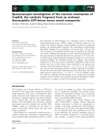
on the island of Hitra (63°38´N). The geo-
graphical distribution of the flocks is shown in
Fig. 1. Thirty-two flocks were found positive,
but only 16 of these had a history of previous
332 S. Stuen & K. Bergström
Acta vet. scand. vol. 42 no. 3, 2001
Figure 1. Geographical distribution of sheep flocks
examined for antibodies to Ehrlichia equi in Norway.
A titre less than 1:40 was considered negative.
᭹ - seropositive flock, ᭺ - seronegative flock
Aust-Agder
TBF infection (Table 1). Symptoms of disease
were not observed in any sampled animal.
Clinical symptoms indicating a TBF infection,
such as arthritis, polyarthritis and sudden death,
were observed in only 6 flocks (8%); 4 of these
had been prophylatically treated with insecti-
cides. Twenty flocks were given prophylactic
treatment against ticks with insecticides / repel-
lents (mainly synthetical pyrethroids); lambs
and adults were treated in 15 flocks, while only
lambs were treated in 5 flocks.
In 78% of the seropositive flocks, more than
80% of the sheep were seropositive and in 91%
of the flocks, more than half of the animals were
seropositive (Table 2).
The antibody titres in 361 lambs and 388 adults
(>1.5 years) were recorded. A total of 129 of the
lambs (35.7% ) and 141 of the adults (36.3%)
were found seropositive (Table 3). However,
among animals that had been grazing on tick in-
fested pasture, 79.6% and 83.9% of lambs and
Serology of granulocytic Ehrlichia infection 333
Acta vet. scand. vol. 42 no. 3, 2001
Table 1. Serological investigation of sheep sera for antibodies to Ehrlichia equi from different counties of Nor-
way.
Number of Number of flocks
positive flocks / Number of Number of with a history of
County total flocks on flocks treated tick-borne fever
number of tick pasture against ticks (during the year
flocks of sampling)
Akershus # 0 / 4 0 0 0 (0)
Aust-Agder 4 / 4 4 3 2 (0)
Buskerud # 0 / 4 0 0 0 (0)
Finnmark # 0 / 4 0 0 0 (0)
Hedmark # 0 / 4 0 0 0 (0)
Hordaland 4 / 4 4 3 0 (0)
Møre og Romsdal 4 / 4 4 1 4 (2)
Nordland 0 / 4 0 0 0 (0)
Nord-Trøndelag 0 / 4 0 0 0 (0)
Oppland # 0 / 4 0 0 0 (0)
Rogaland 3* / 4 3 2 2 (1)
Sogn og Fjordane 4 / 4 4 2 4 (1)
Sør-Trøndelag 3 / 8 4 2 0 (0)
Telemark 4 / 4 4 2 0 (0)
Troms # 0 / 4 0 0 0 (0)
Vest-Agder 4 / 4 4 4 4 (2)
Vestfold 2* / 3 3 1 0 (0)
Østfold 0 / 4 0 0 0 (0)
Total 32 / 75 34 20 16 (6)
# No known occurrence of I. ricinus
* Only one seropositive lamb in one flock
Table 2. Distribution of E. equi antibodies in
seropositive sheep flocks in Norway. Ten animals
were investigated in each flock.
Percentage of seropositive Seropositive flocks
animals
Numbers (%)
100 18 (56)
80-99 7 (22)
50-79 4 (13)
31-49 1 (3)
<30 2* (6)
* Only one seropositive lamb in each flock
adults were found seropositive, respectively.
Significant difference in seroprevalence be-
tween animals of different ages was not found
(Table 4).
Mean antibody titre (log
10
± SD) in seropositive
lambs and adults were 2.59 ± 0.449 and 2.70 ±
0.481, respectively. However, no significant dif-
ferences in mean antibody titres between differ-
ent age groups of seropositive animals were ob-
served (Table 4).
In addition, no significant differences in either
seroprevalence or mean antibody titre values
were found between flocks treated or not treated
with insecticides / repellents (data not shown).
The present investigation indicates that 94% of
sheep flocks on Ixodes pastures were infected
with a granulocytic Ehrlichia infection. The as-
sociation between seropositive flocks and
Ixodes infested pasture shows a very high de-
gree of agreement (p<0.00001) (Table 5).
Discussion
Strong serological cross-reactions between E.
equi, E. phagocytophila and the agent causing
human granulocytic ehrlichiosis (HGE) have
been reported (Dumler et al. 1995, Nicholson et
al. 1997, Pusterla et al. 1997). It is therefore
possible to use any of the 3 closely related
Ehrlichia antigens to get acceptable results in
serosurveys. The titre to a heterologous strain
of Ehrlichia is normally less than against the
homologous strain, but the IgG titres may also
differ noticeably depending on the source of the
antigen (Bjoersdorff et al. 1999, Walls et al.
1999). The sensitivity of the present test could
perhaps have been increased by use of a more
proper antigen, but unfortunately E. phagocy-
tophila was not available for use as antigen in
this study.
All blood samples were collected in October/
November. Earlier investigations indicate that
334 S. Stuen & K. Bergström
Acta vet. scand. vol. 42 no. 3, 2001
Table 3. Reciprocal antibody titres against E. equi
in 361 lambs and 388 adult sheep (>1.5 years) in Nor-
way.
Titre Number of Number of Total (%)
values lambs adults
<40 232 247 479 (64)
40 5 5 10 (1)
80 11 10 21 (3)
160 18 21 39 (5)
320 36 36 72 (10)
640 37 33 70 (9)
1280 12 23 35 (5)
2560 8 7 15 (2)
5120 2 5 7 (1)
10240 0 0 0 (0)
20480 0 1# 1 (0)
Total 361 388 749 (100)
# The highest titre recorded was in a 3.5-year-old sheep.
Table 4. Seroprevalence and mean antibody titres
(log
10
± SD) to granulocytic Ehrlichia in sheep of dif-
ferent ages that had been grazing on Ixodes pastures.
Age Seroprevalence
Mean titre
values*
Number
<1 year 0.80 2.59 ± 0.449 129
1.5 years 0.82 2.59 ± 0.418 37
2.5 years 0.85 2.68 ± 0.387 22
>3 years 0.84 2.79 ± 0.527 82
* Only positive sera included
Table 5. Comparison of E. equi serology and tick
pasture in relation to the number of seropositive
sheep flocks in Norway.
Pasture
Seropositive Seronegative
flocks flocks
Total
Ixodes-infested 32 2 * 34
Ixodes-free 0 41 41
Total 32 43 75
Yates corrected χ
2
= 63.51 (p<0.00001)
* Both flocks were grazing on pasture with an unknown dis-
tribution of I. ricinus; one flock had been prophylactically
treated with synthetic pyrethroids
the antibody titres can be detected for at least 6
months in sheep after the primary infection
(Paxton & Scott 1989), also when E. equi was
used as antigen in the serological test (Stuen et
al. 1998). In humans, serological titres may last
for at least 30 months after an acute HGE in-
fection (Bakken et al. 1997). In horses, serolog-
ical investigations indicate that a positive anti-
body titre to E. equi could persist for more than
12 months in naturally infected horses (Arturs-
son et al. 1999). The persistence of Ehrlichia
antibodies therefore indicates that animals in-
fected during the grazing season would be
found seropositive the following autumn and
winter.
The present study shows that granulocytic
Ehrlichia infected sheep are found on the coast
of southern Norway from Vestfold to Sør-Trøn-
delag (as far north as 63°38´N). No antibodies
to granulocytic Ehrlichia were found on the
southeast, east or northern parts of Norway. The
distribution of seropositive animals in this
study is in accordance with the distribution of I.
ricinus in Norway, although scattered popula-
tions of I. ricinus have been found as far north
as Brønnøysund (65°30´N) (Mehl 1983).
The present results are also in accordance with
earlier reports on the distribution of clinical
cases of TBF in domestic animals (Stuen 1997).
In addition, in June 1997, cattle was found in-
fected with E. phagocytophila for the first time
in Stadsbygd (north of Trondheimsfjorden-
63°32´N), in an area where Babesia divergens
in cattle is common (Schei, personal communi-
cation). The present study indicates that the
area around Trondheimsfjorden is so far the
northernmost limit of Ehrlichia infections in
domestic animals in Norway.
In comparison, babesiosis in cattle in Norway
has been observed as far north as in Nordland
county (65°47´N) (Stuen 1997). This difference
in northern distribution between babesiosis in
cattle and ehrlichiosis in sheep, may be due to
differences in the maintenance of the respective
infections in hosts or vectors. Sheep, wild deer
and small rodents have been proposed as reser-
voir hosts for granulocytic Ehrlichia infection
in Europe (Ogden et al. 1998a, Brouqui 1999),
while B. divergens is regarded to be rather host
specific (Gray & Murphy 1985). Both B. diver-
gens and E. phagocytophila may cause persis-
tent infection in cattle and sheep, respectively
(Joyner & Davies 1967, Foggie 1951, Stuen et
al. 1998), so both infections could be brought
from endemic areas by both ticks or hosts. Both
microorganisms are transmitted by I. ricinus,
the only tick in Norway known to transmit in-
fections to animals (Mehl et al. 1987). E.
phagocytophila is transmitted transstadially in
I. ricinus, and ovarial transmission has not yet
been observed (MacLeod & Gordon 1933, Og-
den et al. 1998b). In contrast, B. divergens in-
fection could persist in
I. ricinus for at least 2
generations even in the abscence of cattle (Don-
nelly & Pierce 1975, Gray & Murphy 1985).
These observations might indicate a greater
chance for maintenance of a B. divergens infec-
tion than a granulocytic Ehrlichia infection in I.
ricinus populations in areas where competent
hosts are sparsely scattered, as along the coast
of northern Norway.
In the present study, 32 out of 34 flocks that
grazed on tick infested pastures were infected
with granulocytic Ehrlichia. The association
between seropositive flocks and Ixodes infested
pastures indicates a high degree of agreement.
In 78% of the seropositive flocks, more than
80% of the sheep were seropositive. These re-
sults indicate a widespread Ehrlichia infection
in areas where I. ricinus populations are pre-
sent. Observations done in UK indicate a nearly
100% probability that a susceptible sheep will
acquire granulocytic Ehrlichia infection on tick
infested pasture (Ogden et al. 1998a). Earlier
investigations indicate that the prevalence of
granulocytic Ehrlichia infection in populations
Serology of granulocytic Ehrlichia infection 335
Acta vet. scand. vol. 42 no. 3, 2001
of I. ricinus varies between different countries
(Brouqui 1999). However, no information on
the prevalence of Ehrlichia infection in I. rici-
nus populations in Norway is available.
No significant differences in antibody titres be-
tween different age groups of sheep were ob-
served in this study. The titre values are in ac-
cordance with E. equi titres, found in expe-
rimentally E. phagocytophila infected lambs, 2
months after the initial infection (Stuen et al.
1998).
No effect of acaricide treatment was observed
on the prevalence of infection or the titre values
in Ehrlichia infected sheep. Most lambs / sheep
were treated only once with acaricides on tick
pastures. Earlier observations indicate that syn-
thetical pyrethroids only give 2 to 3 weeks of
full protection against ticks (Mitchell et al.
1986, Henderson et al. 1987). In addition,
lambs grazing on tick pastures may seroconvert
to E. phagocytophila after 3 weeks of tick ex-
posure, although synthetical pyrethroids have
been applied (Hardeng et al. 1992).
Only half of the seropositive flocks had a
known history of TBF, indicating that granulo-
cytic Ehrlichia infection is underdiagnosed in
sheep flocks on tick infested pastures in Nor-
way. This statement is supported by the fact that
only 20 of 32 seropositive flocks (62.5%) had
been treated prophylatically against TBF. Dis-
ease problems associated with tick infested pas-
ture were only recorded in 6 flocks during the
year of sampling; 4 of these had been treated
with synthetical pyrethroids. These results indi-
cate that some strains of granulocytic Ehrlichia
may have low virulence in sheep, as observed
earlier by Foggie (1951), Tuomi (1967), Stan-
nard et al. (1969) and Stuen et al. (1998). Fog-
gie (1951) and Tuomi (1967) also observed that
isolates of E. phagocytophila from cattle and
sheep in different geographic areas of infection
may vary considerably with regard to their abil-
ity to cross-protect. Antigenic diversity has also
been observed in isolates of the HGE agent
(Asanovich et al. 1997).
Mild or subclinical E. phagocytophila infection
may also be due to breed variations in suscepti-
bility to a TBF infection, as has earlier been re-
ported in sheep (Scott 1983). However, to the
authors knowledge, no such breed differences
have been observed in Norwegian sheep breeds.
Few recorded disease problems may also indi-
cate a recent introduction of TBF in the flock,
since most primary infections of TBF in the
field are not observed due to unobtrusive clini-
cal signs (Scott 1983). The main disease prob-
lems associated with TBF are seen in lambs,
and in sheep purchased from tick-free areas and
put onto tick infested pastures.
In conclusion, the present results indicate that
granulocytic Ehrlichia infection is abundant on
tick infested pastures in Norway. The total
sheep population in Norway during summer
time is around 2.4 million, and the average
flock size is approximately 100 sheep (Trodahl
1998). In 1996, more than 5100 flocks were
treated prophylatically against TBF with tick
repellents / insecticides (Norwegian Animal
Disease Report 1996). However, in the present
investigation only around 60% of the seroposi-
tive flocks had been prophylatically treated
against tick infestation; all animals were treated
in 75% of these flocks. These results indicate
that more than 850 000 sheep in Norway are
grazing on I. ricinus infested pastures and may
be exposed to infection with E. phagocytophila.
It is therefore probable that TBF infection in
sheep may have a wider distribution in Norway
than earlier believed.
Acknowledgements
The authors wish to thank all local veterinarians par-
ticipating in this study by collecting blood samples,
and Pfizer AS and the Norwegian Research Council
for economic support of the study. We also want to
thank Ulla-Britt Wikstrøm for excellent technical as-
sistance.
336 S. Stuen & K. Bergström
Acta vet. scand. vol. 42 no. 3, 2001
References
Artursson K, Gunnarsson A, Wikström U-B, Olsson
Engvall E: A serological and clinical follow-up
in horses with confirmed equine granulocytic
ehrlichiosis. Equine Vet. J. 1999, 31, 473-477.
Asanovich KM, Bakken JS, Madigan JE, Aguero-
Rosenfeld M, Wormser GP, Dumler JS: Antigenic
diversity of granulocytic Ehrlichia isolates from
humans in Wisconsin and New York and a horse
in California. J. infect. Dis. 1997, 176, 1029-
1034.
Bakken JS, Krueth J, Tilden RL, Asanovich, MN,
Asanovich K, Walls J, Dumler JS: Duration of
IFA serologic response in human infected with
the agent of human granulocytic ehrlichiosis
(HGE). Abstract of the IDSA 35th Annual meet-
ing. In: Reviews of infectious diseases 1997, 25,
abst. 73.
Bjoersdorff A, Brouqui P, Eliasson I, Massung RF,
Wittesjö B, Berglund J: Serological evidence of
Ehrlichia infection in Swedish Lyme borreliosis
patients. Scand. J. infect. Dis. 1999, 31, 51-55.
Brodie TA, Holmes PH, Urquhart GM: Some aspects
of tick-borne diseases of British sheep. Vet. Rec.
1986, 118, 415-18.
Brouqui P: Ehrlichiosis in Europe. In: Raoult D,
Brouqui P (eds.) Rickettsiae and rickettsial dis-
eases at the turn of the third millenium, Elsevier,
Paris, 1999, 220-232.
Donnelly J, Pierce MA: Experimental transmission
of Babesia divergens to cattle by the tick Ixodes
ricinus. Int. J. Parasitol. 1975, 5, 363-367.
Dumler JS, Asanovich KM, Bakken JS, Richter P,
Kimsey R, Madigan JE: Serologic cross-reac-
tions among Ehrlichia equi, Ehrlichia phagocy-
tophila, and human granulocytic ehrlichia. J. clin.
Microbiol. 1995, 33, 1098-1103.
Foggie A: Studies on the infectious agent of tick-
borne fever in sheep. J. Path. Bact. 1951, 63, 1-
15.
Gray JS, Murphy TM: Bovine babesiosis in Ireland.
Irish vet. News.1985, 9-14.
Hardeng F, Baalsrud KJ, Øvernes G: Controlling tick
infestations and diseases in sheep by pour-on for-
mulations of synthetic pyrethroids. A field study.
Vet. Res. Comm. 1992, 16, 429-436.
Henderson D, Stevens DP: Cypermethrin pour-on for
the control of ticks (Ixodes ricinus) on sheep. Vet.
Rec. 1987, 121, 317-19.
Joyner LP, Davies SFM: Acquired resistance to
Babesia divergens in experimental calves. J. Pro-
tozol. 1967, 14, 260-262.
Martin SW, Meek AH, Willeberg P: Veterinary epi-
demiology. Principles and methods. Iowa State
University Press, Ames, 1987.
MacLeod J, Gordon WS: Studies in tick-borne fever
of sheep. I. Transmission by the tick Ixodes rici-
nus and the description of the disease produced.
Parasitology 1933, 25, 273-283.
Mehl R: The distribution and host relations of Nor-
wegian ticks (Acari, Ixodides). Fauna Norv. Ser.
B. 1983, 30, 46-51.
Mehl R, Sandven P, Braathen LR:
1987. Skogflåtten
Ixodes ricinus. (The tick Ixodes ricinus). Tidsskr.
Nor. Lægefor. 1987, 107, 1642-1644.
Mitchell GBB, Webster KA, Wright CL: Use of
deltamethrin ´pour on´ for control of the sheep
tick Ixodes ricinus. Vet. Rec. 1986, 119, 156-57.
Nicholson WL, Comer JA, Sumner JW, Gingrich-
Baker C, Coughlin RT, Magnarelli LA, Olson JG,
Childs JE: An indirect immunofluorescence as-
say using a cell culture-derived antigen for detec-
tion of antibodies to the agent of human granulo-
cytic ehrlichiosis. J. clin. Microbiol. 1997, 35,
1510-1516.
Ogden NH, Woldehiwet Z, Hart CA: Granulocytic
ehrlichiosis: an emerging or rediscovered tick-
borne disease? J. med. Microbiol. 1998a, 47,
475-82.
Ogden NH, Bown K, Horrocks BK, Woldehiwet Z,
Bennett M. Granulocytic Ehrlichia infection in
Ixodid ticks and mammals in woodlands and up-
lands of the UK. Med. vet. Entomol. 1998b, 12,
423-429.
Paxton EA, Scott GR: Detection of antibodies of the
agent of tick-borne fever by indirect immunoflu-
orescence. Vet. Microbiol. 1989, 21,133-38.
Pusterla N, Wolfensberger C, Gerber-Bretscher R,
Lutz H: Comparison of indirect immunofluores-
cence for Ehrlichia phagocytophila and Ehrlichia
equi in horses. Equine Vet. J. 1997, 29, 490-492.
Scott GR: Tick-associated infections. In: Martin WR
(ed.) Diseases of sheep. 1st ed. Blackwell Scien-
tific Publications, Oxford, 1983, pp 209-213.
Stannard AA, Gribble DH, Smith RS: Equine ehrli-
chiosis: A disease with similarities to tick-borne
fever and bovine petechial fever. Vet. Rec. 1969,
84, 149-150.
Stuen S: Tick-borne fever (TBF) and secondary in-
fections in sheep. In: Kazár J, Toman R (eds.)
Rickettsiae and rickettsial diseases. Veda,
Bratislava, 1996, 347-349.
Stuen S: Utbredelsen av sjodogg (tick-borne fever) i
Norge. (The distribution of tick-borne fever
Serology of granulocytic Ehrlichia infection 337
Acta vet. scand. vol. 42 no. 3, 2001
(TBF) in Norway). Norsk Vet. Tidsskr. 1997,
109, 83-87.
Stuen S: Sjodogg (tick-borne fever) – et historisk
tilbakeblikk. (Sjodogg (tick-borne fever) – a his-
torical review). Norsk Vet. Tidsskr. 1998, 110,
703-706.
Stuen S, Olsson Engvall E, Artursson K: Persistence
of Ehrlichia phagocytophila infection in lambs in
relation to clinical parameters and antibody re-
sponses. Vet. Rec. 1998, 143, 553-55.
Stuen S, Artursson K, Olsson Engvall E: Experimen-
tal infection of lambs with an equine granulocytic
Ehrlichia species resembling the agent that
causes human granulocytic ehrlichiosis (HGE).
Acta vet. scand. 1998, 39, 491-497.
Trodahl S: Sauen som husdyr (The sheep as a do-
mestic animal). In: Saueboka 2. ed., A/S Land-
bruksforlaget, Oslo 1998, pp 11-27.
Tuomi J: Experimental studies on bovine tick-borne
fever (3) Immunological strain differences. Acta
pathol. microbiol. scand. 1967, 71, 89-100.
Walls JJ, Aguero-Rosenfeld M, Bakken JS, Goodman
JL, Hossain D, Johnson RC, Dumler JS: Inter-
and intralaboratory comparison of Ehrlichia equi
and human granulocytic ehrlichiosis (HGE)
agent strains for serodiagnosis of HGE by the im-
munofluorescent-antibody test. J. clin. Microbiol.
1999, 37, 2968-2973.
Woldehiwet Z, Scott GR: Tick-borne (pasture) fever.
In: Woldehiwet Z, Ristic M (eds): Rickettsial and
chlamydial diseases of domestic animals. Perga-
mon Press, Oxford, 1993: 233-254.
Øverås J: Sjukdom hos sau på Ixodes ricinus infisert
beite. (Diseases of sheep on Ixodes ricinus in-
fested pasture). Norsk Vet. Tidsskr. 1972, 83,
561-67.
Sammendrag
Serologisk undersøkelse med hensyn på granulo-
cyttær Ehrlichia infeksjon hos sau i Norge.
Serologisk undersøkelse med hensyn på antistoffer
mot Ehrlichia equi ble foretatt på 749 sauer, fordelt
på 75 flokker fra hele landet. Totalt ble 361 lam (6-7
måneder gamle) og 388 voksne (>1,5 år) undersøkt.
Seropositive dyr ble funnet fra kysten av Sør-Norge
fra Vestfold til Sør-Trøndelag (så langt nord som
63°38´N). Trettito flokker var seropositive, men
granulocyttær ehrlichiose (sjodogg) hadde bare vært
diagnostisert i halvparten av disse. I 78% av de
seropositive flokkene var mer enn 80% av sauene
seropositive. Totalt var 35,7% og 36,3% av henholds-
vis lam og voksne seropositive. Av de sauene som
hadde gått på Ixodes-infisert beite var imidlertid
79,6% av lammene og 83,9% av de voksne dyra
seropositive. I middel var titret (log
10
± SA) hos sero-
positive lam og voksne henholdsvis 2,59 (± 0,449) og
2,70 (± 0,481). Det var ingen signifikant forskjell i
seroprevalens og titer mellom sau av ulik alder. Nitti-
fire prosent av flokkene på Ixodes-beite var infisert
med granulocyttær Ehrlichia. Det var sterk assosia-
sjon mellom seropositive flokker og forekomst av
Ixodes (p<0,00001). Undersøkelsen tyder på at
granulocyttær ehrlichiose hos sau er under-
diagnostisert i Norge.
338 S. Stuen & K. Bergström
Acta vet. scand. vol. 42 no. 3, 2001
(Received November 27, 1999; accepted March 14, 2001).
Reprints may be obtained from: S. Stuen, Norwegian School of Veterinary Medicine, Department of Sheep and
Goat Research, Kyrkjevegen 332/334, N-4325 Sandnes, Norway. E-mail: , tel: +47 51 60
35 10, fax: +47 51 60 35 09.