Báo cáo y học: " An integrated approach to diagnosis and management of severe haemoptysis in patients admitted to the intensive care unit: a case series from a referral centre" ppsx
Bạn đang xem bản rút gọn của tài liệu. Xem và tải ngay bản đầy đủ của tài liệu tại đây (298.78 KB, 9 trang )
BioMed Central
Page 1 of 9
(page number not for citation purposes)
Respiratory Research
Open Access
Research
An integrated approach to diagnosis and management of severe
haemoptysis in patients admitted to the intensive care unit: a case
series from a referral centre
Muriel Fartoukh*
†1
, Antoine Khalil
†2
, Laurence Louis
†1
, Marie-
France Carette
†2
, Bernard Bazelly
†3
, Jacques Cadranel
†1
, Charles Mayaud
†1
and Antoine Parrot
†1
Address:
1
Service de Pneumologie et Unité de Réanimation Respiratoire, Hôpital Tenon, Assistance Publique – Hôpitaux de Paris and Université
Pierre et Marie Curie, 4 Rue de la Chine, 75020 Paris, France,
2
Service de Radiologie, Hôpital Tenon, Assistance Publique – Hôpitaux de Paris and
Université Pierre et Marie Curie, 4 Rue de la Chine, 75020 Paris, France and
3
Service de Chirurgie Thoracique et Vasculaire, Hôpital Tenon,
Assistance Publique – Hôpitaux de Paris and Université Pierre et Marie Curie, 4 Rue de la Chine, 75020 Paris, France
Email: Muriel Fartoukh* - ; Antoine Khalil - ; Laurence Louis - ;
Marie-France Carette - ; Bernard Bazelly - ;
Jacques Cadranel - ; Charles Mayaud - ; Antoine Parrot -
* Corresponding author †Equal contributors
Abstract
Background: Limited data are available concerning patients admitted to the intensive care unit
(ICU) for severe haemoptysis. We reviewed a large series of patients managed in a uniform way to
describe the clinical spectrum and outcome of haemoptysis in this setting, and better define the
indications for bronchial artery embolisation (BAE).
Methods: A retrospective chart review of 196 patients referred for severe haemoptysis to a
respiratory intermediate care ward and ICU between January 1999 and December 2001. A follow-
up by telephone interview or a visit.
Results: Patients (148 males) were aged 51 (± sd, 16) years, with a median cumulated amount of
bleeding averaging 200 ml on admission. Bronchiectasis, lung cancer, tuberculosis and mycetoma
were the main underlying causes. In 21 patients (11%), no cause was identified. A first-line bronchial
arteriography was attempted in 147 patients (75%), whereas 46 (23%) received conservative
treatment. Patients who underwent BAE had a higher respiratory rate, greater amount of bleeding,
persistent bloody sputum and/or evidence of active bleeding on fiberoptic bronchoscopy. When
completed (n = 131/147), BAE controlled haemoptysis in 80% of patients, both in the short and
long (> 30 days) terms. Surgery was mostly performed when bronchial arteriography had failed and/
or bleeding recurred early after completed BAE. Bleeding was controlled by conservative measures
alone in 44 patients. The ICU mortality rate was low (4%).
Conclusion: Patients with evidence of more severe or persistent haemoptysis were more likely
to receive BAE rather than conservative management. The procedure was effective and safe in
most patients with severe haemoptysis, and surgery was mostly reserved to failure of arteriography
and/or early recurrences after BAE.
Published: 15 February 2007
Respiratory Research 2007, 8:11 doi:10.1186/1465-9921-8-11
Received: 13 May 2006
Accepted: 15 February 2007
This article is available from: />© 2007 Fartoukh et al; licensee BioMed Central Ltd.
This is an Open Access article distributed under the terms of the Creative Commons Attribution License ( />),
which permits unrestricted use, distribution, and reproduction in any medium, provided the original work is properly cited.
Respiratory Research 2007, 8:11 />Page 2 of 9
(page number not for citation purposes)
Background
Haemoptysis may present as a life-threatening condition,
with a mortality rate reaching 80% in the absence of ade-
quate and prompt management [1-4]. The criteria used to
characterize severe haemoptysis are heterogeneous and
ill-defined. They are usually limited to the amount of
blood expectorated within 24–48 hrs and its clinical con-
sequences [5], or to the interventions used [6]. A more
'functional' definition accounting for the respiratory
reserve has also been proposed [6]. Recent surveys suggest
a shift from surgery to bronchial artery embolisation
(BAE) as a first-line procedure in severe haemoptysis
[7,8]. Defining a better standardized management would
be useful to physicians in charge of patients with severe
haemoptysis to improve outcomes and should preferably
take place in or nearby the intensive care unit (ICU).
In this study, we analyzed a large series of unselected
patients with severe haemoptysis referred to a single respi-
ratory intensive care unit with an affiliated intermediate
care ward. Our objectives were to describe the characteris-
tics of the patients managed using one of the three main
initial therapeutic options (conservative measures, BAE or
surgery) and to help better define the role of BAE, accord-
ing to the severity of haemoptysis. The study was con-
ducted in accordance with French law, which does not
require approval of an IRB or the consent of patients for
such retrospective analysis of medical records.
Patients and methods
Patients
The study was conducted between January 1999 and
December 2001 in Tenon hospital, a tertiary university
hospital and referral centre for haemoptysis in Paris,
France. All consecutive patients admitted to the respira-
tory intermediate care ward or ICU for severe haemoptysis
were eligible. Exclusion criteria were iatrogenic bleeding,
bleeding of gastrointestinal and oropharyngeal origin,
heart failure, intra alveolar haemorrhage and incomplete
data. For each patient, the following information were
recorded: baseline demographics, comorbid conditions,
initial clinical presentation and vital signs, laboratory tests
results, chest radiography, fiberoptic bronchoscopy and
CT scan findings when performed, severity of haemopty-
sis, and pre-ICU and in-ICU management. The persistence
or recurrence of bleeding, the patients' ICU and hospital
lengths of stay and their vital status at discharge were
recorded, as well as the occurrence of long-term rebleed-
ing. Patients with recurrent haemoptysis were included at
the first episode only.
Definitions
1. Severity of haemoptysis
The severity of haemoptysis on admission was assessed
according to (i) the cumulated amount of bleeding; (ii)
the consequences of bleeding; (iii) and the presence of
associated severe cardiovascular and pulmonary comor-
bidities. The cumulated amount of bleeding on admission
was assessed from the onset of bleeding until the first
hours of admission to our unit using the following stand-
ardized scale: a spoonful (5 ml), a small filled glass (100
ml) and a large filled glass (200 ml). The consequences of
bleeding were assessed on the need for administration of
local or systemic terlipressin, mechanical ventilation,
vasoactive drugs or blood transfusions before referral or
within the first 24 hours of ICU admission.
2. Cause of haemoptysis
The cause of haemoptysis was diagnosed on the combina-
tion of history, physical examination, chest radiography,
fiberoptic bronchoscopy, CT scan, microbiology and his-
tology when available. Definite causes were bronchiecta-
sis (including inactive tuberculosis), active tuberculosis,
cancer and mycetoma. Pulmonary venous thrombo-
embolic disease, pneumonia and emphysema were classi-
fied as probable causes. Haemoptysis was considered
cryptogenic when no cause was evidenced.
3. Course of haemoptysis
Immediate control of bleeding was defined as a cessation
of bleeding obtained without recurrence until hospital
discharge, whatever the therapeutic option used. Rebleed-
ing was defined as the persistence and/or the recurrence of
bleeding after treatment. Early-onset rebleeding was
defined as occurring within the first 30 days, and late-
onset as rebleeding after one month.
Management
Our approach to initial management favoured conserva-
tive measures and BAE over surgery, whenever possible.
Conservative measures included strict bed rest, nothing by
mouth, and continuous monitoring of oxygen saturation,
respiratory rate, heart rate and arterial blood pressure.
Oxygen was delivered to obtain a pulse oxymetry value >
90%; two large-bore intravenous lines were inserted and
all medications potentially increasing the risk of bleeding
were stopped. Broad-spectrum antibiotics were frequently
administered and no attempt was made to suppress
cough. Bronchoscopic techniques were attempted to con-
trol the bleeding, using cold saline solution lavage, instil-
lation of topical vasoconstrictive agents and/or balloon
tamponade therapy. As the administration of systemic ter-
lipressin may interfere with the success of BAE, its use was
avoided whenever possible.
The selection of BAE as the first-line approach was based
on the presence of severity criteria on admission. A stand-
ardized BAE procedure was used as follows: a catheter was
introduced into the right femoral artery through an intro-
ducer sheath using the Seldinger technique. A 5-French
Respiratory Research 2007, 8:11 />Page 3 of 9
(page number not for citation purposes)
pigtail catheter (Angioflex, biosphere medical, Roissy,
France) with the tip located at the origin of the ascending
aorta was used, and 40 ml of contrast medium was admin-
istered at 20 ml/s. Selective bronchial artery angiography
was then performed, using catheters ranging from 5 to 6.5
French. Embolisation was performed when the bronchial
arteries appeared to be the source of haemoptysis (tortu-
ous hypertrophy, systemic-to-pulmonary shunting,
extravasation of contrast material, or peribronchial hyper-
vascularisation) or when they had a near-normal aspect
but supplied the site of bleeding identified by fiberoptic
bronchoscopy and/or CT scan. The material used for
embolisation was 400- to 1000-µm polyvinyl alcohol par-
ticles and/or gelfoam. A visualisation of an anterior spinal
artery arising from an intercostal artery deriving from the
right bronchointercostal trunk was considered an abso-
lute contraindication to embolisation. Microcatheters
were not used at the time of the study. BAE was considered
successful when bleeding stopped immediately after
embolisation.
Statistical analysis
The patients' demographics, clinical variables and labora-
tory data were analyzed using usual descriptive statistics.
Results were expressed as mean ± standard deviation
(range), unless otherwise stated. Between groups compar-
isons used the Man Whitney U test for categorical varia-
bles, and the chi square test for nominal variables. A p
value below 0.05 was considered statistically significant.
Results
Demographics, clinical features and biology
During the three-year study period, 230 consecutive
patients were referred to our unit for severe haemoptysis.
Thirty-four patients (15%) were excluded because of
bleeding secondary to bronchial biopsies (n = 1), diges-
tive tract bleeding (n = 1), pharyngeal bleeding (n = 1),
heart failure (n = 3), intra alveolar haemorrhage (n = 2)
and incomplete data (n = 26). Overall, 196 patients were
thus included in this study. Most patients (n = 149, 76%)
were referred to our unit from another hospital for consid-
eration of BAE within 24 hours after hospital admission
(1 ± 1.8 days; median 0) because haemoptysis persisted or
worsened. The patients (148 males) were 51 years old.
Cough, persistent bloody sputum and dyspnea were the
main respiratory symptoms on admission. Physical exam-
ination revealed localized crackles in 60% of the cases
(Table 1). There were mild biological consequences of
bleeding regarding blood spillage and gas exchanges
(Table 2).
Severity of haemoptysis
Chronic obstructive pulmonary (n = 50, 26%) and/or car-
diovascular (n = 53, 27%) disease were frequently
recorded. Using our scale, the mean cumulated volume of
blood loss averaged 240 ± 200 ml on admission to our
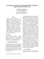
unit (range, 10 to 1000 ml; median 200 ml) (Figure 1).
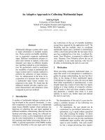
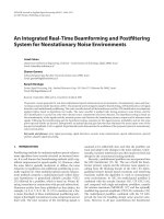
Active tuberculosis, cancer and mycetoma were associated
with a larger volume as compared with the cryptogenic
group (p = 0.03; p = 0.03 and p = 0.003, respectively; Fig-
ure 2.). There were severe consequences of bleeding in 73
patients (37%) leading to the following interventions
prior to the referral or during the first 24 hours of ICU
admission: local (n = 23) or systemic (n = 56) terlipressin,
mechanical ventilation (n = 17), blood transfusion (n =
22), vasoactive drugs support (n = 3) or cardiopulmonary
resuscitation (n = 2). Patients receiving the above men-
tioned interventions had a higher respiratory rate on
admission (24 ± 7 vs. 21 ± 6 per min; p = 0.04), a higher
heart rate (91 ± 20 vs. 85 ± 20 bpm; p = 0.04), a lower
room air partial pressure of oxygen in arterial blood (73 ±
15 vs. 80 ± 17 mm Hg; p = 0.03), a higher cumulated vol-
ume of blood loss (360 ± 240 ml vs. 180 ± 150 ml; p <
0.0001), and a lower haemoglobin value (11.5 ± 2.6 vs.
13.3 ± 2; p < 0.001); they also had more often active
bleeding on bronchoscopy (31/68 vs. 22/114, p =
0.0003), a first-line attempt at bronchial arteriography
(66/73 vs. 81/123; p < 0.0001), a need for surgery (25/73
vs. 9/123; p < 0.0001) and specific aetiologies [mycetoma
(11/73 vs. 3/123; p = 0.002) and cancer (22/73 vs. 11/123;
p < 0.001)], but not a higher frequency of cardiovascular
and pulmonary pre-existing diseases.
Cause of haemoptysis
Bronchiectasis (n = 78, 40%), lung cancer (n = 33, 17%),
active tuberculosis (n = 27, 14%) and mycetoma (n = 14,
7%) accounted for 87% of all causes. Emphysema (n = 10,
5%), pneumonia (n = 6, 3%), pulmonary embolism (n =
2, 1%) and miscellaneous causes (n = 5, 3%) accounted
for the remaining probable causes. In 21 patients (11%),
no cause was evidenced. The cause of bleeding was identi-
fied in 69% (n = 111/162) of patients at bedside when
combining history, comorbid conditions, physical exami-
nation, chest-X-Ray and fiberoptic bronchoscopy find-
ings, as compared with 91% (n = 148/162) after a further
CT scan (p < 0.001). The CT scan examination was espe-
cially useful for diagnosing bronchiectasis.
Management
All patients received conservative measures. Local (n = 6)
or systemic (n = 37) terlipressin, mechanical ventilation
(n = 3) and blood transfusion (n = 3) were administered
to the most severe patients before referral. Forty-three
(22%) patients were receiving aspirin, coumadin or
clopidrogel that may have worsened the bleeding, and
these drugs were temporarily stopped whenever possible.
Broad-spectrum antibiotics were administered to 153
patients (78%). A fiberoptic bronchoscopy was per-
formed within 24 hours of bleeding onset in most
patients (n = 184, 94%). Diffuse and bilateral (n = 13) or
Respiratory Research 2007, 8:11 />Page 4 of 9
(page number not for citation purposes)
Table 1: Clinical characteristics on ICU admission.
Age, years 51 ± 16 (17–89)
Sex Ratio (male:female) 148:48 (3.1:1)
SAPS II score 18 ± 9 (6–48)
McCabe and Jackson categories, n * 133/46/13
Cumulated volume of hemoptysis, ml † 240 ± 200 (10–1000)
< 200 ml, n (%) 86 (45%)
≥ 200 ml, n (%) 107 (55%)
Heart rate,/min 87 ± 20 (48–150)
> 130/min, n (%) 10 (5%)
Systolic Arterial Pressure, mm Hg 135 ± 28 (76–221)
< 100 mm Hg, n (%) 9 (5%)
Spontaneous Ventilation, n (%) 179 (91%)
Mechanical Ventilation [Invasive/Non Invasive], n (%) 17 [16/1] (9%)
Core Temperature, °C 37.3 ± 0.8 (36–40)
> 38.5°C, n (%) 18 (9%)
Respiratory functional signs
Cough, n (%) 116 (73%)
Persistent bloody expectoration, n (%) 107 (69%)
Dyspnea, n (%) 121 (66%)
Purulent expectoration, n (%) 10 (6%)
Chest pain, n (%) 11 (6%)
Physical examination
At least one localized abnormality, n (%) 94 (48%)
Crackles, n 59 (63%)
Results are expressed as mean ± SD (range), unless otherwise stated.
*4 missing data; †3 missing data.
Table 2: Biological variables on ICU admission.
Blood Leukocytes Count, mm
3
9183 ± 3543 (1500–25300)
Platelets Count, mm
3
258 464 ± 105 195 (45000–712000)
< 100 000/mm
3
, n (%) 6 (3%)
Hemoglobin, g/dl 12.6 ± 2.4 (4.6–18.3)
< 10 g/dl, n (%) 29 (15%)
Prothrombin Time, % 88 ± 16 (11–118)
≤ 50%, n (%) 7 (4%)
Activated partial thromboplastin time ratio 1.1 ± 0.2 (0.7–2.2)
≥ 1.5 control, n (%) 7 (4%)
Nitrogen Urea, mmoles/l 5.5 ± 2.7 (1–19)
≥ 10 mmoles/l, n (%) 12 (6%)
Blood gas on room air *
PaO
2
, mm Hg 78 ± 17 (43–100)
PaCO
2
, mm Hg 39 ± 5 (26–63)
pH 7.43 ± 0.05 (7.30–7.50)
SaO
2
, % 95 ± 4 (76–99)
Results are expressed as mean ± SD (range), unless otherwise stated.
*data available for 142 patients.
Respiratory Research 2007, 8:11 />Page 5 of 9
(page number not for citation purposes)
localized endobronchial bleeding (n = 163) was evi-
denced in 176 patients (96%). The bronchoscopic find-
ings revealed a localized active endobronchial bleeding in
53 patients and a localized endobronchial clotting in 41.
Otherwise, a localized endobronchial bleeding was evi-
denced in the upper (n = 43) or lower bronchia (n = 26)
without active bleeding or clotting. In the remaining 8
patients (4%), a few signs of endobronchial blood were
present. Bronchoscopic techniques were combining
blood aspiration and local instillation of cold saline lav-
age. Vasopressors were bronchoscopically delivered in 23
patients, and a balloon was placed in one patient.
A first-line bronchial arteriography was attempted in 147
patients (75%), whereas 46 (23%) received conservative
treatment. Emergency surgery was performed in 3 patients
(bleeding of 700 ml revealing a cancer complicated by a
cardiac arrest; bleeding of 300 ml revealing a cancer
nearby the pulmonary artery; bleeding of 200 ml compli-
cating repeated obstructive pneumonias in a patient diag-
nosed with a cancer) (Figure 3). The following parameters
on admission were associated with the first attempt of
arteriography as opposed to conservative treatment alone:
a higher respiratory rate (23 ± 7 vs. 20 ± 4; p = 0.03), a
greater amount of bleeding (290 ± 205 vs. 80 ± 50; p <
0.0001), a persistent bloody sputum (87/119 vs. 18/35; p
= 0.02), an active bleeding on bronchoscopy (49/141 vs.
3/36; p = 0.002), the identification of a definite cause of
haemoptysis (120/149 definite causes vs. 9/21 cryp-
togenic; p = 0.0005) and the absence of renal impairment
(creatinin, µmol/l; 73 ± 22 vs. 82 ± 25; p = 0.03).
Technical failure of the attempted arteriography occurred
in 15/147 (10%) patients, mostly those with mycetoma
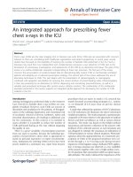
Distribution of the cumulated volume of haemoptysis on ICU admission, according to the first attempt of bronchial arteriogra-phyFigure 1
Distribution of the cumulated volume of haemoptysis on ICU admission, according to the first attempt of
bronchial arteriography. Bronchial arteriography was not attempted in 4 patients with a volume ≥ 200 ml: one patient with
moderate renal insufficiency (cryptogenic haemoptysis of 200 ml) received conservative treatment and emergency surgery was
performed in the 3 other patients.
28
16
2
1
0
1
7
35
42
31
10
20
0
10
20
30
40
50
< 100 100-200 200-300 300-400 400-500 > 500
Volume on ICU adm ission, ml
Patients, no
No first-line bronchial arteriography First line bronchial arteriography
200 ml
Respiratory Research 2007, 8:11 />Page 6 of 9
(page number not for citation purposes)
(n = 4) and cancer (n = 6). This led to either maintaining
conservative measures in 9 patients or to surgery in six; 5
of these 15 patients died within the first month (4/9
patients managed conservatively and 1/6 undergoing sur-
gery). In another patient, bleeding was related to a pulmo-
nary artery aneurysm. Bronchial artery embolisation was
eventually completed in 131/147 patients (89%) leading
to an immediate control of bleeding in 106 patients
(81%), 8 of whom had a secondary scheduled surgery
(Figure 3).
Bleeding recurred in 7/46 patients (15%) managed con-
servatively, 2 of whom received BAE secondarily. Bleeding
recurred in 35/131 patients (27%) receiving completed
BAE. Haemoptysis recurred after 3 ± 3 days (range, 0 to 11
days) in 25 patients, who received conservative treatment
(n = 4), BAE (n = 7) or surgery (n = 14). Mycetoma and
cancer accounted for 50% of the early recurrences. There
were 10 late recurrences (9 ± 4 months; range, 2 to 14
months) managed conservatively (n = 4) or with a second
BAE (n = 3) or surgery (n = 3). Overall, surgery (pneumon-
ectomy, n = 3; lobectomy, n = 11) was performed after 7
± 7.5 days for early recurrences (mycetoma, n = 6; bron-
chiectasis, n = 3; pneumonia, n = 3; cancer, n = 2). A lobec-
tomy was performed for late recurrences 12 ± 5 months
after the initial episode.
Bronchial artery embolisation was associated with a 5%
rate of complications (minor arterial dissection, n = 2; cor-
onary ischemia, n = 2; chest pain, n = 1; transient neuro-
Distribution of the volume of haemoptysis (median, quartile) on admission according to the causeFigure 2
Distribution of the volume of haemoptysis (median, quartile) on admission according to the cause. Plots of the
median, 10th, 25th, 75th, and 90th percentiles as vertical boxes with error bars.
Cause
Volume (median, quartile), ml
0
200
400
600
800
1000
1200
Cryptogenic
Tuberculosis
sequellae
Bronchiectasis
Cancer
Tuberculosis
active
Mycetoma
*p<0.05
Respiratory Research 2007, 8:11 />Page 7 of 9
(page number not for citation purposes)
logical episode, n = 1; and dysphagia; n = 1). The outcome
of these patients was uneventful without further interven-
tion.
Outcome
The lengths of ICU and hospital stay were respectively 5.4
± 4.9 days (range, 0 to 47 days) and 10.7 ± 14 days (range,
0 to 142 days). Lengths of ICU (6 ± 5.4 days vs. 3.8 ± 2.5
days) and hospital stay (11 ± 14.4 days vs. 8.8 ± 12.4 days)
were significantly longer for patients in whom a bronchial
arteriography was first attempted, as compared with
patients receiving conservative treatment alone (all p <
0.01). Therapy was withheld or withdrawn in 12 patients
(6%), 7 of whom died in ICU. The ICU and hospital mor-
tality rates were 4% (n = 8) and 8% (n = 15), respectively.
At short term follow-up (one month), successful control
of haemoptysis was obtained using completed BAE, con-
servative management or surgery in respectively 112
(57%), 44 (22%) and 22 (11%) patients. No recurrence of
haemoptysis occurred in 116 (89%) of the 131 patients in
whom BAE was completed, after a mean (median) follow-
up duration of 20 (8) months.
Discussion
Our study aimed at characterizing the clinical spectrum
and the outcome of a large series of consecutive patients
with severe haemoptysis requiring ICU admission in the
early 2000's. The major aetiologies recorded were bron-
chiectasis, lung cancer, active tuberculosis and mycetoma.
A simple set of clinical variables on admission combined
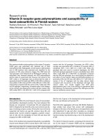
Initial management and short-term outcomeFigure 3
Initial management and short-term outcome. †Life sustaining therapy was withheld/withdrawn in 2/46 patients managed
conservatively and in 10/147 patients in whom BAE was first attempted.
Death
n=2 †
Short term control
of bleeding
n=44
Conservative
measures alone
n=46
Pulmonary
aneurism
n=1
Conservative management
after failure of arteriography
n=9 (4 of whom died) †
Surgery after
failure of arteriography
n=6 (1 of whom died)
Conservative management
after early recurrence
n=4 (3 of whom died) †
Surgery for
early recurrence
n=14
Death after
successfull BAE
n=1 †
Short term control
of bleeding
n=112
Completed bronchial artery
embolisation
n=131
First-line
bronchial arteriography
n=147
Emergency
surgery
n=3
Patients with
severe hemoptysis
n=196
Respiratory Research 2007, 8:11 />Page 8 of 9
(page number not for citation purposes)
with bronchoscopic findings were associated with
attempting a first-line bronchial arteriography. This
approach was applicable to 75% of our patients and led to
an immediate control of bleeding in more than 80% of
them. Although the median cumulated volume of haemo-
ptysis averaged 200 ml on admission, the ICU mortality
rate was low.
Haemoptysis accounted for up to 15% of our admissions.
This high rate reflects in part the fact that both our unit
and the department of radiology of our hospital are refer-
ral centres for haemoptysis. The major criterion for ICU
admission is the amount of blood loss despite the lack of
standardisation for quantifying it, since it is known to be
related to death [5]. Respiratory failure, a substantial drop
of haemoglobin level, and haemodynamic failure all
obviously mandate ICU admission, although their occur-
rence is not specified in most studies. While the usual cri-
teria of severity accounted for a relatively small subset of
our patients, the median cumulated volume of haemopt-
ysis averaged 200 ml on admission and chronic obstruc-
tive pulmonary disease and cardiovascular disease were
frequent.
Bronchiectasis, active tuberculosis and idiopathic haemo-
ptysis were the most frequent diagnoses among a French
cohort of 56 patients with life-threatening haemoptysis
recorded between 1986 and 1996 [6]. In a small recent
series of 29 patients with massive haemoptysis requiring
ICU admission in Singapore between 1997 and 2001,
bronchiectasis, mycetoma, active tuberculosis and cancer
were the main causes identified [9]. In our series, bron-
chiectasis (mainly secondary to inactive tuberculosis),
cancer, active tuberculosis and mycetoma were the lead-
ing causes. Such a distribution of the causes of haemopty-
sis underlines the following points: 1) First, active
tuberculosis still remains a common cause of severe hae-
moptysis in France; 2) Lung cancer appears to be an
emerging cause of haemoptysis requiring ICU admission.
The latter finding is at variance with previous studies by
Mal et al. [6] and Ong et al. [9], which may be related to
the smaller number of patients included in those previous
studies [6,9], the study period [6] and the geographic loca-
tion [9], as well as the current lesser restrictive policy for
ICU admission of cancer patients.
The indications of emergency surgery have gradually been
reduced, because of the reported 20–30% operative mor-
tality rate and improvement in interventional radiology
techniques [1-4,10]. Bronchial artery embolisation is now
considered as the most effective non-surgical first-line
treatment of severe haemoptysis [11,12], although there is
no randomized trial in this field. Bronchoscopy-guided
topical haemostatic tamponade therapy has also been
demonstrated to control haemoptysis with varying suc-
cess rates, using flexible or rigid bronchoscope for the
instillation of procoagulant substances, local injection of
adrenalin solutions, insertion of small calibre catheters or
placement of oxidized regenerated cellulose [13].
In our series, a bronchial arteriography was first attempted
in 75% of patients, whereas only 2% underwent emer-
gency surgery. A relatively large subset of our patients
(23%) was managed conservatively, based on our
approach to assess the amount of blood loss and other cri-
teria of severity on admission. Technical failure of arteri-
ography has been reported in up to 20% of attempts,
although a lower rate is expected with the use of micro
catheters in the near future [6,9,14-17]. Moreover, bleed-
ing recurrences after successful completed BAE range from
0% to 30% and may be influenced by the cause of haemo-
ptysis [18-20]. In our series, the rate and causes of bron-
chial arteriography failure were similar. Haemoptysis
recurred in 27% of patients. There were mostly early recur-
rences, two thirds of which were eventually controlled by
surgery. According to an 'intent to treat analysis', a first-
line arteriography was associated with an immediate con-
trol and a durable cessation of bleeding in 112 (57%) and
116 (59%) patients, respectively. Although other series
reported higher immediate successful rates of BAE for con-
trolling haemoptysis, ranging from 85% to 95%
[16,18,20], it should be noted that no information was
provided on patients in whom the procedure was not
completed [21]. In our series, bleeding was controlled in
112/131 patients (85%) within the first month and in
116/131 patients (89%) after hospital discharge, when
the procedure was completed.
Using a strategy including a routine assessment of the
amount of bleeding with a standardized scale, and pro-
moting BAE over surgery, the outcomes of patients were
good. The ICU mortality rate was low, as reported in
recent series of so-called life-threatening haemoptysis [6].
The limitations of our study are related to its retrospective
nature and to the fact that it was conducted in a referral
centre with an extensive experience of severe haemoptysis
on a heterogeneous patients' group. Nevertheless, our
study reports one of the largest series of medical inpatients
over a short time period and may provide a useful frame-
work for the therapeutic management of haemoptysis in
this clinical setting.
To summarize, a multidisciplinary approach remains the
cornerstone for the management of severe haemoptysis.
Bedside clinical evaluation and early fiberoptic bronchos-
copy may safely screen patients for initial BAE, including
surgical candidates. In the latter, surgery should be post-
poned as much as possible during active bleeding and per-
formed early after control of bleeding. Otherwise, surgery
Publish with Bio Med Central and every
scientist can read your work free of charge
"BioMed Central will be the most significant development for
disseminating the results of biomedical research in our lifetime."
Sir Paul Nurse, Cancer Research UK
Your research papers will be:
available free of charge to the entire biomedical community
peer reviewed and published immediately upon acceptance
cited in PubMed and archived on PubMed Central
yours — you keep the copyright
Submit your manuscript here:
/>BioMedcentral
Respiratory Research 2007, 8:11 />Page 9 of 9
(page number not for citation purposes)
should be reserved to cases of failure of interventional
radiology and/or uncontrolled bleeding despite embolisa-
tion. Further prospective studies are needed to confirm
the safety and the reproducibility of such a therapeutic
approach; this approach may also be influenced by the
use of the multi detector row helical CT scan, which can
depict accurately the bronchial and non bronchial arter-
ies, prior to the embolisation.
Competing interests
The author(s) declare that they have no competing inter-
ests.
Financial support
None
Authors' contributions
Dr Fartoukh had full access to the data and takes respon-
sibility for the integrity of the data and the accuracy of the
data analysis.
Study concept and design: Fartoukh, Cadranel.
Acquisition of data: Parrot, Louis, Fartoukh.
Analysis and interpretation of data: Fartoukh, Parrot,
Mayaud, Cadranel.
Drafting of the manuscript: Fartoukh, Parrot, Carette, Kha-
lil.
Critical revision of the manuscript for important intellectual
content: Parrot, Khalil, Carette, Bazelly.
Statistical analysis: Fartoukh, Parrot, Cadranel.
Study supervision: Fartoukh.
References
1. Crocco JA, Rooney JJ, Fankushen DS: Massive hemoptysis. Arch
Intern Med 1968, 121:495-498.
2. Garzon AA, Gourin A: Surgical management of massive hemo-
ptysis. Ann Surg 1977, 137:267-271.
3. Garzon AA, Gourin A: Surgical management of massive hemo-
ptysis: a 10-year experience. Ann Surg 1978, 187:267-271.
4. Sehhat S, Oreizie M, Moinedine K: Massive pulmonary hemor-
rhage: surgical approach as choice of treatment. Ann Thorac
Surg 1978, 25:12-15.
5. Dweik RA, Stoller JK: Role of bronchoscopy in massive hemop-
tysis. Clinics in chest medicine 1999, 20:89-105.
6. Mal H, Rullon I, Mellot F, Brugiere O, Sleiman C, Menu Y, Fournier M:
Immediate and long-term results of bronchial artery embol-
ization for life-threatening hemoptysis. Chest 1999,
115:996-1001.
7. Haponik EF, Chin R: Hemoptysis: clinicians' perspectives. Chest
1990, 97:469-475.
8. Haponik EF, Fein A, Chin R: Managing life-threatening hemopt-
ysis. Has anything really changed? Chest 2000, 118:1431-1435.
9. Ong TH, Eng P: Massive hemoptysis requiring intensive care.
Intensive Care Med 2003, 29:317-320.
10. Gourin A, Garzon AA: Operative treatment of massive hemo-
ptysis. Ann Thorac Surg 1974:52-60.
11. Remy J, Arnaud A, Fardou H: Treatment of hemoptysis by
embolization of bronchial arteries. Radiology 1977, 122:33-37.
12. Lordan JL, Gascoigne A, Corris PA: The pulmonary physician in
critical care. Illustrative case 7: assessment and manage-
ment of massive haemoptysis. Thorax 2003, 58:814-819.
13. Valipour A, Kreuzer A, Koller H, Koessler W, Burghuber OC: Bron-
choscopy-guided topical hemostatic tamponade therapy for
the management of life-threatening hemoptysis. Chest 2005,
127:2113-2118.
14. Rabkin JE, Astafjef VI, Gothman LN: Transcatheter embolization
in the management of pulmonary hemorrhage. Radiology
1987, 163:361-365.
15. Ufkacker R, Kaemmerer A, Picon PD: Bronchial artery embolisa-
tion in the management of hemoptysis: technical aspects
and long-term results. Radiology 1985, 157:637-644.
16. Cremaschi P, Nascimbene C, Vitulo P: Therapeutic embolization
of bronchial artery: a successful treatment in 209 cases of
relapse hemoptysis. Angiology 1993, 44:295-299.
17. Tanaka N, Yamakado K, Murashima S, Takeda K, Matsumura K, Nak-
agawa T, Takano K, Ono M, Hattori T: Superselective bronchial
artery embolization for hemoptysis with a coaxial micro-
catheter system. J Vasc Interv Radiol 1997, 8:65-70.
18. Hayakawa K, Tanaka F, Torizuka T: Bronchial artery emboliza-
tion for hemoptysis: immediate and long-term results. Cardi-
ovasc Intervent Radiol 1992, 15:154-159.
19. Knott-Craig CJ, Oostuizen JD, Rossouw G: Management and
prognosis of massive hemoptysis. J Thorac Cardiovascular Surg
1993, 105:394-397.
20. Ramakantan R, Bandekar VG, Gandhi MS: Massive hemoptysis due
to pulmonary tuberculosis: control with bronchial artery
embolization. Radiology 1996, 200:691-694.
21. White RI: Bronchial artery embolotherapy for control af
acute hemoptysis. Chest 1999, 115:912-915.