Báo cáo khoa học: " Causes of Stillbirth and Time of Death in Swedish Holstein Calves Examined Post Mortem" potx
Bạn đang xem bản rút gọn của tài liệu. Xem và tải ngay bản đầy đủ của tài liệu tại đây (130.05 KB, 10 trang )
Berglund B, Steinbock L, Elvander M: Causes of stillbirth and time of death in
Swedish Holstein calves examined post mortem. Acta vet. scand. 2003, 44, 111-120.
– This study was initiated due to the observation of increasing and rather high levels of
stillbirths, especially in first-calving Swedish Holstein cows (10.3%, 2002). Seventy-six
Swedish Holstein calves born to heifers at 41 different farms were post mortem exam-
ined in order to investigate possible reasons for stillbirth and at what time in relation to
full-term gestation they had occurred. The definition of a stillborn calf was dead at birth
or within 24 h after birth after at least 260 days of gestation. Eight calves were consid-
ered as having died already in uterus. Slightly less than half of the examined calves
(46.1%) were classified as having died due to a difficult calving. Four calves (5.3%) had
different kinds of malformations (heart defects, enlarged thymus, urine bladder defect).
Approximately one third of the calves (31.6%) were clinically normal at full-term with
no signs of malformation and born with no indication of difficulties at parturition or any
other reason that could explain the stillbirth. The numbers of male and female calves
were rather equally distributed within the groups. A wide variation in post mortem
weights was seen in all groups, although a number of the calves in the group of clini-
cally normal calves with unexplained reason of death were rather small and, compared
with e.g. those calves categorised as having died due to a difficult calving, their average
birth weight was 6 kg lower (39.9±1.7 kg vs. 45.9±1.5 kg, p≤0.01). It was concluded that
the cause of stillbirth with a non-infectious aetiology is likely to be multifactorial and
difficult calving may explain only about half of the stillbirths. As much as one third of
the calves seemed clinically normal with no obvious reason for death. This is a target
group of calves that warrants a more thorough investigation in further studies.
Autopsy; congenital defects; dairy calves.
Acta vet. scand. 2003, 44, 111-120.
Acta vet. scand. vol. 44 no. 3-4, 2003
Causes of Stillbirth and Time of Death in Swedish
Holstein Calves Examined Post Mortem
By B. Berglund
1
, L. Steinbock
1
and M. Elvander
2
1
Department of Animal Breeding and Genetics, Centre for Reproductive Biology in Uppsala, Swedish Univer-
sity of Agricultural Sciences,
2
National Veterinary Institute, Uppsala, Sweden.
Introduction
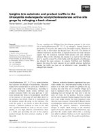
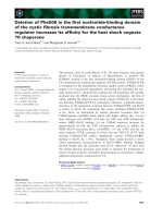
In recent years, we have seen a remarkable in-
crease in the incidence of stillbirths in the
Swedish Black and White Breed (SLB) concur-
rent with increased imports of North American
Holstein genes. Today, the SLB breed can be re-
garded as a Swedish Holstein Breed. Stillbirth
is commonly defined as a calf that dies just
prior to, during, or within 12-48 h of parturi-
tion. The problem is most pronounced in
heifers and during the past 20 years an increase
from about 6% to 10.3% has occurred. In con-
trast, in older SLB cows and in heifers and cows
of the Swedish Red and White Breed (SRB),
the increase has been of much lower magnitude
with an overall incidence of around 5%
(Swedish Dairy Association, 2003). See Fig. 1
for a description of the stillbirth rate during
1982-2002.
An increasing stillbirth rate is probably due to a
multifactorial cause. The problems appear to be
rather different today, compared with earlier ex-
perience, since stillbirths in calves born to
heifers seem to be less closely related to high
birth weight and to difficult calving than they
used to be (Berglund & Philipsson 1992). It
was concluded in that study that calf vitality ap-
pears to be of growing importance in the still-
born syndrome. Sluijter et al. (1990) and Simp-
son (1990) also reported increasing incidences
of stillbirths in heifer calvings in the Nether-
lands and in England, respectively. In the Dutch
study it was found that the placenta was usually
expelled together with the calf, which other-
wise is a quite unusual feature. In the English
study, evidence of trauma, often very severe
with rib fractures, was found in the majority of
dead calves.
During late bovine pregnancy several hormones
are involved to maintain and develop a success-
ful result with a live calf. Changes in endocrine
profiles in late bovine pregnancy have been
related to abortions and stillbirths. Measure-
ment of bovine pregnancy-specific protein B
(bPSPB), pregnancy associated glycoprotein
(PAG), and oestrone sulphate have been found
useful for monitoring placental functions and
indirectly also foetal viability (Dobson et al.
1993, Patel et al. 1997, Beckers et al. 1999,
Kornmatitsuk et al. 2003).
The outcome of pregnancies can also be af-
fected by pathogens causing stillbirths and
abortions. Sweden is free from several serious
infectious agents such as brucellosis, lep-
tospirosis and IBR. Presence of Bovine Viral
Diarrhoea Virus (BVDV) and Neospora can-
inum may play a role for neonatal mortality and
calves that are very weak and die soon after
birth. N. caninum infection causes stillbirth and
abortion throughout pregnancy, but abortion at
5 to 6 months of gestation is most common
112 B. Berglund et al.
Acta vet. scand. vol. 44 no. 3-4, 2003
Figure 1. Annual statistics of stillbirth rate 1982-2002 in Swedish Holstein (SLB) and Swedish Red and White
(SRB) breeds (Swedish Dairy Association).
(Dubey 1999). The prevalence of N. caninum is
low in Sweden (Björkman et al. 2000). BVDV
is important for early abortions (2-5 months)
(Larsson et al. 1994). An eradication pro-
gramme of BVDV started in 1993 (Lindberg &
Alenius 1999) with a rapid decrease in herd
prevalence, and today 95% of the herds are de-
clared free from the infection. Giri et al. (1990)
used endotoxins to induce abortions. The high-
est sensitivity for abortions was in the first
trimester, but termination of pregnancy can oc-
cur at any stage of gestation.
Incompatibility between calf size and dam size,
as well as pelvic and vulvar conformation, are
factors likely to have an important impact on
stillbirths. A prolonged but not necessarily dif-
ficult calving caused by, e.g. weak labour, might
be a risk factor for calf mortality. For stillborn
calves, the time interval from onset of labour
until calving was completed was approximately
twice as long as for liveborn calves (Berglund
et al. 1987).
A large genetic variation in calving traits be-
tween daughter groups of SLB bulls is also ev-
ident (Berglund & Philipsson 1992, Steinbock
et al. 2003). Genetic defects, e.g. a larger num-
ber of sublethal genes, lower the viability at
birth and might be one explanation of the in-
creased stillbirth rates. Some examples of re-
cently identified genetic defects in the Holstein
breed are Complex Vertebral Malformation
(CVM), bulldog syndrome, Bovine Dilated
CardioMyoPathy (BDCMP) and Bovine Leu-
kocyte Adhesion Deficiency (BLAD). Other
defects, which we do not know about yet, might
be present and new defects will certainly turn
up in future.
Furthermore, increasing average herd sizes,
which most likely gives less time for supervi-
sion of calving, might result in a larger propor-
tion of difficult calvings being recorded as still-
births. The large number of stillborn calves is
both an ethical and an economic problem. The
economic problem comprises loss of the calf,
lower fertility of the dam at the next breeding,
longer calving to conception interval, and a ten-
dency for milk production to be decreased
(Chassagne et al. 1999).
This study was initiated due to the observation
of increasing and rather high levels of still-
births, especially in first-calving Holstein cows.
The objective was to investigate possible rea-
sons for stillbirths and to establish the time they
had occurred in relation to full term. Therefore,
a post mortem examination of Swedish Hol-
stein calves born to heifers was carried out. In-
creased knowledge of the background to these
problems is important both for management
and breeding purposes in the effort to reduce
stillbirths and calving problems.
Materials and methods
Farmers from 200 Swedish Holstein herds were
asked during the years 1994-1996 to send still-
born calves from primiparous cows for post
mortem examination. The definition of a still-
born calf was dead at birth or within 24 h of
birth after at least 260 days of gestation. Alto-
gether 76 Swedish Holstein calves were exam-
ined for reasons of stillbirth, including estab-
lishment of the approximate time of death. The
calves were born at 41 different farms and all
were singletons except for 2 pairs of twins. The
post mortem examinations were performed in 3
different regions in southern Sweden Halmstad
(Svelab), Kristianstad (Svelab) and Skara
(AnalyCen) at laboratories with the required
competence and facilities for pathological/
anatomical investigations. A further prerequi-
site was that the laboratories were located rea-
sonably close to the herds.
A standard post mortem examination protocol
was developed for this purpose. The protocol
included information on sex, weight and length
of foetus (crown rump length). An assessment
was made if the foetus was alive during parturi-
Stillbirth and death in calves 113
Acta vet. scand. vol. 44 no. 3-4, 2003
tion or dead in uterus, and if so, approximately
for how long before expulsion. Calves with
signs of dehydration and mummification were
regarded as having been dead for more than one
week before expulsion and calves with subcuta-
neous oedema and hydropsies in the chest and
abdominal cavity for less than one week.
Calves that had died during the process of birth
showed signs of partially inflated lungs, and/or
subcutaneous, subdural or internal bleedings.
A macroscopic investigation of the most impor-
tant organs (heart, liver, lungs, kidneys, spleen,
brain and of navel region) was performed.
Lungs were checked as to whether they were in-
flated or not. Samples from lungs, liver heart
and muscle were routinely taken for histologi-
cal examination when there was a reason to sus-
pect changes. Visible signs of inflammatory
changes were recorded and samples for relevant
microbiological examination were taken. Pla-
centa was checked when available. Congenital
defects/malformations visible as severe ana-
tomic deformities were recorded.
The study was part of a larger study where
preparing for calving and the calving process of
about 4000 first calving Swedish Holstein cows
was followed by detailed observations recorded
in calving reports (Berglund 1996). These con-
tained information about age and pedigree of
heifer, insemination dates and pedigree of bull,
calving date, single or twin birth, calving ease
(1=easy, 2=normal, 3=difficult), attitude of
calf, status of cow after calving and retained
placenta. In addition, the calving reports also
contained information from the herdsman on
the sex of calf, birth weight, viability (born
alive or dead/died within 24 h) and on any visi-
ble malformation.
Data from all sources were compiled to form
cause and time of death categories based on the
presence of one or several of the criteria in each
category as follows:
Difficult calving
Trauma associated with technical assistance at
delivery such as subcutaneous, subdural or in-
ternal bleedings, and external lesions. Abun-
dant amounts of mucus in the respiratory tract
which may be a sign of ceased placenta func-
tion before the birth process was completed,
resulting in suffocation of the calf. In cases of
uncertainty in interpreting the pathological
findings, the report of a difficult calving from
the herdsman was used to strengthen the as-
sessment.
Intrauterine death
Subcutaneous oedema and hydropsies in chest
and abdominal cavity or dehydration/mummifi-
cation. The state of decomposition as an indica-
tor of how long the calf has been dead in uterus.
114 B. Berglund et al.
Acta vet. scand. vol. 44 no. 3-4, 2003
Table 1. Cause and time of death categories and sex of calf for 76 stillborn Swedish Holstein calves.
Cause and time of death N % Male calf Female calf
Difficult calving 35
a
46.1 16 16
Intrauterine death 8 10.5 3 5
Clinically normal, unknown cause of death 24
b
31.6 10 12
Malformations 4 5.3 2 2
Unspecified infections 2 2.6 2 0
Unknown cause/not possible to categorise 3
b
3.9 0 1
a, b
recording of sex missing for
a
3 calves,
b
2 calves
Clinically normal at full-term, unknown cause
of death
Clinically normal (well-formed) calves. Lack
of external signs of difficult calving. Foetal or
partial neonatal atelectasis. No malformations.
Malformations
Calves with visible malformations. Apart from
the specific malformation no consideration was
paid to whether the calves were clinically nor-
mal at full-term or not.
Unspecified infections
Calves with unspecified infections (born after
normal gestation length).
Unknown cause/not possible to categorise
Not fulfilling the criteria for clinically normal
at full-term, unknown cause of death. Full in-
formation such as, e.g. age of calf missing.
The GLM procedure in the SAS package (SAS
institute Inc., 2000) was used to analyse the ef-
fects of sex of calf and cause and time of death
category on birth weights.
Results
Table 1 shows the distribution of calves in dif-
ferent cause-and-time of death categories.
Slightly less than half of the calves (n=35) had
signs of a difficult calving (subcutaneous, sub-
dural or internal bleedings). Among these, 8
calves also had reports from the herdsman of a
difficult calving. Eight calves were regarded as
already dead in uterus, one of them for more
than one week. No macroscopic findings indi-
cated the cause of death in this group. Four
calves (5.3%) were malformed. The malforma-
tions found were: one calf with enlarged thy-
mus (5cm×15cm), 2 calves with heart defects
(chamber septum defect and persistent foramen
ovale), and one calf with a urine bladder defect.
Two calves died within 24 h of birth after full-
term gestation due to peritonitis and gastro-en-
teritis. Approximately one third of the calves
were clinically normal at full-term with no
signs of malformations and born with no indi-
cations of difficulties at parturition or any other
reason that could explain the stillbirth. The re-
maining 3 calves could not be categorised,
partly due to the lack of certain data. The num-
bers of male and female calves within each
cause and time of death category were rather
equally distributed (Table 1).
From the calving reports a note made by the
herdsman on the ease of calving was made in
connection with 46 parturitions, 13 of which
were classified as difficult. The post-mortem
examination showed that 3 calves out of the 76
examined had fractured ribs (1) or fractured
spinal column (2). There were no reports from
the herdsmen of expulsion of the placenta to-
gether with the calf. Seven cases of retained
placenta were reported. These were rather
evenly distributed over the different cause and
time of death categories.
Stillbirth and death in calves 115
Acta vet. scand. vol. 44 no. 3-4, 2003
Table 2. Post mortem weights of calves (kg) in different cause-and-time of death categories.
Cause-and-time of death N Mean SD Min. Max.
Difficult calving 32 44.9 7.6 32 62
Intrauterine death 6 44.7 11.4 31 60
Clinically normal, unknown cause of death 23 40.2 8.4 24 55
Malformations 4 45.0 15.0 23 56
Unspecified infections 2 47.5 3.5 45 50
Unknown cause/not possible to categorise 2 38.0 2.8 36 40
The examined calves weighed from 23 to 62 kg
(n=69), with an average body weight of 43.2 kg
(SD=8.7 kg). In Table 2, the average weight at
post-mortem inspection of calves in the differ-
ent cause and time of death categories are
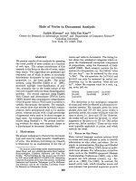
shown. Fig. 2 shows the frequencies of calves in
different categories of death distributed over
birth weight classes. Calves were grouped in 3
categories: difficult calving, clinically normal
with unknown cause of death, and a third group
comprising the other causes of death. A wide
variation in post mortem weights can be seen
for all groups. The clinically normal calves
with unknown cause of death had a centre of
gravity shifted to the left, indicating slightly
lower weights.
The analysis of variance showed that the overall
difference in birth weights between the differ-
ent cause and time categories of deaths was
non-significant. But when the group of calves
categorised as clinically normal at full-term
with unknown cause of death was compared
with the category of calves with difficult calv-
ing, the former group was 6 kg lighter
(39.9±1.7 kg vs. 45.9±1.5 kg, p≤0.01). There
was a significant effect of sex of calf on birth
weight, male calves weighing 45.4±2.0 kg and
female calves 38.3±2.4 kg (p≤0.001).
Discussion
The classification of calves in different cause
and time of death categories was primarily
based on the pathological findings in the post
mortem examination. Since data on the calving
process were collected under field conditions in
several different farms, all calvings were not
supervised. This means that the finding of a
dead calf does not necessarily mean that the
calf was born dead. This is also why the com-
monly used definition of a stillborn calf in-
cludes a certain time period after birth. Further-
more, the criteria for the judgement of calving
difficulty is likely to vary between herdsmen.
This is probably also an explanation of why not
all calves from calvings reported as difficult by
the farmer fell into this category as a result
from the pathological investigation. Thirteen of
all calvings were reported as having been diffi-
cult by the farmer. Based on pathological find-
ings eight calves from these 13 calvings were
classified in the category difficult calving.
Slightly less than half of the examined calves
were classified as having died due to a difficult
calving. This corresponds well with what has
been recorded by herdsmen in larger field ma-
terials where less than half of the stillborn
calves were from an easy or a normal calving
116 B. Berglund et al.
Acta vet. scand. vol. 44 no. 3-4, 2003
0
5
10
15
20
25
30
35
40
45
<25
25-32 33-40 41-48 49-56 >56
weight, kg
% calves
Difficult calving
Clinically normally developed
unknown cause of death
Intrauterine death, malformation,
unspecified infections
Figure 2. Frequencies (%) of calves in the different cause-and-time of death categories distributed over birth
weight classes.
(Berglund & Philipsson 1992, Steinbock et al.
2003). Genetic analysis of stillbirths also shows
that half of the variation in stillbirth still re-
mains after adjusting for calving difficulties
(Steinbock et al. 2003). Thus, only about half of
the variation in stillbirths is explained by diffi-
cult calving. Difficult calving is normally more
common in connection with the birth of male
calves (e.g. Steinbock et al. 2003). In the pre-
sent study, the distribution of sexes in this cate-
gory of death was even but the large difference
in birth weights between male- and female
calves was mainly due to some very large male
calves in this category.
One of the signs used to classify a difficult calv-
ing was subcutaneous bleedings as these might
be a sign of trauma in connection with the ex-
pulsion of the calf. Another possibility might
also be that the bleedings had already started in
the uterus due to a sub-optimal environment.
About one tenth of the stillborn calves were
classified as dead in uterus. The cause of death
is not known and could only be speculated upon
as dysfunction of the placenta, hormonal
changes or a nonvisible defect in the calf, or
some combination of these. These calves had
about the same average birth weight as calves in
the total material. Thus, generally there was
probably no intrauterine growth retardation,
which often is the case when there is a dysfunc-
tion of placenta (except for possibly one calf
weighing 31 kg classified as having been dead
less than one week in uterus). Smyth et al.
(1999) found that calves with leptospiral anti-
gens detected in the placenta were significantly
lighter by an average of 6 to 10 kg than calves
with no antigen in the placenta.
The incidence of malformed calves was consid-
ered rather normal and in accordance with what
has been reported in earlier studies. In a
Swedish investigation of 104 aborted calf foe-
tuses (from 2.5-9 months of gestation) during
1987-1988, 6 calves (5.8%) were malformed
(Elvander 1991). The various types of malfor-
mations both in that study and in the present
study were in line with what has previously
been reported.
There was a wide variation in birth weight of
the stillborn calves, although they were proba-
bly, on average, slightly heavier than live born
calves are. In the inquiry study, stillborn calves
(n=261) were only 1.6 kg heavier than the aver-
age weight of all calves (n=2825). Calves born
at a parturition classified as difficult (n=345)
were as much as 4.0 kg heavier than calves born
at a normal calving (unpublished results).
For as many as one third of the calves, no obvi-
ous cause of death could be seen. They were ap-
parently born clinically normal after full term.
A number of these calves had a rather low birth
weight. Dysfunction of the placenta might be
one reason for small, but otherwise apparently
normally developed, calves. In a recent Swedish
study (Kornmatitsuk et al. 2003) comprising 96
calvings no differences in the morphology of
the foetal membranes could be observed, how-
ever, between cows with viable calves com-
pared to cows with stillbirths. A weak calf syn-
drome characterised by the birth of full-term
calves which either failed to breathe or did not
breathe for longer than 10 min was recorded by
Rice et al. (1986). This syndrome was sug-
gested to be called stillbirth/perinatal weak calf
syndrome (SB/WCS) by Smyth et al. (1992).
However, in these studies from Northern Ire-
land many of the weak calves were born to mul-
tiparous cows, and consequently this situation
differs from ours. An abnormal thyroid is a
common finding in SB/WCS, but McCoy et al.
(1995) found that iodine and selenium supple-
mentation did not lower the incidence of
SB/WCS. Nor could an experimental iodine de-
ficiency induce SB/WCS (McCoy et al. 1997),
and it was concluded that other factors were
likely to contribute. It is not known if the cause
of death in the present study was due to any hor-
Stillbirth and death in calves 117
Acta vet. scand. vol. 44 no. 3-4, 2003
monal deficiencies since endocrinology was not
specifically studied and thus beyond the scope
of this study.
Calves with the genetic defect CVM have a re-
duced viability and a lower body weight com-
pared with normal calves. Embryonic mortality
occurs soon after conception and throughout
the entire gestation period, and recent Danish
studies (Nielsen et al. 2003) show that very few
calves are born alive at full-term gestation. Ac-
cording to our recordings, we had no case of
CVM in our material.
There were very few calves with signs of severe
trauma such as rib fractures in our material, and
we had no reports of placentas expelled to-
gether with the calves, and thus our conditions
(e.g. herd management) also appear to differ
from those described in connection with weak
calves in heifer calvings from England (Simp-
son 1990) and from the Netherlands (Sluijters
et al. 1990).
Stillbirth rates for primiparous Holstein cows in
Sweden are now close to the levels recently re-
ported from the United States by Meyer et al.
(2001). They also reported a slightly negative,
but non significant, genetic trend in perinatal
survival. Increased knowledge of genetical as
well as non genetical possible causes of still-
births is therefore important. Furthermore, it is
important to consider the calving traits in the
breeding evaluation of bulls and to use bulls
that inherit a low rate of stillbirth and calving
difficulty. Additionally it is important to have
national control programs for congenital ge-
netic defects to avoid multiplication of deleteri-
ous genes. Moreover it is important that all mal-
formations are reported.
In conclusion, the cause of a stillbirth of a non-
infectious aetiology is likely to be multifacto-
rial and difficult calving may explain only about
half of the stillbirths from primiparous cows.
As many as one third of the calves seemed to be
clinically normal and born at full-term with no
obvious reason for death, which could be re-
garded as a surprisingly large proportion of the
calves. Although a wide variation in birth
weight was seen in all categories of death, these
calves were, on average, lighter than the other
calves. This is a target group of calves that war-
rants a more thorough investigation in further
studies.
Acknowledgements
We wish to express our gratitude to all farmers who
submitted data for this study, and to the veterinary
staff at Svelab in Halmstad (Helena Johansson, Karl-
Gustav Kolmodin), Svelab in Kristianstad (Christer
Gyllerup, Sten Vesterlund) and AnalyCen in Skara
(Anders Linder, Helena Wejsfelt) for their excellent
help in performing the post mortem examinations.
The Swedish Farmers´ Foundation for Agricultural
Research (SLF), AGRIA Insurance Company,
Swedish Board of Agriculture and Swedish Animal
Health Service (Ingvar Friede) are acknowledged for
contributing to the financing of this study.
References
Beckers JF, Drion PV, Garbayo JM, Perenyi Z,
Zarrouk A, Sulon J, Remy B, Szenci O: Pregnancy
associated glycoproteins in ruminants: inactive
members of the aspartic proteinase family. Acta
Vet. Hung., 1999, 47, 461-469.
Berglund B: Ongoing research on the causes of vari-
ation in calving performance and stillbirths in
Swedish dairy cattle. International Workshop on
Genetic Improvement of Functional Traits in Cat-
tle, Jan 21-23, 1996, Gembloux, Belgium. Inter-
bull Bulletin No 12: 78-83.
Berglund B, Philipsson, J: Increasing stillbirth rates
in the Swedish Friesian population. Paper in 43rd
Ann. Meet. of the European Ass. for Anim. Prod.,
Madrid, 14-17 Sept. 1992.
Berglund B, Philipsson J, Danell Ö: External signs of
preparation for calving and course of parturition
in Swedish dairy cattle breeds. Anim. Repr. Sci.,
1987, 15, 61-79.
Björkman C, Alenius S, Emanuelson U, Uggla, A:
Neospora Caninum and Bovine Virus Diarrhoea
Virus infections in Swedish dairy cows in relation
to abortion. The Veterinary Journal, 2000, 159,
201-206.
Chassagne M, Barnouin J, Chacornac JP: Risk fac-
118 B. Berglund et al.
Acta vet. scand. vol. 44 no. 3-4, 2003
tors for stillbirth in Holstein heifers under field
conditions in France: a prospective survey. Theri-
ogenology, 1999, 51, 1477-1488.
Dobson H, Rowan TG, Kippax IS, Humblot P: As-
sessment of fetal number, and fetal and placental
viability throughout pregnancy in cattle. Theri-
ogenology, 1993, 40, 411-425.
Dubey JP: Neosporosis in cattle: biologic and eco-
nomic impact. Journal of the American Veteri-
nary Medical Association, 1999, 214, 1160-
1163.
Elvander M: Aborter hos nötkreatur. [Abortions in
Swedish dairy cattle]. Paper in SHS Djurhälso-
konferens 24-25 April 1991. Svensk Mjölk SE-
631 84 Eskilstuna, Sweden. 1991. (In Swedish).
Giri SN, Emau P, Cullor JS, Stabenfeldt GH, Bruss
ML, BonDurant RH, Osburn BI: Effects of
endotoxin infusion on circulating levels of
eicosanoids, progesterone, cortisol, glucose and
lactic acid, and abortion in pregnant cows. Vet.
Microbiol., 1990, 21, 211-231.
Kornmatitsuk B, Franzén G, Gustafsson H, Kindahl
H: Endocrine measurements and calving perfor-
mance of Swedish Red and White and Swedish
Holstein dairy cattle with respect to stillbirth.
Submitted for publication 2003.
Larsson B, Niskanen R, Alenius, S: Natural infection
with bovine virus diarrhoea virus in a dairy herd:
A spectrum of symptoms including early repro-
ductive failure and retained placenta. Anim. Re-
prod. Sci., 1994, 36, 37-48.
Lindberg A, Alenius S: Principles for eradication of
bovine viral diarrhoea virus (BVDV) infections
in cattle populations. Vet. Microbiol., 1999, 64,
197-222.
McCoy MA, Smyth JA, Ellis WA, Kennedy DG: Par-
enteral iodine and selenium supplementation in
stillbirth/perinatal weak calf syndrome. Vet.
Rec., 1995, 136, 124-126.
McCoy MA, Smyth JA, Ellis WA, Kennedy DG: Still-
birth/Perinatal weak calf syndrome. Cattle Prac-
tice, 1997, 5, 31-34.
Meyer CL, Berger PJ, Thompson JR, Sattler CG: Ge-
netic evaluation of Holstein sires and maternal
grandsires in the United States for perinatal sur-
vival. J. Dairy Sci., 2001, 84, 1246-1254.
Nielsen US, Aamand GP, Andersen O, Bendixen C,
Nielsen VH, Agerholm JS: Effects of complex
vertebral malformation on fertility traits in Hol-
stein cattle. Livest. Prod. Sci., 2003, 79, 233-238.
Patel OV, Takahashi T, Takenouchi N, Hirako M,
Sasaki N, Domeki, I.: Peripheral cortisol levels
throughout gestation in the cow: effect of stage of
gestation and foetal number. Br. Vet. J., 1997,
152, 425-432.
Rice DA, McMurray CH, Kennedy S, Ellis WA: Lack
of effect of selenium supplementation on the in-
cidence of weak calves in dairy herds. Vet. Rec.,
1986, 119, 571-573.
SAS Institute Inc.: SAS version 8.1. SAS Institute
Inc., Cary, NC, USA. 2000.
Simpson VR: Weak calf syndrome. Vet. Rec., 1990,
127, 459.
Sluijter FJH, Zimmer GM, Wouda W: Weak calf syn-
drome. Vet. Rec., 1990, 127, 355.
Smyth JA, Fitzpatrick DA, Ellis WA: Stillbirth/perina-
tal calf syndrome: a study of calves infected with
Leptospira. Vet. Rec., 1999, 19, 539-542.
Smyth JA, McNamee PT, Kennedy DG, McCullough
SJ, Logan EF, Ellis WA: Stillbirth/perinatal weak
calf syndrome: preliminary pathological, micro-
biological and biochemical findings. Vet. Rec.,
1992, 130, 237-240.
Steinbock L, Näsholm A, Berglund B, Johansson K,
Philipsson, J: Genetic effects on stillbirth and
calving difficulty in Swedish Holsteins at first
and second calving. J. Dairy Sci. 2003, 86, 2228-
2235.
Swedish Dairy Association:
Husdjursstatistik. [Cat-
tle Statistics] 2003. Svensk Mjölk, SE-631 84 Es-
kilstuna, Sweden. 2003.
Sammanfattning
Obduktionsstudie av dödfödda SLB-kalvar - dödsor-
saker och dödstidpunkt.
Andelen dödfödda kalvar har ökat och den genom-
snittliga frekvensen hos förstakalvande SLB är nu
10.3% (2002). Detta utgjorde bakgrunden till att
denna studie initierades där 76 dödfödda SLB-kalvar
från förstakalvare i 41 olika besättningar obducera-
des med syfte att fastställa dödsorsak samt tidpunkt i
förhållande till födseln. Definitionen av en dödfödd
kalv var att den efter fullgången dräktighet (minst
260 dagar) var död vid födseln eller inom 24 timmar
efter födseln. Åtta kalvar (10,5%) bedömdes ha dött
redan i livmodern. Knappt hälften av kalvarna
(46,1%) bedömdes ha dött på grund av svår kalvning.
Fyra kalvar (5,3%) hade olika typer av missbild-
ningar (2 hjärtmissbildningar, 1 förstorad thymus, 1
urinblåsedefekt). Nära en tredjedel av kalvarna
(31,6%) var fullt utvecklade och utan några tecken på
missbildningar, kalvningssvårigheter eller andra or-
Stillbirth and death in calves 119
Acta vet. scand. vol. 44 no. 3-4, 2003
saker som kunde förklara dödfödseln. Fördelningen
av tjur- resp. kvigkalvar inom de olika kategorierna
var i stort sett lika. Variationen i födelsevikter var stor
inom alla kategorier, men den genomsnittliga födel-
sevikten var ca 6 kilo lägre för den tredjedel kalvar
som föddes fullt utvecklade med oförklarad dödsor-
sak. Det konkluderades att orsaken till dödfödslar
(med en icke-infektiös sjukdomshistoria) är multi-
faktoriell och att svåra kalvningar bara förklarar
cirka hälften av dödfödslarna. Att det hos en så stor
andel som en tredjedel av kalvarna tycks finnas nå-
gon form av oförklarat vitalitetsproblem gör att orsa-
kerna till dödsfallen för denna grupp av kalvar behö-
ver studeras ytterligare i framtida studier.
120 B. Berglund et al.
Acta vet. scand. vol. 44 no. 3-4, 2003
(Received February 11, 2003; accepted June 25, 2003).
Reprints may be obtained from: B. Berglund, Department of Animal Breeding and Genetics, Centre for Repro-
ductive Biology in Uppsala, Swedish University of Agricultural Sciences, SE-750 07 Uppsala, Sweden. E-mail:
, tel: +46 18671973, fax: +46 18672648.