Báo cáo y học: "IL-13-induced proliferation of airway epithelial cells: mediation by intracellular growth factor mobilization and ADAM17" doc
Bạn đang xem bản rút gọn của tài liệu. Xem và tải ngay bản đầy đủ của tài liệu tại đây (790.35 KB, 12 trang )
BioMed Central
Page 1 of 12
(page number not for citation purposes)
Respiratory Research
Open Access
Research
IL-13-induced proliferation of airway epithelial cells: mediation by
intracellular growth factor mobilization and ADAM17
Brian W Booth
1,2
, Tracy Sandifer
1,3
, Erika L Martin
1
and Linda D Martin*
1
Address:
1
Department of Molecular Biomedical Sciences, North Carolina State University, Raleigh, NC, USA,
2
Mammary Biology and
Tumorigenesis Laboratory, National Cancer Institute, National Institutes of Health, Bethesda, MD, USA and
3
Department of Epidemiology, School
of Public Health and Community Medicine, University of Washington, Seattle, WA, USA
Email: Brian W Booth - ; Tracy Sandifer - ; Erika L Martin - ;
Linda D Martin* -
* Corresponding author
Abstract
Background: The pleiotrophic cytokine interleukin (IL)-13 features prominently in allergic and
inflammatory diseases. In allergic asthma, IL-13 is well established as an inducer of airway
inflammation and tissue remodeling. We demonstrated previously that IL-13 induces release of
transforming growth factor-α (TGFα) from human bronchial epithelial cells, with proliferation of
these cells mediated by the autocrine/paracrine action of this growth factor. TGFα exists as an
integral membrane protein and requires proteolytic processing to its mature form, with a
disintegrin and metalloproteinase (ADAM)17 responsible for this processing in a variety of tissues.
Methods: In this study, normal human bronchial epithelial (NHBE) cells grown in air/liquid
interface (ALI) culture were used to examine the mechanisms whereby IL-13 induces release of
TGFα and cellular proliferation. Inhibitors and antisense RNA were used to examine the role of
ADAM17 in these processes, while IL-13-induced changes in the intracellular expression of TGFα
and ADAM17 were visualized by confocal microscopy.
Results: IL-13 was found to induce proliferation of NHBE cells, and release of TGFα, in an
ADAM17-dependent manner; however, this IL-13-induced proliferation did not appear to result
solely from ADAM17 activation. Rather, IL-13 induced a change in the location of TGFα expression
from intracellular to apical regions of the NHBE cells. The apical region was also found to be a site
of significant ADAM17 expression, even prior to IL-13 stimulation.
Conclusion: Results from this study indicate that ADAM17 mediates IL-13-induced proliferation
and TGFα shedding in NHBE cells. Furthermore, they provide the first example wherein a cytokine
(IL-13) induces a change in the intracellular expression pattern of a growth factor, apparently
inducing redistribution of intracellular stores of TGFα to the apical region of NHBE cells where
expression of ADAM17 is prominent. Thus, IL-13-induced, ADAM17-mediated release of TGFα,
and subsequent epithelial cell proliferation, could contribute to the epithelial hypertrophy, as well
as other features, associated with airway remodeling in allergic asthma.
Published: 9 July 2007
Respiratory Research 2007, 8:51 doi:10.1186/1465-9921-8-51
Received: 23 August 2006
Accepted: 9 July 2007
This article is available from: />© 2007 Booth et al; licensee BioMed Central Ltd.
This is an Open Access article distributed under the terms of the Creative Commons Attribution License ( />),
which permits unrestricted use, distribution, and reproduction in any medium, provided the original work is properly cited.
Respiratory Research 2007, 8:51 />Page 2 of 12
(page number not for citation purposes)
Background
Growth factors and cytokines serve integral functions in
physiological processes as diverse as proliferation, differ-
entiation, angiogenesis, immune responses and disease
progression [1-3]. In a process impacting many cell types
such as an immune response, the relationship between
cytokines and growth factors can influence the response of
tissues that become surrounded by an inflammatory
milieu [3]. Similarly, cytokines and growth factors serve to
ultimately enhance or resolve inflammation-induced
changes in biological structures [4,5]. Such a coordinated
relationship between the cytokine interleukin-13 (IL-13)
and the growth factor, transforming growth factor-α
(TGFα), was demonstrated previously by our laboratory
in normal human bronchial epithelial (NHBE) cells. In
these cells, IL-13 was found to induce proliferation via the
autocrine/paracrine activity of epithelium-derived TGFα
[6].
IL-13, produced by CD4
+
T cells, is categorized as a Th2
cytokine based on its roles in immune function [7]. IL-13
is also known to be a central mediator of the allergic asth-
matic phenotype, exerting numerous effects on airway
epithelial cells [8]. Specifically, IL-13 has been shown to
play a role in the development of mucous cell hyperplasia
[9-11], in activating matrix metalloproteinases [12], and
in inducing expression of epithelium-derived growth fac-
tors (i.e. TGFα [6], TGFβ [13]) and chemokines (i.e.
eotaxin [14], MCP-3 [15]). These released factors, in turn,
affect neighboring epithelial cells as well as other cell
types within the airway walls such as fibroblasts and
smooth muscle cells [16]. While it is well documented
that epithelial cells, including those of the airways, pro-
duce and release growth factors [17], the mechanism, or
mechanisms, regulating cytokine-induced release of
growth factors has not been fully elucidated.
TGFα is a growth factor that helps control essential bio-
logical processes such as development, differentiation,
and proliferation [18-20], with its overexpression contrib-
uting to a variety of disease states. Specifically, overexpres-
sion of TGFα has been implicated in the development of
mammary, squamous, and renal carcinomas, melanomas,
hepatomas, glioblastomas [21,22], and in the induction
of pulmonary fibrosis or emphysema [23,24].
The release of mature TGFα requires proteolytic cleavage
of a membrane-associated pro-peptide. This process,
termed shedding, is usually accomplished by the ADAM
(a
disintegrin and metalloproteinase) family member,
TNFα converting enzyme (TACE or ADAM17) [25].
ADAM17 appears to be activated by protein kinase C
(PKC) [26], nitric oxide (NO) [27] and extracellular sig-
nal-regulated kinase (Erk) [28]. Although cytokines are
known to activate PKC, NO and Erk in a variety of cells
[29], direct cytokine-induced activation of ADAM17 has
yet to be documented. ADAM17 does, however, have the
capacity to mediate cytokine-inducible events such as
MUC5AC expression, as demonstrated in an airway epi-
thelial cell line (NCI-H292) [30]. Furthermore, IL-13-
induced mucin gene and protein expression can be
blocked by a broad-spectrum inhibitor of MMP/ADAM in
differentiated NHBE cells [31].
ADAM17 is known to be expressed on the surface of cells,
and has been observed in perinuclear compartments as is
the ADAM17-cleavable protein, TNFα [32]. Another
ADAM17 target, TGFα, also has been found stored in
intracellular granules in monocytes, neutrophils [33], and
eosinophils [34]. It is not known, though, whether these
intracellular stores of growth factor are mobilized in
response to stimuli that induce shedding.
In this study, we use primary NHBE cells differentiating in
air/liquid interface (ALI) culture to explore potential rela-
tionships between IL-13, ADAM17, and TGFα in the
mechanism controlling IL-13-induced proliferation. Spe-
cifically, we demonstrate that IL-13-induced proliferation
of NHBE cells requires ADAM17; however, the mechanis-
tic link between IL-13 and TGFα shedding seems to
involve more than a simple increase in ADAM17 activity.
Rather, we show that IL-13 appears to mobilize intracellu-
lar TGFα to the apical region of the cells where the cleav-
age enzyme ADAM17 is expressed in abundance.
Materials and methods
Cell culture and experimental protocol
NHBE cells (Cambrex, Walkersville, MD) were grown on
Transwell membranes as described previously [35]. Media
was changed every other day until the cells reached con-
fluence, at which time the apical medium was removed to
establish an ALI. Thereafter, the basolateral medium was
changed daily. All experimentation was carried out on day
7–9 after ALI establishment. At this point, mature secre-
tory cells are present in these differentiating cultures and
the cells respond with maximal proliferation to IL-13 (10
ng/ml) as determined previously [6]. Concentrations of
TGFα (5 ng/ml) and neutralizing antibodies (0.2 μg/ml)
used were based on studies utilizing similar compounds
in NHBE cells ([6]; X Fu and LD Martin, unpublished
results). A range of concentrations of rhADAM17 (50 - 0.1
ng/ml) as well as TIMP1 and TIMP3 (100 - 0.5 μg/ml;
R&D Systems, Minneapolis, MN) were examined for effec-
tiveness in modulating IL-13-induced proliferation or
TGFα shedding in NHBE cells. The lowest possible con-
centrations that yielded repeatable results with little
impact on constitutive growth or growth factor release
were used for final experiments in this study. All experi-
ments were repeated a minimum of three times using cells
from at least two human donors (except the RT-PCR
Respiratory Research 2007, 8:51 />Page 3 of 12
(page number not for citation purposes)
which was done once). One representative experiment is
shown in each Figure.
ELISA
Following experimental treatments, media samples were
collected and analyzed with commercially-available TGFα
or IL-8 ELISA kits according to manufacturer's instructions
(R&D Systems, Minneapolis, MN).
Proliferation assays
[
3
H]-thymidine incorporation assays were performed as
described previously [6]. Cells were exposed for 24 hrs to
IL-13 (10 ng/ml) and/or specific reagents as described. To
perform manual cell counts, NHBE cells were liberated
from the Transwell membranes with warm Versene (Invit-
rogen, Grand Island, NY) for 5–10 min at 37°C and
counted using a hemacytometer.
Antisense assays
Antisense oligonucleotides were utilized following a pro-
tocol modified from Li et al [36]. Briefly, NHBE cells in
ALI culture were exposed to varying concentrations of
antisense oligonucleotides directed against ADAM17,
scrambled oligonucleotides as a control, or transfection
reagent alone (FuGene6; Roche, Indianapolis, IN). All
cells were treated for 3 days with the oligonucleotides,
with FuGene6 added only on the first day at the manufac-
turer's suggested concentration. On the third day, the cells
were exposed to IL-13, media (control) or TGFα for 24
hrs, with media samples collected and cells counted.
Phosphorothioate-modified oligonucleotides were syn-
thesized by Invitrogen (Rockville, MD). ADAM17 anti-
sense sequence was 5'-CCG CCT CAT GTT CCC GT-3'
[Genbank: NM_003183
]. The scrambled sequence was 5'-
TGC GCC ATC TCG CTC TC-3'.
Immunoprecipitation
Total protein was extracted from NHBE cells using RIPA
buffer containing Roche Complete protease inhibitor
cocktail (1 mM EDTA; 1% NP-40; 0.5% sodium deoxy-
cholate, 0.1% SDS, 30 μg/ml pancreas extract, 3 μg/ml
pronase, 0.8 μg/ml thermolysin, 1.5 μg/ml chymotrypsin,
0.2 μg/ml trypsin, and 1.0 mg/ml papain). These lysates
were incubated overnight with primary antibody at 4°C
with shaking. A 50% slurry of Protein A was then added
and incubated for 3 hrs. The resulting pellet was washed 5
times in buffer and mixed 1:1 with 2× SDS gel loading
buffer (100 mM Tris-Cl, pH 6.8; 4% SDS, 0.2%
bromophenol blue, 20% glycerol, 200 mM β-mercap-
toethanol). Western analysis was then performed.
Western analysis
Total protein in 2× SDS gel loading buffer was boiled for
5 min, and separated via SDS-PAGE on 10–20% precast
gradient gels (Bio-Rad, Hercules, CA). Proteins were trans-
ferred to a nitrocellulose membrane (Bio-Rad, Hercules,
CA) that was then blocked in 5% nonfat milk/PBS for 1 hr
at room temperature. Membranes were hybridized with
primary anti-ADAM17 antibody (R&D Systems, Minneap-
olis, MN) at a concentration of 1:1000 in 5% nonfat milk/
PBS overnight at 4°C. The membranes were then washed
twice (30 min each) with 0.01% Tween-20/PBS at room
temperature. After the second wash, the membrane was
exposed to HRP-conjugated secondary antibody diluted
1:5000 in 5% nonfat milk/PBS for 45 min at room tem-
perature. Washes were repeated and bands visualized with
ECL (Amersham, Buckinghamshire, UK). The blots were
stripped using a commercially available kit (Chemicon
International, Temecula, CA) and then rehybridized with
an anti-actin primary antibody (Santa Cruz Biotechnol-
ogy, Santa Cruz, CA) to verify equal protein loading.
RT-PCR
Total RNA was extracted from NHBE cells with TRI Rea-
gent (Sigma, St. Louis, MO) and reverse transcribed using
specific oligonucleotides and the First Strand cDNA Syn-
thesis Kit for RT-PCR (AMV) (Roche, Indianapolis, IN) in
accordance with manufacturer's guidelines. Effort was
made to use the amount of cDNA in each PCR that would
provide a product in the linear range of the reaction in 35
cycles. PCR reactions were carried out using Taq polymer-
ase (Boeringher Mannheim, Mannheim, Germany) in a
Perkin Elmer GenAmp PCR System 2400. PCR products
were separated by electrophoresis through a 2% agarose
gel and visualized by staining with ethidium bromide.
Primers used were ADAM17 forward-ACCTGAAGAGCTT-
GTTCATCGAG, ADAM17 reverse-CCATGAAGTGTTC-
CGATAGATGTC [Genbank: NM_003183
]; β-actin
forward-TCGACAACGGCTCCGGCA, β-actin reverse-
CGTACATGGCTGGGGTGT [Genbank: BC014861
].
Confocal microscopy
At each time point, 2 control cultures were exposed to
media and 2 experimental cultures to IL-13 (10 ng/ml).
Following treatment, the NHBE cells were fixed on the
Transwell inserts with 4% formalin. All staining was car-
ried out in the Transwell inserts. Cells were washed with
PBS, permeabilized with 0.2% Triton X-100 in PBS, and
reacted with primary antibodies, either anti-TGFα or anti-
ADAM17, followed by a 45 min incubation in the dark
with appropriate secondary antibodies tagged with Alexa
488 (for use with anti-ADAM17) or Alexa 594 (for use
with anti-TGFα) (Molecular Probes, Eugene, OR). Mem-
branes containing the cells were then removed from the
Transwell inserts and mounted on glass slides in Vectash-
ield mounting media (Vector Laboratories, Burlingame,
CA). Cells were visualized with a Nikon Eclipse TE2000-E
confocal microscope via a Plan Apo 60× water immersion
objective. The entire experiment, from cell growth
through microscopy, was repeated 3 times, resulting in 6
Respiratory Research 2007, 8:51 />Page 4 of 12
(page number not for citation purposes)
samples per experimental and 6 samples per control, time
point. Each sample was divided into quadrants and 250 to
300 cells per quadrant were examined qualitatively to
gain a general understanding of the expression patterns at
each time point.
Confocal quantitative analyses
Six to nine scans per control or experimental time point
were chosen randomly from the captured Z-stack confocal
microscopy images. Five to 10 cells per scan were exam-
ined. Three areas [apical/middle/basal] within each cell
were inspected to determine whether more TGFα or more
ADAM17 was expressed in each area. The Z-stack images
had been generated using a constant Z-stack interval. In
each Z-stack, the first image was from just above the tran-
swell membrane at the basal cellular surface and the last
image was at the cell's apical surface. Thus, "apical" and
"basal" refer to the apical-most and basal-most images in
the Z-stack from a single cell, while "middle" is defined as
the image halfway between the apical-most and basal-
most images from a single cell. With examination of
approximately 100 cells (50 control and 50 experimental)
per time point, about 97% of the cells were found to have
essentially two expression patterns [apical/middle/basal]:
[TGFα/TGFα/ADAM17] or [ADAM17/ADAM17/TGFα ].
Using only these 97% of cells, final percentages of cells
exhibiting each pattern were calculated.
Statistical analysis
Experimental data were analyzed for significance by one-
way analysis of variance (ANOVA), with post-test correc-
tion for multiple comparisons where appropriate. Differ-
ences between treatments were considered significant at p
< 0.05. Data are shown as mean ± standard error of the
mean (SEM).
Results
ADAM17 induces TGF
α
-mediated proliferation of NHBE
cells
Research from our laboratory indicates that IL-13 initiates
proliferation of NHBE cells via a TGFα/EGFR (epidermal
growth factor receptor) autocrine/paracrine growth loop
[6]. Since ADAM17 is known to cleave membrane-
inserted pro-TGFα to its mature form in a number of cell
types [25,37,38], we determined whether ADAM17 could
act similarly in NHBE cells to mediate proliferation in a
TGFα-dependent manner. Treatment of NHBE cells with
exogenous recombinant human (rh) ADAM17 resulted in
an increase of soluble TGFα in the surrounding medium
(Fig. 1a). ADAM17 also induced cellular proliferation as
did IL-13 and TGFα (Fig. 1b). These results indicate that
NHBE cells express TGFα on the extracellular membrane
in a form that is amenable to proteolytic cleavage by
ADAM17. Next we determined if the proliferation
observed following exposure to rhADAM17 was occurring
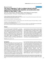
ADAM17-induced proliferation is mediated by TGFαFigure 1
ADAM17-induced proliferation is mediated by TGFα.
a) NHBE cells were treated with rhADAM17 (10 ng/ml) for
1 hr after which surrounding medium was analyzed for the
presence of TGFα by ELISA (n = 3, *p < 0.05 vs. CON). b)
NHBE cells were treated with rhADAM17 (10 ng/ml), IL-13
(10 ng/ml), or TGFα (5 ng/ml) for 24 hrs. [
3
H]-thymidine
incorporation was used as a measure of proliferation (n = 6,
*p < 0.05 vs. CON). c) NHBE cells were treated with IL-13
(10 ng/ml), ADAM17 (10 ng/ml) or ADAM17 plus neutraliz-
ing anti-TGFα antibody (0.2 μg/ml) for 24 hrs, with [
3
H]-thy-
midine incorporation used as a measure of proliferation (n =
6, *p < 0.05 vs. CON).
sTGF
α
α
α
α (pg/ml)
0
10
20
30
40
50
CON
ADAM17
*
a
b
c
Cpm / culture
0
10000
20000
30000
40000
*
*
Cpm / culture
0
10000
30000
50000
*
A
D
A
M
1
7
C
O
N
I
L
-
1
3
*
*
T
G
F
α
α
α
α
I
L
-
1
3
CO
N
A
D
A
M
1
7
AD
A
M
1
7
+
a
n
t
i
-
T
GF
α
α
α
α
Respiratory Research 2007, 8:51 />Page 5 of 12
(page number not for citation purposes)
via cleavage of surface expressed TGFα, rather than via
cleavage of another growth factor. The addition of neutral-
izing anti-TGFα antibody attenuated the proliferative
effect induced by exogenous rhADAM17 (Fig. 1c) suggest-
ing that rhADAM17 cleaves surface-expressed TGFα, that
in turn induces proliferation of the epithelial cells.
ADAM17 mediates IL-13-induced proliferation of NHBE
cells
After determining that exogenous ADAM17 can induce
cellular proliferation mediated by TGFα in NHBE cells, we
determined whether endogenous ADAM17 is involved in
IL-13-induced proliferation of these cells. First, the effects
of various inhibitors of ADAM17 on IL-13-induced shed-
ding of TGFα were examined. Tissue inhibitor of metallo-
proteinase (TIMP)-3 is a documented inhibitor of
ADAM17 [39,40], while a related family member, TIMP-
1, has been found to have no effect on ADAM17 [41]. Fur-
thermore, the differential inhibition of ADAM17 by the
two TIMPs is useful to distinguish the action of ADAM17
from that of ADAM10, whose activity can be inhibited by
both TIMP-3 and TIMP-1 [41]. In the current study, TIMP-
3 was found to attenuate IL-13-induced TGFα shedding,
while TIMP-1 did not have an inhibitory effect (Fig. 2a).
Additionally, anti-ADAM17 antibodies also blocked IL-
13-induced TGFα shedding (Fig. 2b). Thus, these data
support the role of ADAM17 in mediating IL-13-induced
TGFα shedding in NHBE cells.
To confirm the requirement of ADAM17 in mediating IL-
13-induced TGFα shedding, and to determine whether
ADAM17 is similarly required for IL-13-induced NHBE
cell proliferation, cells were exposed to antisense oligonu-
cleotides directed against ADAM17 or to scrambled oligo-
nucleotides as a control. Scrambled oligonucleotides had
little effect on ADAM17 expression in a culture exposed to
media and in another culture exposed to IL-13; however,
in the same experiment, decreased expression of ADAM17
was easily discernible in comparable cultures exposed to
antisense oligonucleotides directed against the protease
(Fig. 3a). In cultures similarly exposed in this same exper-
iment, ADAM17 antisense oligonucleotides inhibited IL-
13-induced NHBE cell proliferation (Fig. 3b) and inhib-
ited IL-13-induced, as well as constitutive, shedding of
TGFα (Fig. 3c). ADAM17 antisense oligonucleotides,
however, did not inhibit TGFα-induced proliferation (Fig.
3b). In all experiments, scrambled oligonucleotides had
no significant effect on growth of control cells or on their
constitutive release of TGFα (Figs. 3b and 3c). Further-
more, while the presence of scrambled or ADAM17 anti-
sense oligonucleotides reduced the maximal level of
proliferation inducible by IL-13 or TGFα, only the
ADAM17 antisense oligonucleotides were capable of
blocking IL-13-induced proliferation with specificity, as
these oligonucleotides had no effect on TGFα-induced
proliferation (Fig. 3b). Taken together, these results sup-
port the requirement of endogenous ADAM17 for IL-13-
induced proliferation of NHBE cells, and confirm that
ADAM17 plays a role in the shedding of TGFα in NHBE
cells.
IL-13-induced effects are not mediated solely via
activation of ADAM17
Since ADAM17 appeared to mediate IL-13-induced TGFα
shedding and proliferation in NHBE cells, we wanted to
determine whether these effects were due to a simple IL-
13-induced increase in ADAM17, or its activity. The
amount of steady-state mRNA coding for ADAM17 in
control or IL-13-treated cells was found to be the same fol-
lowing 4 or 24 hrs of treatment (Fig. 4a). Next the amount
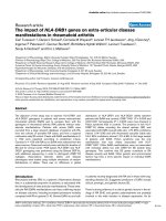
Inhibitors of ADAM17 attenuate IL-13-induced shedding of TGFαFigure 2
Inhibitors of ADAM17 attenuate IL-13-induced shed-
ding of TGFα. NHBE cells were exposed to control media,
inhibitors of ADAM17, IL-13 or IL-13 plus inhibitors for 1 hr.
a) NHBE cells were exposed to either control media (no
inhibitor), TIMP-1 or TIMP-3 (both at 2 μg/ml) for 30 min
prior to treatment with IL-13 (10 ng/ml) or control media.
The inhibitors were also included during the treatment
period. After the 1 hr treatment, supernatants were exam-
ined for TGFα shedding via ELISA (n = 4, *p < 0.05 vs. corre-
sponding control, †p < 0.05 vs. IL-13 alone). Light gray bars =
TIMP-1; Dark gray bars = TIMP-3. b) NHBE cells were
exposed to control media, anti-ADAM17 antibodies, IL-13,
or IL-13 plus anti-ADAM17 for 1 hr. Supernatants were then
examined for shed TGFα via ELISA (n = 6, *p < 0.05 vs.
media control, †p < 0.05 vs. IL-13 alone).
TIMP-1
TIMP-3
No Inhibitor
0
4
8
12
16
IL-13
*
*
†*
Control
a
0
20
40
60
0 0 10 10 IL-13 (ng/ml)
0 0.2 0 0.2 anti-ADAM17 (μ
μμ
μg/ml)
*
†
b
sTGF
α
α
α
α (pg/ml)
sTGF
α
α
α
α (pg/ml)
*
Respiratory Research 2007, 8:51 />Page 6 of 12
(page number not for citation purposes)
of ADAM17 protein was examined. This protein exists in
two forms, an inactive, latent form and an active form
[32]. Conversion to the active form requires proteolytic
cleavage of the enzyme, resulting in removal of a 20-kDa
section of the protein. The amount of latent ADAM17 in
NHBE cells varied little in response to control media or IL-
13 over a time course of 5 min to 24 hrs (Fig. 4b). The
amount of active ADAM17 in control cells during this
time period also varied little, while slightly less active
ADAM17 was observed at early time points in IL-13-
treated cells. The amount of active ADAM17 in these
treated cells, however, was similar to control levels at the
latter time points (1 to 24 hrs) (Fig. 4b). Thus, while IL-13
may induce a small, transient decrease in the amount of
active ADAM17, the quantity of active protein is no
greater than that observed in control cells at time points
when IL-13 induces an increase in soluble TGFα (i.e.
approximately 60 min in this study (Fig. 4c), and as early
as 15 min in a previous study [6]). These data show that
IL-13 does not induce a dramatic alteration in the amount
of ADAM17 mRNA, latent ADAM17, or active ADAM17 in
NHBE cells.
Since activation of ADAM17 and ADAM17-mediated
shedding can be induced via PKC stimulation [26,42], we
tried to enhance the shedding of TGFα by exposing NHBE
cells to phorbol-12-myristate 13-acetate (PMA), a known
activator of PKC and well-characterized inducer of TGFα
ectodomain shedding [43], at a concentration shown pre-
viously to enhance TGFα shedding in a pulmonary
mucoepidermoid carcinoma cell line (NCI-H292) [30].
Exposure of the NHBE cells to PMA, however, did not
yield an increase in soluble TGFα (Fig. 4c) or cellular pro-
liferation (Fig. 4d), even though IL-13 could still induce
these events. The NHBE cells did respond to the PMA,
however, as secretion of IL-8, a process known to be PKC-
dependent in NHBE cells [44], was enhanced while IL-13
had no effect on IL-8 secretion (Fig. 4e). Thus, these
results suggest that the mechanism mediating IL-13-
induced release of soluble TGFα from NHBE cells differs
from the PKC-mediated mechanism responsible for TGFα
shedding in NCI-H292 cells, an event which appears to
involve direct activation of ADAM17 by PKC [30]. Thus, it
appears that although the IL-13-induced increase in TGFα
shedding, as well as the IL-13-induced proliferation, is
mediated by ADAM17 in NHBE cells, these events do not
occur solely via an IL-13-induced increase in ADAM17 or
its activity.
IL-13 induces apical movement of intracellular TGF
α
An alternate mechanism whereby IL-13 could increase the
amount of TGFα shed from NHBE cells would be for the
cytokine to promote the release of pre-formed, intracellu-
lar growth factor. NHBE cells are already known to release
pre-formed mucin proteins (the glycoprotein component
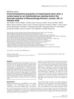
Blocking endogenous ADAM17 inhibits IL-13-induced effectsFigure 3
Blocking endogenous ADAM17 inhibits IL-13-induced
effects. Antisense oligonucleotides directed against ADAM17
(antisense), or corresponding scrambled oligonucleotides (scram-
bled), were added to NHBE cell cultures for 2 days. Cultures con-
taining no oligonucleotides received the transfection reagent
(FuGene6) during this time. On the third day, cells were exposed
to control media, IL-13 (10 ng/ml), or TGFα (5 ng/ml), with or
without the addition of the scrambled or antisense oligonucle-
otides for 24 hrs. a) Total protein was extracted from a single cul-
ture from each treatment group and from the FuGene-only
control group. ADAM17 was immunoprecipitated from these
extracts and subjected to Western analysis (A = antisense oligo-
nucleotides; Sc = scrambled oligonucleotides; 10 μM). The per-
centage of ADAM17 in experimental cultures compared to a
FuGene-only exposed culture (Fugene) was determined by densit-
ometry as indicated (left panel). The right panel was overexposed
to verify the location of the two, expected ADAM17 bands. Both
blots reveal decreased expression of ADAM17 in the two cultures
exposed to antisense oligonucleotides. b) Cell number was deter-
mined as a measure of proliferation (n = 6, *p < 0.05 compared to
appropriate control, †p < 0.01 compared to appropriate IL-13-
treated, scrambled oligo sample), and c) the amount of TGFα in
the supernatant was quantified via ELISA (n = 4, *p < 0.05 com-
pared to appropriate control, †p < 0.01 compared to appropriate
treated, scrambled oligo sample).
Fugene Media Media
Fugene IL-13 IL-13 IL-13
b
Cells / culture x 10
5
0
4
8
12
16
0 5 2.5 5 2.5
μM oligo
Antisense Scrambled
*
*
*
*
*
*
*
Con
*
†
TGFα
c
Antisense Scrambled
0 10 5 10 5 μM oligo
sTGFα (pg/ml)
0
5
10
15
20
†
*
*
*
Con IL-13
a
ADAM17
Sc + A +
Sc + A +
Sc + A + IL-13
IL-13 IL-13
% Fugene
0
100
50
IL-13
Respiratory Research 2007, 8:51 />Page 7 of 12
(page number not for citation purposes)
of airway mucus) upon stimulation with various inflam-
matory mediators [36,45]. Under such conditions, gran-
ules containing the mucin proteins are thought to be
mobilized rapidly to the cell surface where the proteins
are secreted [36]. To determine whether a similar mecha-
nism mediates IL-13-induced release of TGFα, confocal
microscopy was used to examine the location of TGFα and
its sheddase, ADAM17, in NHBE cells exposed to IL-13 or
control media over a 4-hr time course. (Quantitative
results from this study are shown in Table 1.)
Untreated NHBE cells (data not shown), or NHBE cells
exposed only to control media (Figs. 5 and 6a; Table 1),
were found to express TGFα and ADAM17 constitutively.
The majority of the growth factor (TGFα) was localized to
the interior of the epithelial cells, with ample expression
observed in the basal and central regions of the cells. Little
expression of TGFα was observed in apical cellular
regions. By contrast, ADAM17 was expressed throughout
the cytoplasm, although the majority of this enzyme was
expressed in portions of the cytoplasm adjacent to the cell
membrane, with expression particularly prominent in the
apical region of the epithelial cells. In fact, about 80% of
control cells exhibited this pattern of expression which
remained relatively unchanged as NHBE cells were
exposed to fresh media for 15 min, 30 min, 1 hr, or 4 hrs
(Figs. 5 and 6a; Table 1). More precisely, the percentage of
media-exposed, control cells exhibiting this expression
pattern (TGFα interior with ADAM17 highly expressed in
the apical region) at these time points was 81%, 82%,
71%, and 88%, respectively (see Table 1). The cross-sec-
tion and Z-stack video images of the control cells (Fig. 6a
and Additional file 1, respectively) as well as an illustra-
tion of a control cell (Fig. 6b) summarize the observed
location of TGFα (red) and ADAM17 (green) in cells with-
out IL-13 stimulation.
While exposure of NHBE cells to IL-13 for 15 min did not
alter the location of TGFα expression compared to its loca-
tion within control cells (Fig. 5), continued exposure to
IL-13 for 30 min or more did induce an alteration in the
location of TGFα expression. Specifically, at 30 min,
patches of TGFα were less defined within the cytoplasm,
with almost no TGFα expression detectable in the basal
areas of IL-13-exposed cells. Rather, the majority of the
growth factor was expressed in the apical region and on
the apical surface of the NHBE cells (Fig. 5). This pattern
of apical TGFα localization was observed in 46% of the IL-
13-treated cells compared to just 18% of the control cells
(Table 1). While IL-13 induced increased apical localiza-
tion of TGFα, apical localization of ADAM17 was
Table 1: Percentage of NHBE cells with specified patterns of
TGFα/ADAM17 expression following IL-13 stimulation.
EXPRESSION PATTERN
Time point % Cells with
Apical TGFα
% Cells with Apical
ADAM17
15 min CON 19 81
IL-13 20 80
30 min CON 18 82
IL-13 46 54
60 min CON 29 71
IL-13 35 65
4 hrs CON 12 88
IL-13 298
Cells were examined by confocal microscopy to determine whether
expression of TGFα or ADAM17 was greater in the apical-most,
middle and basal-most sections of the Z-stack images. "Expression
pattern" refers to expression noted within [apical/middle/basal]
regions on an NHBE cell. "Apical TGFα " refers to a pattern of
[TGFα/TGFα/ADAM17]. "Apical ADAM17" refers to a pattern of
[ADAM17/ADAM17/TGFα].
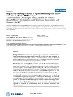
IL-13-induced effects are not due solely to activation of ADAM17Figure 4
IL-13-induced effects are not due solely to activation
of ADAM17. a) NHBE cells were exposed to IL-13 (10 ng/
ml) or control media for 4 or 24 hrs, and steady-state mRNA
levels of ADAM17 and β-actin determined via RT-PCR.
Ethidium bromide-stained gels of PCR products are shown.
b) NHBE cells were treated with control media or IL-13 for
the times indicated. Total protein from these cells was exam-
ined for ADAM17 expression via Western blot. Membranes
were chemically stripped and rehybridized to detect β-actin
as a control for equal protein loading. c) NHBE cells were
treated with control media, IL-13, or PMA (10 nM) for 1 hr
and the supernatants examined for soluble TGFα via ELISA
(n = 4, *p < 0.05 compared to control). d) NHBE cells were
treated for 24 hrs with control media, IL-13, or PMA (10
nM), and [
3
H]-thymidine incorporation determined as a
measure of proliferation (n = 6, *p < 0.05 compared to con-
trol). e) Secretion of IL-8 from NHBE cells was examined by
ELISA following 1 hr exposure to control media, IL-13, or
PMA (10 nM) (n = 6, *p < 0.05 compared to control).
e
IL-8 (ng/ml)
0
5
10
15
20
25
CON IL-13 PMA
*
CON IL-13 PMA
Cpm/culture
0
20000
40000
60000
80000
10000
*
d
c
0
10
20
30
40
50
CON IL-13 PMA
*
sTGF
α
α
α
α (pg/ml)
a
β
ββ
β-actin
ADAM17
- + - + IL-13
4 24 hrs
b
Con
IL-13
Time (min)
5 15 30 60 360 1440
Active
β
ββ
β-actin
β
ββ
β-actin
ADAM17 Latent
ADAM17 Latent
Active
Respiratory Research 2007, 8:51 />Page 8 of 12
(page number not for citation purposes)
observed in fewer cells (54% compared to 82% of control
cells) following IL-13 exposure, with the enzyme now
found to a greater extent in the middle and basal regions
of the NHBE cells. Thus, it would appear that when NHBE
cells are exposed to IL-13, localization of TGFα shifts to
the apical region of these cells within 15 to 30 min. Such
a finding would be consistent with the movement of
TGFα from its intracellular region of constitutive expres-
sion (middle and basal) into the apical region of these
cells, a region where prominent ADAM17 expression is
observed constitutively.
Following exposure of NHBE cells to IL-13 for 60 min, the
expression patterns of both TGFα and ADAM17 remained
similar to those observed in cells exposed to IL-13 for 30
min (Fig. 6a; Table 1; see Additional file 2); more treated
cells expressed TGFα in their apical regions (35% com-
pared to 29% of control cells) while fewer treated cells
expressed ADAM17 apically (65% compared to 71% of
control cells). However, the percentage of affected cells
appeared somewhat intermediate between the 15 min
and the 30 min-treated values. This finding may suggest
that the TGFα, whose apical expression was induced by IL-
13, is beginning to be cleaved from the cell, while
ADAM17 is being internalized.
Following a 4-hr exposure to IL-13, little TGFα remains
within most of the NHBE cells. In fact, 98% of the treated
cells, compared to 88% of the control cells, express mainly
ADAM17 with little to no TGFα expression found at any
level within the cells. The relatively small amount of
growth factor that is present appears to be expressed in the
intracellular regions where TGFα was maintained prior to
stimulation (middle or basal region of the cells). Con-
versely, more control cells (12%) express TGFα in their
apical regions compared to IL-13-treated cells (2%). This
dramatic shift from 35 – 46% of IL-13-treated cells
expressing TGFα apically at 30 – 60 min, to just 2% of the
Summary of TGFα and ADAM17 expression patterns induced by IL-13Figure 6
Summary of TGFα and ADAM17 expression patterns
induced by IL-13. a) Confocal images (y-z plane; apical to
basal cross-section) of NHBE cells exposed for 60 min to
media alone (control) or IL-13 (10 ng/ml). See Additional files
1 and 2 for movies of Z-stack images (basal to apical) taken
from a control culture and an IL-13-treated culture, respec-
tively, at this time point. TGFα (red) and ADAM17 (green);
scale bars represent 10 μm. b) Illustration summarizing
expression patterns of TGFα and ADAM17 observed via
confocal microscopy in IL-13-treated NHBE cells at the times
indicated. Colors represent TGFα (red) and ADAM17
(green).
60 Min
Control
IL-13 Treated
a
b
TGFα and ADAM17 expression patterns are consistent with IL-13-induced movement of TGFαFigure 5
TGFα and ADAM17 expression patterns are consist-
ent with IL-13-induced movement of TGFα. Confocal
microscopy was used to determine the cellular distribution
of TGFα and ADAM17 in NHBE cells following stimulation
with IL-13 for various lengths of time. Representative images
from cultures of NHBE cells treated with media only (con-
trol) or IL-13 (10 ng/ml) for 15 or 30 min are shown. NHBE
cultures were imaged in Z-stack mode from the basal to the
apical boundaries of the cells. Images shown are x-y planes
(large squares) halfway between the basal-most and the api-
cal-most images, bordered by corresponding y-z planes
(shown at right of x-y plane) and x-z planes (shown at bot-
tom of x-y plane). The y-z and x-z plane images are from the
sites indicated by the white arrows at the bottom and the
right of the x-y plane images, respectively. a → b denotes the
apical (a) to basal (b) direction as it relates to the x-z and y-z
planes. TGFα (red) and ADAM17 (green); scale bars repre-
sent 10 μm.
Control IL-13 Treated
15 Min
b a
b
a
30 Min
Respiratory Research 2007, 8:51 />Page 9 of 12
(page number not for citation purposes)
treated cells expressing it at 4 hrs, is consistent with the
apical TGFα being cleaved and released from the cells.
Taken together, the confocal images (examples provided
in Figs. 5 and 6a) and quantitative analysis (Table 1) of
TGFα and ADAM17 expression in NHBE cells support the
conclusion that IL-13 can induce movement of a stored
growth factor (TGFα) from the central and basal cytoplas-
mic regions to the apical region of airway epithelial cells,
where it is cleaved by ADAM17. Fig. 6b illustrates the tim-
ing of this inducible translocation, with an increase in
TGFα near the apical surface observed by 30 – 60 min of
IL-13 exposure, with the growth factor co-localizing with
ADAM17 in this region. By 4 hrs of IL-13 exposure, very
little TGFα is observed within the cells, likely due to its
being cleaved from the apical surface by ADAM17, follow-
ing its IL-13-induced translocation.
Discussion
In this study, we report what appears to be the first
cytokine-induced redistribution of a growth factor (TGFα)
from an intracellular store to the apical surface of a cell,
where a protease required for shedding of the growth fac-
tor (ADAM17) is prominently expressed. Having demon-
strated previously that IL-13-induced proliferation of
NHBE cells is mediated by TGFα [6], this report extends
those results by establishing that ADAM17 is required for
both IL-13-induced proliferation and TGFα shedding in
these cells. This conclusion is supported by data demon-
strating that the proliferation and growth factor shedding
are inhibited by antisense oligonucleotides directed
against ADAM17, while rhADAM17-induced prolifera-
tion of NHBE cells can also be blocked with neutralizing
anti-TGFα antibodies. In examining the mechanism
whereby IL-13 induces these ADAM17-mediated events, a
dramatic activation of ADAM17 was not observed; rather,
IL-13 induced a change in the location of TGFα expression
in 30 to 60 min, with expression shifted to the apical
region of the NHBE cells where significant ADAM17
expression is observed constitutively. A slight increase in
the expression of ADAM17 was also observed within the
middle and basal regions of the cells following IL-13 stim-
ulation; this observed increase may be relative, as it is pos-
sible that apically-located sheddase is released along with
the cleaved growth factor. Alternatively, ADAM17 may be
internalized, an event known to occur with PMA-stimula-
tion [46].
While a short exposure to IL-13 appears to induce a rapid
redistribution of TGFα in NHBE cells, by 4 hrs of exposure
to the cytokine only a small amount of the growth factor
is observed within the cells and that within the basal
region. While low-level synthesis of TGFα may occur con-
tinuously in NHBE cells, regardless of stimulation, it is
also possible that new intracellular stores of the growth
factor must be synthesized following IL-13-induced cleav-
age of apically-located TGFα.
Implications of the novel IL-13-induced mechanism
directing TGFα to the apical region/surface of NHBE cells
are broad-reaching, having the potential to provide
insight not only into the role of epithelial cells in allergic
asthma, but also into the impact of intracellular growth
factor pools in a variety of cell types and diseases. Such
intracellular stores are known to exist in neutrophils and
monocytes where TGFα appears to be stored in mem-
brane-bound compartments [33]. Intracellular stores of
EGF have been similarly reported in human submandibu-
lar and parotid glands [47,48]. There is not, however, a
complete understanding of the cellular mechanisms acti-
vating these stores, particularly in response to inflamma-
tory stimuli.
By contrast, some growth factors, rather than being stored
in intracellular compartments, are known to sort to vari-
ous surfaces of polarized epithelial cells immediately fol-
lowing translation. For example, in Madin-Darby canine
kidney cells, pro-TGFα sorts to the basolateral surface in a
process requiring specific domains within the newly trans-
lated protein [49] and interaction with specific cytoplas-
mic proteins [50]. Similar sorting of another EGF family
ligand, heregulin-α, also appears to occur in human bron-
chial epithelial cells [51]. EGF, however, has been found
to sort to both apical and basolateral surfaces of polarized
epithelial cells where it is released into the medium sur-
rounding the cells. Differential activation of this growth
factor then occurs due either to the presence, or activity, of
metalloproteinases within the extracellular compartments
around the cells [52].
In a similar fashion, the constitutive expression of acti-
vated ADAM17, occurring mainly in defined apical and
lateral regions of NHBE cells, could result in constitutive
release of TGFα during exponential and stationary growth
of these cells. Constitutive release of TGFα is observed in
unstimulated NHBE cells in vitro [6], where it appears to
be mediated by ADAM17 (Fig. 3c). Although the present
study does not distinguish the continuous presence of a
small amount of TGFα in the cell membrane from a slow
sorting of intracellular growth factor to this membrane, it
does indicate that TGFα present in the membrane of a
resting cell can be cleaved when it encounters activated
ADAM17. Specifically, addition of a large excess of exoge-
nous rhADAM17, which ensures a high probability of
cleaving all TGFα in the membrane, results in a significant
increase in soluble TGFα compared to control levels (Fig.
1a). This cleavage and release of TGFα by exogenous
ADAM17 is similar to that observed previously using cell
membrane preparations [38].
Respiratory Research 2007, 8:51 />Page 10 of 12
(page number not for citation purposes)
While constitutive release of TGFα may be important for
general maintenance of an epithelial barrier, it is the
inducible nature of TGFα redistribution that likely con-
tributes to the role of airway epithelial cells as rapid "effec-
tors" following a provocation, such as inhalation of an
allergen to which the host is sensitized. By maintaining
intracellular reserves of growth factors, and perhaps other
molecules, as well as the constitutive expression of pro-
teases that activate these factors, the reaction time in
response to inflammatory stress and other epithelial inju-
ries can be minimized. This inducible system also pro-
vides a number of safeguards to ensure the cell will be
both equipped to respond to a stimulus and to direct that
response in a specified manner. For example, the mainte-
nance of intracellular growth factor reserves eliminates the
possibility of surface-tethered molecules being inadvert-
ently cleaved prior to their being needed for response to a
specific biological insult. Such unintentional cleavage
events could occur as neutrophil elastase or other pro-
teases become present in the airway as a natural response
due to infiltration of inflammatory cells following inhala-
tion of everyday irritants. If growth factors were expressed
constitutively in large amounts on airway epithelial cells,
such proteases might liberate ligands such as TGFα, result-
ing in unwarranted consequences such as upregulation of
mucin gene expression [53] or unnecessary proliferation.
The IL-13-inducible, apparent movement of TGFα from
intracellular basal regions to the apical region/surface of
NHBE cells could also have evolved as a way to lessen the
impact of TGFα on cell types which underlie the epithe-
lium. By keeping the ligand and sheddase separated phys-
ically within the epithelial cells, cleavage of the growth
factor is prevented; even direct PKC activation, an event
known to enhance ADAM17 activity and subsequent
shedding [26,43], was incapable of inducing TGFα release
above constitutive levels in this study. Inducible move-
ment of the growth factor into the apical region where
activated ADAM17 is present, however, would direct the
shedding of TGFα exclusively from the apical surface of
the NHBE cells toward neighboring epithelial cells, or res-
ident and infiltrating inflammatory cells within the epi-
thelium, rather than toward the basally-located
fibroblasts or smooth muscle cells. In this manner, the IL-
13-induced mechanism may provide a means of maximiz-
ing the presence of growth factor near damaged epithelial
cells in an inflamed airway, enhancing the probability of
epithelial barrier restoration without induction of remod-
eling features such as fibrosis or smooth muscle hyperpla-
sia. A related mechanism has been suggested previously
when heregulin-α was observed to be present exclusively
in the apical membrane of human airway epithelia while
its receptors, erbB2-4, were found to be present only on
the basolateral membrane [51]. This arrangement appears
to allow for ligand-receptor interaction only after epithe-
lial integrity is disrupted, or when the tight junctions
between cells are opened.
Conclusion
In NHBE cells, IL-13-induced proliferation and TGFα
shedding are mediated by ADAM17. Surprisingly, IL-13
does not seem to regulate these events by inducing a dra-
matic activation of ADAM17; rather, the cytokine appears
to initiate a change in location of TGFα expression to the
apical region of the cells where ADAM17 is prominently
expressed. Thus, the cytokine appears to induce redistri-
bution of an intracellular store of TGFα into a location
where ADAM17 is expressed constitutively, thereby direct-
ing the apical cleavage and shedding of the growth factor.
Since growth factors exhibit their functions during many
stages of development, cellular differentiation, the heal-
ing process, and inflammatory responses, the finding that
stored growth factors can be released from cells in
response to cytokines is likely to have far-reaching impact.
Such cytokine-induced release may prove essential for
restorative biological functions, yet also mediate deleteri-
ous cellular outcomes as growth factor levels are enhanced
repeatedly during chronic inflammation. Thus, while the
precise mechanism whereby IL-13 induces the movement
of TGFα to the apical surface of NHBE cells remains to be
elucidated, unraveling such a mechanism will likely pro-
vide diverse therapeutic targets for the prevention of air-
way remodeling or the enhancement of epithelial repair.
Abbreviations
IL = interleukin
TGFα = transforming growth factor alpha
ADAM = a disintegrin and metalloproteinase
TNFα = tumor necrosis factor alpha
TACE = TNFα converting enzyme
NHBE = normal human bronchial epithelial
ALI = air/liquid interface
ELISA = enzyme-linked immunosorbent assay
PKC = protein kinase C
NO = nitric oxide
MAP kinase = mitogen activated protein kinase
Erk = extracellular signal regulated kinase
Respiratory Research 2007, 8:51 />Page 11 of 12
(page number not for citation purposes)
PMA = phorbol-12-myristate 13-acetate
rh = recombinant human
EGFR = epidermal growth factor receptor
TIMP = tissue inhibitor of metalloproteinase
Competing interests
The author(s) declare that they have no competing inter-
ests.
Authors' contributions
BWB performed the majority of the studies, participated
in study design and data interpretation, and drafted the
manuscript. TS prepared RNA samples as well as per-
formed and interpreted the RT-PCRs. ELM helped design,
and performed the quantitative analyses of the confocal
studies. LDM provided input and oversight regarding all
aspects of study design and interpretation of results. She
also was responsible for revising and finalizing the manu-
script. All authors read and approved the final manu-
script.
Additional material
Acknowledgements
The authors wish to thank Eve E. Kingsley Booth for exceptional graphic
assistance; Anne L. Crews for technical assistance and helpful discussions;
Drs. Jonathan M. Horowitz and Kenneth B. Adler for helpful discussions;
and Lane Roebuck for editorial assistance. This work was supported by
National Institute of Health grant R01 HL66236 (LDM) and the state of
North Carolina. Portions of this work were performed in partial fulfillment
of the requirements for the degree of Doctor of Philosophy in Comparative
Biomedical Sciences (BWB).
References
1. McDonald DM: Angiogenesis and remodeling of airway vascu-
lature in chronic inflammation. Am J Respir Crit Care Med 2001,
164:S39-45.
2. Fowlkes JL, Winkler MK: Exploring the interface between met-
allo-proteinase activity and growth factor and cytokine bioa-
vailability. Cytokine Growth Factor Rev 2002, 13:277-287.
3. Cohn L, Elias JA, Chupp GL: Asthma: mechanisms of disease
persistence and progression. Annu Rev Immunol 2004,
22:789-815.
4. Chung KF: The role of airway smooth muscle in the pathogen-
esis of airway wall remodeling in chronic obstructive pulmo-
nary disease. Proc Am Thorac Soc 2005, 2:347-354.
5. Podolsky DK: Mucosal immunity and inflammation. V. Innate
mechanisms of mucosal defense and repair: the best offense
is a good defense. Am J Physiol 1999, 277:G495-G499.
6. Booth BW, Adler KB, Bonner JC, Tournier F, Martin LD: Inter-
leukin-13 induces proliferation of human airway epithelial
cells in vitro via a mechanism mediated by transforming
growth factor-alpha. Am J Respir Cell Mol Biol 2001, 25:739-743.
7. Zurawski G, de Vries JE: Interleukin 13, an interleukin 4-like
cytokine that acts on monocytes and B cells, but not on T
cells. Immunol Today 1994, 15:19-26.
8. Wills-Karp M: Interleukin-13 in asthma pathogenesis. Immunol
Rev 2004, 202:175-190.
9. Grunig G, Warnock M, Wakil AE, Venkayya R, Brombacher F, Ren-
nick DM, Sheppard D, Mohrs M, Donaldson DD, Locksley RM, Corry
DB: Requirement for IL-13 independently of IL-4 in experi-
mental asthma. Science 1998, 282:2261-2263.
10. Wills-Karp M, Luyimbazi J, Xu X, Schofield B, Neben TY, Karp CL,
Donaldson DD: Interleukin-13: central mediator of allergic
asthma. Science 1998, 282:2258-2261.
11. Kuperman DA, Huang X, Koth LL, Chang GH, Dolganov GM, Zhu Z,
Elias JA, Sheppard D, Erle DJ: Direct effects of interleukin-13 on
epithelial cells cause airway hyperreactivity and mucus over-
production in asthma. Nat Med 2002, 8:885-889.
12. Lanone S, Zheng T, Zhu Z, Liu W, Lee CG, Ma B, Chen Q, Homer RJ,
Wang J, Rabach LA, Shipley JM, Shapiro SD, Senior RM, Elias JA:
Overlapping and enzyme-specific contributions of matrix
metalloproteinases-9 and -12 in IL-13-induced inflammation
and remodeling. J Clin Invest 2002, 110:463-474.
13. Wen FQ, Kohyama T, Liu X, Zhu YK, Wang H, Kim HJ, Kobayashi T,
Abe S, Spurzem JR, Rennard SI: Interleukin-4- and interleukin-13-
enhanced transforming growth factor-beta2 production in
cultured human bronchial epithelial cells is attenuated by
interferon-gamma. Am J Respir Cell Mol Biol 2002, 26:484-490.
14. Li L, Xia Y, Nguyen A, Lai YH, Feng L, Mosmann TR, Lo D: Effects of
Th2 cytokines on chemokine expression in the lung: IL-13
potently induces eotaxin expression by airway epithelial
cells. J Immunol 1999, 162:2477-2487.
15. Blackburn MR, Lee CG, Young HW, Zhu Z, Chunn JL, Kang MJ, Ban-
erjee SK, Elias JA: Adenosine mediates IL-13-induced inflam-
mation and remodeling in the lung and interacts in an IL-13-
adenosine amplification pathway. J Clin Invest 2003,
112:332-344.
16. Davies DE, Holgate ST: Asthma: the importance of epithelial
mesenchymal communication in pathogenesis. Inflamma-
tion and the airway epithelium in asthma. Int J Biochem Cell Biol
2002, 34:1520-1526.
17. Holgate ST: Epithelial damage and response. Clin Exp Allergy
2000, 30:37-41.
18. Podolsky DK: Regulation of intestinal epithelial proliferation:
a few answers, many questions. Am J Physiol 1993,
264:G179-G186.
19. Kumar VH, Ryan RM: Growth factors in the fetal and neonatal
lung. Front Biosci 2004, 9:464-480.
20. Berkowitz EA, Seroogy KB, Schroeder JA, Russell WE, Evans EP,
Riedel RF, Phillips HK, Harrison CA, Lee DC, Leutteke NC:
Charac-
terization of the mouse transforming growth factor-alpha
gene: its expression during eyelid development and in waved
1 tissues. Cell Growth Differ 1996, 7:1271-1282.
Additional file 1
Confocal Z-stack images of NHBE cells exposed to control media for 60
min. NHBE cells were exposed to control media for 60 min, reacted with
fluorescent-tagged antibodies against TGF
α
(red) and ADAM17 (green),
as described in Materials and methods, and then imaged by confocal
microscopy. Shown is a video of the Z-stack images beginning with the
basal-most section of the NHBE cells and ending with the apical-most sec-
tion.
Click here for file
[ />9921-8-51-S1.zip]
Additional file 2
Confocal Z-stack images of NHBE cells exposed to IL-13 for 60 min.
NHBE cells were exposed to IL-13 (10 ng/ml) for 60 min, reacted with
fluorescent-tagged antibodies against TGF
α
(red) and ADAM17 (green),
as described in Materials and methods, and then imaged by confocal
microscopy. Shown is a video of the Z-stack images beginning with the
basal-most section of the NHBE cells and ending with the apical-most sec-
tion.
Click here for file
[ />9921-8-51-S2.zip]
Publish with BioMed Central and every
scientist can read your work free of charge
"BioMed Central will be the most significant development for
disseminating the results of biomedical research in our lifetime."
Sir Paul Nurse, Cancer Research UK
Your research papers will be:
available free of charge to the entire biomedical community
peer reviewed and published immediately upon acceptance
cited in PubMed and archived on PubMed Central
yours — you keep the copyright
Submit your manuscript here:
/>BioMedcentral
Respiratory Research 2007, 8:51 />Page 12 of 12
(page number not for citation purposes)
21. Jhappan C, Stahle C, Harkins RN, Fausto N, Smith GH, Merlino GT:
TGF alpha overexpression in transgenic mice induces liver
neoplasia and abnormal development of the mammary
gland and pancreas. Cell 1990, 61:1137-1146.
22. Sandgren EP, Luetteke NC, Palmiter RD, Brinster RL, Lee DC: Over-
expression of TGF alpha in transgenic mice: induction of epi-
thelial hyperplasia, pancreatic metaplasia, and carcinoma of
the breast. Cell 1990, 61:1121-1135.
23. Hardie WD, Piljan-Gentle A, Dunlavy MR, Ikegami M, Korfhagen TR:
Dose-dependent lung remodeling in transgenic mice
expressing transforming growth factor-alpha. Am J Physiol
2001, 281:L1088-L1094.
24. Hardie WD, Le Cras TD, Jiang K, Tichelaar JW, Azhar M, Korfhagen
TR: Conditional expression of transforming growth factor-
alpha in adult mouse lung causes pulmonary fibrosis. Am J
Physiol Lung Cell Mol Physiol 2004, 286(4):L741-9.
25. Peschon JJ, Slack JL, Reddy P, Stocking KL, Sunnarborg SW, Lee DC,
Russell WE, Castner BJ, Johnson RS, Fitzner JN, Boyce RW, Nelson
N, Kozlosky CJ, Wolfson MF, Rauch CT, Cerretti DP, Paxton RJ,
March CJ, Black RA: An essential role for ectodomain shedding
in mammalian development. Science 1998, 282:1281-1284.
26. Doedens JR, Mahimkar RM, Black RA: TACE/ADAM-17 enzy-
matic activity is increased in response to cellular stimula-
tion. Biochem Biophys Res Commun 2003, 308:331-338.
27. Zhang Z, Kolls JK, Oliver P, Good D, Schwarzenberger PO, Joshi MS,
Ponthier JL, Lancaster JR: Activation of tumor necrosis factor-
alpha-converting enzyme-mediated ectodomain shedding
by nitric oxide. J Biol Chem 2000, 275:15839-15844.
28. Diaz-Rodriguez E, Montero JC, Esparis-Ogando A, Yuste L, Pandiella
A: Extracellular signal-regulated kinase phosphorylates
tumor necrosis factor alpha-converting enzyme at threonine
735: a potential role in regulated shedding. Mol Biol Cell 2002,
13:2031-2044.
29. L'Allemain G: Deciphering the MAP kinase pathway. Prog
Growth Factor Res 1994, 5:291-334.
30. Shao MX, Ueki IF, Nadel JA: Tumor necrosis factor alpha-con-
verting enzyme mediates MUC5AC mucin expression in cul-
tured human airway epithelial cells. Proc Natl Acad Sci 2003,
100:11618-11623.
31. Yoshisue H, Hasegawa K: Effect of MMP/ADAM inhibitors on
goblet cell hyperplasia in cultured human bronchial epithe-
lial cells. Biosci Biotechnol Biochem 2004, 68:2024-2031.
32. Schlondorff J, Becherer JD, Blobel CP: Intracellular maturation
and localization of the tumour necrosis factor alpha conver-
tase (TACE). Biochem J 2000, 347:131-138.
33. Calafat J, Janssen H, Stahle-Backdahl M, Zuurbier AE, Knol EF, Egesten
A: Human monocytes and neutrophils store transforming
growth factor-alpha in a subpopulation of cytoplasmic gran-
ules. Blood 1997, 90:1255-1266.
34. Egesten A, Calafat J, Knol EF, Janssen H, Watz TM: Subcellular
localization of transforming growth factor-alpha in human
eosinophil granulocytes. Blood 1996, 87:3910-3918.
35. Krunkosky TM, Fischer BM, Martin LD, Jones N, Akley NJ, Adler KB:
Effects of TNF-alpha on expression of ICAM-1 in human air-
way epithelial cells in vitro. Signaling pathways controlling
surface and gene expression. Am J Respir Cell Mol Biol 2000,
22:685-692.
36. Li Y, Martin LD, Spizz G, Adler KB: MARCKS protein is a key
molecule regulating mucin secretion by human airway epi-
thelial cells in vitro. J Biol Chem 2001, 276:40982-40990.
37. Borrell-Pages M, Rojo F, Albanell J, Baselga J, Arribas J: TACE is
required for the activation of EGFR by TGF-alpha in tumors.
EMBO J 2003, 22:1114-1124.
38. Lee DC, Sunnarborg SW, Hinkle CL, Myers TJ, Stevenson MY, Russell
WE, Castner BJ, Gerhart MJ, Paxton RJ, Black RA, Chang A, Jackson
LF: TACE/ADAM17 processing of EGFR ligands indicates a
role as a physiological convertase. Ann NY Acad Sci 2003,
995:22-38.
39. Amour A, Slocombe PM, Webster A, Butler M, Knight CG, Smith BJ,
Stephens PE, Shelley C, Hutton M, Knauper V, Docherty AJ, Murphy
G: TNF-alpha converting enzyme (TACE) is inhibited by
TIMP-3. FEBS Lett 1998, 435:39-44.
40. Lee MH, Knauper V, Becherer JD, Murphy G: Full-length and N-
TIMP-3 display equal inhibitory activities toward TNF-alpha
convertase. Biochem Biophys Res Commun
2001, 280:945-950.
41. Amour A, Knight CG, Webster A, Slocombe PM, Stephens PE, Knau-
per V, Docherty AJ, Murphy G: The in vitro activity of ADAM-10
is inhibited by TIMP-1 and TIMP-3. FEBS Lett 2000,
473:275-279.
42. Arribas J, Lopez-Casillas F, Massague J: Role of the juxtamem-
brane domains of the transforming growth factor-alpha pre-
cursor and the beta-amyloid precursor protein in regulated
ectodomain shedding. J Biol Chem 1997, 272:17160-17165.
43. Arribas J, Massague J: Transforming growth factor-alpha and
beta-amyloid precursor protein share a secretory mecha-
nism. J Cell Biol 1995, 128:433-441.
44. Wyatt TA, Heires AJ, Sanderson SD, Floreani AA: Protein kinase C
activation is required for cigarette smoke-enhanced C5a-
mediated release of interleukin-8 in human bronchial epithe-
lial cells. Am J Respir Cell Mol Biol 1999, 21:283-288.
45. Park JA, He F, Martin LD, Li Y, Chorley BN, Adler KB: Human neu-
trophil elastase induces hypersecretion of mucin from well-
differentiated human bronchial epithelial cells in vitro via a
protein kinase C{delta}-mediated mechanism. Am J Pathol
2005, 167:651-661.
46. Doedens JR, Black RA: Stimulation-induced down-regulation of
tumor necrosis factor-alpha converting enzyme. J Biol Chem
2000, 275:14598-14607.
47. Cossu M, Perra MT, Piludu M, Lantini MS: Subcellular localization
of epidermal growth factor in human submandibular gland.
Histochem J 2000, 32:291-294.
48. Lantini MS, Piludu M, Cossu M: Subcellular localization of epider-
mal growth factor in human parotid gland. Histochem J 2001,
33:427-431.
49. Dempsey PJ, Meise KS, Coffey RJ: Basolateral sorting of trans-
forming growth factor-alpha precursor in polarized epithe-
lial cells: characterization of cytoplasmic domain
determinants. Exp Cell Res 2003, 285:159-174.
50. Franklin JL, Yoshiura K, Dempsey PJ, Bogatcheva G, Jeyakumar L,
Meise KS, Pearsall RS, Threadgill D, Coffey RJ: Identification of
MAGI-3 as a transforming growth factor-alpha tail binding
protein.
Exp Cell Res 2005, 303:457-470.
51. Vermeer PD, Einwalter LA, Moninger TO, Rokhlina T, Kern JA, Zab-
ner J, Welsh MJ: Segregation of receptor and ligand regulates
activation of epithelial growth factor receptor. Nature 2003,
422:322-326.
52. Dempsey PJ, Meise KS, Yoshitake Y, Nishikawa K, Coffey RJ: Apical
enrichment of human EGF precursor in Madin-Darby canine
kidney cells involves preferential basolateral ectodomain
cleavage sensitive to a metalloprotease inhibitor. J Cell Biol
1997, 138:747-758.
53. Kohri K, Ueki IF, Nadel JA: Neutrophil elastase induces mucin
production by ligand-dependent epidermal growth factor
receptor activation. Am J Physiol 2002, 283:L531-L540.