Báo cáo y học: " Vascular endothelial growth factor: an angiogenic factor reflecting airway inflammation in healthy smokers and in patients with bronchitis type of chronic obstructive pulmonary disease?" ppsx
Bạn đang xem bản rút gọn của tài liệu. Xem và tải ngay bản đầy đủ của tài liệu tại đây (459.38 KB, 8 trang )
BioMed Central
Page 1 of 8
(page number not for citation purposes)
Respiratory Research
Open Access
Research
Vascular endothelial growth factor: an angiogenic factor reflecting
airway inflammation in healthy smokers and in patients with
bronchitis type of chronic obstructive pulmonary disease?
Nikoletta Rovina*
1,2
, Andreas Papapetropoulos
2
, Androniki Kollintza
2
,
Makrina Michailidou
1
, Davina CM Simoes
2
, Charis Roussos
1,2
and
Christina Gratziou
1,2
Address:
1
Asthma and Allergy Center, Pulmonary and Critical Care Department, Evgenidion Hospital, Medical School, University of Athens,
Greece and
2
"G. P. Livanos" and "M. Simos" Laboratories, Department of Critical Care and Pulmonary Services, Evangelismos Hospital, University
of Athens, Greece
Email: Nikoletta Rovina* - ; Andreas Papapetropoulos - ;
Androniki Kollintza - ; Makrina Michailidou - ; Davina CM Simoes - ;
Charis Roussos - ; Christina Gratziou -
* Corresponding author
Abstract
Background: Patients with bronchitis type of chronic obstructive pulmonary disease (COPD) have raised
vascular endothelial growth factor (VEGF) levels in induced sputum. This has been associated with the
pathogenesis of COPD through apoptotic and oxidative stress mechanisms. Since, chronic airway inflammation is
an important pathological feature of COPD mainly initiated by cigarette smoking, aim of this study was to assess
smoking as a potential cause of raised airway VEGF levels in bronchitis type COPD and to test the association
between VEGF levels in induced sputum and airway inflammation in these patients.
Methods: 14 current smokers with bronchitis type COPD, 17 asymptomatic current smokers with normal
spirometry and 16 non-smokers were included in the study. VEGF, IL-8, and TNF-α levels in induced sputum were
measured and the correlations between these markers, as well as between VEGF levels and pulmonary function
were assessed.
Results: The median concentrations of VEGF, IL-8, and TNF-α were significantly higher in induced sputum of
COPD patients (1,070 pg/ml, 5.6 ng/ml and 50 pg/ml, respectively) compared to nonsmokers (260 pg/ml, 0.73 ng/
ml, and 15.4 pg/ml, respectively, p < 0.05) and asymptomatic smokers (421 pg/ml, 1.27 ng/ml, p < 0.05, and 18.6
pg/ml, p > 0.05, respectively). Significant correlations were found between VEGF levels and pack years (r = 0.56,
p = 0.046), IL-8 (r = 0.64, p = 0.026) and TNF-α (r = 0.62, p = 0.031) levels both in asymptomatic and COPD
smokers (r = 0.66, p = 0.027, r = 0.67, p = 0.023, and r = 0.82, p = 0.002, respectively). No correlation was found
between VEGF levels in sputum and pulmonary function parameters.
Conclusion: VEGF levels are raised in the airways of both asymptomatic and COPD smokers. The close
correlation observed between VEGF levels in the airways and markers of airway inflammation in healthy smokers
and in smokers with bronchitis type of COPD is suggestive of VEGF as a marker reflecting the inflammatory
process that occurs in smoking subjects without alveolar destruction.
Published: 15 July 2007
Respiratory Research 2007, 8:53 doi:10.1186/1465-9921-8-53
Received: 28 March 2007
Accepted: 15 July 2007
This article is available from: />© 2007 Rovina et al; licensee BioMed Central Ltd.
This is an Open Access article distributed under the terms of the Creative Commons Attribution License ( />),
which permits unrestricted use, distribution, and reproduction in any medium, provided the original work is properly cited.
Respiratory Research 2007, 8:53 />Page 2 of 8
(page number not for citation purposes)
Background
Chronic obstructive pulmonary disease (COPD) is charac-
terized by slowly, progressive and largely irreversible air-
flow limitation due to chronic bronchitis, emphysema, or
both [1]. Long-term cigarette smoking is the most impor-
tant risk factor that may initiate the disease, as a result of
inflammatory cells into the lung (leading to chronic air-
way inflammation), imbalance between proteolytic and
anti-proteolytic activity, oxidative stress and apoptosis
[2].
The appearance of chronic progressive airflow limitation
in part reflects lung remodeling [3]. Vascular endothelial
growth factor (VEGF) is the most potent directly acting
regulator of angiogenesis [4,5], and a trophic factor that is
required for the survival of endothelial cells, inducing
endothelial cell proliferation [6], while its withdrawal
leads to endothelial cell apoptosis [7,8]. VEGF's expres-
sion is often excessive in chronic inflammation and fibro-
sis, and it has been implicated in the pathogenesis of
emphysema through apoptotic and oxidative stress mech-
anisms [9-11]. Cigarette smoking may upregulate VEGF,
as suggested by an acute increase of VEGF plasma levels
during smoking [12]. Although there is increasing evi-
dence of the implication of VEGF in the pathogenesis of
COPD its role at different stages of the disease seems to be
controversial; it is suggested that it has a detrimental func-
tion in the bronchi and a protective role in the alveoli
[13].
Although chronic inflammation is considered the hall-
mark of COPD, little data exists about the role of VEGF in
the inflammatory process involved in the pathogenesis of
the disease, especially in current smokers. On this basis,
the aim of this study was not only to assess VEGF levels in
induced sputum of healthy smokers and of smokers with
bronchitis type of COPD, but to further assess smoking as
a potential cause of raised airway VEGF levels in bronchi-
tis type of COPD, and to test the association between
VEGF levels in induced sputum and airway inflammation
in these subjects.
Methods
Subjects
Fourteen smokers with COPD, seventeen asymptomatic
healthy current smokers and sixteen non-smoking con-
trols were included in the study. All asymptomatic smok-
ers were lifelong smokers (> 15 pack-years), with no
history of lung disease, no chronic respiratory symptoms,
and normal spirometry. All COPD patients were current
smokers (> 15 pack-years), with chronic cough and spu-
tum production over at least 3 months for 2 successive
years, and irreversible airflow limitation (reversibility <
10% predicted forced expiratory volume in 1 second
(FEV
1
) after 200 μg of inhaled salbutamol). All patients
satisfied the ERS criteria [14] for COPD and were selected
according to the Global Initiative for Chronic Obstructive
Lung Disease (GOLD) [15] to fulfill the criteria for GOLD
stages I and II COPD (FEV
1
/FVC ≤ 0.7, and FEV
1
≥ 0.8 and
0.8 ≤ FEV
1
≤ 0.7, respectively) and to have no evidence of
emphysema, based on high-resolution computed tomo-
graphic scans of the lungs and the diffusing capacity of
lung for carbon monoxide (DLCO).
All participants met the following criteria: no use of
inhaled or oral corticosteroids in the previous 6 months,
no atopy (negative skin prick tests for 10 common aeroal-
lergens), and no respiratory tract infection 1 month prior
to the study. None of the COPD patients was ever hospi-
talized due to an exacerbation of COPD. None of the
healthy non-smokers and smokers was receiving either
long acting bronchodilators or leukotriene modifiers.
Nine out of fourteen COPD patients were under treatment
with inhaled tiotropium and inhaled short acting beta
agonists per need, five were receiving inhaled short acting
beta agonists or ipratropium per need, and none was
receiving long acting bronchodilators or leukotriene mod-
ifiers. Before each measurement subjects were asked not to
use long or short-acting β
2
agonists and/or ipratropium at
least 12 hours prior to the tests, and tiotropium 48 hours
prior to the tests.
All subjects gave informed consent for participation in the
study, which was approved by the Hospital ethics com-
mittee.
Measurements
All subjects visited the hospital on 3 separate days, at least
2 days apart. Lung function tests (flow-volume curves,
reversibility test, diffusing lung capacity for carbon mon-
oxide (DLCO), measurement of arterial blood gases, skin
prick tests, and sputum induction were performed.
Lung function
Lung function (FEV
1,
FEV
1
/FVC) was measured with a dry
wedge spirometer (Masterscreen, Jaeger, Hoechberg, Ger-
many) according to standardized guidelines [16], by the
same technician using the same spirometer. Reversibility
test was performed 20 minutes after inhalation of 200 μg
salbutamol via a metered dose inhaler. The diffusing lung
capacity for carbon monoxide (DLCO) was measured by
the single breath method at least twice (Masterscreen,
Jaeger, Hoechberg, Germany).
Sputum induction and processing
Sputum was induced by inhalation of hypertonic saline
aerosol and processed as described previously [17].
Briefly, 15 minutes after salbutamol inhalation (200 μg),
normal saline 0.9% and then hypertonic saline (3%, 4%
and 5%) nebulized by an ultrasonic nebulizer (ULTRA-
Respiratory Research 2007, 8:53 />Page 3 of 8
(page number not for citation purposes)
NEB 2000, DeVilbiss Heathcare INC, Somerset, USA) was
inhaled for each concentration over a period of 7 minutes.
Subjects were encouraged to cough deeply after the 7-
minute intervals. All subjects produced an adequate aliq-
uot of sputum which was processed within 2 hours after
termination of the induction. The sputum was diluted
threefold with phosphate buffer solution containing
dithiothreitol (final concentration: 1 mmol/L) and centri-
fuged at 790 g for 4 minutes (4°C), and the pellet was
resuspended. Slides were made by using cytospin (Cyt-
ospin 3, Shandon, INC, Pittsburgh, USA). Two sputum
cytospin slides were stained with May-Grünwald-Giemsa
for differential cell counts. Counting of 400 non-squa-
mous cells took place in a blinded way by one technician.
Sputum samples containing > 20% of squamous cells
were excluded from analysis as indication of poor cyt-
ospin quality. The supernatant was stored at -80°C for
subsequent assay for IL-8, TNF-α, and VEGF concentra-
tions, which were measured using an enzyme-linked
immunosorbent assay kit (ELISA) (R & D Systems, Minne-
apolis, Minnesota, USA).
Statistical analysis
Data were expressed in mean (± SD) or median values.
Inflammatory markers and VEGF were expressed in
median values and inter-quartile range. Differences
between subjects' groups were initially assessed by
Kruskal-Wallis test, and if significant, the Mann-Whitney
rank test was then assessed. Correlations between inflam-
matory cells and mediators in sputum, smoking character-
istics or lung function parameters were calculated with
Spearman's rank correlation test. Statistical analysis was
not influenced by values at the lower limits of detection
since the non-parametric tests used were based on ranks
of values.
A p value of less than 0.05 was considered significant.
Results
Clinical characteristics of subjects participated in the
study are in Table 1. All subjects were matched for age,
and smokers had similar mean values for smoking pack
years, arterial oxygen tension, DLCO (% pred), FRC (%
pred), RV (% pred), and TLC (% pred). However, FEV
1
and FEV
1
/FVC were significantly lower (p < 0.001, Table
1) in COPD smokers (mean ± SD, 68 ± 11%), compared
to healthy non smokers (106 ± 12%) and asymptomatic
smokers (101 ± 9%).
Sputum
The median (inter-quartile range) total number of cells in
COPD smokers) was higher (though not significantly, p >
0.05) compared to asymptomatic smokers and signifi-
cantly higher (p < 0.05) compared to non smokers (Table
2). Smokers with COPD had higher percentage of sputum
neutrophils compared to asymptomatic smokers (p <
0.05), and non smokers (p < 0.05). In contrast, the per-
centage of sputum macrophages was significantly lower in
COPD smokers compared to asymptomatic smokers (p <
0.05), and non smokers (p < 0.05) (Table 2).
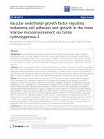
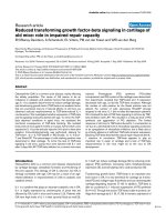
The concentration of VEGF in induced sputum was signif-
icantly higher in COPD smokers than in asymptomatic
smokers (p = 0.024) and healthy non-smokers (p = 0.002)
(Figure 1).
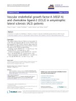
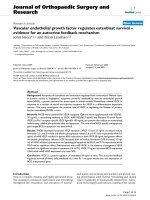
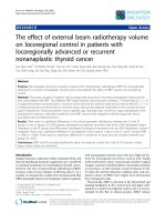
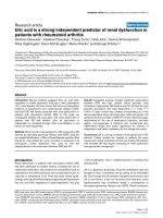
Levels of IL-8 and TNF-α in induced sputum of COPD
smokers [5.6 ng/ml (2.3–10), and 50 pg/ml [17–75],
respectively] were higher compared to asymptomatic
smokers [1.27 ng/ml (0.72–3.2), p = 0.021, and 18.6 pg/
ml [10–35], p = 0.322, respectively] and non smoking
subjects [0.73 ng/ml (0.6–1.4), p = 0.000, and 15.4 pg/ml
[9–25], p = 0.014, respectively] (Figures 2, 3).
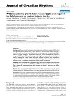
The VEGF levels in induced sputum in both groups of
smokers (asymptomatic and COPD smokers) were signif-
icantly correlated with smoking pack years, with IL-8 and
TNF-α levels (Table 3, Figures 4a and 4b).
No significant correlation was found between VEGF levels
in sputum and pulmonary function parameters in COPD
patients.
Discussion
The main finding of this study is that cigarette smoking is
an important determinant of vascular endothelial growth
factor (VEGF) upregulation in the airways, as assessed by
VEGF's levels in induced sputum, since it correlated signif-
icantly with pack years but not with other clinical and
functional parameters. Furthermore, this VEGF upregula-
tion correlated positively with increased levels of inflam-
matory mediators, such as interleukin-8 (IL-8) and tumor
necrosis factor-α (TNF-α) in sputum not only in mild
COPD smokers, but also in asymptomatic smokers.
It has been long recognized that exposure to cigarette
smoke causes cellular oxidative stress and release of
inflammatory mediators in the airways of healthy subjects
and that these effects can be both acute and chronic [18-
20]. Compared to healthy non-smokers the degree of air-
way inflammation is higher in COPD patients irrespective
of whether these patients are current or ex-smokers [21-
25]. In bronchitis type of COPD airway inflammation is
characterized by an influx of inflammatory cells, predom-
inantly neutrophils, macrophages, and CD8+ T lym-
hocytes, into the airway walls [26], and is associated with
structural alterations including an increase in the amount
of smooth muscle and connective tissue in the airway wall
[27]. Furthermore, previous studies have indicated that
pulmonary arteries in patients with chronic bronchitis
Respiratory Research 2007, 8:53 />Page 4 of 8
(page number not for citation purposes)
have increased adventitial infiltration of activated T lym-
phocytes [28,29]. Therefore, active airway inflammation
might affect pulmonary vascular remodeling in chronic
bronchitis. In turn, angiogenesis of bronchial vasculature,
has been shown to increase the recruitment of inflamma-
tory cells and the exudation of mediators in the airways
[4,27], resulting in a vicious cycle of intracellular signal-
ling between inflammatory and angiogenesis mediators.
Vascular endothelial growth factor (VEGF) is the most
potent directly acting regulator of angiogenesis [4,5]
which is produced by various cell types. Macrophages,
neutrophils, epithelial cells, fibroblasts, and smooth mus-
cle cells are all important sources of VEGF in inflamed tis-
sue [30]. Many inflammatory mediators [prostaglandin E
1
(PGE
1
), PGE
2
, TNF-α, IL-1, IL-6, IL-8, nitric oxide, and
platelet-activating factor], and pathophysiological condi-
tions (hypoxia, pulmonary hypertension) have been
shown to induce the expression of VEGF, angiogenesis, or
both [30,31]. Interestingly, in our study elevated levels of
VEGF were found both in asymptomatic and COPD
smokers, indicating that the stimulus of chronic exposure
to smoke might be the mainstream trigger for the VEGF
upregulation. Cigarette smoking may upregulate the
expression of VEGF in the airways, as suggested by acute
increase in VEGF levels during smoking [32]. Although
the increase in neutrophils by smoking may explain the
increase of VEGF, it may also be the result of increased
VEGF. The lung epithelia can produce VEGF which could
act as a chemokine for neutrophils [33,34].
Conklin and colleagues [35] have also shown that in cur-
rent smokers, nicotine and cotinine upregulate VEGF in
endothelial cells, while Wright and colleagues [36] dem-
onstrated upregulation of VEGF gene expression and its
receptor (Flk-1) in pulmonary arteries of rats exposed to
cigarette smoke.
Although, increased airway VEGF levels were found in
both groups of smokers (healthy and COPD smokers)
these were significantly higher in COPD smokers, proba-
bly due to the effects of current smoking superimposed
upon the ongoing underlying inflammatory process of
COPD. In the present study, higher number of inflamma-
tory cells, percentage of neutrophils and levels of IL-8 and
Table 1: Clinical characteristics and lung function parameters of study subjects
No Healthy non-smokers 16 Asymptomatic smokers 17 COPD smokers n = 14
Age (years) 46 ± 11 47 ± 8 54 ± 9
Smoking (pack-years) 0 34 ± 6 45 ± 17
FEV
1
(% pred) 106 ± 12* 101 ± 9* 68 ± 11
FVC (% pred) 107 ± 10* 108 ± 11* 85 ± 15
FEV
1
/FVC (%) 85 ± 6* 79 ± 6* 64 ± 5
FEF
25–75
(% pred) 94 ± 31* 79 ± 20* 32 ± 10
RV (% pred) 99 ± 13 92 ± 9 85 ± 25
TLC (% pred) 97 ± 8 96 ± 8 89 ± 12
FRC (% pred) 98 ± 16 95 ± 14 95 ± 15
DLCO (% pred) 91 ± 9 85 ± 10 89 ± 11
PaO
2
(mm Hg) - 96 ± 11 81 ± 14
Values are expressed as mean ± SD.
COPD: chronic obstructive pulmonary disease; FEV
1
: forced expiratory volume in one second; FVC: forced vital capacity; FEF
25–75
: forced
expiratory flow 25–75; RV: residual volume; TLC: total lung capacity; FRC: forced residual capacity; DLCO: diffusing lung capacity for carbon
monoxide; PaO
2
: partial pressure of oxygen, arterial.
*p < 0.05, asymptomatic smokers and healthy non-smokers vs COPD smokers
Table 2: Sputum inflammation in asymptomatic and COPD smokers as compared to healthy non-smokers
No Healthy non-smokers 16 Asymptomatic smokers 17 COPD smokers 14
Total no of cells × 10
4
28.5 (23–53)* 41 (27–115) 56 (25–133)
Cell concentration ml × 10
4
14 (11–18)* 22.5 (13–57) 28 (12–66)
Macrophages, % 56 (50–68)* 48 (42–62)* 22 (14–43)
Neutrophils, % 37 (27–47)* 48.5 (34–55)* 70 (47–82)
Lymphocytes, % 3.4 (1.6–5.6) 2.5 (0.6–5.5) 2.5 (0.21–6.4)
Eosinophils, % 0.46 (0.32–0.6) 0.35 (0.1–0.9) 1.25 (0.58–3.2)
Values are median (inter-quartile range).
*p < 0.05 vs COPD smokers; ¶p < 0.05 vs asymptomatic smokers
Respiratory Research 2007, 8:53 />Page 5 of 8
(page number not for citation purposes)
TNF-α were demonstrated in the induced sputum of
COPD smokers compared to asymptomatic smokers, in
concordance with previous observations [24,25,37,38].
The role of TNF-α and IL-8 in smoking induced airway
diseases has been demonstrated in several studies [22-24].
IL-8 is a potent activator of neutrophils [39], while TNF-α
is a powerful pro-inflammatory cytokine that is a key
mediator of inflammation, and has an important role in
fibrogenesis [40]. TNF-α activates macrophages, and epi-
thelial and mesenchymal cells to produce various inflam-
matory cell chemo-attractants such as IL-8, MCP-1, and
leukotriene B
4
[41,42], and has been implicated in the
smoke induced influx of macrophages and connective tis-
sue breakdown [43]. It is suggested that the higher levels
of VEGF found in COPD smokers might be the result of a
cross talk between VEGF and inflammatory mediators
participating in the underlying ongoing pathophysiologic
procedure of the disease. The close correlation found
between VEGF levels and inflammatory mediators which
interfere with smoking related airway disease supports
this suggestion and indicates that VEGF may be actively
implicated in the pathogenesis of COPD commencing at
the initial stages of the disease. However, our results can-
not establish whether the increased levels of VEGF are the
cause or the consequence of increased inflammatory
mediators' levels found in the sputum of both groups of
smokers (healthy and COPD smokers). Hypoxia, a major
factor involved in the induction of VEGF gene expression
does not seem to be implicated here, since the majority of
our patients had normal arterial oxygen tension.
Another aspect reasoning this observation could be that
VEGF and its receptor system may contribute to the main-
tenance of endothelial and epithelial cell viability in
response to injury caused by smoking. This hypothesis is
supported by the finding of Kranenburg and colleagues
Levels of VEGF (pg/ml), expressed as median values (inter-quartile range) in induced sputum of healthy non-smokers (A), asymptomatic smokers (B) and COPD smokers (C)Figure 1
Levels of VEGF (pg/ml), expressed as median values (inter-
quartile range) in induced sputum of healthy non-smokers
(A), asymptomatic smokers (B) and COPD smokers (C). *p
< 0.05, for healthy non-smokers vs COPD smokers; **p <
0.05, for asymptomatic vs COPD smokers.
Levels of IL-8 (ng/ml), expressed as median values (inter-quartile range) in induced sputum of healthy non-smokers (A), asymptomatic smokers (B) and COPD smokers (C)Figure 2
Levels of IL-8 (ng/ml), expressed as median values (inter-
quartile range) in induced sputum of healthy non-smokers
(A), asymptomatic smokers (B) and COPD smokers (C). *p
< 0.05, for healthy non-smokers vs COPD smokers; **p <
0.05, for asymptomatic vs COPD smokers.
Levels of TNF-α (pg/ml), expressed as median values (inter-quartile range) in induced sputum of healthy non-smokers (A), asymptomatic smokers (B) and COPD smokers (C)Figure 3
Levels of TNF-α (pg/ml), expressed as median values (inter-
quartile range) in induced sputum of healthy non-smokers
(A), asymptomatic smokers (B) and COPD smokers (C). *p
< 0.05, for healthy non-smokers vs COPD smokers.
Respiratory Research 2007, 8:53 />Page 6 of 8
(page number not for citation purposes)
[44] that increased VEGF expression and their receptors
(VEGFR-1, also called FLT-1, and VEGFR-2 also called
KDR/Flk-1) were demonstrated in ex-smoking patients
with COPD in comparison with ex-smoking healthy con-
trol subjects. Furthermore, in COPD patients, increased
numbers of macrophages with increased KDR/Flk-1 and
TGF-β expression were found in the bronchiolar airway
epithelium [45]. Taken together, these data suggest that
TGF-β-VEGF represents a molecular link between inflam-
matory cell infiltration at sites of smoking-induced injury
contributing to airway remodeling in COPD through tis-
sue repair mechanisms. It seems that in the lungs of
COPD patients interactions between inflammatory, oxi-
dative stress and apoptotic mechanisms most probably
take place. Recenty, Kanazawa and colleagues [46] sug-
gested that VEGF levels in induced sputum could be pos-
sibly used as a non-invasive marker of pulmonary vascular
remodeling in patients with bronchitis type of COPD,
indicating that VEGF may have a potential role in the
pathogenesis of the vascular changes that take place in this
group of patients. The results of this study support the
findings of the present study, since Kanazawa and col-
leagues hypothesized that vascular remodeling in the pul-
monary arteries of bronchitis type of COPD could be
related to the inflammatory process caused by smoking
and disease.
In the present study no correlation was found between air-
flow limitation and VEGF. A possible explanation could
be that our patients, who had mild bronchitis type of
COPD, were axiomatically homogeneous for disease
severity. In the study of Kanazawa and colleagues [13], the
COPD patients had much lower FEV
1,
and impaired
DLCO compared to our subjects. Furthermore they all
were ex-smokers. This is the first study examining the cor-
relation of VEGF with pulmonary function and inflamma-
tory mediators in current smokers with mild COPD and
intact alveolar structure. If our study included COPD
patients with a wider degree of airflow obstruction, a cor-
relation between VEGF levels and functional parameters
(i.e.FEV
1
and FEV
1
/FVC) might be demonstrated.
Conclusion
In conclusion, cigarette smoking seems to be the major
determinant of vascular endothelial growth factor (VEGF)
upregulation in the airways even before the occurrence of
respiratory symptoms. The fact that VEGF upregulation
positively correlates with the increased levels of inflam-
Spearman's rank correlation: VEGF and IL-8 levels in induced sputum of asymptomatic (r = 0.636, p = 0.026) and COPD smok-ers (r = 0.673 and p = 0.023)Figure 4
Spearman's rank correlation: VEGF and IL-8 levels in induced sputum of asymptomatic (r = 0.636, p = 0.026) and COPD smok-
ers (r = 0.673 and p = 0.023). 4b. Spearman's rank correlation: VEGF and TNF-α levels in induced sputum of asymptomatic (r
= 0.622, p = 0.031) and COPD smokers (r = 0.818 and p = 0.002).
Respiratory Research 2007, 8:53 />Page 7 of 8
(page number not for citation purposes)
matory mediators in sputum not only in bronchitis type
of COPD smokers but also in asymptomatic smokers,
may indicate that VEGF plays an important signalling role
linking the inflammatory milieu with changes in bron-
chial epithelium and endothelium quite early in smokers'
airways. It is likely that, in the progression of COPD, phe-
nomena that are the result of complex regulatory abnor-
malities play a central role, in which VEGF might be a key
factor.
Competing interests
All authors of this paper declare that they have no finan-
cial or other potential conflicts of interest concerning the
subject of this manuscript.
Authors' contributions
NR and MM performed all the clinical measurements of
the study. NR, AP and CG provided intellectual input,
writing and review of the data and paper. NK and DS ana-
lysed the sputum samples. CR reviewed the paper. All
authors read and approved the final manuscript.
Acknowledgements
The authors would like to gratefully thank Christina Sotiropoulou for addi-
tional statistical review.
References
1. Pauwels RA, Buist AS, Calverley PM, Jenkins CR, Hurd SS: Global
strategy for the diagnosis, management, and prevention of
chronic obstructive pulmonary disease. NHLBI/WHO Glo-
bal Initiative for Chronic Obstructive Lung Disease (GOLD)
Workshop summary. Am J Respir Crit Care Med 2001,
163:1256-1276.
2. Barnes PJ, Shapiro SD, Pauwels RA: Chronic obstructive pulmo-
nary disease: molecular and cellular mechanisms. Eur Respir J
2003, 22:672-688.
3. Jeffery PK: Remodeling in asthma and chronic obstructive
lung disease. Am J Respir Crit Care Med 2001, 16:S28-S38.
4. Leung DW, Cachianes G, Kuang WJ, Goeddel DV, Ferrara N: Vascu-
lar endothelial growth factor is a secreted angiogenic
mitogen. Science 1989, 246:1306-1309.
5. Chetta A, Zanini A, Foresis A, Díppolito R, Tipa A, Castagnaro A, Bar-
aldos S, Neri M, Saetta M, Olivieri D: Vascular endothelial growth
factor up-regulation and bronchial wall remodeling in
asthma. Clin Exp Allergy 2005, 35:1437-1442.
6. Neufeld G, Cohen T, Gengrinovitch S, Poltorak Z: Vascular
endothelial growth factor (VEGF) and its receptors. FASEB J
1999, 13:9-22.
7. Aoshiba K, Yokohori N, Nagai A: Alveolar wall apoptosis causes
lung destruction and emphysematous changes. Am J Respir Cell
Mol Biol 2003, 28:555-562.
8. Kasahara Y, Tuder RM, Taraseviciene-Stewart L, Le Cras TD, Abman
S, Hirth PK, Waltenberger J, Voelkel NF: Inhibition of VEGF
receptors causes lung cell apoptosis and emphysema. J Clin
Invest 2000, 106:1311-1319.
9. Kanazawa H, Yoshikawa J: Elevated oxidative stress and recipro-
cal reduction of vascular endothelial growth factor levels
with severity of COPD. Chest 2005, 128:3191-3197.
10. Kasahara Y, Tuder RM, Cool CD, Lynch DA, Flores SC, Voelkel NF:
Endothelial cell death and decreased expression of vascular
endothelial growth factor and vascular endothelial growth
factor receptor 2 in emphysema. Am J Respir Crit Care Med 2001,
163:737-744.
11. Sakao S, Tatsumi K, Hashimoto T, Igari H, Shino Y, Shirasawa H, Kuri-
yama T: Vascular endothelial growth factor and the risk of
smoking-related COPD. Chest 2003, 124:323-327.
12. Wasada T, Kawahara R, Katsumori K, Naruse M, Omori Y: Plasma
concentration of immunoreactive vascular endothelial
growth factor and its relation to smoking. Metabolism 1998,
47:27-30.
13. Kanazawa H, Asai K, Hirata K, Yoshikawa J: Possible effects of vas-
cular endothelial growth factor in the pathogenesis of
chronic obstructive pulmonary disease. Am J Med 2003,
114:354-358.
14. Siafakas NM, Vermeire P, Pride NB, Paoletti P, Gibson J, Howard P,
Yernault JC, Decramer M, Higenbottam T, Postma DS, Rees J: Opti-
mal assessment and management of chronic obstructive pul-
monary disease (COPD). The European Respiratory Society
Task Force. Eur Respir J 1995, 8:1398-1420.
15. Global initiative for Chronic Obstructive Lung Disease NIH.
Definitions chapter I in Global Initiative for Chronic
Obstructive Lung Disease. GOLD 2003:5-10.
16. Quanjer PH, Tammeling GJ, Cotes JE, Pedersen OF, Peslin R, Yernault
JC: Lung volumes and forced ventilatory flows. Report Work-
ing Party Standardization of Lung Function Tests, European
Community for Steel and Coal. Official Statement of the
European Respiratory Society. Eur Respir J Suppl 1993, 16:5-40.
17. Rutgers SR, Timens W, Kaufmann HF, van der Mark TW, Koeter GH,
Postma DS: Comparison of induced sputum with bronchial
wash, bronchoalveolar lavage and bronchial biopsies in
COPD. Eur Respir J 2000, 15:109-115.
18. Mullen JBM, Wright JL, Wiggs BR, Pare PD, Hogg JC: Reassessment
of inflammation of airways in chronic bronchitis. B Med J 1985,
291:1235-1239.
Table 3: Spearman's rank correlations between VEGF levels in induced sputum and smoking pack-years, airway obstruction, and
airway inflammation in asymptomatic and COPD smokers
Asymptomatic smokers n = 17 COPD smokers n = 14
r p-value r p-value
VEGF levels in induced
sputum
Smoking pack-years 0.56 0.046 0.66 0.027
FEV
1
, % pred -0.20 NS -0.13 NS
DLCO, % pred 0.524 NS 0.00 NS
Macrophages in sputum, % -0.261 NS 0.046 NS
Neutrophils in sputum, % 0.246 NS 0.430 NS
IL-8 in sputum, ng/ml 0.636 0.026 0.673 0.023
TNF-α in sputum, pg/ml 0.622 0.031 0.818 0.002
FEV
1
: forced expiratory volume in one second; DLCO: diffusing lung capacity for carbon monoxide; VEGF: vascular endothelial growth factor; IL-8:
interleukin-8; TNF-α: tumor necrosis factor alpha.
Publish with BioMed Central and every
scientist can read your work free of charge
"BioMed Central will be the most significant development for
disseminating the results of biomedical research in our lifetime."
Sir Paul Nurse, Cancer Research UK
Your research papers will be:
available free of charge to the entire biomedical community
peer reviewed and published immediately upon acceptance
cited in PubMed and archived on PubMed Central
yours — you keep the copyright
Submit your manuscript here:
/>BioMedcentral
Respiratory Research 2007, 8:53 />Page 8 of 8
(page number not for citation purposes)
19. Van der Vaart H, Postma DS, Timens W, Ten Hacken NHT: Acute
effects of cigarette smoke on inflammation and oxidative
stress: a review. Thorax 2004, 59:713-721.
20. Saetta M, Di Stefano A, Maestrelli P, Ferraresso A, Drigo R, Potena A,
Ciaccia A, Fabbri LM: Activated T-lymphocytes and macro-
phages in bronchial mucosa of subjects with chronic bronchi-
tis. Am Rev Respir Dis 1993, 147:301-306.
21. O'Shaughnessy TC, Ansari TW, Barnes NC, Jeffery PK: Inflamma-
tion in bronchial biopsies of subjects with chronic bronchitis:
inverse relationship of CD8+ T lymphocytes with FEV1. Am
J Respir Crit Care Med 1997, 155:852-857.
22. Turato G, Di Stefano A, Maestrelli P, Mapp CE, Ruggieri MP, Roggeri
A, Fabbri LM, Saetta M: Effect of smoking cessation on airway
inflammation in chronic bronchitis. Am J Respir Crit Care Med
1995, 152:1262-1267.
23. Linden M, Rasmussen JB, Piitulainen E, Tunek A, Larson M, Tegner H,
Venge P, Laitinen LA, Brattsand R: Airway inflammation in smok-
ers with nonobstructive and obstructive chronic bronchitis.
Am Rev Respir Dis 1993, 148:1226-1232.
24. Willemse BWM, ten Hacken NHT, Rutgers B, Postma DS, Timens W:
Association of current smoking with airway inflammation in
chronic obstructive pulmonary disease and asymptomatic
smokers. Respir Res 2005, 6:38.
25. Keatings VM, Collins PD, Scott DM, Barnes PJ: Differences in inter-
leukin-8 and tumor necrosis factor-alpha in induced sputum
from patients with chronic obstructive pulmonary disease or
asthma. Am J Respir Crit Care Med 1996, 153:530-534.
26. Saetta M, Turato G, Maestrelli P, Mapp CE, Fabbri LM: Cellular and
structural bases of chronic obstructive pulmonary disease.
Am J Respir Crit Care Med 2001, 163:1304-9.
27. Rennard I: Inflammation and repair processes in chronic
obstructive lung disease. Am J Respir Crit Care Med 1999,
160:S12-6.
28. Hale KA, Ewing SL, Gosnell BA, Niewoehner DE: Lung disease in
long-term cigarettes smokers with and without chronic air-
flow obstruction. Am Rev Respir Dis 1984, 130:716-21.
29. Santos S, Peinado VI, Ramirez J, Melgosa T, Roca J, Rodriguez-Roisin
R, Barbera JA: Characterization of pulmonary vascular remod-
eling in smokers and patients with mild COPD. Eur Respir J
2002, 19:632-8.
30. Puxeddu I, Ribatti D, Crivellato E, Levi-Schaffer F: Mechanisms of
asthma and allergic inflammation. Mast cells and eosi-
nophils: a novel link between inflammation and angiogenesis
in allergic diseases. J Allergy Clin Immunol 2005, 116:531-536.
31. Voelkel NF, Cool C, Taraceviene-Stewart L, Geraci MW, Yeager M,
Bull T, Kasper M, Tuder RM: Janus face of vascular endothelial
growth factor: the obligatory survival factor for lung vascular
endothelium controls precapillary artery remodeling in
severe pulmonary hypertension. Crit Care Med 2002, 30(5
suppl):S251-6.
32. Wright JL, Tai H, Churg A: Cigarette smoke induces persisting
increases of vasoactive mediators in pulmonary arteries. Am
J Respir Cell Mol Biol 2004, 31:501-9.
33. Tetley TD: Inflammatory cells and chronic obstructive pulmo-
nary disease. Curr Drug Targets Inflamm Allergy 2005, 4(6):607-18.
Review
34. Morozumi T, Kubota T, Sugita N, Itagaki M, Yoshie H: Alterations
of gene expression in human neutrophils induced by smoking
cessation. J Clin Periodontol 2004, 33:1110-1116.
35. Conklin BS, Zhao W, Zhong D-S, Chen CH: Nicotine and cotinine
upregulate vascular endothelial growth factor expression in
endothelial cells. Am J Pathol 2002, 160:413-418.
36. Wright JL, Tai H, Dai J, Churg A: Cigarette smoke induces rapid
changes in gene expression in pulmonary arteries. Lab Invest
2002, 82:1391-1398.
37. Thompson AB, Daughton D, Robbins RA, Ghafouri MA, Oehlerking
M, Rennard SI: Intraluminar airway inflammation in chronic
bronchitis. Characterization and correlation with clinical
parameters. Am Rev Respir Dis 1989, 140:1527-1537.
38. Takanashi S, Hasegawa Y, Kanehira Y, Yamamoto K, Fujimoto K,
Satoh K, Okamura K: Interleukin-10 level in sputum is reduced
in bronchial asthma, in COPD and in smokers. Eur Respir J
1999, 14:309-314.
39. Peveri P, Walz A, Dewald B, Baggiolini M: A novel neutrophil-acti-
vating factor produced by human mononuclear phagocytes.
J Exp Med 1988, 167:1547-1559.
40. Piguet PF, Collart MA, Grau GE, Kapanci Y, Vassalli P: Tumor
necrosis factor/cachectin plays a key role in bleomycin-
induced pneumopathy and fibrosis. J Exp Med 1989,
170:655-663.
41. Wewers MD, Gadek JL, Marsh EB: Pro-inflammatory cytokines.
In: Crystal RG, Barnes PJ, West JB, Weibel ER, editors. In The
lung 2nd edition. Philadelphia: Lippincott-Raven; 1997:117-132.
42. Fugita M, Shannon JM, Irvin CG, Fagan KA, Cool C, Augustin A, Mason
RJ: Overexpression of tumor necrosis factor-α produces an
increase in lung volumes and pulmonary hypertension. Am J
Physiol 2001, 280:L39-L49.
43. Churg A, Dai J, Tai H, Xie C, Wright J: Tumor necrosis factor-α is
central to acute cigarette smoke-induced inflammation and
connective tissue breakdown. Am J Respir Crit Care Med 2002,
166:849-854.
44. Kranenburg AR, De Boer QI, Alagappan VKT, Sterk PJ, Sharma HS:
Enhanced bronchial expression of vascular endothelial
growth factor and receptors (Flk-1 and Flt-1) in patients with
chronic obstructive pulmonary disease. Thorax 2005,
60:106-113.
45. Fehrenbach J, Kasper M, Haase M, Schuh D, Muller M: Differential
immunolocalization of VEGF in rat and human adult lung,
and in experimental rat lung fibrosis: light fluorescence and
electron microscopy. Anat Rec 1999, 254:61-73.
46. Kanazawa H, Asai K, Nomura S: Vascular endothelial growth fac-
tor as a non-invasive marker of pulmonary vascular remod-
eling in patients with bronchitis-type of COPD. Respir Res
2007, 8:22.