Báo cáo y học: " Endothelin-1 in exhaled breath condensate of allergic asthma patients with exercise-induced " pps
Bạn đang xem bản rút gọn của tài liệu. Xem và tải ngay bản đầy đủ của tài liệu tại đây (521.66 KB, 9 trang )
BioMed Central
Page 1 of 9
(page number not for citation purposes)
Respiratory Research
Open Access
Research
Endothelin-1 in exhaled breath condensate of allergic asthma
patients with exercise-induced bronchoconstriction
Ziemowit Zietkowski*, Roman Skiepko, Maria M Tomasiak and
Anna Bodzenta-Lukaszyk
Address: Department of Allergology and Internal Medicine, Medical University of Bialystok, Poland
Email: Ziemowit Zietkowski* - ; Roman Skiepko - ; Maria M Tomasiak - ;
Anna Bodzenta-Lukaszyk -
* Corresponding author
Abstract
Background: Exercise-induced bronchoconstriction (EIB) is a highly prevalent condition, whose
pathophysiology is not well understood. Endothelins are proinflammatory, profibrotic, broncho-
and vasoconstrictive peptides which play an important role in the development of airway
inflammation and remodeling in asthma. The aim of the study was to evaluate the changes in
endothelin-1 levels in exhaled breath condensate following intensive exercise in asthmatic patients.
Methods: The study was conducted in a group of 19 asthmatic patients (11 with EIB, 8 without
EIB) and 7 healthy volunteers. Changes induced by intensive exercise in the concentrations of
endothelin-1 (ET-1) in exhaled breath condensate (EBC) during 24 hours after an exercise
challenge test were determined. Moreover, the possible correlations of these measurements with
the results of other tests commonly associated with asthma and with the changes of airway
inflammation after exercise were observed.
Results: In asthmatic patients with EIB a statistically significant increase in the concentration of ET-
1 in EBC collected between 10 minutes and 6 hours after an exercise test was observed. The
concentration of ET-1 had returned to its initial level 24 hours after exercise. No effects of the
exercise test on changes in the concentrations of ET-1 in EBC in either asthmatic patients without
EIB or healthy volunteers were observed. A statistically significant correlation between the
maximum increase in ET-1 concentrations in EBC after exercise and either baseline F
ENO
and the
increase in F
ENO
or BHR to histamine 24 hours after exercise in the groups of asthmatics with EIB
was revealed.
Conclusion: The release of ET-1 from bronchial epithelium through the influence of many
inflammatory cells essential in asthma and interactions with other cytokines, may play an important
role in increase of airway inflammation which was observed after postexercise bronchoconstriction
in asthmatic patients.
Background
The airway response to exercise in most asthmatic patients
has been known as a postexercise fall in lung function fol-
lowed by a spontaneous recovery. This classical response
Published: 31 October 2007
Respiratory Research 2007, 8:76 doi:10.1186/1465-9921-8-76
Received: 24 March 2007
Accepted: 31 October 2007
This article is available from: />© 2007 Zietkowski et al; licensee BioMed Central Ltd.
This is an Open Access article distributed under the terms of the Creative Commons Attribution License ( />),
which permits unrestricted use, distribution, and reproduction in any medium, provided the original work is properly cited.
Respiratory Research 2007, 8:76 />Page 2 of 9
(page number not for citation purposes)
is labelled as exercise-induced bronchoconstriction (EIB).
Despite the wide prevalence and clinical significance of
EIB, the mechanisms responsible for it have yet to be
clearly described [1]. Also the findings related to the par-
ticipation of inflammatory mediators in either the main-
tenance or induction of bronchoconstriction provoked by
exercise are conflicting [2].
Endothelins are proinflammatory, profibrotic, broncho-
and vasoconstrictive peptides. Endothelin-1 (ET-1) has
been demonstrated in the airway epithelial and endothe-
lial cells and is involved in the pathogenesis of bronchial
asthma. ET-1 accelerates DNA synthesis and cellular pro-
liferation in human lung fibroblasts. It is also suggested
that ET-1 influences asthmatic inflammation, provoking
concentration and proliferation of bronchial smooth
muscle cells and subepithelial fibrosis. This leads to air-
way remodeling and severe bronchial hyperreactivity [3].
Recent studies suggest the essential role of ET-1 in bron-
choconstriction, mucus secrection, and plasma exudation
[4-7].
In our previous reports, we suggest that during exercise-
induced bronchoconstriction, changes in the function of
the pulmonary endothelium occur [8]. Based on these
findings, it is considered that the release of inflammatory
mediators, such as endothelin-1, as well as adhesion mol-
ecules, through enhancing the migration of inflammatory
cells as well as interactions with other cytokines essential
in asthma, may contribute to the exacerbation of asth-
matic inflammation in the airways and bronchial hyperre-
activity after exercise.
The airway epithelium is involved in allergic inflamma-
tory processes, producing and releasing endothelins,
cytokines, chemokines, and growth factors, as well as
eicosanoides active in the pathophysiology of airway dis-
eases [9]. This study was designed to clarify the possible
role of ET-1 released from bronchial epithelial cells in the
pathogenesis of EIB, particular in the inflammatory basis
of this condition. ET-1 levels were measured in exhaled
breath condensate (EBC), collecting by cooling exhaled
air – noninvasive procedure, easily performed and effort
independent, a rapid method for obtaining samples from
the lower respiratory tract [10].
The aim of the study was to evaluate the changes in ET-1
in EBC following intensive exercise in asthmatic patients
and to establish the possible correlation of these measure-
ments with the parameters of airway inflammation and
their changes after exercise.
Materials and methods
Patients
The study was conducted on a group of 19 mild allergic
asthma patients. Asthma was diagnosed according to the
criteria recommended by the GINA 2002 [11]. All patients
had been in a stable condition, free from acute exacerba-
tions and respiratory tract infections for the previous two
months. Patients with other factors which could change
F
ENO
levels (except for asthma, features of atopy, or aller-
gic rhinitis) were excluded. In all patients the tests were
performed out of pollen season. Prior to the beginning of
this study, patients were allowed to take short- and long-
acting β
2
-agonists. Asthmatic patients who had been
treated with drugs other than β
2
-agonists (inhaled ster-
oids, antileucotrienes) in the past three months, were
excluded from the study. F
ENO
measurement, skin prick
tests with commonly encountered aeroallergens (house
dust mites, trees, weeds, grasses, cat, Alternaria and
Cladosporium), flow/volume spirometry, and a bronchial
provocation test with histamine were performed on each
asthmatic patient before qualifying for the exercise test.
Seven healthy volunteers were used as a negative control
group. All of them underwent F
ENO
, flow/volume spirom-
etry, and skin prick tests with common aeroallergens.
They had FEV
1
> 80% predicted. They were free of respira-
tory tract infection for 2 months prior to the study and
from other significant illnesses known to affect F
ENO
meas-
urements. Asthma patients and healthy volunteers were
non-smokers and during the last year have not been pas-
sive smokers.
Total IgE and peripheral blood eosinophilia were deter-
mined in all asthmatic patients and healthy volunteers. In
all asthmatic patients and healthy volunteers, an exercise
test on the bicycle ergometer was performed.
24 hours after exercise, measurement of F
ENO
and a bron-
chial provocation test with histamine were performed.
The study protocol was approved by the Ethics of Research
Committee of the Medical University of Bialystok, agree-
ment number: R-I-003/80/2006. Informed consent was
obtained from every patient entered into the study.
Measurements
Exhaled nitric oxide (F
ENO
) was measured in all of the
asthma patients and healthy subjects by the chemilumi-
nescence technique using a Sievers 280i NO Analyzer
(Boulder, Colorado, USA). The measurements were per-
formed at an expiratory flow of 50 ml/s [12]. The duration
of exhalation had to be at least 6 seconds to produce a sta-
ble NO level for 3 seconds. All subjects had three recorded
F
ENO
measurements. Repeated measurements were per-
formed until the 3 values agreed within 10% of the mean.
Respiratory Research 2007, 8:76 />Page 3 of 9
(page number not for citation purposes)
The mean value of the three measurements was recorded
as the final F
ENO
level.
The baseline spirometry was performed using a Master-
Screen Pneumo PC spirometer (Jaeger, Hoechberg, Ger-
many). Spirometry was performed according to ATS
standards [13]. FEV
1
(forced expiratory volume in one sec-
ond) was evaluated. Before the examination the patients
did not take any medications that could change spirome-
try results. The highest value from three technically satis-
factory attempts was attached.
A non-specific bronchial provocation test with histamine
(BPT) was carried out according to the method described
by Ryan et al [14]. Provocation was performed using a De
Vilbiss nebuliser 646 (Viasys Healthcare GmbH, Hoech-
berg, Germany) at an air pressure of 0.15 MPa linked to a
Rosenthal-French dosimeter (Baltimore, USA). The results
were presented as PC
20
FEV
1
– concentration of histamine,
which causes a decrease in FEV
1
of exactly 20% in compar-
ison to initial values.
An exercise test was performed on a bicycle ergometer for
9 minutes with a fixed work load adjusted to increase the
heart rate to 85% of the maximum predicted for the age of
each patient [15]. Basic spirometric parameters were
recorded before, and immediately after, the exercise test,
and 1, 5, 10, 15, 20, and 60 minutes after completion of
exercise. Those patients whose maximum decrease in FEV
1
was greater than 15% were considered to have EIB.
EBC was collected by using a condensing chamber (Eco-
Screen; Erich Jaeger GmbH, Hoechberg, Germany).
Exhaled air entered and left the chamber through one-way
valves and the inlet and outlet, thus keeping the chamber
closed. A low temperature inside the condensing chamber
throughout the collection time produced a cooling down
sample. The temperature of collection was around 0°C
[10,16]. Exhaled breath collections were performed
before, 10, 30, 60 minutes, 6 and 24 hours after the exer-
cise challenge test. Patients were instructed to breathe tid-
ally for 10 minutes with nose clip. The respiratory rate
ranged from 15–20 breaths/minute. Patients were asked
to swallow their saliva periodically and to temporalily dis-
continue collection if they needed to cough. At the end of
collection 1.5- to 3.5 ml aliquots of condensate were
transferred to Eppendorf tubes and immediately frozen.
Samples were stored at -80°C [17].
Serum total IgE concentrations was measured using
ImmunoCAP™ Technology (Pharmacia Diagnostics, Upp-
sala, Sweden). Blood eosinophil count was measured
using a hematologic analyzer (Coulter Electronics GmbH,
Miami, Florida, USA). Concentrations of ET-1 in EBC
were determined using enzyme immunoassay kits for
quantitative determination (ET-1 – Biomedica Gruppe,
Vienna, Austria). Detection limit (0 fmol/ml + 3 SD): 0.02
fmol/ml.
Analysis
Statistical significance was analyzed by using analysis of
variance (ANOVA). All values were expressed as means ±
SD; p values < 0.05 were considered significant. PC
20
val-
ues were logarithmically transformed for analysis. The
relationship between studied parameters was assayed by
correlation. Pearson's linear correlation coefficient was
used.
Results
Characteristics of patients and healthy volunteers are pre-
sented in table 1. Table 1.
Table 1: Characteristics of study subjects and healthy volunteers
Characteristics. Dimension. Patients with EIB. Patients without EIB. Differences between
asthma patients with
and without EIB.
Healthy volunteers.
Number of patients 11 8 7
Sex F/M 7/4 5/3 4/3
Age Years 27.36 ± 7.50 31.63 ± 5.40 p = 0.19 28.40 ± 4.90
Duration of symptoms Years 3.70 ± 4.63 4.12 ± 3.54 p = 0.32
Baseline FEV
1
% predicted 95.63 ± 18.54 92.25 ± 8.61 p = 0.63 106.85 ± 9.73
Maximum decrease in FEV
1
after exercise % 25.8 ± 13.5 3.6 ± 1.9 p = 0.0003 0.71 ± 3.2*
+
Log PC20hist FEV
1
mg/ml -0.59 ± 1.16 -0.05 ± 0.55 p = 0.24
Blood eosinophil count cells/mm
3
239 ± 138 157 ± 66 p = 0.14 51 ± 26*
+
Serum total IgE kU/L 358 ± 322 171 ± 69 p = 0.13 65 ± 31*
+
Baseline F
ENO
ppB 98.90 ± 55.37 66.62 ± 23.05 p = 0.21 18.00 ± 5.59*
+
Baseline ET-1 fmol/ml 0.88 ± 0.24 0.74 ± 0.25 p = 0.29 0.59 ± 0.18*
Data are presented as mean ± SD
FEV
1
– forced expiratory volume in one second
PC20histamine FEV
1
– provocative concentration of histamine that caused a 20% fall in FEV
1
* Values significantly different from patients with EIB, p < 0.05
+
Values significantly different from patients without EIB, p < 0.05
Respiratory Research 2007, 8:76 />Page 4 of 9
(page number not for citation purposes)
In the studied group of asthmatics, 11 patients had a pos-
itive and 8 had a negative exercise test. In none of the
healthy volunteers were spirometric indices worse after
exercise.
Blood eosinophilia, baseline F
ENO
and total IgE were sta-
tistically significantly higher in both groups of asthmatics
compared with healthy volunteers. In the group of
patients with positive exercise tests compared to patients
without EIB we observed higher blood eosinophil counts,
serum levels of total IgE and baseline F
ENO
, but these dif-
ferences were not statistically significant.
We revealed statistically significant higher levels of ET-1 in
EBC in all studied asthmatic patients compared with
healthy controls (0.83 fmol/ml ± 0.24 vs. 0.59 ± 0.18, p =
0.02). There was no statistically significant difference
between the concentration of ET-1 in EBC before exercise
in asthmatics patients with EIB in comparison to asthmat-
ics without EIB (0.88 fmol/ml ± 0.24 vs. 0.74 ± 0.25, p =
0.29). In the group of healthy volunteers we observed the
lowest levels of ET-1 in EBC, but this difference was statis-
tically significant only comparing with asthmatics with
EIB (asthma with EIB vs. healthy volunteers: 0.59 fmol/ml
± 0.18, p = 0.018; asthma without EIB vs. healthy volun-
teers: p = 0.13).
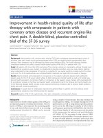
A statistically significant increase in the concentration of
ET-1 in asthmatic patients with EIB was observed (10 min
after exercise: 1.64 fmol/ml ± 1.27, 30 min after exercise:
2.91 fmol/ml ± 1.18, 60 min after exercise: 2.38 fmol/ml
± 0.89, 6 hours after exercise: 1.69 fmol/ml ± 0.78,) (p <
0.001). The concentration of ET-1 had returned to the ini-
tial level 24 hours after exercise (0.98 fmol/ml ± 0.65). No
effects of the exercise test on changes in the concentrations
of ET-1 in EBC in either asthmatic patients without EIB or
healthy volunteers were observed. Figure 1.
There were no statistically significant correlations between
the baseline concentrations of ET-1 in EBC and other
studied parameters in either group of asthmatic patients
or the healthy volunteers and the decrease in FEV
1
after
exercise in asthmatics with EIB.
24 hours after the exercise test, in the group of asthmatics
with EIB, a statistically significant increase in F
ENO
(before
exercise: 98.90 ppB ± 55.37; 24 hours after exercise:
119.18 ± 64.39; p = 0.034) and BHR to histamine (log
Concentrations of ET-1 in EBC at rest, and subsequent changes which were observed during the 24 hours after exercise test in groups of patients with asthma and healthy volunteersFigure 1
Concentrations of ET-1 in EBC at rest, and subsequent changes which were observed during the 24 hours after exercise test in
groups of patients with asthma and healthy volunteers.
Respiratory Research 2007, 8:76 />Page 5 of 9
(page number not for citation purposes)
PC
20
FEV
1
before exercise: -0.59 mg/ml ± 1.16; 24 hours
after exercise: -0.95 ± 1.03; p = 0.0009) was revealed. Fig-
ure 2, Figure 3. Such changes were not observed in the
group of asthmatic patients without EIB (F
ENO
before exer-
cise: 66.62 ppB ± 23.05; 24 hours after exercise: 67.87 ±
23.03; p = 0.25; log PC
20
FEV
1
before exercise: -0.053 mg/
ml ± 0.55; 24 hours after exercise: -0.0511.62 ± 0.59; p =
0.99). In neither group of asthmatics did we detect signif-
icant changes in FEV
1
24 hours after exercise.
A statistically significant correlation between the maxi-
mum increase in ET-1 concentrations in EBC after exercise
and either baseline F
ENO
(r = 0.64, p = 0.03) and the
increase in F
ENO
(r = 0.83, p = 0.001) or the increase of
BHR (expressed as decrease in logPC
20
FEV
1
; r = -0.61, p =
0.04) 24 hours after exercise in the groups of asthmatics
with EIB was revealed. Figure 4.
Discussion
The findings related to the participation of inflammatory
mediators in either the maintenance or induction of bron-
choconstriction provoked by exercise are conflicting.
However, many reports demonstrate that EIB could have
an inflammatory basis [18]. There is no information con-
cerning the late consequences of many years of respiratory
tract stimulation by exercise-induced bronchoconstric-
tion. Epithelial remodeling was previously described in
ski athletes who developed asthma symptoms and bron-
chial hyperreactivity after repeated bouts of exercise in
cold dry air [19].
In our previous studies we revealed that bronchoconstric-
tion following an exercise challenge in asthmatics leads to
pulmonary endothelium changes, which in turn activate
and release mediators (such as endothelin-1), causing the
increase of airway inflammation and, as a consequence,
airway remodeling [8].
In human airways, immunoreactive ET-1 is located princi-
pally in the bronchial epithelium and its expression at this
site is increased in asthma [7,20]. The study of Black et al
has indicated that airway epithelium could produce and
release endothelin [21]. Elevated BAL fluid levels of ET-1
have been observed in asthmatics when compared with
normal control subjects – the highest levels being found
in patients with the most severe disease [22,23]. Except for
human bronchial epithelial cells [24], ET-1 is produced by
vascular endothelial cells [25], and inflammatory cells
such as macrophages [26], mast cells [27], as well as alve-
olar epithelial cells [28].
Many interactions between ET-1 and other cytokines
essential in asthma have been described. Xu et al have
demonstrated that tumor necrosis factor-α (TNF-α) – an
important mediator in initiating airway inflammation by
activating the secretion of cytokines from a variety of cells
– induces secretion of ET-1 from cultured bronchial
smooth muscle cells [29]. ET-1 can induce expression of
granulocyte-macrophage colony-stimulating factor (GM-
CSF) in human lung fibroblasts and, through this, could
directly affect recruitment of eosinophils in the airways
[29]. Cunningham et al have reported that ET-1 stimulates
monocytes to release GM-CSF, IL-6, IL-8, IL-1, TNFα, and
Changes in F
ENO
24 hours after exercise in the groups of asthmatic patientsFigure 2
Changes in F
ENO
24 hours after exercise in the groups of asthmatic patients.
Respiratory Research 2007, 8:76 />Page 6 of 9
(page number not for citation purposes)
TGF-α [30]. ET-1 induces the proliferation and fibrosis of
airway smooth muscle cells. The interaction between ET-1
and other cytokines which are growth factors for bron-
chial subepithelial myofibroblasts may play a key role in
remodeling in asthmatic patients, which is the conse-
quence of repeated episodes of epithelial damage and
repair in asthmatic inflammation [31]. In response to
mechanical stresses similar to those occuring in vivo dur-
ing airway constriction, increases in soluble levels of ET-1
and TGF-β1 have been observed [32].
ET-1 may contribute significantly to the remodeling of the
airway by slowing epithelial cell migration as well as
increasing proliferation of airway fibroblasts and smooth
muscle cells. In turn, this process results in delayed repair
and enhanced fibroblast activation and remodeling. The
damage of asthmatic airways by enviromental agents and
allergens may be additionally increased by slower repair
mechanisms in which ET-1 may be involved [33].
A number of studies have reported increased BAL fluid ET-
1 levels in asthma patients, suggesting that this peptide
may contribute to the elevated resting bronchomotor tone
in this disease [23]. However, Makker et al do not support
the hypothesis that ET-1 is involved in the bronchocon-
strictor response induced in vivo by hyperosmolar saline
[34]. The endobronchial hypertonic saline challenge does
not completely reflect changes occurring in airways during
and after postexercise bronchoconstriction, and the
authors of this study could perform the determinations
only few minutes after the application of hypertonic
saline. Also Redington et al do not support the hypothesis
that allergen exposure in asthma results in immediate
release of endothelin. However, release at later time-
points, and a role for endothelin in late-phase bronchoc-
onstriction, are not excluded by the authors because the
levels of ET-1 in BAL fluid were measured only 10 minutes
after the endobronchial allergen challenge [35].
The aim of the present study was the assessment of the
changes of ET-1 levels in EBC during the first 24 hours
after postexercise bronchoconstriction. Exhaled breath
condensate, collecting by cooling exhaled air, is a nonin-
vasive, easily performed, effort independent and rapid
method for obtaining samples from the lower respiratory
tract. EBC contains a large number of mediators including
leukotrienes, prostaglandins, adenosine, and 8-isopros-
tane. Concentrations of these mediators have proved to be
a useful noninvasive method for the assessment and mon-
itoring of airway inflammation. EBC collection is well tol-
erated by patients, can be performed repeatedly at short
intervals, and does not alter airway function or inflamma-
tion [16]. Therefore this method makes possible the
observation of the dynamic of changes in ET-1 levels. The
monitoring of ET-1 levels 24 hours after exercise using
noninvasive methods and correlations of obtained results
with other markers of airway inflammation have made
possible the assessment of the participation of this medi-
ator not only in acute bronchoconstriction, but first of all
in the increase of airway inflammation during postexer-
cise bronchoconstriction.
Changes in BHR to histamine expressed as the histamine logPC
20
24 hours after exercise in the groups of asthmatic patientsFigure 3
Changes in BHR to histamine expressed as the histamine logPC
20
24 hours after exercise in the groups of asthmatic patients.
Respiratory Research 2007, 8:76 />Page 7 of 9
(page number not for citation purposes)
In the previous studies elevated levels of other inflamma-
tory mediators (such as adenosine and Cys-LT) in EBC
were observed in asthmatics with EIB. Csoma et al
revealed pronounced increase in adenosine level in EBC
during EIB in asthmatic patients and this increase was
related to the degree of bronchospasm [36]. Carraro et al
observed higher baseline EBC Cys-LT in asthmatic chil-
dren with EIB and these values correlated with the
decrease in FEV1 after exercise [37].
In the present study, the highest baseline concentration of
ET-1 was observed in asthmatic patients with postexercise
bronchoconstriction. However, the statistically significant
changes in the levels of this parameter were demonstrated
only in comparison with the group of healthy volunteers.
This minute difference could be the consequence of the
fact, that the study was performed in the group of mild
asthmatics with short time-course of the disease. Only in
group of patients with EIB was a statistically significant
increase in ET-1 levels in EBC collected between 10 min-
utes and 6 hours after exercise observed. The maximum
increase of ET-1 was correlated with baseline exhaled
nitric oxide levels – which has become a more and more
appreciable criterium for the evaluation of airway inflam-
mation [38] – as well as with the increase of F
ENO
and
bronchial hyperreactivity to histamine, 24 hours after
exercise.
Conclusion
This study was performed to clarify the possible role of ET-
1 in the pathogenesis of EIB, particular in the inflamma-
tory basis of this condition and the remodeling of the air-
ways. We show that, as a result of intensive exercise
leading to bronchoconstriction, the increase in ET-1 level
in EBC occurs. Based on these findings, it is considered
that the release of endothelin-1 through interactions with
other cytokines and the influence on many airway cells
essential in asthma, may contribute to the exacerbation of
asthmatic inflammation in the airways and bronchial
hyperreactivity after exercise. This process is not presented
in asthmatics, in whom post-exercise bronchoconstriction
does not occur. Prevention of post-exercise bronchocon-
Correlations between the maximum increase in ET-1 in EBC and either baseline F
ENO
or changes in F
ENO
and BHR to histamine 24 hours after exercise in the group of asthmatic patients with EIBFigure 4
Correlations between the maximum increase in ET-1 in EBC and either baseline F
ENO
or changes in F
ENO
and BHR to histamine
24 hours after exercise in the group of asthmatic patients with EIB.
Respiratory Research 2007, 8:76 />Page 8 of 9
(page number not for citation purposes)
striction by proper anti-inflammatory treatment may play
a crucial role in limiting the effect of EIB on airway inflam-
mation as well as remodeling in asthmatic patients.
Competing interests
The authors declare that they have no competing interests
in the publication of the manuscript. This work was sup-
ported by research grant No 3-35523P from the Medical
University of Bialystok, Poland.
Authors' contributions
ZZ conceived the trial, participated in its design, study
procedures, interpretation of results, performed the statis-
tical analysis and helped to draft the manuscript. RS par-
ticipated in the study procedures, laboratory tests and
helped to draft the manuscript. MMT participated in the
study procedures and helped to draft the manuscript. AB-
L participated in study design, interpretation of results
and helped to draft the manuscript. All of the authors read
and approved the final manuscript.
Acknowledgements
We would like to thank all the study participants.
References
1. Anderson SD, Daviskas E: The mechanism of exercise-induced
asthma is J Allergy Clin Immunol 2000, 106:453-459.
2. Peroni DG, Boner AL: Exercise-induced asthma: is there space
for late-phase reactions? Eur Respir J 1996, 9:1335-1338.
3. Xu J, Zhong NS: Mechanisms of bronchial hyperresponsive-
ness: The interaction of endothelin-1 and other cytokines.
Respirology 1999, 4:413-417.
4. Uchida Y, Ninimiya H, Sakamoto T, Lee JY, Endo T, Namura A, Haseg-
awa S, Hirata F: ET-1 released histamine from guinea pig pul-
monary but not peritoneal mast cells. Biochem Biophys Res
Commun 1992, 189:1169-1201.
5. Shimura S, Ishihara H, Satoh M, Masuda T, Nagaki N, Sasaki H, Tak-
ishima T: Endothelin regulation of mucus secretion from
feline tracheal submucosal glands. Am J Physiol 1992,
262:308-313.
6. Sofia M, Mormile M, Faraone S, Alifano M, Zofra S, Romano L, Carratu
L: Increased endothelin-like immunoreactive material on
bronchoalveolar lavage fluid, from patients with bronchial
asthma and patients with interstitial lung disease. Respiration
1993, 60:89-95.
7. Springall DR, Howarth PH, Counihan H, Djukanovic R, Holgate ST,
Polak JM: Endothelin immunoreactivity of airway epithelium
in asthmatic patients. Lancet 1991, 337:697-701.
8. Zietkowski Z, Bodzenta-Lukaszyk A, Tomasiak MM, Skiepko R,
Szmitkowski M, Mroczko B: The role of endothelium-derived
mediators in exercise-induced bronchoconstriction. Int Arch
Allergy Immunol 2007, 143:299-310.
9. Van der Velden VHJ, Savelkoul HFJ, Versnel MA: Bronchial epithe-
lium: morphology, function, and pathophysiology in asthma.
Eur Cytokine Netw 1988, 9:585-597.
10. Horvath I, Hunt J, Barnes PJ: Exhaled breath condensate: meth-
odological recommendations and unresolved questions. Eur
Respir J 2005, 26:523-548.
11. Global Initiative for Asthma. Global strategy for asthma manage-
ment and prevention: NHLBI/WHO Report; publication 02-3569 2002.
12. American Thoracic Society/American Lung Association
Recommendations for On-line Measurement of Exhaled
Nitric Oxide in Adults and the Recommendations for On-
line, Offline and Nasal Expired Nitric Oxide Measurements
in Children. Am J Respir Crit Care Med 1999, 160:2104-2117.
13. American Thoracic Society: Lung function testing: selection of
reference values and interpretative strategies. Am Rev Respir
Dis 1991, 144:1202-1218.
14. Ryan G, Dolovich MB, Roberts RS, Frith PA, Juniper EF, Hargreave FE,
Newhouse MT: Standardization of inhalation provocation
tests: two techniques of aerosol generation and inhalation
compared. Am Rev Respir Dis 1981, 123:195-199.
15. Eggleston PA: Methods of exercise challenge. J Allergy Clin Immu-
nol 1984, 73:666-669.
16. Rahman I, Biswas S: Non-invasive biomarkers of oxidative
stress: reproducibility and methodological issues. Redox Rep
2004, 9(3):125-143.
17. Multu GM, Garey KW, Robbins RA, Damiger LH, Rubinstein I: Col-
lection and analysis of exhaled breath condensate in humans.
Am Respir Crit Care Med 2001, 164:731-737.
18. Hallstrand TS, Moody MW, Wurfel MM, Schwartz LB, Henderson
WR, Aitken ML: Inflammatory basis of exercise-induced bron-
choconstriction. Am J Respir Crit Care Med 2005, 172:679-686.
19. Karjalajnen EM, Laitinen A, Sue-Chu M, Altraja A, Bjermer L, Laitinen
LA: Evidence of airway inflammation and remodeling in ski
athletes with and without bronchial hyperresponsiveness to
metacholine. Am J Respir Crit Care Med 2000, 161:2086-2091.
20. Redington AE, Springall DR, Meng QH, Tuck AB, Holgate ST, Polak
JM, Howarth PH: Immunoreactive endothelin in bronchial
biopsy specimens: increased expression in asthma and mod-
ulation by corticosteroid therapy. J Allergy Clin Immunol 1997,
100:544-552.
21. Black PN, Ghatei MA, Takahashi K: Formation of endothelin by
cultured airway epithelial cells. FEBS Lett 1989, 255:129-132.
22. Gawlik R, Jastrzebski D, Ziora D, Jarzab J: Concentration of
endothelin in plasma and BAL fluid from asthmatic patients.
J Physiol Pharmacol 2006:103-110.
23. Redington AE, Springall DR, Ghatei MA, Lau LC, Bloom SR, Holgate
ST, Polak JM, Howarth PH: Endothelin in bronchoalveolar lav-
age fluid and its relation to airflow obstruction in asthma. Am
J Respir Crit Care Med 1995, 151:1034-1049.
24. Mattoli S, Mezzetti G, Riva L, Allegra F, Fasoli A: Specific binding of
endothelin on human bronchial smooth muscle cells in cul-
ture and secretion of endothelin-like material from bron-
chial epithelial cells. Am J Resp Cell Mol Biol 1990, 3:103-108.
25. Yanagisava M, Kurihara S, Kurima Y: A novel potent vasoconstric-
tor peptide produced by vascular endothelial cells. Nature
1988, 332:411-415.
26. Ehrenreich HR, Anderson CH, Fox P: Endothelins, peptides with
potent vasoactive properties, are produced by human mac-
rophages. J Exp Med 1990, 172:1741-1748.
27. Ehrenreich HR, Bur M, Rottem L: Endothelins belong to the
assortment of mast cell derived and mast cell-bound
cytokines. New Biol 1991, 4:147-151.
28. Luscher TF: Endothelin. J Cardiovasc Pharmacol 1991, 18:15-22.
29. Xu J, Zhong NS: The interaction of tumor necrosis factor alfa
and endothelin-1 in pathogenic models of asthma. Clin Exp
Allergy 1997, 27:568-573.
30. Cunningham ME, Huribal M, Bala RJ, McMillen MA: Endothelin-1
and endothelin-4 stimulate monocyte production of
cytokines. Crit Care Med 1997, 25:958-964.
31. Zhang S, Smartt H, Holgate ST, Roche WR: Growth factors
secreted by bronchial epithelial cells control myofibroblasts
proliferation: An in vitro co-culture model of airway remod-
eling in asthma. Lab Invest 1999, 79:395-405.
32. Gandhi CR, Kuddus RH, Uemura T, Rao AS: Endothelin stimulates
transforming growth factor-beta1 and collagen synthesis in
stellate cells from control but not cirrhotic rat liver. Eur J
Pharmacol 2000, 406:311-318.
33. Dosanjh A, Zuraw B: Endothelin-1 (ET-1) decreases human
bronchial epithelial cell migration and proliferation: implica-
tions for airway remodeling in asthma. J Asthma 2003,
40:883-886.
34. Makker HK, Springall DR, Redington AE, Ghatei MA, Bloom SR, Polak
JM, Howarth PH, Holgate ST: Airway endothelin levels in
asthma: influence of endobronchial hypertonic saline chal-
lenge. Clin Exp Allergy 1999, 29:241-247.
35. Redington AE, Springall DR, Ghatei MA, Madden J, Bloom SR, Frew
AJ, Polak JM, Holgate ST, Howarth PH: Airway endothelin levels
in asthma: influence of endobronchial allergen challenge and
Publish with BioMed Central and every
scientist can read your work free of charge
"BioMed Central will be the most significant development for
disseminating the results of biomedical research in our lifetime."
Sir Paul Nurse, Cancer Research UK
Your research papers will be:
available free of charge to the entire biomedical community
peer reviewed and published immediately upon acceptance
cited in PubMed and archived on PubMed Central
yours — you keep the copyright
Submit your manuscript here:
/>BioMedcentral
Respiratory Research 2007, 8:76 />Page 9 of 9
(page number not for citation purposes)
maintenance corticosteroid therapy. Eur Respir J 1997,
10:1026-1032.
36. Csoma Z, Huszar E, Vizi E, Vass G, Szabo Z, Herjavecz I, Kollai M,
Horvath I: Adenosine level in exhaled breath increases during
exercise-induced bronchoconstriction. Eur Respir J 2005,
25:873-878.
37. Carraro S, Corradi M, Zanconato S, Alinovi R, Pasquale MF, Zacchello
F, Baraldi E: Exhaled breath condensate cysteinyl leukotrienes
are increased in children with exercise-induced bronchocon-
striction. J Allergy Clin Immunol 2005, 115:764-770.
38. Smith AD, Cowan JO, Filsell S, McLachlan C, Monti-Sheehan G, Jack-
son P, Taylor DR: Diagnosing asthma. Comparisons between
exhaled nitric oxide measurements and conventional tests.
Am J Respir Crit Care Med 2004, 169:473-478.