Báo cáo y học: " Different effects of deep inspirations on central and peripheral airways in healthy and allergen-challenged mice" ppt
Bạn đang xem bản rút gọn của tài liệu. Xem và tải ngay bản đầy đủ của tài liệu tại đây (769.43 KB, 12 trang )
BioMed Central
Page 1 of 12
(page number not for citation purposes)
Respiratory Research
Open Access
Research
Different effects of deep inspirations on central and peripheral
airways in healthy and allergen-challenged mice
Sofia Jonasson*
1
, Linda Swedin
2
, Maria Lundqvist
1
, Göran Hedenstierna
1
,
Sven-Erik Dahlén
2
and Josephine Hjoberg
1
Address:
1
Department of Medical Sciences, Clinical Physiology, Uppsala University, Uppsala, Sweden and
2
The National Institute of
Environmental Medicine, Division of Physiology, Karolinska Institutet, Stockholm, Sweden
Email: Sofia Jonasson* - ; Linda Swedin - ; Maria Lundqvist - ;
Göran Hedenstierna - ; Sven-Erik Dahlén - ;
Josephine Hjoberg -
* Corresponding author
Abstract
Background: Deep inspirations (DI) have bronchodilatory and bronchoprotective effects in
healthy human subjects, but these effects appear to be absent in asthmatic lungs. We have
characterized the effects of DI on lung mechanics during mechanical ventilation in healthy mice and
in a murine model of acute and chronic airway inflammation.
Methods: Balb/c mice were sensitized to ovalbumin (OVA) and exposed to nebulized OVA for 1
week or 12 weeks. Control mice were challenged with PBS. Mice were randomly selected to
receive DI, which were given twice during the minute before assessment of lung mechanics.
Results: DI protected against bronchoconstriction of central airways in healthy mice and in mice
with acute airway inflammation, but not when OVA-induced chronic inflammation was present. DI
reduced lung resistance induced by methacholine from 3.8 ± 0.3 to 2.8 ± 0.1 cmH
2
O·s·mL
-1
in
healthy mice and 5.1 ± 0.3 to 3.5 ± 0.3 cmH
2
O·s·mL
-1
in acute airway inflammation (both P < 0.001).
In healthy mice, DI reduced the maximum decrease in lung compliance from 15.9 ± 1.5% to 5.6 ±
0.6% (P < 0.0001). This protective effect was even more pronounced in mice with chronic
inflammation where DI attenuated maximum decrease in compliance from 44.1 ± 6.6% to 14.3 ±
1.3% (P < 0.001). DI largely prevented increased peripheral tissue damping (G) and tissue elastance
(H) in both healthy (G and H both P < 0.0001) and chronic allergen-treated animals (G and H both
P < 0.0001).
Conclusion: We have tested a mouse model of potential value for defining mechanisms and sites
of action of DI in healthy and asthmatic human subjects. Our current results point to potent
protective effects of DI on peripheral parts of chronically inflamed murine lungs and that the
presence of DI may blunt airway hyperreactivity.
Published: 28 February 2008
Respiratory Research 2008, 9:23 doi:10.1186/1465-9921-9-23
Received: 3 January 2008
Accepted: 28 February 2008
This article is available from: />© 2008 Jonasson et al; licensee BioMed Central Ltd.
This is an Open Access article distributed under the terms of the Creative Commons Attribution License ( />),
which permits unrestricted use, distribution, and reproduction in any medium, provided the original work is properly cited.
Respiratory Research 2008, 9:23 />Page 2 of 12
(page number not for citation purposes)
Background
Mice are increasingly being used to develop in vivo models
for studying airway physiology and airway inflammation.
Exposure to aerosolized antigen in animals mimics the
chronic inflammatory characteristics of human asthma
and prolonged exposure to allergen has been suggested to
be of importance for the development of airway hyperre-
activity and remodeling in asthma [1,2].
Deep inspirations (DI) have been shown in human sub-
jects to cause a decrease in airway resistance, to have bron-
choprotective effects in healthy subjects, and to reverse
bronchoconstriction [3-8]. The effectiveness of a deep
inspiration is related to the number of DI before adminis-
tration of a bronchoconstricting stimulus [4]. There is
convincing evidence that both bronchodilatory and bron-
choprotective actions of DI are deficient or absent in the
asthmatic lung and it has been proposed that a lack of
bronchoprotective or bronchodilatory effects of DI may
play a major role as an underlying abnormality leading to
airway hyperreactivity in asthma [5,7,9-13].
In this study, we aimed at characterizing the effects of DI
on lung mechanics during mechanical ventilation in
healthy mice and in mice exposed to allergen to simulate
asthma and we describe both a murine OVA model for
acute inflammation and a model for chronic inflamma-
tion that may resemble chronic airway inflammation in
humans. Our goals were to investigate if these mouse
models could be used to identify the site of action of DI
and whether it is a good model of response to DI in nor-
mal and asthmatic subjects.
Methods
Animals
Female Balb/c mice (Charles River, Sulzfeld, Germany,
and Taconic (M&B), Denmark) were used in this study.
They were housed in plastic cages with absorbent bedding
material and were maintained on a 12 h daylight cycle.
Food and water were provided ad libitum. Their care and
the experimental protocols were approved by the
Regional Ethics Committee on Animal Experiments in
Sweden (Stockholm N348/05 and Uppsala C86/5).
Healthy mice were 12 weeks of age and weighed 20.5 ±
0.2 g and animals included in the acute airway inflamma-
tion study were 9 weeks of age and weighed 18.9 ± 0.2 g
when airway physiology was assessed. Animals included
in the chronic airway inflammation study were 8 weeks
old when the inflammatory protocol started and 22 weeks
old and weighed 22.0 ± 0.2 g when airway physiology was
assessed.
Preparation of animals
The mice were anesthetized with an intraperitoneal (i.p.)
injection of pentobarbital sodium (90 mg·kg
-1
, from
local suppliers). They were tracheostomized with an 18-
gauge cannula and mechanically ventilated in a quasi-
sinusoidal fashion with a small animal ventilator (FlexiV-
ent
®
, Scireq, Montreal, PQ, Canada) at a frequency of 2.5
Hz and a tidal volume (V
T
) of 12 mL·kg
-1
body weight.
Once ventilation was established bilateral holes were cut
in the chest wall so that pleural pressure would equal
body surface pressure and so that the rib cage would not
interfere with lung movement. This enabled strict lung
mechanics measurements. Positive end-expiratory pres-
sure (PEEP) of 3 cmH
2
O was applied by submerging the
expiratory line in water. Four sigh maneuvers at three
times the tidal volume were performed when beginning
the experiment to establish stable baseline lung mechan-
ics and ensure a similar volume history before the experi-
ments. The lateral tail vein was cannulated for intravenous
(i.v.) injections. The mice were then allowed a five min
resting period before the experiment began.
Analysis of lung mechanics
Dynamic lung mechanics were measured by applying a
sinusoidal standardized breath and analyzed using the
single compartment model and multiple linear regres-
sion, giving us lung resistance (R
L
) and compliance (C
L
)
[14]. More thorough evaluations of lung mechanics were
made using Forced Oscillation Technique (FOT). During
the forced oscillatory maneuver the ventilator piston
delivers 19 superimposed sinusoidal frequencies, ranging
from 0.25 to 19.625 Hz, during 4 s (prime 4), at the
mouse's airway opening. Harmonic distortion in the sys-
tem is avoided by using mutually prime frequencies [15].
Knowing the dynamic calibration signal characteristics,
the Fourier transformations of the recordings of pressure
and volume displacement within the ventilator cylinder
can be used (P
cyl
and V
cyl
) to calculate the respiratory sys-
tem input impedance (Zrs) [16]. Fitting the Zrs to an
advanced model of respiratory mechanics, the constant
phase model [15], allows partitioning of lung mechanics
into central and peripheral components. The primary
parameters obtained are the Newtonian resistance (R
N
), a
close approximation of resistance in the central airways;
tissue damping (G), closely related to tissue resistance and
reflecting energy dissipation in the lung tissues; and tissue
elastance (H), characterizing tissue stiffness and reflecting
energy storage in the tissues [14,17-19].
Experimental Protocols
Common for all mice studied, lung mechanics measure-
ments were assessed every fifth min during a 30 min pro-
tocol (Figure 1A). Mice were randomly selected to receive
DI, that was given twice during the minute before assess-
ment of lung mechanics, DI is defined as incremental
increase and decrease of three times V
T
during a period of
16 s. Mice not receiving DI, were given normal ventilation
for 16 s.
Respiratory Research 2008, 9:23 />Page 3 of 12
(page number not for citation purposes)
Healthy mice
Healthy mice were allocated into the following groups:
1) the TIME group: To investigate the effect of time, lung
mechanics were assessed at five min intervals in mice ran-
domly selected to receive DI (TIME+DI, n = 6) or no DI
(TIME, n = 6).
2) the PBS group: This group received i.v. injections of
2000 µL·kg
-1
phosphate buffered saline (PBS, pH 7.4,
Sigma-Aldrich, St. Louis, MO, USA) containing 10 U·ml
-
1
of heparin. Mice either received DI before each injection
and measurement of lung mechanics (PBS+DI, n = 6), or
received no DI (PBS, n = 6). PBS was given six times, at five
min intervals, lung mechanics were measured immedi-
ately before and after the injections at the same time
points used for the TIME group.
3) the MCH group: To assess airway responsiveness this
group was given incremental doses of MCh (MCh, acetyl-
β-methylcholine chloride, Sigma-Aldrich) i.v. (0 = PBS,
0.03, 0.1, 0.3, 1, and 3 mg·kg
-1
) at five min intervals.
MCh was diluted in PBS with 10 U·ml
-1
of heparin, and a
volume of 2000 µL·kg
-1
was given at each injection. Lung
mechanics were measured immediately before and after
the injections at the same time points used for the TIME
and PBS groups. Control mice received no DI before the
MCh doses (MCH, n = 8), while another group of mice
received DI before the injection of MCh (MCH+DI, n = 6).
R
L
and C
L
were measured immediately after each DI or
normal ventilation. To further evaluate the ability of DI to
reverse a fall in C
L
, we calculated the total fall from base-
line to the last measurement of C
L
, denoted ∆C
L
(Figure
1B).
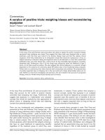
Schematic presentation of study design and graph describing tracings and measurements of lung complianceFigure 1
Schematic presentation of study design and graph describing tracings and measurements of lung compliance. (A) Experimental
protocol. R&Cscan is a program for measuring lung resistance and compliance with the single compartment model. A pertur-
bation of forced oscillation was performed for 4 s (Prime 4, Zrs measurements) and was used in the acute 17-day (OVA'17 and
PBS'17 animals) and chronic 98-day protocol (OVA'98 and PBS'98 animals). During A → F, methacholine (MCh) or phosphate
buffered saline (PBS) was administrated or nothing was given. MCh or PBS was administrated 20 s after last DI. (B) Tracings of
lung compliance (C
L
) obtained by R&Cscan indicating measurement points for C
L
(A → F) and ∆C
L
with and without deep
inspirations (DI).
0 5 10 15 20 25 30
0.00
0.01
0.02
0.03
0.04
0.05
A
F
∆C
L
=F-A
BCDE
no DI
DI
Time
(min)
C
L
(cmH
2
O
⋅
mL
-1
)
A
B
wait 5 min
2DI
2DI
2DI
2DI
2DI
2DI
R&Cscan
R&Cscan
R&Cscan
R&Cscan
R&Cscan
R&Cscan
wait 5 min
AB C D E F
2DI
2DI
2DI
2DI
R&Cscan
R&Cscan
R&Cscan
R&Cscan
R&Cscan
R&Cscan
Prime 4
Prime 4
Prime 4
Prime 4
Prime 4
Prime 4
Respiratory Research 2008, 9:23 />Page 4 of 12
(page number not for citation purposes)
Acute allergen-challenged, OVA- or PBS-treated mice
Acute airway inflammation was induced by intraperito-
neal injections of 10 µg ovalbumin (OVA, Sigma-Aldrich)
emulsified in Al(OH)
3
(Sigma-Aldrich) on day 0 and day
7. Mice were then challenged with 1% OVA diluted in
phosphate-buffered saline (PBS, Sigma-Aldrich). Animals
were exposed to aerosolized OVA for 30 min, on day 14,
15 and 16. Aerosol exposure was performed in a chamber
coupled to a nebulizer (DeVilbiss UltraNeb
®
, Sunrise
Medical Ltd, U.K.). The chamber was divided into pie-
shaped compartments with individual boxes for each ani-
mal, providing equal and simultaneous exposure to aller-
gen. The experiment ended with assessment of lung
mechanics on day 17, 24 h after last allergen exposure.
Control mice were sensitized with OVA i.p. and chal-
lenged with aerosolized PBS using the same protocol as
for OVA described above.
The effects of DI on lung mechanics were investigated
after the 17-day protocol in OVA and PBS challenged mice
in a fashion similar to that described above for healthy
unchallenged mice in the MCH group. Besides, OVA and
PBS challenged mice received immediately after each DI
or normal ventilation for 16 s, a shorter 4 s perturbation
of forced oscillation (Prime 4), followed by the injection.
Mice were given one of four treatments:
1) PBS-challenged mice that were given DI (PBS'17+DI, n
= 8) before injection of incremental doses of MCh i.v.
(from 0 to 3 mg·kg
-1
).
2) Another group of PBS-challenged mice that did not
receive any DI (PBS'17, n = 7).
3) OVA-challenged mice that were given DI (OVA'17+DI,
n = 8) before injection of incremental doses of MCh i.v.
(from 0 to 3 mg·kg
-1
).
4) Another group of OVA-challenged mice that did not
receive any DI (OVA'17, n = 10).
Chronic allergen-challenged, OVA- or PBS-treated mice
Chronic airway inflammation was induced using the
same protocol as for acute OVA described above. How-
ever, animals were exposed to aerosolized OVA for 30
min, three days a week between day 14 and 93. Five days
after last allergen exposure, the experiment ended with
assessment of lung mechanics on day 98. Control mice
were sensitized using the same protocol as for acute OVA
described above and challenged with aerosolized PBS.
The effect of DI on lung mechanics were investigated after
the 98-day protocol in OVA and PBS-challenged mice in a
fashion similar to that described above for healthy
unchallenged mice in the MCH group. Besides, OVA and
PBS challenged mice also received a shorter 4 s perturba-
tion of forced oscillation (Prime 4), followed by the injec-
tion. Mice were given one of four treatments:
1) PBS-challenged mice that were given DI (PBS'98+DI, n
= 5) before injection of incremental doses of MCh i.v.
(from 0 to 3 mg·kg
-1
).
2) Another group of PBS-challenged mice that did not
receive any DI (PBS'98, n = 6).
3) OVA-challenged mice that were given DI (OVA'98+DI,
n = 5) before injection of incremental doses of MCh i.v.
(from 0 to 3 mg·kg
-1
).
4) Another group of OVA-challenged mice that did not
receive any DI (OVA'98, n = 6).
Bronchoalveolar lavage
After completion of the lung mechanics experiment, mice
subjected to the 17-day and the 98-day protocol respec-
tively were exsanguinated and subjected to bronchoalveo-
lar lavage (BAL). The lungs were lavaged three times via
the tracheal tube with a total volume of 1 mL PBS contain-
ing 0.6 mM EDTA (EDTA, Ethylenediaminetetraacetic
acid, Sigma-Aldrich). The BAL fluid was then immediately
centrifuged (10 min, 4°C, 1200 rpm). After removing the
supernatant, the cell pellet was resuspended in 100 µL of
red cell lysis buffer containing 0.15 M NH
4
Cl, 1.0 mM
KHCO
3
, and 0.1 mM EDTA for 2 min at room tempera-
ture. The suspension was then diluted with 1 mL PBS and
recentrifuged (10 min, 4°C, 1200 rpm). Leukocytes were
counted manually in a hemacytometer so that 50,000
cells could be loaded and centrifuged using a cytospin
centrifuge. Cytocentrifuged preparations were stained
with May-Grünwald-Giemsa and differential cell counts
of pulmonary inflammatory cells (macrophages, neu-
trophils, lymphocytes, and eosinophils) were determined
using standard morphological criteria and counting 3 ×
100 cells per cytospin preparation. The total number of
each cell type was then calculated and expressed as
number of cells per mL of BAL fluid.
Histological evaluation of the chronic allergen-challenged
lungs
Following BAL, the lungs were inflated with 4% parafor-
maldehyde solution to a pressure of 20 cmH
2
O without
removing the lungs from the chest. After 1 h the trachea
was tied off, the lungs were stored at 4°C overnight in 4%
paraformaldehyde, then washed several times in ethanol
and stored in 70% ethanol at 4°C until time for embed-
ding. After embedding in paraffin, the tissue was cut into
5 µm sections and mounted on positively charged slides.
To assess inflammatory cell infiltration the sections were
deparaffinized, dehydrated, and stained with hematoxylin
Respiratory Research 2008, 9:23 />Page 5 of 12
(page number not for citation purposes)
and eosin (H&E). H&E stained sections were examined by
bright field microscopy (Nikon Eclipse TS100, Nikon
Instruments Inc., Melville, N.Y, USA) and images were
captured with a Nikon DS digital camera system (Tekno
Optik AB, Stockholm, Sweden).
Statistical analysis
Results are presented as mean ± standard error of mean
(SEM). Statistical significance was assessed by parametric
methods using two-way analysis of variance (ANOVA) to
analyze differences between groups, followed by Bonfer-
roni post hoc test. When appropriate, one-way ANOVA or
Student's unpaired t-test was used. A statistical result with
P < 0.05 was considered significant. Statistical analysis
and preparations of graphs were performed with Graph-
Pad Prism (version 4.0 GraphPad software Inc., San
Diego, CA, USA).
Results
Healthy mice
MCh increased R
L
, from baseline 0.33 ± 0.01 to 3.8 ± 0.3
cmH
2
O·s·mL
-1
(P < 0.001) at the highest dose of MCh
(Figure 2A). DI significantly reduced the maximum R
L
from 3.8 ± 0.3 to 2.8 ± 0.1 cmH
2
O·s·mL
-1
(P < 0.001, Fig-
ure 2A). R
L
did not change from baseline in TIME or PBS
groups, (no MCh provocation), with or without DI (P >
0.05).
C
L
was measured immediately before injections of PBS or
MCh. In the TIME group, receiving no i.v. injections and
no DI, C
L
decreased by 9.3 ± 0.8% from baseline to the last
measurement point (∆C
L
, Figure 2B). A similar decline
was seen in the PBS group, receiving PBS injections with-
out DI, where C
L
decreased by 6.9 ± 1.6% (P > 0.05, Figure
2B). In the MCH group, receiving incremental doses of
MCh without DI, C
L
decreased by 15.9 ± 1.5%, the decline
being significantly larger than in the TIME and PBS groups
(P < 0.05 and P < 0.001 respectively, Figure 2B). DI signif-
icantly protected against the reduction in C
L
in the
MCH+DI group, where the decline in C
L
was attenuated to
5.6 ± 0.6% (P < 0.0001, Figure 2B). Although displaying a
tendency to protection, DI had no significant attenuating
effect on the decrease in C
L
in either the TIME+DI (4.0 ±
1.9%, P > 0.05) or the PBS+DI group (3.8 ± 1.1%, P >
0.05, Figure 2B).
Bronchoalverolar lavage and histology
Mice undergoing the 17-day or 98-day ovalbumin chal-
lenge protocol, the OVA'17 and OVA'98 group respec-
tively, had clear signs of airway inflammation compared
to control animals. OVA'17 group had approximately a 6-
fold increase in total BAL cell count and OVA'98 had a 5-
fold increase compared to control groups (both P <
0.001). Animals in the OVA'17 had a significant higher
BAL cell count than OVA'98 (P < 0.03). Differential BAL
cell count confirmed an inflammatory profile with mark-
edly increased counts of macrophages, eosinophils, neu-
trophils, and lymphocytes in both acute and chronic
challenged OVA groups. The OVA'17 animals had a
higher number of eosinophils than OVA'98 animals
(Table 1).
Effects of deep inspirations (DI) in healthy mice; (A) lung resistance (R
L
) in mice given incremental doses of methacholine (MCH group), and (B) the effect of DI on lung compliance (C
L
) presented as ∆C
L
Figure 2
Effects of deep inspirations (DI) in healthy mice; (A) lung resistance (R
L
) in mice given incremental doses of methacholine
(MCH group), and (B) the effect of DI on lung compliance (C
L
) presented as ∆C
L
. Values are mean ± SEM, * P < 0.05, ** P <
0.01, *** P < 0.001.
PBS 0. 03 0. 1 0. 3 1 3
0
1
2
3
4
5
MCH n=8
MCH+DI n=6
***
[MCh]
(mg⋅kg
-1
)
R
L
(cmH
2
O
⋅
s
⋅
mL
-1
)
A
B
Respiratory Research 2008, 9:23 />Page 6 of 12
(page number not for citation purposes)
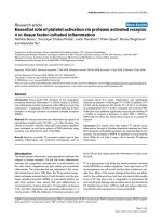
OVA'98 group had also clear signs of remodeling, light
microscopic examination of hematoxylin and eosin sec-
tions from OVA'98 and PBS'98 animals revealed an eosi-
nophilic inflammation in the OVA-treated animals with a
patchy distribution of eosinophils surrounding the air-
ways and within the alveolar spaces. OVA'98 animals also
revealed a significantly increased perivascular inflamma-
tion (Figure 3).
Table 1: Differential cell counts in bronchial alveolar lavage from animals having undergone an ovalbumin challenge protocol (OVA'17
and OVA'98) or a control protocol with phosphate buffered saline (PBS'17 and PBS'98).
PBS'17 (n = 15) OVA'17 (n = 17) PBS'98 (n = 11) OVA'98 (n = 11)
Macrophages 73 600 ± 4 100 147 000 ± 7 500
¤
68 000 ± 5 500 168 000 ± 10 700 *
Eosinophils 0 222 100 ± 39 700
¤
0 76 000 ± 26 500 *
Neutrophils 2 300 ± 500 3 900 ± 2 100 2 500 ± 2 000 52 500 ± 11 000 *
Lymphocytes 9100 ± 2400 23 000 ± 3 500
¤
900 ± 350 23 500 ± 6 500 *
Values are mean ± SEM.
¤
P < 0.05 vs. PBS'17, * P < 0.05 vs. PBS'98.
Representative histological sections (hematoxylin and eosin stained) from healthy control animals in the PBS'98 group (picture A and B) and from animals having undergone a 98-day ovalbumin challenge protocol, the OVA'98 group (picture C and D)Figure 3
Representative histological sections (hematoxylin and eosin stained) from healthy control animals in the PBS'98 group (picture
A and B) and from animals having undergone a 98-day ovalbumin challenge protocol, the OVA'98 group (picture C and D).
Examination of sections from OVA'98 animals revealed a significant inflammation surrounding the airways and within the alve-
olar spaces. PBS'98 did not show any signs of inflammation.
AB
CD
Respiratory Research 2008, 9:23 />Page 7 of 12
(page number not for citation purposes)
Acute allergen-challenged mice
Lung resistance and compliance
In PBS'17 mice, MCh induced bronchoconstriction with a
maximum R
L
of 3.6 ± 0.2 cmH
2
O·s·mL
-1
. After DI, R
L
was
significantly lower, 2.5 ± 0.2 cmH
2
O·s·mL
-1
(P < 0.0001,
Figure 4A). In OVA'17 mice, MCh induced bronchocon-
striction with a maximum R
L
of 5.1 ± 0.3 cmH
2
O·s·mL
-1
.
After DI, R
L
was significantly lower, 3.5 ± 0.3
cmH
2
O·s·mL
-1
(P < 0.0001, Figure 4B). In the OVA'17
group, MCh induced higher bronchoconstriction than the
PBS'17 group, (P < 0.0001).
In the PBS'17 group, C
L
decreased by 12.5 ± 3.2% from
baseline to the last dose of MCh (Figure 5). Animals
treated with DI, the PBS'17+DI group, had a significantly
smaller decrease in C
L
(2.5 ± 1.6%, P < 0.05). In the
OVA'17 group without DI, the decrease in C
L
was larger
than in the PBS-treated animals (15.9 ± 2.3%, NS, Figure
5). In OVA-treated animals receiving DI, the OVA'17+DI
group, the decrease in C
L
was largely prevented (2.7 ±
3.4%, P < 0.001, Figure 5).
Peripheral lung mechanics
During bronchial reactivity assessment the 4 s perturba-
tion of forced oscillation (Prime 4) before each dose of
PBS and MCh revealed significant differences in Newto-
nian resistance (R
N
) between OVA'17 and PBS'17 groups
(23.3 ± 3.6% and 8.6 ± 4.5% respectively, P < 0.01). Treat-
ing animals with DI significantly lowered R
N
at each dose
of PBS and MCh in OVA'17 group (OVA'17+DI, 10.5 ±
2.8%, P < 0.01). DI did not have any effect in the PBS'17
group (PBS'17+DI, 8.2 ± 3.9%, P > 0.05).
In the PBS'17 group, tissue elastance (H) increased by 9.4
± 4.6% from baseline to the last dose of MCh. There was
no protective effect on H in animals treated with DI,
PBS'17+DI group. In the OVA'17 group without DI, H was
two times higher than in the PBS'17 group (20.7 ± 3.1%,
P < 0.0001). In the OVA'17+DI group, DI largely pre-
vented the increase in H (8.5 ± 1.9%, P < 0.0001). There
were no differences in tissue damping (G) in the PBS'17
group and the OVA'17 group (26.5 ± 4.4% and 14.5 ±
4.5% respectively, P > 0.05). DI prevented the increase in
G in the OVA'17 group but not in the PBS'17 group
(OVA'17+DI, 15.0 ± 2.2%, P < 0.05 and PBS'17+DI 4. 7 ±
3.1%, NS)
Chronic allergen-challenged mice
Lung resistance and compliance
In PBS'98 mice, MCh induced bronchoconstriction with a
maximum R
L
of 3.8 ± 0.2 cmH
2
O·s·mL
-1
. After DI, R
L
was
significantly lower, 2.4 ± 0.2 cmH
2
O·s·mL
-1
(P < 0.001,
Figure 6A). This protective effect of DI against bronchoc-
onstriction was totally abolished in OVA treated mice
(OVA'98, 3.7 ± 1.1 cmH
2
O·s·mL
-1
and OVA'98+DI, 4.3 ±
0.4 cmH
2
O·s·mL
-1
respectively, P > 0.05, Figure 6B).
In the PBS'98 group, C
L
decreased by 18.1 ± 1.2% from
baseline to the last dose of MCh (Figure 5). Animals
treated with DI, the PBS'98+DI group, had a significantly
smaller decrease in C
L
(9.7 ± 1.0%, P < 0.001). In the
OVA'98 group without DI, the decrease in C
L
was more
than double that in PBS-treated animals (44.1 ± 6.6%, P <
0.001, Figure 5). In OVA-treated animals receiving DI, the
The effect of deep inspirations (DI) on lung resistance (R
L
) in healthy mice (PBS'17) and in animals with acute airway inflamma-tion (OVA'17 group)Figure 4
The effect of deep inspirations (DI) on lung resistance (R
L
) in healthy mice (PBS'17) and in animals with acute airway inflamma-
tion (OVA'17 group). Values are mean ± SEM, ** P < 0.01, *** P < 0.001.
PBS 0.03 0.1 0.3 1 3
0
1
2
3
4
5
6
PBS´17 n=7
PBS´17+DI n=8
***
***
**
[MCh] (mg⋅kg
-1
)
R
L
(cmH
2
O
⋅
s
⋅
mL
-1
)
PBS 0.03 0.1 0.3 1 3
0
1
2
3
4
5
6
OVA´17+DI n= 8
OVA´17 n=10
***
**
***
[MCh]
(mg⋅kg
-1
)
R
L
(cmH
2
O
⋅
s
⋅
mL
-1
)
AB
Respiratory Research 2008, 9:23 />Page 8 of 12
(page number not for citation purposes)
The effect of deep inspirations (DI) on lung compliance (C
L
) presented as ∆C
L
Figure 5
The effect of deep inspirations (DI) on lung compliance (C
L
) presented as ∆C
L
. DI attenuated the fall in ∆C
L
in both healthy
mice (PBS'17) and in mice with acute airway inflammation (OVA'98). Mice with chronic airway inflammation, the OVA'98
group, had significantly larger fall in ∆C
L
than healthy control animals, the PBS'98 group. DI attenuated the fall in ∆C
L
in both
groups, OVA'98 and PBS'98. Values are mean ± SEM, * P < 0.05, ** P < 0.01, *** P < 0.001.
The effect of deep inspirations (DI) on lung resistance (R
L
) in healthy mice (PBS'98) and in mice with chronic airway inflamma-tion (OVA'98 group)Figure 6
The effect of deep inspirations (DI) on lung resistance (R
L
) in healthy mice (PBS'98) and in mice with chronic airway inflamma-
tion (OVA'98 group). Values are mean ± SEM, *** P < 0.001.
PBS 0.03 0.1 0.3 1 3
0
1
2
3
4
5
6
PBS´98 n=6
PBS´98+DI n=5
***
***
[MCh] (mg⋅kg
-1
)
R
L
(cmH
2
O
⋅
s
⋅
mL
-1
)
PBS 0.03 0.1 0.3 1 3
0
1
2
3
4
5
6
OVA´98+DI n=5
OVA ´98 n=6
[MCh] (mg⋅kg
-1
)
R
L
(cmH
2
O
⋅
s
⋅
mL
-1
)
B
A
Respiratory Research 2008, 9:23 />Page 9 of 12
(page number not for citation purposes)
OVA'98+DI group, the decrease in C
L
was largely pre-
vented (14.3 ± 1.3%, P < 0.001).
Peripheral lung mechanics
During bronchial reactivity assessment the 4 s perturba-
tion of forced oscillation (Prime 4) before each dose of
PBS and MCh revealed no significant differences in R
N
between OVA'98 and PBS'98 groups. Treating animals
with DI significantly lowered R
N
at each dose of PBS and
MCh in both groups (P < 0.0001, Figure 7). In the PBS'98
group, tissue elastance (H) increased by 16.7 ± 2.3% from
baseline to the last dose of MCh (Figure 8). Animals
treated with DI, PBS'98+DI group, had a significantly
smaller increase in H (3.5 ± 2.0%, P < 0.0001). In the
OVA'98 group without DI, H was three times higher than
in the PBS'98 group (51.1 ± 7.5%, P < 0.0001). In the
OVA'98+DI group, DI largely prevented the increase in H
(14.7 ± 1.1%, P < 0.0001).
In the OVA'98 group without DI, the increase in tissue
damping (G) (Figure 9) from baseline was four times
greater than in the PBS'98 group (108.1 ± 20% and 25.9 ±
4.97%, respectively, P < 0.0001). In the OVA'98+DI
group, DI largely prevented the increase in tissue damping
(25.0 ± 1.2%, P < 0.0001), while there were no differences
in tissue damping between the PBS'98 and PBS'98+DI
groups.
Discussion
We have investigated the effects of deep inspirations (DI)
in healthy mice, in mice with acute airway inflammation
and in mice with chronic airway inflammation and
remodeling. Our major findings are that: 1) DI had a
marked effect on lung resistance after MCh-challenge in
healthy mice and in acute allergen-challenged mice, but
not in mice with chronic inflammation; 2) DI protects
against the decrease in lung compliance that occurs both
spontaneously over time and after intravenous injections
Measurements of Newtonian resistance (R
N
) were per-formed with forced oscillation technique (Prime 4 perturba-tion, Zrs measurements) before each injection of phosphate buffered saline or methacholineFigure 7
Measurements of Newtonian resistance (R
N
) were per-
formed with forced oscillation technique (Prime 4 perturba-
tion, Zrs measurements) before each injection of phosphate
buffered saline or methacholine. P values for each significant
R
N
value for each group; * P < 0.05, ** P < 0.01, *** P < 0.001
vs. same group without DI. Values are mean ± SEM.
PBS 0. 03 0. 1 0. 3 1 3
0. 00
0. 05
0. 10
0. 15
0. 20
0. 25
0. 30
0. 35
0. 40
OVA´98 n=6
OVA´98+DI n=5
PBS´98+DI n=5
PBS´98 n=6
**
*
**
***
**
**
*
[MCh] (mg⋅kg
-1
)
R
N
(cmH
2
O
⋅
s
⋅
mL
-1
)
Measurements of tissue elastance (H) were performed with forced oscillation technique (Prime 4 perturbation, Zrs measurements) before each injection of phosphate buffered saline or methacholineFigure 8
Measurements of tissue elastance (H) were performed with
forced oscillation technique (Prime 4 perturbation, Zrs
measurements) before each injection of phosphate buffered
saline or methacholine. Values are mean ± SEM, * P < 0.05, **
P < 0.01, *** P < 0.001 vs. all other groups.
PBS 0.03 0.1 0.3 1 3
0
5
10
15
20
25
30
35
40
OVA´98 n=6
OVA´98+DI n=5
PBS´98 n=6
***
***
**
*
PBS´98+DI n=5
***
**
*
[MCh] (mg⋅kg
-1
)
H
(cmH
2
O
⋅
mL
-1
)
Measurements of tissue damping (G) were performed with forced oscillation technique (Prime 4 perturbation, Zrs measurements) before each injection of phosphate buffered saline or methacholineFigure 9
Measurements of tissue damping (G) were performed with
forced oscillation technique (Prime 4 perturbation, Zrs
measurements) before each injection of phosphate buffered
saline or methacholine. Values are mean ± SEM, ** P < 0.01,
*** P < 0.001 vs. all other groups.
PBS 0.03 0.1 0.3 1 3
0
5
10
15
20
OVA´98 n=6
OVA´98+DI n=5
PBS´98 n=6
**
***
PBS´98+DI n=5
[MCh] (mg⋅kg
-1
)
G
(cmH
2
O
⋅
mL
-1
)
Respiratory Research 2008, 9:23 />Page 10 of 12
(page number not for citation purposes)
of PBS or MCh; 3) DI has a major impact on peripheral
airway and tissue physiology, protecting against MCh-
induced increases in tissue elastance (H) in both animals
with acute and chronic inflammation and also in healthy
mice undergoing the 98-day protocol; 4) DI totally abol-
ishes MCh-induced increases in tissue damping (G) seen
in mice with acute and chronic inflammation.
This mouse model has potential value for defining mech-
anisms and sites of action of DI and our goals were to
investigate if this mouse model could be used to identify
the site of action of DI. We have implemented both the
constant phase model (the low-frequency oscillation tech-
nique) and the single compartment model to characterize
the effect of a DI. The constant phase model has the capac-
ity to partition the respiratory properties into central and
peripheral airways and also pure tissue properties
[15,17,19]. In this study animals were of varying age
depending on the duration of the different protocols. This
could have possible effects on mouse lung mechanics [20-
23], we solved this by having matched controls.
The airway protective effects of DI are similar to what has
also been seen in other animal studies [24-27] and in
humans [5-7,28]. The mechanisms underlying this bron-
choprotective effect are not clear, but several hypotheses
have been put forward as to how DI confers bronchopro-
tection [9], in which the main mechanisms have been sug-
gested to be neural, nitric oxide (NO)-mediated, or
mechanical. Scichilone et al. [7] suggested that DI could
reduce bronchoconstriction through inhibition of cholin-
ergic tone or activation of nonadrenergic, noncholinergic
(NANC) system, and it has been suggested that airway
stretch could cause release of substances such as NO [29]
or cyclooxygenase products [30]. Mechanical explana-
tions involve different theories, the simplest one being
that stretching airway smooth muscle disrupts cross
bridges, thereby reducing force generation. Fredberg et al.
[31,32] suggested that asthmatic smooth muscle becomes
"frozen" due to excessive latch bridge formation and that
DI may detach these latch bridges, which provides an
opportunity for normal cross-bridges. On the other hand,
Gunst and co-workers [33,34] contend that cross-bridge
properties cannot account for this, and that it is rather due
to the plastic organization of contractile filaments in
smooth muscle, allowing for adaptation to stretch [34].
This idea is in line with Wang and Paré [9] who proposed
that DI initiate an adaptive process involving dissembly of
contractile filaments, thereby allowing for reorganization
of the contractile apparatus and better adaptation to the
new smooth muscle cell length. In spite of recent investi-
gations and new theories on the behavior of smooth mus-
cle cells in response to stretch and mechanical forces [35-
37], the cellular and subcellular mechanisms behind DI
and bronchial responsiveness remain undefined. The cur-
rent study provides a model for further investigation of
the mechanisms.
Using short acute OVA challenge protocols [38], mice
develop inflammation almost completely localized to the
proximal airways, while chronic exposure to OVA leads to
inflammation throughout the lung [39,40]. In the current
study, mice were subjected to a 1-week or a 12-week OVA
inflammation protocol and we found clear signs of
inflammation and after the 12-week protocol there was
also airway remodeling. Our results indicate that our 98-
day long chronic inflammation model resembles human
asthma more than an acute model does because of more
peripheral inflammation in the lung after chronic chal-
lenge. When Wegmann [39] ran a similar protocol,
chronic inflammation and remodeling were seen to
involve peripheral airways, compared with acute inflam-
mation that mainly involved proximal airways. Xisto et al
[40] found inflammatory cell infiltration and remodeling
of the central as well as the peripheral airways and lung
parenchyma after a chronic inflammation protocol. Con-
trary to what Wegmann [39] and Xisto [40] reported, we
could not detect any increased responsiveness to MCh in
the chronically inflamed animals not receiving DI as com-
pared with healthy mice and mice with acute airway
inflammation. Possible explanations for this may be due
to the use of a shorter OVA protocol [40] or to differences
between assessing airway function with body-plethys-
mography [41] and our measurements of lung resistance.
While cautiously interpreting responses based on the
body plethysmography technique and refraining from
directly comparing enhanced pause system and lung
resistance [18,42], there is in a study by McMillan et al [1]
a trend toward less reactivity after a long term chronic
OVA-protocol that resembles our findings. Another expla-
nation to our findings in the chronic inflammation could
be that these animals induced a tolerance against OVA
[43] and this could lead to a decreased responsiveness to
MCh. Our results are also in line with human studies,
where airway response to MCh is similar in healthy and
asthmatic subjects when no DI is allowed [13], a phenom-
enon directly linked to narrowing of the conducting air-
ways [44]. This has led us to believe that our mouse model
of chronic airway inflammation closely resembles human
asthma with respect to several points. Our present results
show that DI protects from MCh-induced increase in lung
resistance in healthy mice and in acute airway inflamma-
tion, but not in mice with chronic inflammation. The lack
of protective effect against increased lung resistance in
chronically inflamed mice is in line with human studies
where DI gives asthmatic patients no protection against
MCh-induced bronchoconstriction [5,6].
Most investigations of murine models of airway inflam-
mation have focused on bronchial responsiveness and
Respiratory Research 2008, 9:23 />Page 11 of 12
(page number not for citation purposes)
remodeling of more central airways. Recent reports show
that the peripheral airways and parenchyma play a more
important role in pathophysiology than expected. Lund-
blad et al [45] and Wagers et al [46] have recently shown
that increased airway reactivity in OVA inflamed mice is
entirely due to exaggerated closure of peripheral airways
and that excessive narrowing is due to purely geometric
reasons.
Inflammation of distal airways and lung parenchyma
directly affects lung physiology by increasing tissue
elastance and resistance, as well as by elevating pulmo-
nary static and dynamic elastance [40]. In our current
study, acute inflammation increased lung resistance and
reduced lung compliance. There was no effect on tissue
damping but an effect on tissue elastance. When applying
DI before MCh-challenges, we saw a strong protective
effect on lung resistance and lung compliance. DI had a
significant protective effect on tissue elastance, while tis-
sue damping was already low and was not altered by DI.
However, chronic inflammation reduced lung compli-
ance, while increasing tissue elastance and tissue damp-
ing. When applying DI before MCh-challenges, we saw
stronger protective effects on these peripheral parameters
in animals with chronic inflammation than in the acute
inflammation. In healthy animals, DI had a significant
protective effect on lung compliance and tissue elastance,
while tissue damping was already low and was not altered
by DI. This indicates that DI has a stronger effect on
peripheral tissue in the chronic airway inflammation and
that the protective effect of DI on lung resistance is greater
in acute airway inflammation. Our results in the chronic
airway inflammation are in line with those of Schweitzer
et al [24], who showed that DI in Brown Norway rats, pro-
tected against MCh-induced increases in respiratory sys-
tem elastance, but not resistance. Similar results were
previously found by Hirai and Bates [25], who showed
that DI, in healthy Sprague-Dawley rats, was neither bron-
chodilatory nor bronchoprotective, but indeed had a sig-
nificant effect on both tissue damping and tissue
elastance.
Conclusion
In summary, we have found that presence of DI may blunt
bronchoconstriction of central airways in healthy mice
and in acute airway inflammation, but not when chronic
inflammation is present. We have presented a murine
OVA model that in many ways resembles human chronic
airway inflammation. Many human studies suggest that
DI is not bronchoprotective in asthmatic subjects, which
is in line with our current findings in the chronic inflam-
mation model. However, our present results point to very
potent protective effects in the peripheral parts of the
chronically inflamed murine lung and it is conceivable
that this could also play a major role on overall lung
health in asthma patients. This model of chronic airway
inflammation should pave the way for investigations of
mechanisms that may help identify new targets for thera-
pies in chronic airway inflammation and asthma.
Competing interests
The author(s) declare that they have no competing inter-
ests.
Authors' contributions
SJ carried out the animal experiments and drafted the
manuscript. LS carried out the histological evaluation and
the cellular data. ML carried out animal experiments. JH,
SED and GH participated in the study design, coordina-
tion and helped to draft the manuscript. All authors read
and approved the final manuscript.
Acknowledgements
This work was supported by the Swedish Heart-Lung Association and the
Swedish Medical Research Council.
References
1. McMillan SJ, Lloyd CM: Prolonged allergen challenge in mice
leads to persistent airway remodelling. Clin Exp Allergy 2004,
34(3):497-507.
2. Cui ZH, Skoogh BE, Pullerits T, Lotvall J: Bronchial hyperrespon-
siveness and airway wall remodelling induced by exposure to
allergen for 9 weeks. Allergy 1999, 54(10):1074-1082.
3. Brown RH, Mitzner W: Airway response to deep inspiration:
role of inflation pressure. J Appl Physiol 2001, 91(6):2574-2578.
4. Brusasco V, Crimi E, Barisione G, Spanevello A, Rodarte JR, Pellegrino
R: Airway responsiveness to methacholine: effects of deep
inhalations and airway inflammation. J Appl Physiol 1999,
87(2):567-573.
5. Kapsali T, Permutt S, Laube B, Scichilone N, Togias A: Potent bron-
choprotective effect of deep inspiration and its absence in
asthma. J Appl Physiol 2000, 89(2):711-720.
6. Scichilone N, Kapsali T, Permutt S, Togias A: Deep inspiration-
induced bronchoprotection is stronger than bronchodila-
tion. Am J Respir Crit Care Med 2000, 162(3 Pt 1):910-916.
7. Scichilone N, Permutt S, Togias A: The lack of the bronchopro-
tective and not the bronchodilatory ability of deep inspira-
tion is associated with airway hyperresponsiveness. Am J
Respir Crit Care Med 2001, 163(2):413-419.
8. Sundblad BM, Larsson K: Effect of deep inhalations after a bron-
chial methacholine provocation in asthmatic and non-asth-
matic subjects. Respir Med 2002, 96(7):477-481.
9. Wang L, Pare PD: Deep inspiration and airway smooth muscle
adaptation to length change. Respir Physiol Neurobiol 2003,
137(2-3):169-178.
10. Fredberg JJ: Airway obstruction in asthma: does the response
to a deep inspiration matter? Respir Res 2001, 2(5):273-275.
11. Jensen A, Atileh H, Suki B, Ingenito EP, Lutchen KR: Selected con-
tribution: airway caliber in healthy and asthmatic subjects:
effects of bronchial challenge and deep inspirations. J Appl
Physiol 2001, 91(1):506-15; discussion 504-5.
12. Fish JE, Ankin MG, Kelly JF, Peterman VI: Regulation of broncho-
motor tone by lung inflation in asthmatic and nonasthmatic
subjects. J Appl Physiol 1981, 50(5):1079-1086.
13. Skloot G, Permutt S, Togias A: Airway hyperresponsiveness in
asthma: a problem of limited smooth muscle relaxation with
inspiration. J Clin Invest 1995, 96(5):2393-2403.
14. Irvin CG, Bates JH: Measuring the lung function in the mouse:
the challenge of size. Respir Res 2003, 4(1):4.
15. Hantos Z, Daroczy B, Suki B, Nagy S, Fredberg JJ: Input impedance
and peripheral inhomogeneity of dog lungs. J Appl Physiol 1992,
72(1):168-178.
Publish with BioMed Central and every
scientist can read your work free of charge
"BioMed Central will be the most significant development for
disseminating the results of biomedical research in our lifetime."
Sir Paul Nurse, Cancer Research UK
Your research papers will be:
available free of charge to the entire biomedical community
peer reviewed and published immediately upon acceptance
cited in PubMed and archived on PubMed Central
yours — you keep the copyright
Submit your manuscript here:
/>BioMedcentral
Respiratory Research 2008, 9:23 />Page 12 of 12
(page number not for citation purposes)
16. Gomes RF, Shen X, Ramchandani R, Tepper RS, Bates JH: Compar-
ative respiratory system mechanics in rodents. J Appl Physiol
2000, 89(3):908-916.
17. Tomioka S, Bates JH, Irvin CG: Airway and tissue mechanics in a
murine model of asthma: alveolar capsule vs. forced oscilla-
tions. J Appl Physiol 2002, 93(1):263-270.
18. Bates JH, Irvin CG: Measuring lung function in mice: the pheno-
typing uncertainty principle. J Appl Physiol 2003,
94(4):1297-1306.
19. Bates JH, Lutchen KR: The interface between measurement
and modeling of peripheral lung mechanics. Respir Physiol Neu-
robiol 2005.
20. Bozanich EM, Janosi TZ, Collins RA, Thamrin C, Turner DJ, Hantos
Z, Sly PD: Methacholine responsiveness in mice from 2 to 8 wk
of age. J Appl Physiol 2007, 103(2):542-546.
21. Bozanich EM, Collins RA, Thamrin C, Hantos Z, Sly PD, Turner DJ:
Developmental changes in airway and tissue mechanics in
mice. J Appl Physiol 2005, 99(1):108-113.
22. Busse PJ, Zhang TF, Srivastava K, Schofield B, Li XM: Effect of ageing
on pulmonary inflammation, airway hyperresponsiveness
and T and B cell responses in antigen-sensitized and -chal-
lenged mice. Clin Exp Allergy 2007, 37(9):1392-1403.
23. Hirai T, Hosokawa M, Kawakami K, Takubo Y, Sakai N, Oku Y, Chin
K, Ohi M, Higuchi K, Kuno K, et al.: Age-related changes in the
static and dynamic mechanical properties of mouse lungs.
Respir Physiol 1995, 102(2-3):195-203.
24. Schweitzer C, Demoulin B, Bello G, Bertin N, Leblanc AL, Marchal F:
Deep inhalation prevents the respiratory elastance response
to methacholine in rats. Pediatr Res 2006, 59(5):646-649.
25. Hirai T, Bates JH: Effects of deep inspiration on bronchocon-
striction in the rat. Respir Physiol 2001, 127(2-3):201-215.
26. Chapman RW, Skeans S, Lamca J, House A, Hey JA, Celly C: Effect
of histamine, albuterol and deep inspiration on airway and
lung tissue mechanics in cynomolgus monkeys. Pulm Pharmacol
Ther 2005, 18(4):243-249.
27. Gunst SJ, Shen X, Ramchandani R, Tepper RS: Bronchoprotective
and bronchodilatory effects of deep inspiration in rabbits
subjected to bronchial challenge. J Appl Physiol 2001,
91(6):2511-2516.
28. Skloot G, Togias A: Bronchodilation and bronchoprotection by
deep inspiration and their relationship to bronchial hyperre-
sponsiveness. Clin Rev Allergy Immunol 2003, 24(1):55-72.
29. Bannenberg GL, Gustafsson LE: Stretch-induced stimulation of
lower airway nitric oxide formation in the guinea-pig: inhibi-
tion by gadolinium chloride. Pharmacol Toxicol 1997, 81(1):13-18.
30. Gao Y, Vanhoutte PM: Responsiveness of the guinea pig trachea
to stretch: role of the epithelium and cyclooxygenase prod-
ucts. J Appl Physiol 1993, 75(5):2112-2116.
31. Fredberg JJ, Inouye D, Miller B, Nathan M, Jafari S, Raboudi SH, Butler
JP, Shore SA: Airway smooth muscle, tidal stretches, and
dynamically determined contractile states. Am J Respir Crit
Care Med 1997, 156(6):1752-1759.
32. Fredberg JJ, Inouye DS, Mijailovich SM, Butler JP: Perturbed equilib-
rium of myosin binding in airway smooth muscle and its
implications in bronchospasm. Am J Respir Crit Care Med 1999,
159(3):959-967.
33. Gunst SJ: Contractile force of canine airway smooth muscle
during cyclical length changes. J Appl Physiol 1983,
55(3):759-769.
34. Shen X, Wu MF, Tepper RS, Gunst SJ: Mechanisms for the
mechanical response of airway smooth muscle to length
oscillation. J Appl Physiol 1997, 83(3):731-738.
35. Gunst SJ, Fredberg JJ: The first three minutes: smooth muscle
contraction, cytoskeletal events, and soft glasses. J Appl Physiol
2003, 95(1):413-425.
36. Fabry B, Fredberg JJ: Remodeling of the airway smooth muscle
cell: are we built of glass? Respir Physiol Neurobiol 2003, 137(2-
3):109-124.
37. Deng L, Trepat X, Butler JP, Millet E, Morgan KG, Weitz DA, Fred-
berg JJ: Fast and slow dynamics of the cytoskeleton. Nat Mater
2006, 5(8):636-640.
38. Hjoberg J, Shore S, Kobzik L, Okinaga S, Hallock A, Vallone J, Subra-
maniam V, De Sanctis GT, Elias JA, Drazen JM, Silverman ES: Expres-
sion of nitric oxide synthase-2 in the lungs decreases airway
resistance and responsiveness. J Appl Physiol 2004,
97(1):249-259.
39. Wegmann M, Fehrenbach H, Fehrenbach A, Held T, Schramm C,
Garn H, Renz H: Involvement of distal airways in a chronic
model of experimental asthma. Clin Exp Allergy 2005,
35(10):1263-1271.
40. Xisto DG, Farias LL, Ferreira HC, Picanco MR, Amitrano D, Lapa
ESJR, Negri EM, Mauad T, Carnielli D, Silva LF, Capelozzi VL, Faffe DS,
Zin WA, Rocco PR: Lung parenchyma remodeling in a murine
model of chronic allergic inflammation. Am J Respir Crit Care
Med 2005, 171(8):829-837.
41. Hamelmann E, Schwarze J, Takeda K, Oshiba A, Larsen GL, Irvin CG,
Gelfand EW: Noninvasive measurement of airway responsive-
ness in allergic mice using barometric plethysmography. Am
J Respir Crit Care Med 1997, 156(3 Pt 1):766-775.
42. Adler A, Cieslewicz G, Irvin CG: Unrestrained plethysmography
is an unreliable measure of airway responsiveness in BALB/c
and C57BL/6 mice. J Appl Physiol 2004, 97(1):286-292.
43. Van Hove CL, Maes T, Joos GF, Tournoy KG: Prolonged inhaled
allergen exposure can induce persistent tolerance. Am J Respir
Cell Mol Biol 2007, 36(5):573-584.
44. Brown RH, Croisille P, Mudge B, Diemer FB, Permutt S, Togias A:
Airway narrowing in healthy humans inhaling methacholine
without deep inspirations demonstrated by HRCT. Am J
Respir Crit Care Med 2000, 161(4 Pt 1):1256-1263.
45. Lundblad LK, Thompson-Figueroa J, Allen GB, Rinaldi L, Norton RJ,
Irvin CG, Bates JH: Airways Hyperresponsiveness in Allergi-
cally Inflamed Mice: The Role of Airway Closure. Am J Respir
Crit Care Med 2007.
46. Wagers S, Lundblad LK, Ekman M, Irvin CG, Bates JH: The allergic
mouse model of asthma: normal smooth muscle in an abnor-
mal lung? J Appl Physiol 2004, 96(6):2019-2027.