Báo cáo y học: "Regulation of the cd38 promoter in human airway smooth muscle cells by TNF-α and dexamethasone" pot
Bạn đang xem bản rút gọn của tài liệu. Xem và tải ngay bản đầy đủ của tài liệu tại đây (902.29 KB, 14 trang )
BioMed Central
Page 1 of 14
(page number not for citation purposes)
Respiratory Research
Open Access
Research
Regulation of the cd38 promoter in human airway smooth muscle
cells by TNF-α and dexamethasone
Krishnaswamy G Tirumurugaan
†1
, Bit Na Kang
†1
, Reynold A Panettieri
2
,
Douglas N Foster
3
, Timothy F Walseth
4
and Mathur S Kannan*
1,5
Address:
1
Department of Veterinary and Biomedical Sciences, College of Veterinary Medicine, University of Minnesota, St. Paul, MN, USA,
2
Department of Medicine, University of Pennsylvania, Philadelphia, PA, USA,
3
Department of Animal Science, University of Minnesota, St. Paul,
MN, USA,
4
Department of Pharmacology, College of Medicine, University of Minnesota, Minneapolis, MN, USA and
5
Department of Pediatrics,
College of Medicine, University of Minnesota, Minneapolis, MN, USA
Email: Krishnaswamy G Tirumurugaan - ; Bit Na Kang - ;
Reynold A Panettieri - ; Douglas N Foster - ; Timothy F Walseth - ;
Mathur S Kannan* -
* Corresponding author †Equal contributors
Abstract
Background: CD38 is expressed in human airway smooth muscle (HASM) cells, regulates intracellular
calcium, and its expression is augmented by tumor necrosis factor alpha (TNF-α). CD38 has a role in
airway hyperresponsiveness, a hallmark of asthma, since deficient mice develop attenuated airway
hyperresponsiveness compared to wild-type mice following intranasal challenges with cytokines such as
IL-13 and TNF-α. Regulation of CD38 expression in HASM cells involves the transcription factor NF-κB,
and glucocorticoids inhibit this expression through NF-κB-dependent and -independent mechanisms. In
this study, we determined whether the transcriptional regulation of CD38 expression in HASM cells
involves response elements within the promoter region of this gene.
Methods: We cloned a putative 3 kb promoter fragment of the human cd38 gene into pGL3 basic vector
in front of a luciferase reporter gene. Sequence analysis of the putative cd38 promoter region revealed
one NF-κB and several AP-1 and glucocorticoid response element (GRE) motifs. HASM cells were
transfected with the 3 kb promoter, a 1.8 kb truncated promoter that lacks the NF-κB and some of the
AP-1 sites, or the promoter with mutations of the NF-κB and/or AP-1 sites. Using the electrophoretic
mobility shift assays, we determined the binding of nuclear proteins to oligonucleotides encoding the
putative cd38 NF-κB, AP-1, and GRE sites, and the specificity of this binding was confirmed by gel
supershift analysis with appropriate antibodies.
Results: TNF-α induced a two-fold activation of the 3 kb promoter following its transfection into HASM
cells. In cells transfected with the 1.8 kb promoter or promoter constructs lacking NF-κB and/or AP-1
sites or in the presence of dexamethasone, there was no induction in the presence of TNF-α. The binding
of nuclear proteins to oligonucleotides encoding the putative cd38 NF-κB site and some of the six AP-1
sites was increased by TNF-α, and to some of the putative cd38 GREs by dexamethasone.
Conclusion: The EMSA results and the cd38 promoter-reporter assays confirm the functional role of NF-
κB, AP-1 and GREs in the cd38 promoter in the transcriptional regulation of CD38.
Published: 14 March 2008
Respiratory Research 2008, 9:26 doi:10.1186/1465-9921-9-26
Received: 5 December 2007
Accepted: 14 March 2008
This article is available from: />© 2008 Tirumurugaan et al; licensee BioMed Central Ltd.
This is an Open Access article distributed under the terms of the Creative Commons Attribution License ( />),
which permits unrestricted use, distribution, and reproduction in any medium, provided the original work is properly cited.
Respiratory Research 2008, 9:26 />Page 2 of 14
(page number not for citation purposes)
Background
CD38 is a pleiotropic protein that has enzymatic and
receptor functions [1-3]. It is a ~45-kDa glycosylated
transmembrane protein, with an extracellular domain
that has an enzyme activity which generates cyclic ADP-
ribose (cADPR) and ADPR from nicotinamide adenine
dinucleotide (NAD) [1]. CD38 is expressed in different
cells including airway smooth muscle (ASM) cells, where
its expression is confined to the plasma membrane [4]. In
ASM cells, CD38/cADPR signaling has a role in the regu-
lation of intracellular calcium ([Ca
2+
]
i
) [5-7]. Previous
studies from our laboratory showed that CD38 expression
and its enzymatic activities are augmented by TNF-α and
IL-13, cytokines that are implicated in the pathogenesis of
inflammatory airway diseases such as asthma [5,8]. The
regulation of CD38 expression by TNF-α requires NF-κB
activation and involves MAPK signaling in ASM cells
[9,10].
Glucocorticoids are used in the treatment of asthma [11]
which regulate gene expression via the glucocorticoid
receptor (GR)[12]. Upon activation by ligand binding, the
GR translocates to the nucleus and acts either as a tran-
scription factor or as an inhibitor of transcription factors
such as NF-κB or AP-1. We have previously shown that
TNF-α-induced CD38 expression in ASM cells is inhibited
by glucocorticoids through a mechanism that involves
decreased NF-κB activation [9].
Regulation of the CD38 gene has also been investigated in
human myeloid cells [13]. In these cells, CD38 expression
is induced by retinoic acid through the retinoic acid
response element located within the first intron of the
cd38 gene. Response elements for other transcription fac-
tors, including AP-1 have been described in other cell sys-
tems, including osteoblasts and osteoclasts [14] and in
these cell lines, TNF-α-induced activation of a cd38 pro-
moter fragment requires an intact AP-1 site. Sequence
analysis of a 3 kb putative cd38 promoter fragment (Gen-
Bank Accession # DQ091293
) cloned from a human
erythropoietic cell line (K562 cells) in our laboratory
revealed binding sites for NF-κB, AP-1, and GR (summa-
rized in Table 1). To determine whether CD38 expression
in human ASM cells is regulated by TNF-α and glucocorti-
coid response elements (GREs), we measured the binding
of transcription factors and the GR to their respective
putative sites within this promoter region. Our results
demonstrate that TNF-α causes increased binding to the
NF-κB site and to some of the AP-1 sites, and that muta-
tions in either of the binding sites abolish promoter acti-
vation. Dexamethasone increases the binding of GR to
some of the GRE sites within the promoter and abolishes
promoter activation induced by TNF-α. These results dem-
onstrate that TNF-α regulates CD38 expression transcrip-
tionally through NF-κB and AP-1, and glucocorticoids
decrease this expression possibly by binding to GREs
within the promoter and/or also by decreased NF-κB- and
AP-1-mediated transcription.
Methods
Materials
Tris base, glucose, HEPES and TNF-α were purchased from
Sigma Chemical (St. Louis, MO). Hanks' balanced salt
solution (HBSS) and Dulbecco's modified Eagle's
medium (DMEM), Trizol, Lipofectamine™ 2000, Super-
script III reverse transcriptase and the 1 kb DNA ladder
were obtained from Invitrogen (Carlsbad, CA). Dual-Luci-
ferase Reporter assay system, pGL3 basic vector, pRL-TK
plasmid, GoTaq
R
Green Master Mix and EMSA kit were
purchased from Promega (Madison, WI). QuickChange
Site-Directed Mutagenesis kit was obtained from Strata-
gene (La Jolla, CA). The nuclear extraction kit was pur-
chased from Active Motif (Carlsbad, CA). Recombinant
human glucocorticoid receptor protein (RP-500) was
obtained from Affinity Bioreagents (Golden, CO). Anti-
bodies for p65 or p50 subunit of NF-κB, c-jun and c-fos
were purchased from Santa Cruz Biotechnology (Santa
Cruz, CA).
Promoter-luciferase reporter constructs and site directed
mutagenesis
Genomic DNA was isolated from the human erythro-
leukemia cell line K562 and approximately 3 kb of the
cd38 promoter was amplified by PCR using the following
primers: 3181F 5'-TGATGCCTCCTGTTGGGGGTCTA-3'
and 3181R 5'-CGGGAAAGCGCTTGGTGGTG-3' (Gen-
Bank Acc. No. DQ091293
). The reverse primer (3181R)
was phosphorylated using T4 polynucleotide kinase and
PCR was performed under the following conditions: 94°C
for 3 min denaturing, then 30 cycles of 94°C for 50 s,
59.6°C for 50 s, 72°C for 90 s, and a final extension at
72°C for 10 min to yield a 3240 bp fragment. A truncated
1.8 kb promoter was also amplified employing the same
Table 1: Putative binding sites for AP-1, NF-B and GRE in the
cd38 promoter.
NF-B binding site Location Designator References
GGGATTCCTC -1728 to -1719 NF-CD38 (46)
AP-1 sites Location Designator References
TGAATCA -2915 to -2909 AP-1–6 (47,48)
TTGGTCA -2835 to -2829 AP-1–5 (49,50)
TTGACTCAT -2798 to -2789 AP-1–4 (51)
AACTACA -1041 to -1035 AP-1–3 (52)
TGCCTCA -993 to -987 AP-1–2 (49)
TGAGGCA -151 to -145 AP-1–1 (49)
GRE sites Location Designator References
TGTTCT -2662 to -2658 GRE-4 (53)
TGTTCT -1398 to -1393 GRE-3 (53)
TGTTCT -1069 to -1063 GRE-2 (53)
TGTTCT -881 to -875 GRE-1 (53)
Respiratory Research 2008, 9:26 />Page 3 of 14
(page number not for citation purposes)
PCR program with annealing at 60°C using the primer
pairs 1378F 5'-GCATGCATATGTTCATTGTAGCACTAT-3'
and 3181R 5'-CGGGAAAGCGCTTGGTGGTG-3' which
was phosphorylated using T4 polynucleotide kinase. The
resulting 3 kb and 1.8 kb PCR fragments were gel purified,
cloned into pCR 3.1 Uni vector (Invitrogen) and the
reverse orientation was confirmed by sequencing at the
Advanced Genetic Analysis Center, University of Minne-
sota. The 3 kb and 1.8 kb (truncated) positive clones were
digested with HindIII/EcoRV and ligated into SmaI/Hin-
dIII digested pGL3 basic vector (Promega, WI, USA). This
enabled cloning of the larger and truncated promoter frag-
ments in the forward orientation to drive the expression
of the luciferase reporter gene. The 3 kb and the truncated
cd38 promoter fragments in the pGL3 basic vector were
confirmed by nucleotide sequence analysis. To mutate the
putative NF-κB and AP-1 binding sites, primers for
mutated NF-κB and AP-1 binding sites were designed
(Table 2). Putative binding sites are underlined and
mutated sequences are shown in bold font. Mutations of
the putative NF-κB or AP-1 binding sites in the promoter
constructs were performed by the QuickChange Site-
Directed Mutagenesis Method (Strategene, La Jolla, CA)
using Pfu Turbo polymerase. Template DNAs were
digested with the methylation-dependent restriction
enzyme DpnI. Bacteria were then transformed with DpnI-
digested DNA, and the cloned mutated constructs were
confirmed by sequencing.
Sequence analysis of the cd38 promoter
The GeneQuest module of Lasergene 6.0 program from
DNASTAR was used to identify the potential transcription
factor binding sites in the cd38 promoter. The 3 kb
sequence of the cd38 promoter was analyzed using
GeneQuest for the potential transcription factor binding
sites using tfd.dat file. Analysis revealed six AP-1 binding
sites, one NF-κB binding site and four GRE binding sites
within the cd38 promoter. The putative transcription fac-
tor binding sites on the cd38 promoter are shown in Table
1.
Human Airway Smooth Muscle Cell culture
Human airway smooth muscle (HASM) isolated from the
trachealis muscle and propagated as described previously
[9,10]. were used in this study. The cells were plated at a
density of 1.0 × 10
4
cells/cm
2
and were cultured in DMEM
supplemented with 10% FBS, 2 mM L-glutamine, 100 U/
ml of penicillin, 0.1 mg/ml of streptomycin, and 0.25 µg/
ml of amphotericin B. HASM cells were transfected as
described below, then 24 hrs following transfection they
were growth-arrested by maintaining them for at least 24
hrs in arresting medium containing no serum, but in the
presence of transferrin and insulin prior to TNF-α (50 ng/
ml) or dexamethasone (1 µM) treatment and measure-
ment of luciferase reporter activity.
DNA Transfections
Transient transfections were performed with Lipo-
fectamine™ 2000 according to the manufacturer's instruc-
tions. Cells (0.5–1 × 10
5
) in 500 µl of growth medium
without antibiotics were plated one day before transfec-
tion. For the transfection, 0.8 µg of the vector DNA and 2
µl of Lipofectamine™ 2000 in 50 µl of Opti-MEM
®
were
mixed gently and incubated for 5 min at room tempera-
ture. Diluted DNA and lipofectamine were mixed and
incubated for 20 min at room temperature to form com-
plexes which were added to each well, and incubated at
37°C for 6 hrs. The cells were growth-arrested 24 hrs fol-
lowing transfection before exposing to TNF-α and dexam-
ethasone. The cells were collected for luciferase reporter
activity (described below).
Luciferase reporter gene transactivation assay
Reporter gene assays were performed 24 hrs after transfec-
tion. Cell lysates were subjected to the Dual-Luciferase
Reporter assay system and luciferase activities were meas-
ured with a luminometer (Lumat LB9507; Berthold).
Cells were washed twice with phosphate-buffered saline
(PBS) with no calcium and magnesium, and covered (0.1
ml/well) with Passive Lysis Buffer (Promega). The cells
were then scraped, the lysate transferred to microcentri-
fuge tubes, which was mixed by vortexing for 15 s, then
passed a few times through a needle and used for the
reporter assay. A 20 µl aliquot of the lysate was mixed with
100 µl of luciferase assay reagent and placed in a lumi-
nometer to measure the firefly luciferase activity. The flu-
orescence was quenched by the addition of the Stop and
Glo buffer and Renilla luciferase activity was measured
after a 2 second delay. Firefly luciferase activities were nor-
malized to Renilla luciferase activity to account for trans-
fection efficiency. Samples were analyzed in triplicate and
the experiment was repeated at least twice.
Table 2: Sequences of the primers for the cd38 putative NF-κB and AP1–4 binding sites.
NFκB-mut-F 5'-GTGGAAGACAGTATGGCGATTCCTCAAAGATCTAGAACC-3' 39 bp
NFκB-mut-R 5'-GGTTCTAGATCTTTGAGGAATCGCCATACTGTCTTCCAC-3' 39 bp
AP1–4-mut-F 5'-CTTGGCATCATCTTTGACT
TGTCTCTTTCTTGCAAATGC-3' 39 bp
AP1–4-mut-R 5'-GCATTTGCAAGAAAGAGA
CAAGTCAAAGATGATGCCAAG-3' 39 bp
The putative NF-κB and AP1–4 binding sites are underlined and the mutated sequences are shown in bold font.
Respiratory Research 2008, 9:26 />Page 4 of 14
(page number not for citation purposes)
Nuclear protein extraction
Nuclear extracts were prepared from growth-arrested
HASM cells at confluence. The media were aspirated and
washed with ice-cold PBS containing phosphatase inhibi-
tors and the cells were scraped in 3 ml of the same buffer.
The cells were pelletted by centrifugation at 1000 × g for 5
minutes and the supernatant discarded. The cells were
resuspended in 500 µl 1× hypotonic buffer by pipetting
several times, transferred to a chilled microcentrifuge tube
and incubated for 15 mins on ice. Detergent (25 µl) was
added, vortexed for 10 sec and pelleted by centrifugation
at 14,000 × g for 30 sec at 4°C. The supernatant was
removed and the nuclear pellet was resuspended in 50 µl
of complete lysis buffer and vortexed for 10 sec. The mix-
ture was incubated on ice for 30 min, vortexed briefly and
pelleted at 14,000 × g for 10 min at 4°C. The supernatant
(nuclear fraction) was aliquoted, protein content meas-
ured and stored at -80°C until use.
Electrophoretic mobility shift assay (EMSA)
The protein concentration of the nuclear extract was quan-
titated using the Bradford protein assay (Bio-Rad, Her-
cules, CA). EMSA was performed as described earlier
[9,10]. The double-stranded oligonucleotides containing
the consensus binding sites for NF-κB, AP-1, GRE and the
putative cd38 binding sites (as shown in Table 3) were
labeled with [γ-
32
P]ATP (3,000 Ci/mmol at 10 mCi/ml)
by T4 Polynucleotide Kinase (Promega, Madison, WI).
Nuclear extracts (5 µg) from HASM cells or 1 µg of recom-
binant human GR protein were incubated for 30 min at
room temperature with 0.4 pmol of double-stranded
32
P-
labeled oligonucleotide containing the consensus binding
sites in a total volume of 10 µl in a buffer containing 20%
glycerol, 5 mM MgCl
2
, 2.5 mM EDTA, 2.5 mM DTT, 250
mM NaCl, 50 mM Tris-HCl (pH 7.5), and 0.25 mg/ml
poly (dI-dC). After 30 min at room temperature, samples
were separated on a nonreducing 4% polyacyrlamide gel
using 0.5 M TBE buffer. The gels were dried and autoradi-
ography carried out with intensifying screens at -70°C. To
confirm specificity of the EMSA, competition assays were
performed with a 100-fold excess of unlabeled NF-κB or
AP-1 probe, or the SP-1 probe as a nonspecific competitor.
Gel super shift assays were performed to confirm the spe-
cificity of the EMSA using anti-p65 or -p50 subunit of NF-
κB, and anti-c-jun and anti-c-fos antibodies.
Statistical analysis
HASM cells isolated from three different donors were used
in the experiments. The experiments involving EMSA and
transient transfections of the constructs were repeated
three times. The samples were compared by one-way
ANOVA with Bonferroni's test for multiple comparisons.
GraphPad PRISM statistical software program was used
for statistical analyses and significance established at P
value of ≤ 0.05.
Results
NF-
κ
B, AP-1 and Glucocorticoid Receptor binding to the
cd38 promoter
To investigate the transcriptional regulation of CD38
expression in HASM cells, we cloned a putative 3 kb pro-
moter fragment (GenBank Acc. No. DQ091293
) from
K562 cells into the pGL3 basic vector. The cd38 promoter
sequence was examined for the presence of typical con-
sensus elements using the GeneQuest module of Laser-
gene 6.0 program from DNASTAR. We identified six AP-1,
one NF-κB, and four GRE motifs which are shown in
Table 1. Using the electrophoretic mobility shift assay
(EMSA), we examined whether transcription factors from
HASM nuclear extracts or recombinant human GR pro-
teins can bind to these putative binding sites following
exposure of cells to TNF-α and dexamethasone. Oligonu-
cleotides were synthesized from putative NF-κB, AP-1 and
GRE binding sites (Table 3). The specificity of the EMSA
was confirmed by competition experiments using unla-
beled oligonucleotide sequences and gel supershift assays
Table 3: Sequences of the Oligonucleotides used in the EMSAs.
NF-κB consensus 5'-AGTTGAGGGGACTTTCCCAGGC-3' 22 bp
NF-CD38 5'-AGTATGGGGATTCCTCAAAGAT-3' 22 bp
AP-1 consensus 5'-CGCTTGATGACTCA
GCCGGAA-3' 21 bp
AP1–1 5'-GGAACTCTGAGGCA
AGGGGTT-3' 21 bp
AP1–2 5'-GCTTTTCTGCCTCA
GAGTCTT-3' 21 bp
AP1–3 5'-CTAGCCTAACTACA
ATTGGCC-3' 21 bp
AP1–4 5'-ATCATCTTTGACTCAT
CTCTTTC-3' 21 bp
AP1–5 5'-CCTTCCTTTGGTCA
GTTACAC-3' 21 bp
AP1–6 5'-CAATTCTTGAATCA
TGCCTCT-3' 21 bp
GRE consensus 5'-TAGAGGATCTGTACA
GGATGTTCTAGAT-3' 28 bp
GRE1 5'-AATGTCACAGATGTTCT
CTTAATAAAGA-3' 28 bp
GRE2 5'-TTCCGAACTTCTGTTCT
GTTTCCCTCAA-3' 28 bp
GRE3 5'-AAGCACTGCCATGTTCT
CACTTATAAGT-3' 28 bp
GRE4 5'-GCCATTGTAACTGTTCT
CCATCCTTATC-3' 28 bp
* The putative binding sites for the different transcription factors in the proximal promoter region of cd38 are underlined and in bold font.
Respiratory Research 2008, 9:26 />Page 5 of 14
(page number not for citation purposes)
TNF-α-induced activation and specific binding of NF-κB to the consensus and cd38 putative binding sites in HASM cellsFigure 1
TNF-α-induced activation and specific binding of NF-κB to the consensus and cd38 putative binding sites in
HASM cells. Electrophoretic mobility gel shift demonstrating binding of nuclear proteins obtained from either control
(untreated) or TNF-α-treated (50 ng/ml) HASM cells to labeled oligonucleotides corresponding the consensus (NF-κB-consen-
sus) or putative cd38 (NF-CD38) NF-κB binding sequences. Note NF-κB binding (indicated by horizontal arrow) in samples
obtained from TNF-α-treated cells. Binding specificity was confirmed using a 100-fold excess of unlabeled oligonucleotide cor-
responding to either the consensus or putative sequences. Binding to the consensus and putative cd38 sequences is abolished
by excess unlabeled putative sequence (shown by vertical arrows). SP1 oligonucleotides were used as a nonspecific competitor
to confirm the specificity of the binding. FP: Free Probe in this and subsequent figures; T: TNF-α. Representative of 4 different
assays.
Respiratory Research 2008, 9:26 />Page 6 of 14
(page number not for citation purposes)
using specific antibodies. TNF-α increased the specific
binding of nuclear proteins to consensus (Figure 1) as
well as putative cd38 NF-κB sites (Figure 1), which was
effectively competed with excess unlabeled consensus or
putative sequences (Figure 1). EMSA also demonstrated
that TNF-α increased the specific binding of nuclear pro-
teins to the AP-1 consensus oligonucleotide sequence
(Figure 2) and the putative cd38 AP-1 sites 1, 4 and 6
(referred to as AP1–1, AP1–4 and AP1–6 respectively),
with the strongest binding to AP1–4 (Figure 2). Strong
competition for binding to the consensus AP-1 sequence
was observed with excess unlabeled AP1–4 sequence (Fig-
ure 3). AP-1 binding to the putative AP1–4 was confirmed
by a gel supershift assay with anti-c-fos antibodies (Figure
3).
Glucocorticoid receptor (GR) binding to consensus GRE
and putative GREs from cd38 sequences were performed
using nuclear extract obtained from dexamethasone-
treated HASM cells. Dexamethasone increased the bind-
ing of nuclear proteins to putative cd38 GRE sites 1, 3
(slight increase) and 4, but not to the GRE site 2 (Figure
4). This binding was inhibited with the respective excess
unlabeled oligonucleotide sequences. To examine the
direct binding of GR to putative GRE sites, we performed
EMSA with recombinant human GR protein. There was
binding of recombinant GR to labeled oligonucleotide
putative cd38 GRE sites 1, 3 and 4 (Figure 5) as well as
consensus GRE sequence (Figure 6). The binding of GR to
the putative cd38 GRE sites 1, 3 and 4 was inhibited by
excess unlabeled oligonucleotide sequences (Figure 5).
Furthermore, the GR binding to the labeled consensus
GRE sequence was inhibited by excess unlabeled cd38
putative GRE1, but not by the other putative GRE
sequences (Figure 6) as well as by GRE-TAT, a GRE site
from tyrosine aminotransferase gene (Figures 6). There
was no binding of GR to an irrelevant sequence, as shown
by a lack of binding to CREB binding sites (Figure 6). The
specificity of GR binding to the consensus GRE sequence
was further substantiated by gel supershift with an anti-
GR antibody. The EMSA with HASM nuclear extract and
putative GRE sites showed several binding complexes
(Figure 4), which is not unexpected since GR is known to
interact with many co-activators in the nucleus [15,16].
TNF-α-induced activation of AP-1 in HASM cellsFigure 2
TNF-α-induced activation of AP-1 in HASM cells. Binding of nuclear proteins to labeled oligonucleotides corresponding
to the AP-1 consensus (A) or putative cd38 (B) binding sequences. The specificity of binding was confirmed with excess unla-
beled consensus or putative AP-1 oligonucleotide sequences as a specific competitor, and SP1 as a nonspecific competitor.
Anti-c-jun or -c-fos antibodies was used for the gel supershift assay. Panel A: TNF-α-induced increased binding to the consen-
sus AP-1 sequence (horizontal arrow) and gel supershift in the presence of an anti-c-Jun antibody (c-Jun). Note decreased bind-
ing in the presence of unlabeled consensus AP-1 (AP1) or with mutated AP-1 (AP-1 mut). Panel B: TNF-α-induced increased
binding to labeled putative cd38 AP-1 sites 1, 4 and 6 (indicated by arrows and labeled AP1–1, AP1–4 and AP1–6 respectively),
with the strongest binding to AP1–4.
A
B
Respiratory Research 2008, 9:26 />Page 7 of 14
(page number not for citation purposes)
Activation of the cd38 promoter requires NF-
κ
B and AP-1,
and is inhibited by dexamethasone
The EMSA studies revealed that TNF-α increased the bind-
ing of nuclear proteins to the putative NF-κB site, and to
some of the putative AP-1 sites in the cd38 promoter. Like-
wise, dexamethasone increased the binding of nuclear
proteins selectively to some of the putative cd38 GREs. To
investigate whether TNF-α modulates cd38 promoter
activity directly, HASM cells were transiently transfected
with a cd38 promoter-driven luciferase reporter construct.
In the initial studies, we used the 3 kb promoter (Figure 7)
and a truncated 1.8 kb promoter that lacks the NF-κB site
and the AP1–4 site that exhibited very strong binding fol-
lowing TNF-α treatment. HASM cells were transiently
transfected with the promoter constructs and luciferase
activity was determined following exposure to TNF-α.
TNF-α caused an increase in luciferase activity of the 3 kb
promoter, but not the truncated 1.8 kb promoter, and
dexamethasone decreased the TNF-α-induced activation
of the 3 kb promoter (Figure 8)). To determine the tran-
scription factor binding sites within the 3 kb promoter
that are involved in the regulation of CD38 expression,
HASM cells were transfected with site directed mutated
constructs. For these studies, cd38 promoter luciferase
constructs mutated at the NF-κB site or the AP1–4 site, or
at both of these sites were used. Following exposure to
TNF-α, luciferase activity was abolished in the promoter
constructs with mutations of either the NF-κB or the AP1–
4 sites, or mutation in both the sites (Figure 8). The EMSA
results and the decreased activation of the promoter with
mutations (that lack the NF-κB and the dominant AP1–4
binding sites) confirm a functional role for NF-κB and
AP1–4 in the transcriptional regulation of CD38. Gluco-
corticoid regulation also involves binding to cd38 GREs
and inhibition of NF-κB- and AP-1-dependent transcrip-
tion.
Discussion
Airway hyperresponsiveness to non-specific stimuli is a
hallmark of asthma. In this regard, airway smooth muscle
has a role in the regulation of airflow and in maintaining
airway caliber. Airway smooth muscle contractility
TNF-α-induced activation and specific binding of AP-1 to the consensus and cd38 putative binding sites in HASM cellsFigure 3
TNF-α-induced activation and specific binding of AP-1 to the consensus and cd38 putative binding sites in
HASM cells. Left Panel: Nuclear protein binding to AP-1 consensus sequence and competition for AP-1 binding with unla-
beled oligonucleotide consensus (AP-1 con) and putative AP-1 sequences (labeled AP1–1 to AP1–6). Note decreased binding
with AP-1 con, and AP1–4 and AP1–6 unlabeled sequences. Right Panel: Nuclear protein binding to labeled oligonucleotide
AP1–4 sequence (arrow on left), which is abolished in the presence of excess unlabeled oligonucleotide AP1–4 sequence
(labeled AP1–4), but not by a non-specific competitor (SP1). Gel supershift with anti-c-fos antibodies (arrow and labeled Fos).
Representative of 4 different assays.
Respiratory Research 2008, 9:26 />Page 8 of 14
(page number not for citation purposes)
requires the elevation of intracellular calcium and the
CD38/cADPR signaling pathway has a central role in cal-
cium homeostasis [7]. A previous study from our labora-
tory demonstrated that CD38 expression is up-regulated
by the proinflammatory cytokine TNF-α resulting in an
increased intracellular calcium response to multiple ago-
nists [5]. The increased CD38 expression is down-regu-
lated by the anti-inflammatory glucocorticoid
dexamethasone through inhibition of NF-κB [9]. In this
study, we characterized a 3 kb fragment that functions as
a promoter of the cd38 gene. We also show that the cd38
promoter contains one NF-κB, six AP-1, and four GRE
putative binding sites. TNF-α caused activation of the 3 kb
promoter fragment, which is decreased when the NF-κB
and/or the AP1–4 sites were mutated. The EMSA studies
confirmed direct binding of NF-κB and AP-1 to putative
cd38 binding sites. Dexamethasone reversed the TNF-α-
induced activation of the 3 kb promoter and increased the
binding of GR to consensus and putative cd38 GREs.
These studies demonstrate an important role of NF-κB
and AP-1 in the regulation of CD38 expression in HASM
cells. Furthermore, glucocorticoids decrease CD38 expres-
sion transcriptionally by directly binding to the putative
cis-acting binding sites and also by interfering with the
transcription factors.
The cd38 gene has been localized on chromosome 4 in
human and chromosome 5 in the mouse [17]. The CD38
protein is encoded by a >80 kb length gene comprising of
8 exons. Studies from other laboratories have revealed
binding sites for several transcription activating factors in
the cd38 gene [17,18]. Previous studies have shown the
Specific binding of GR to cd38 putative GRE binding sitesFigure 4
Specific binding of GR to cd38 putative GRE binding sites. Electrophoretic mobility gel shift assays demonstrating bind-
ing of nuclear proteins obtained from control or dexamethasone-treated HASM cells to labeled oligonucleotide putative cd38
GRE sites. To confirm specificity of binding, unlabeled oligonucleotide putative cd38 GRE sequences were used as a specific
competitor. Dexamethasone induced binding of nuclear proteins to oligonucleotides corresponding to the cd38 putative GRE
binding sequences 1, 3 and 4 (labeled GRE1 to GRE4), and decreased binding in the presence of the respective unlabeled oligo-
nucleotide sequences. The binding to GRE3 is weaker compared to the other putative GRE motifs. Note that there is no
increase in nuclear protein binding to GRE2 by dexamethasone compared to untreated control.
Respiratory Research 2008, 9:26 />Page 9 of 14
(page number not for citation purposes)
absence of a canonical TATA or CAAT box sequences in
the cd38 promoter region, suggesting that transcription
can be initiated at multiple sites [19]. However, TATA-less
promoters with transcription start sites such as an initiator
(Inr) sequence or binding sites for the PU.1 transcription
factor have been described in myeloid and B cells [20].
The G/C rich region upstream of ATG may also support
the initiation of transcription. In addition, consensus
binding sites for T cell transcription factor (TCF-1α), Ig
gene box enhancer motifs (µE1, µE5 and κE2), nuclear
factor-IL-6 and IFN-responsive factor-1 have been
described [21]. Kishimoto et al [13] have reported the
DR5 repeat (TGACCCgaaagTGCCCC) within intron 1,
which has a role in retinoic acid induction of CD38
expression in HL-60 cells. Studies from other laboratories
have revealed a ~900 bp CpG island spanning exon 1 and
the 5' end of intron 1 with a binding sequence for Sp1, a
transcription factor that regulates the constitutive expres-
sion of CD38 [22]. Furthermore, a glucocorticoid
response element and an estrogen binding motif have also
been described in the promoter region of cd38 [22]. In
support of a functional role of the estrogen binding motif
within the promoter, our previous studies demonstrate
the up-regulation of CD38 expression by estrogen in uter-
ine smooth muscle [23-25]. Taken together, it is likely the
transcriptional regulation of CD38 expression by these
hormones may have a physiological role in uterine motil-
ity.
Inflammatory cytokines such as TNF-α, IL-1β and IFN-γ
play an important role in diseases such as asthma [26,27].
Previous investigations have demonstrated that the levels
of inflammatory cytokines are elevated in the bronchoal-
veolar lavage fluid obtained from asthmatic subjects
[26,27]. TNF-α has been shown to increase the expression
of a variety of genes resulting in functional changes in air-
way smooth muscle cells [28,29]. Recent investigations
from our laboratory have shown that the inflammatory
cytokines increase the expression of CD38 in human air-
way smooth muscle cells [5,7,8]. The regulation of CD38
expression by TNF-α in HASM cells involves NF-κB and
AP-1 activation and signaling through the p38 and JNK
Binding of recombinant glucocorticoid receptor (GR) to cd38 putative GRE sequencesFigure 5
Binding of recombinant glucocorticoid receptor (GR) to cd38 putative GRE sequences. Binding of recombinant
glucocorticoid receptor (GR) to cd38 putative GRE sequences showing increased binding to GRE sequences 1, 3 and 4, and
competition for binding with the respective unlabeled oligonucleotide sequences (indicated by arrows).
Respiratory Research 2008, 9:26 />Page 10 of 14
(page number not for citation purposes)
Binding of recombinant glucocorticoid receptor (GR) to consensus GRE sequencesFigure 6
Binding of recombinant glucocorticoid receptor (GR) to consensus GRE sequences. Binding of recombinant GR to
labeled consensus GRE sequence (lane 2 and indicated by horizontal arrow), competition for binding with cd38 putative GRE
sequences (labeled GRE1 to GRE4, lanes 7–10), and gel supershift with anti-GR antibodies (Anti-GR, lane 5). Note decreased
binding in the presence of either 100- (100 × GRE-con, lane 3) or 200- (200 × GRE-con, lane 4) fold excess unlabeled consen-
sus sequence or 100-fold GRE-TAT (lane 11, vertical arrow), a known GRE binding sequence. Competition assays with excess
unlabeled cd38 putative GRE sequences reveal decreased binding only in the presence of the GRE1 (lane 7, vertical arrow).
Note gel supershift in the presence of an anti-GR antibody (shown as anti-GR). Lanes on extreme right show no specific bind-
ing of GR to an irrelevant binding site (shown here for CREB, lanes 12 and 13). Representative of 4 assays.
Respiratory Research 2008, 9:26 />Page 11 of 14
(page number not for citation purposes)
MAP kinases [9,10]. TNF-α-induced CD38 expression in
airway smooth muscle cells involves signaling via the
TNFR1 receptor and IFNβ that is generated in response to
TNF-α [30]. Thus, the induction of CD38 expression by
TNF-α may involve regulation by multiple transcription
factors such as interferon regulatory factor-1, NF-κB, AP-1
and possibly others. In this context, sequence analysis of
the cloned human cd38 promoter also reveals 4 putative
binding sites for the transcription factor c/EBPβ, three of
which are within a region upstream of the NF-κB site. The
1.8 kb truncated promoter construct that was not acti-
vated by TNF-α also contains these c/EBPβ sites. The role,
if any, of this transcription factor in the regulation of
CD38 expression in HASM cells remains to be deter-
mined.
Glucocorticoids are used extensively as anti-inflammatory
therapy in asthma [11] and their mechanism(s) of action
are complex [31]. The nuclear translocation of the GR
complex and its binding to specific DNA motifs results in
both transactivation and repression of a variety of genes
[12,32-34]. The presence of GREs provides a basis for tran-
scriptional regulation of CD38 expression. The GR com-
plex also interferes with NF-κB binding to DNA [35,36].,
thereby decreasing the expression of genes that are regu-
lated by this transcription factor. We have previously dem-
onstrated inhibition of NF-κB activation by
dexamethasone in HASM cells exposed to TNF-α [9]. This
inhibition results from decreased NF-κB expression and
increased IκB expression following exposure to dexameth-
asone. This mechanism of regulation of NF-κB activation
has been described in other cell systems [33,37]. In pre-
liminary studies, we have also noticed decreased AP-1
activation in TNF-α-stimulated cells by dexamethasone.
The mechanism of glucocorticoid-mediated reduction of
CD38 expression may involve steric hindrance for the
binding of NF-κB and AP-1 to their binding sites and/or
interference with transactivation. The actions of glucocor-
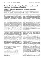
The cloned 3 kb cd38 promoter showing the location of the putative binding sites for NF-κB, AP-1 and GR (labeled NF-κB, AP-1 and GRE)Figure 7
The cloned 3 kb cd38 promoter showing the location of the putative binding sites for NF-κB, AP-1 and GR
(labeled NF-κB, AP-1 and GRE). Location of the putative binding sites for NF-κB, AP-1, and GRE on the cd38 promoter,
the 3 kb (Full) promoter, the truncated 1.8 kb promoter (Truncated), and promoter constructs with mutations in the binding
sites for NF-κB or AP1–4 or both binding sites (NF-AP-mut, NF-mut and AP-mut). The promoter was cloned in front of a luci-
ferase reporter gene in the pGL3 plasmid and was used to transfect HASM cells.
Respiratory Research 2008, 9:26 />Page 12 of 14
(page number not for citation purposes)
ticoids have been demonstrated for the NF-κB- and AP-1-
mediated regulation of other genes [34,38-43].
In this study, we have identified 4 glucocorticoid response
elements in the putative promoter region of the cd38 gene
as well as response elements for AP-1 and NF-κB (Table
1). Inhibition of NF-κB or AP-1 activation, or MAPK sign-
aling using pharmacological and molecular tools has con-
firmed their role in the regulation of CD38 expression
[9,10]. The identified putative sites for AP-1 and GRE also
exhibit strong binding in EMSA upon exposure to TNF-α
and dexamethasone respectively. The AP1–4 site (residing
between -2798 to -2789 bp) that shows very strong bind-
ing also appears to be functionally important in the acti-
vation of the promoter, since mutation of this site
profoundly affected TNF-α-induced activation of CD38
expression. With respect to NF-κB, mutation of the only
identifiable binding site also resulted in abolition of
CD38 transcription. It is worth noting that binding to this
site was weak compared to the consensus NF-κB sequence
binding, although competition with the unlabelled puta-
tive sequence effectively abolished the strong binding to
the consensus sequence. In the presence of dexametha-
sone, there was complete reversal of TNF-α-induced acti-
vation of the promoter, indicating direct transcriptional
regulation of CD38 expression by glucocorticoids in
HASM cells. These findings implicate the importance of
NF-κB and AP-1, and the GRE within the proximal pro-
moter region in the regulation of CD38 gene expression.
The results of promoter transfections and EMSAs with
cd38 putative GREs demonstrate transcriptional repres-
sion of CD38 expression by glucocorticoids. However,
glucocorticoids are also known to repress gene expression
in HASM cells through inhibition of histone acetylation
[44]. Evidence for glucocorticoid resistance of CD38
expression in HASM cells has also been reported when a
combination of cytokines is used as the stimulus as
opposed to the single stimulus used in the present study.
In this context, a recent study showed that in the com-
bined presence of TNF-α and IFN-γ or IFN-β, CD38
expression in HASM cells becomes refractory to glucocor-
ticoids [45]. The mechanism appears to involve induction
of the dominant negative GR-β. Thus, the glucocorticoid
regulation of CD38 expression in airway smooth muscle
cells is very complex and appears to depend on the stimu-
lus or combination of stimuli used.
In a recent study, Sun et al described the structure of the
promoter region of rabbit cd38 and provided evidence for
the functional regulation of CD38 expression in osteob-
Activation of the cd38 promoter in the HASM cellsFigure 8
Activation of the cd38 promoter in the HASM cells. Luciferase activity was measured as an index of promoter activation
with the Renilla luciferase activity (pRL-TK) to normalize for transfection efficiency. The normalized luciferase activity is
expressed as the fold change compared to the control. Left Panel: Activation of the full length promoter and the truncated
promoter. TNF-α (T) causes activation of the 3 kb promoter as compared to control (C), which is inhibited in the presence of
dexamethasone (D+T). Truncated promoter: There is no activation of the truncated promoter by TNF-α. Right Panel: TNF-
α causes activation of the 3 kb promoter (3 kb), but not the constructs with mutations in NF-κB or/and AP-1 site 4 (labelled
NF-AP mut, NF mut and AP mut). Representative of 3 different assays.
Respiratory Research 2008, 9:26 />Page 13 of 14
(page number not for citation purposes)
last and osteoclast cell lines [14]. In a region encompass-
ing 1.5 kb of the promoter obtained from a rabbit
genomic DNA library, the authors identified potential
binding sites for SP-1, AP-1, and AP-4. Using promoter-
reporter assays similar to those described in the present
studies, with a 1.5 kb promoter and several deletion
mutants, they were able to demonstrate a functional AP-1
site in the 1.0 kb promoter fragment. There also appears
to be cell-type specific activation of the promoter as
shown by studies with deletion mutagenesis.
Conclusion
In the present study, we describe NF-κB and AP-1 binding
motifs within the cd38 promoter that exhibit very strong
binding of nuclear proteins, mutations of which decrease
promoter activation and hence may be functionally rele-
vant. Our results also support the role of multiple tran-
scription factors in the regulation of CD38 expression in
HASM cells. Furthermore, we demonstrate a direct tran-
scriptional control of CD38 expression by glucocorti-
coids, although we have not identified specific GREs
within the proximal promoter region involved in this reg-
ulation. The fact that CD38 expression is regulated by
cytokines and transcription factors that are implicated in
asthma, and inhibited by glucocorticoids which are a
mainstay of asthma therapy makes this an attractive ther-
apeutic target.
Competing interests
The author(s) declare that they have no competing inter-
ests.
Authors' contributions
KGT and BNK contributed equally to the studies and
should be considered co-first authors. KGT cloned the
human cd38 promoter fragments and carried out the
sequence alignment. BNK carried out the electrophoretic
mobility shift assays and the promoter activation assays.
Both KGT and BNK drafted the manuscript. DNF, TFW
and MSK conceived of the investigations, helped in the
design of the experiments, and helped to draft the final
manuscript. RAP participated in the study by providing
well-characterized human airway smooth muscle cells
and helped in the draft of the manuscript.
Acknowledgements
This study was supported by National Institutes of Health Grants HL-
057498 (to M.S. Kannan), DA-11806 (to T.F. Walseth), HL-081824 and
National Institute of Environmental Health Sciences (NIEHS) ES0135080
grants (to R.A. Panettieri), and a Grant-in-Aid from the University of Min-
nesota Graduate School (to M.S. Kannan).
References
1. Lee HC: Enzymatic functions and structures of CD38 and
homologs. Chem Immunol 2000, 75:39-59.
2. Lee HC, Graeff RM, Walseth TF: ADP-ribosyl cyclase and CD38.
Multi-functional enzymes in Ca+2 signaling. Adv Exp Med Biol
1997, 419:411-9.
3. Mehta K, Shahid U, Malavasi F: Human CD38, a cell-surface pro-
tein with multiple functions. FASEB J 1996, 10:1408-17.
4. White TA, Johnson S, Walseth TF, Lee HC, Graeff RM, Munshi CB,
Prakash YS, Sieck GC, Kannan MS: Subcellular localization of
cyclic ADP-ribosyl cyclase and cyclic ADP-ribose hydrolase
activities in porcine airway smooth muscle. Biochim Biophys
Acta 2000, 1498:64-71.
5. Deshpande DA, Walseth TF, Panettieri RA, Kannan MS: CD38/cyclic
ADP-ribose-mediated Ca2+ signaling contributes to airway
smooth muscle hyper-responsiveness. FASEB J 2003, 17:452-4.
6. White TA, Kannan MS, Walseth TF: Intracellular calcium signal-
ing through the cADPR pathway is agonist specific in porcine
airway smooth muscle. FASEB J 2003, 17:482-4.
7. Deshpande DA, White TA, Dogan S, Walseth TF, Panettieri RA, Kan-
nan MS: CD38/cyclic ADP-ribose signaling: role in the regula-
tion of calcium homeostasis in airway smooth muscle. Am J
Physiol Lung Cell Mol Physiol 2005, 288:L773-88.
8. Deshpande DA, Dogan S, Walseth TF, Miller SM, Amrani Y, Panettieri
RA, Kannan MS: Modulation of calcium signaling by inter-
leukin-13 in human airway smooth muscle: role of CD38/
cyclic adenosine diphosphate ribose pathway. Am J Respir Cell
Mol Biol 2004, 31:36-42.
9. Kang BN, Tirumurugaan KG, Deshpande DA, Amrani Y, Panettieri
RA, Walseth TF, Kannan MS: Transcriptional regulation of CD38
expression by tumor necrosis factor-alpha in human airway
smooth muscle cells: role of NF-kappaB and sensitivity to
glucocorticoids. FASEB J 2006, 20:1000-2.
10. Tirumurugaan KG, Jude JA, Kang BN, Panettieri RA, Walseth TF, Kan-
nan MS: TNF-alpha induced CD38 expression in human air-
way smooth muscle cells: role of MAP kinases and
transcription factors NF-kappaB and AP-1. Am J Physiol Lung
Cell Mol Physiol 2007, 292:L1385-95.
11. Barnes PJ: Drugs for asthma. Br J Pharmacol 2006, 147(Suppl
1):S297-303.
12. Beato M: Gene regulation by steroid hormones. Cell 1989,
56:335-44.
13. Kishimoto H, Hoshino S, Ohori M, Kontani K, Nishina H, Suzawa M,
Kato S, Katada T: Molecular mechanism of human CD38 gene
expression by retinoic acid. Identification of retinoic acid
response element in the first intron. J Biol Chem 1998,
273:15429-34.
14. Sun L, Iqbal J, Zaidi S, Zhu LL, Zhang X, Peng Y, Moonga BS, Zaidi M:
Structure and functional regulation of the CD38 promoter.
Biochem Biophys Res Commun 2006, 341:804-9.
15. Sugiyama T, Scott DK, Wang JC, Granner DK: Structural require-
ments of the glucocorticoid and retinoic acid response units
in the phosphoenolpyruvate carboxykinase gene promoter.
Mol Endocrinol 1998, 12:1487-98.
16. Scott DK, Stromstedt PE, Wang JC, Granner DK: Further charac-
terization of the glucocorticoid response unit in the phos-
phoenolpyruvate carboxykinase gene. The role of the
glucocorticoid receptor-binding sites. Mol Endocrinol 1998,
12:482-91.
17. Ferrero E, Saccucci F, Malavasi F: The making of a leukocyte
receptor: origin, genes and regulation of human CD38 and
related molecules. Chem Immunol 2000, 75:1-19.
18. Mehta K: Retinoid-mediated signaling in CD38 antigen
expression. Chem Immunol 2000, 75:20-38.
19. Nata K, Takamura T, Karasawa T, Kumagai T, Hashioka W, Tohgo A,
Yonekura H, Takasawa S, Nakamura S, Okamoto H: Human gene
encoding CD38 (ADP-ribosyl cyclase/cyclic ADP-ribose
hydrolase): organization, nucleotide sequence and alterna-
tive splicing. Gene 1997, 186:285-92.
20. Ernst P, Smale ST: Combinatorial regulation of transcription. I:
General aspects of transcriptional control. Immunity 1995,
2:311-9.
21. Ferrero E, Malavasi F: Human CD38, a leukocyte receptor and
ectoenzyme, is a member of a novel eukaryotic gene family
of nicotinamide adenine dinucleotide+-converting enzymes:
extensive structural homology with the genes for murine
bone marrow stromal cell antigen 1 and aplysian ADP-ribo-
syl cyclase. J Immunol 1997, 159:3858-65.
Publish with BioMed Central and every
scientist can read your work free of charge
"BioMed Central will be the most significant development for
disseminating the results of biomedical research in our lifetime."
Sir Paul Nurse, Cancer Research UK
Your research papers will be:
available free of charge to the entire biomedical community
peer reviewed and published immediately upon acceptance
cited in PubMed and archived on PubMed Central
yours — you keep the copyright
Submit your manuscript here:
/>BioMedcentral
Respiratory Research 2008, 9:26 />Page 14 of 14
(page number not for citation purposes)
22. Ferrero E, Saccucci F, Malavasi F: The human CD38 gene: poly-
morphism, CpG island, and linkage to the CD157 (BST-1)
gene. Immunogenetics 1999, 49:597-604.
23. Dogan S, Deshpande DA, Kannan MS, Walseth TF: Changes in
CD38 expression and ADP-ribosyl cyclase activity in rat
myometrium during pregnancy: influence of sex steroid hor-
mones. Biol Reprod 2004, 71:97-103.
24. Dogan S, White TA, Deshpande DA, Murtaugh MP, Walseth TF, Kan-
nan MS: Estrogen increases CD38 gene expression and leads
to differential regulation of adenosine diphosphate (ADP)-
ribosyl cyclase and cyclic ADP-ribose hydrolase activities in
rat myometrium. Biol Reprod 2002, 66:596-602.
25. Dogan S, Deshpande DA, White TA, Walseth TF, Kannan MS: Regu-
lation of CD 38 expression and function by steroid hormones
in myometrium. Mol Cell Endocrinol 2006, 246:101-6.
26. Riffo-Vasquez Y, Pitchford S, Spina D: Cytokines in airway inflam-
mation. Int J Biochem Cell Biol 2000, 32:833-53.
27. Broide DH, Lotz M, Cuomo AJ, Coburn DA, Federman EC, Wasser-
man SI: Cytokines in symptomatic asthma airways. J Allergy Clin
Immunol 1992, 89:958-67.
28. Ammit AJ, Lazaar AL, Irani C, O'Neill GM, Gordon ND, Amrani Y,
Penn RB, Panettieri RA Jr: Tumor necrosis factor-alpha-induced
secretion of RANTES and interleukin-6 from human airway
smooth muscle cells: modulation by glucocorticoids and
beta-agonists. Am J Respir Cell Mol Biol 2002, 26:465-74.
29. Amrani Y, Chen H, Panettieri RA Jr: Activation of tumor necrosis
factor receptor 1 in airway smooth muscle: a potential path-
way that modulates bronchial hyper-responsiveness in
asthma? Respir Res 2000, 1:49-53.
30. Tliba O, Panettieri RA Jr, Tliba S, Walseth TF, Amrani Y: Tumor
necrosis factor-alpha differentially regulates the expression
of proinflammatory genes in human airway smooth muscle
cells by activation of interferon-beta-dependent CD38 path-
way. Mol Pharmacol 2004, 66:322-9.
31. Chikanza IC, Kozaci D, Chernajovsky Y: The molecular and cellu-
lar basis of corticosteroid resistance. J Endocrinol 2003,
179:301-10.
32. Scheinman RI, Cogswell PC, Lofquist AK, Baldwin AS Jr: Role of
transcriptional activation of I kappa B alpha in mediation of
immunosuppression by glucocorticoids. Science 1995,
270:283-6.
33. Auphan N, DiDonato JA, Rosette C, Helmberg A, Karin M: Immu-
nosuppression by glucocorticoids: inhibition of NF-kappa B
activity through induction of I kappa B synthesis. Science 1995,
270:286-90.
34. Bamberger CM, Schulte HM, Chrousos GP: Molecular determi-
nants of glucocorticoid receptor function and tissue sensitiv-
ity to glucocorticoids. Endocr Rev 1996, 17:245-61.
35. Verma IM, Stevenson JK, Schwarz EM, Van Antwerp D, Miyamoto S:
Rel/NF-kappa B/I kappa B family: intimate tales of associa-
tion and dissociation. Genes Dev 1995, 9:2723-35.
36. Karin M: New twists in gene regulation by glucocorticoid
receptor: is DNA binding dispensable? Cell 1998, 93:487-90.
37. Doucas V, Shi Y, Miyamoto S, West A, Verma I, Evans RM: Cytoplas-
mic catalytic subunit of protein kinase A mediates cross-
repression by NF-kappa B and the glucocorticoid receptor.
Proc Natl Acad Sci USA 2000, 97:11893-8.
38. Yang-Yen HF, Chambard JC, Sun YL, Smeal T, Schmidt TJ, Drouin J,
Karin M: Transcriptional interference between c-Jun and the
glucocorticoid receptor: mutual inhibition of DNA binding
due to direct protein-protein interaction. Cell 1990,
62:1205-15.
39. Schule R, Rangarajan P, Kliewer S, Ransone LJ, Bolado J, Yang N,
Verma IM, Evans RM: Functional antagonism between oncopro-
tein c-Jun and the glucocorticoid receptor. Cell 1990,
62:1217-26.
40. Brostjan C, Anrather J, Csizmadia V, Stroka D, Soares M, Bach FH,
Winkler H: Glucocorticoid-mediated repression of NFkappaB
activity in endothelial cells does not involve induction of Ika-
ppaBalpha synthesis. J Biol Chem 1996, 271:19612-6.
41. McKay LI, Cidlowski JA: Cross-talk between nuclear factor-
kappa B and the steroid hormone receptors: mechanisms of
mutual antagonism. Mol Endocrinol 1998, 12:45-56.
42. Jonat C, Rahmsdorf HJ, Park KK, Cato AC, Gebel S, Ponta H, Herrlich
P: Antitumor promotion and antiinflammation: down-modu-
lation of AP-1 (Fos/Jun) activity by glucocorticoid hormone.
Cell 1990, 62:1189-204.
43. Kerppola TK, Luk D, Curran T: Fos is a preferential target of glu-
cocorticoid receptor inhibition of AP-1 activity in vitro. Mol
Cell Biol 1993, 13:3782-91.
44. Nie M, Knox AJ, Pang L: beta2-Adrenoceptor agonists, like glu-
cocorticoids, repress eotaxin gene transcription by selective
inhibition of histone H4 acetylation. J Immunol 2005,
175:478-86.
45. Tliba O, Cidlowski JA, Amrani Y: CD38 expression is insensitive
to steroid action in cells treated with tumor necrosis factor-
alpha and interferon-gamma by a mechanism involving the
up-regulation of the glucocorticoid receptor beta isoform.
Mol Pharmacol 2006, 69:588-96.
46. Lenardo MJ, Baltimore D: NF-kappa B: a pleiotropic mediator of
inducible and tissue-specific gene control. Cell 1989, 58:227-9.
47. Cousin E, Medcalf RL, Bergonzelli GE, Kruithof EK: Regulatory ele-
ments involved in constitutive and phorbol ester-inducible
expression of the plasminogen activator inhibitor type 2
gene promoter. Nucleic Acids Res 1991, 19:3881-6.
48. Lee W, Mitchell P, Tjian R: Purified transcription factor AP-1
interacts with TPA-inducible enhancer elements. Cell 1987,
49:741-52.
49. Lopez-Bayghen E, Vega A, Cadena A, Granados SE, Jave LF, Gariglio
P, Alvarez-Salas LM: Transcriptional analysis of the 5'-noncod-
ing region of the human involucrin gene. J Biol Chem 1996,
271:512-20.
50. Minta JO, Fung M, Turner S, Eren R, Zemach L, Rits M, Goldberger G:
Cloning and characterization of the promoter for the human
complement factor I (C3b/C4b inactivator) gene. Gene 1998,
208:17-24.
51. Risse G, Jooss K, Neuberg M, Bruller HJ, Muller R: Asymmetrical
recognition of the palindromic AP1 binding site (TRE) by Fos
protein complexes. EMBO J 1989, 8:3825-32.
52. Sweetser MT, Hoey T, Sun YL, Weaver WM, Price GA, Wilson CB:
The roles of nuclear factor of activated T cells and ying-yang
1 in activation-induced expression of the interferon-gamma
promoter in T cells. J Biol Chem 1998, 273:34775-83.
53. Beato M: Gene regulation by steroid hormones. Cell 1989,
56:335-44.