Báo cáo y học: "Surprising negative association between IgG1 allotype disparity and anti-adalimumab formation: a cohort study" doc
Bạn đang xem bản rút gọn của tài liệu. Xem và tải ngay bản đầy đủ của tài liệu tại đây (390.82 KB, 7 trang )
RESEARCH ARTIC LE Open Access
Surprising negative association between IgG1
allotype disparity and anti-adalimumab
formation: a cohort study
Geertje M Bartelds
1
, Els de Groot
2
, Michael T Nurmohamed
1,3
, Margreet HL Hart
2
, Peter H van Eede
4
,
Carla A Wijbrandts
5
, Jakob BA Crusius
6
, Ben AC Dijkmans
1,3
, Paul Peter Tak
5
, Lucien Aarden
2
, Gerrit J Wolbink
1,2*
Abstract
Introduction: The human monoclonal antibody adalimumab is known to induce an anti-globulin response in
some adalimumab-treated patients. Antibodies against adalimumab (AAA) are associated with non-response to
treatment. Immunoglobulins, such as adalimumab, carry allotypes which represent slight differences in the amino
acid sequences of the constant chains of an IgG molecule. Immunoglobulins with particular IgG (Gm) allotypes are
racially distributed and could be immunogenic for individuals who do not express these allotypes. Therefore, we
investigated whether a mismatch in IgG allotypes between adalimumab and IgG in adalimumab-treated patients is
associated with the developmen t of AAA.
Methods: This cohort study consisted of 250 adalimumab-treated rheumatoid arthritis (RA) patients. IgG allotypes
were determined for adalimumab and for all patients. Anti-idiotype antibodies against adalimumab were measured
with a regular radio immunoassay (RIA), and a newly developed bridging enzyme linked immunosorbent assay
(ELISA) was used to measure anti-allotype antibodies against adalimumab. The association between AAA and the
G1m3 and the G1m17 allotypes was determined. For differences between groups we used the independent or
paired samples t-test, Mann-Whitney test or Chi square/Fisher’s exact test as appropriate. To investigate the
influence of confounders on the presence or absence of AAA a multiple logistic regression-analysis was used.
Results: Adalimumab carries the G1m17 allotype. No anti-allotype antibodies against adalimumab were detected.
Thirty-nine out of 249 patients had anti-idiotype antibodies against adalimumab (16%). IgG allotypes of RA patients
were associated with the frequency of AAA: patients homozygous for G1m17 had the highest frequency of AAA
(41%), patients homozygous for G1m3 the lowest frequency (10%), and heterozygous patients’ AAA frequency was
14% (P = 0.0001).
Conclusions: An allotype mismatch between adalimumab and IgG in adalimumab-treated patients did not lead to
a higher frequency of AAA. On the contrary, patients who carried the same IgG allotype as present on the
adalimumab IgG molecule, had the highest frequency of anti-adalimumab antibodies compared to patients whose
IgG allotype differed from adalimumab. This suggests that the allotype of adalimumab may not be highly
immunogenic. Furthermore, patients carrying the G1m17-allotype might be more prone to antibody responses.
Introduction
Treatment with monoclonal antibodies (mAbs) is known
to induce anti-mAb antibodies, leading to a diminished
treatment response [1-5]. The general structure of all
antibodies is very similar; it consist s of a constant and a
variable regio n, the variable region determines the idio-
type. The anti-adalimumab antibodies (AAA) measured
in previous studies are anti-idiotype antibodies, directed
against the idiotype of adalimumab [1,6]. The constant
region is almost identical in all antibodies of t he same
isotype, but differs in antibodies of different isotypes
(for example, IgA, IgM, IgG, IgE, IgD). However, within
* Correspondence:
1
Department of Rheumatology, Jan van Breemen Institute, Dr. Jan van
Breemenstraat 2, 1056AB Amsterdam, The Netherlands
Full list of author information is available at the end of the article
Bartelds et al. Arthritis Research & Therapy 2010, 12:R221
/>© 2010 Bartelds et al.; licensee BioMed Central Ltd. This is a n open access article distributed under the terms of the Creative Commons
Attribution License (http://creative commons.org/licenses/by/2.0), which permits unrestricted use, distribution, and reproduction in
any medium, provided the original work is properly cited.
an immunoglobulin of a certain isotype, allotypes repre-
sent slight differences in the amino acid sequences of
the constant heavy or light chains of different indivi-
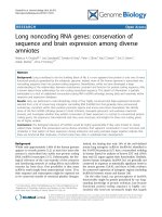
duals (Figure 1) [7]. There are different allotypes for
IgG1, IgG2 and IgG3 and no allotypes have been found
for IgG4. Allotypes are inherited in a codominant Men-
delian way, in fixed combinations called haplotypes.
Allotypes expressed on the constant region of IgG heavy
chain are designated as Gm (Genetic markers) together
with the subclass. Allotypes of heavy g1chainsare
defined as G1m allotypes, allo types of heavy g2chains
as G2m allotypes, and of heavy g3 chains as G3m allo-
types. The Gm system is unique in its ability to charac-
terize human populations by specific sets of haplotypes.
Specific Gm haplotypes are found in African, Caucasian
and Mongoloid populations. In a Caucasian population
(
a
)
(b)
CH1
CH2
CH3
CL
VH
VL
CH1
CH2
CH3
CL
VH
VL
G1m3,17
G1m2
G1m1
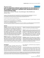
Figure 1 Basic i mmunoglobulin structure and IgG1 allotypes. (a).Basicimmunoglobulinstructure.CH1,2and3aretheconstantheavy
chains. CL is the constant light chain. VH is the variable heavy chain and VL the variable light chain which together form the variable domain of
the immunoglobulin, a specific antigen binding site, also referred to as the idiotype. (b). IgG1 allotypes [7]. The white residues in the constant
parts are those residues which differ by allotype in human IgG1. There is a Lys (G1m17) for Arg (G1m3) change at codon 214 in the CH1
domain, an Asp 356 Leu 358 (G1m1) for Glu 356 Met 358 (nG1m1) in the CH3 domain and a Gly 431 (G1m2) for Ala (nG1m2) also in the CH3
domain. The nG1m1 and nG1m2 are not “true” allotypes because these amino acid residues are present in other IgG subclasses and are not
expected to be immunogenic in the individual.
Bartelds et al. Arthritis Research & Therapy 2010, 12:R221
/>Page 2 of 7
the G1m1,17 (or G1m(a,z)) allotype is much less fre-
quent (0.15 to 0.35) than G1m3 (or G1m(f)) (0.65 to
0.85) [8]. Therefore, serologically defined allotypes differ
widely within and between population groups [9].
Allotypic markers can therefore differ b etween indi-
viduals and i mmunoglobulins with certain allotypes
can be immunogenic when injected into individuals
whose immunoglobulins lack the allotype. Treatment
with monoclonal antibodies with a certain allotype can
lead to the formation of anti-allo type antibodies. The
allotypes of a panel of licensed m Abs was determined
and adalimumab expresses the G1m1,17-allotype [9].
The risk to provoke antibody resp onses as a result of
allo-immunization has been described in a review [9].
MAb treatment of patients may lead to both allo-
immunization and/or xeno-immunization that result in
antisera that may recognize isotypic, allotypic and idio-
typic epitop es.
The association between anti-infliximab antibodies
and immunoglobulin allotypes was recently investigated
[10]. Infliximab expresses the G1m1,17-allotype, the
hypothesis of this study was that patients without the
G1m1,17-allotype were more likely to develop anti-
infliximab antibodies. However, no association was
found between the patients’ allotypes and the presence
or concentration o f anti-infliximab antibodies. The
authors pose the question whether this would also be
the case for humanized or fully human antibodies,
because in chimeric antibodies the murine variable
domain could dominate the antibody response. This
might not be the case for humanized or human mono-
clonal antibodies.
In th is study we investigated whether an I gG allotype
mismatch between adalimumab- and adalimumab-trea-
ted patients is associated with a higher frequency of
AAA.
Materials and methods
Patients
All 250 consecutive unrelated RA patients were included
in a prospective observational c ohort at the outpatient
clinicsoftheDepartmentsofRheumatologyoftheJan
van Breemen Institute and the Acade mic Medical Cen-
ter in Amsterdam. All patients fulfilled the American
College of Rheumatology 1987 revised criteria for RA
[11], and had active disease, indicated by a disease activ-
ity score in 28 joints (DAS28) of ≥3.2 despite earlier
treatment with two disease modifying anti-rheumatic
drugs (DMARDs) including metho trexate (MTX) at a
dosage of 25 mg weekly or at the maximal tolerable
dosage, according to the Dutch consensus statement on
the initiation and continuation of TNF blocking therapy
in RA [12]. Patients were treated with either adalimu-
mab and concomitant DMARD therapy, or adalimumab
monotherapy. All patients used adalimumab 40 mg sub-
cutaneously every other week. In patients with an inade-
quate response as judged by the treating rheumatologist,
the dosing freq uency of adalimum ab could be increased
to 40 mg per week. The study was approved by the
Medical Ethics Committee of the Slotervaart Hospital,
BovenIJ Hospital, the Jan van Breemen Institute, and the
Academic Medical Center/University of Amsterdam. All
patients gave written informed consent.
Clinical response to adalimumab
Disease activity was assessed at baseline and after
28 weeks of therapy using the DAS28 score. Clinical
response was assessed by the decrease in DAS28 score
(ΔDAS28) and the European League Against Rheuma-
tism (EULAR) response criteria [13].
The measurement of human IgG1 allotypes
ThemostprevalentallotypesoftheG1msystemwere
measured: G1m1, G1m3 and G1m17.
Immunoglobulin allotypes were determined by an
enzyme-linked immunosorbent assay (ELISA) using spe-
cific antibodies against Gm markers. All incubations
were at room temperature. Plates were coated for two
hours with 0.5 μg/ml of a mouse monoclonal antibod y
to human IgG1 (MH161-1, Sanquin, Amsterdam, The
Netherlands). A fter washing, the plates were incubated
for one hour with serum of interest, and diluted 1:1000
in PTG buffer (PBS, 0.2% gelatine, 0.02% Tween). After-
ward washing plates were incubated for one hour with
allot ype-specific biotinylated monoclonal antibodies. For
that purpose we used anti-G1m3 (%A1), ant i-G1m17
(5F10) and anti-G1m1 (MG102-A2 at 10 ng/ml (San-
quin). After washing, the plates were incubated with
polymerTsed st reptavidin-horseradis h peroxidase (poly-
HRP). N on-bound streptavidin-poly-HRP was removed
by washing and the amount of bound streptavidin was
measured by incubating the plates with tetr amethylben-
zidine (TMB), the substrate for HRP. The reaction was
stopped with H
2
SO
4
. Absorption at 450 nm was deter-
mined in a microtiter plate reader. The results of the
unknown sera were compared with the sera with known
allotypes.
Measurement of antibodies against adalimumab
Serum samples were collected at baseline and just prior
to an injection with adalimumab after 4, 16 and 28
weeks. The presence of AAA was determined at all time
points between baseline and 28 weeks. AAA were
detected with a radio immunoassay (RIA). One micro
litre of serum diluted in PBS/0.3% bovine serum albu-
min (BSA) (PA buffer) was incubated over night with 1
mg Sepharose-immobilized protein A (GE Healthcare,
Chalfont, St. Giles, UK) in a final volume of 800 μl.
Bartelds et al. Arthritis Research & Therapy 2010, 12:R221
/>Page 3 of 7
Subsequently the samples were washed with PBS 0.005%
Tween and specific ADA binding was detected by o/n
incubation with 20.000 dpm (approximately 1 ng) 1,25I
labeled F(ab)2 adalimumab diluted in Freeze buffer
(Sanquin). The unbound label was removed by washing,
and protein A bound radioactivity was measured. When
binding was higher than 25% of the input, sera were
further titrated. Antibody levels were compared to a
standard serum containing anti-drug antibody levels and
expressed in arbitrary units (AU). One AU corresponds
to approximately 12 ng. The mean cut-off value was set
at 12 AU/ml which was derived from 100 healthy
donors. Assay specificity was demonstrated by the
absence of AAA in 25 sera containing high-titres anti-
infliximab antibodies from patients not treated with ada-
limumab. In the assays we did not find cross reactivity.
Recently, patient sera were tested in a bioassay, which
confirmed the specificity and validity of the RIA [14].
Patients were defined as positive for anti-adalimumab
antibodies if tit res were above 12 AU/ml on at least one
occasion, in combina tion with serum adalimumab levels
below 5.0 mg/L. All baseline samples before the start of
treatment were negative.
Measurement of anti-allotype antibodies against
adalimumab/infliximab
We searched for anti-allotype antibodies using a two-
sided assay/bridging ELISA . Sera were tested for their
capacity to make a bridge between coated adalimumab
and biotinylated adalimumab. To that end plates were
coated with 0.5 μg/ml adalimumab in phosphate buf-
fered saline. After washing, the plates were incubated
with patient sera. After washing, the plates were incu-
bated with biot inylated adalimumab at 5 ng/ml. After
washing, the plate s were incubated with streptavidin-
poly-HRP and developed as described above. All sera
that were positive in the RIA were also positive in this
bridging ELISA. Adalimumab and inf liximab have the
same allotype (G1m1,17). We reasoned that if sera were
positive due to anti-allotype antibodies, these sera
should also be positive if adalimumab was replaced by
infliximab. Therefore, sera were also tested for their
capacity to bridge infliximab with adalimu mab and ada-
limumab with infliximab. As controls we used rabbit
anti-adalimumab-idiotype, rabbit anti-infliximab-idiotype
and a monoclonal antibody to human IgG (MH16-1).
Statistical analysis
For statistical analysis SPSS version 16.0 (SPSS Inc., Chi-
cago, Illinois, USA) was used. We chose to analyze the
association among AAA and the G1m3 and the G1m17
allotypes at codon 214 (Figure 1), since these allotypes
correspondwithasingleaminoacidchangeandhaplo-
type construction is not required. For differences
between groups we used the independent or paired sam-
ples t-test, Mann-Whitney test or Chi square/Fisher’s
exact test as appropriate. To investigate the influence of
confounders on the presence or absence of AAA, a mul-
tiple logistic regression-analysis was used. Variables con-
sidered potential confounders were chosen from all
available baseline variables and were determined for
every analysis specifically, based on differences between
groups includ ed in the analysis. Variables were included
in the regression model as confounders if the beta chan-
ged 10% or more after inclusion of the variable in the
model. The threshold for significance was set at
P < 0.05. To analyze clinical response we used the last
observation carried forward for patients who stopped
treatment due to non-response or adverse events, and
for patients who had received increased adalimumab
dosing frequency.
Results
Patient characteristics
Patient characteristics are shown in Tab le 1. Of the 250
patients enrolled in the study, six (2%) discontinued ada-
limumab treatment after four weeks of therapy, and 16
(6%) stopped treatment after Week 16. Ten patients
(4%) stopped due to treatment failure, nine (4%) because
of adverse events and three (1%) were l ost to follow-up.
Twenty-one patients (8%) had an increased dosing fre-
quency before 28 weeks to 40 mg adalimumab per
week; in these patients the last DAS28 before dose
increase was carried forward to 28 weeks.
Clinical response
The mean DAS28 after 28 weeks of adalimumab therapy
decreased from 5.2 ± 1.2 at baseline to 3.7 ± 1.5 (P =
0.0001). There were 63 (25%) non-responders, 105
(42%) moderate responders, and 82 (33%) good respon-
ders according to the EULAR response criteria.
Association between allotypes and anti-adalimumab
antibodies
Thirty-nine out of 249 patients had antibodies against
adalimumab (16%); in one patient AAA could not be
determined. Patients without AAA had a significantly
greater DAS28 improv ement than patients with AAA
(ΔDAS28 = 1.7 versus ΔDAS28 = 0.5, P = 0.0001).
Adalimumab and infliximab have the same allotype
G1m1,17 [9,10]. Nevert heless we observed that all se ra
positive in the assay for antibodies to adalimumab
(hence the adalimumab-adalimumab combination) were
neg ative in the assay for anti-inflixim ab (the infliximab-
infliximab combination) as well as in the assay where
adalimumab was combined with infliximab. The anti-
IgG was strongly positive in all three assays. Our con-
clusion is that t hese patients do not make anti-allotype
Bartelds et al. Arthritis Research & Therapy 2010, 12:R221
/>Page 4 of 7
antibodies and that all AAA’ s are due to anti-idiotypic
antibodies.
There was a significant association between the G1m3
and G1m17 allotypes and antibodies against adalimu-
mab (Table 2). After adjustment for MTX dose in logis-
ticregressionthecarriageofmoreG1m17alleleswas
significantly associated with a higher frequency of anti-
bodies against adalimumab (P = 0.0001; OR = 2.639;
95% CI = 1.608 to 4.332). Baseline characteristics for the
three groups wit h G1m3 and G1m17 allotyp es are
shown in Table 3.
Discussion
Our h ypothesis was that a mismatch between the allo-
type of adalimumab, G 1m1,17, and the allotypes of the
IgG o f adalimumab treated RA patients would be asso-
ciated with a higher frequency of anti-adalimumab anti-
bodies. This was not the case. The first explanation for
this lack of association could be that neither of the
assays we used was able to d etect anti-allotype antibo-
dies. Our RIA for the detection of AAA is designed to
detect anti-idiotype antibodies. In this assay a solution
containing pepsine treated polyclonal IgG Freeze buffer
is added, as a result anti-allotype antibodies are not
detected. However, without Freeze buffer anti-allotype
antibodies also were not de tected. No anti-allo type anti-
bodies were detected with the bridging ELISA. It might
be possible that the bridging ELISA was not able to
detect anti-allotype antibodies, due to low titers, epitope
masking or steric hindrance. Another explanation could
be that anti-allotype antibodies are not developed or
that the quantity of the anti-allotype antibody response
is not large enough to be detected. The allotypes of ada-
limumab may not be highly immunogenic, and could be
only a minor antigen compared to the idiotype of adali-
mumab. Patients who were homozygous for G1m3, for
whom the allotype of adalimumab theoretically would
be immunogenic, had a clinical response that did not
differ from patients who carried the G1m17-allotype
after adjustment for having anti-idiotype antibodies
Table 1 Demographic and clinical characteristics at
baseline
Total population
n = 250
Demographics
Age, years 52 ± 13
Female, no. (%) 197 (79)
DMARD therapy
Prior DMARDs (no.) 3.4 ± 1.6
Methotrexate use, no. (%) 199 (80)
Methotrexate dose (mg/wk) 23 (15 to 25)
Prednisone use, no. (%) 82 (33%)
Prednisone dose (mg/day) 7.5 (5 to 10)
Disease status
Disease duration (years) 8 (4 to 17)
Rheumatoid factor positive, no. (%) 179 (72)
Erosive disease, no. (%) 194 (78)
Erythrocyte sedimentation rate (mm/h) 30 ± 23
C-reactive protein (mg/dl) 11 (5 to 24)
DAS28 5.2 ± 1.2
Mean values ± SD, median and interquartile range, or percentages are shown.
DAS28, Disease Activity Score in 28 joints; DMARD, diseases modifying anti
rheumatic drug.
Table 2 Association between G1m3 and G1m17 allotypes
and antibodies against adalimumab
G1m phenotype AAA - AAA +
3,3 108 (90%) 12 (10%)
3,17 83 (86%) 14 (14%)
17,17 19 (59%) 13 (41%)
AAA -, anti-adalimumab antibodies negative; AAA +, anti-adalimumab
antibodies positive.
Patient numbers and corresponding percentages are shown. P = 0.0001 for
the whole table.
Table 3 Demographic and clinical characteristics at
baseline
G1m phenotype
3,3 3,17 17,17
n = 120 n =98 n =32
Demographics
Age, years 53 ± 14 53 ± 12* 48 ± 12*
Female, no. (%) 95 (79) 74 (76) 28 (88)
DMARD therapy
Prior DMARDs (no.) 3.5 ± 1.6 3.4 ± 1.7 3.3 ± 1.6
Methotrexate use, no. (%) 95 (79) 81 (83) 23 (72)
Methotrexate dose
(mg/wk)
21 (15 to 25) 25 (15 to 25) 25 (17.5 to
25)
Prednisone use, no. (%) 39 (33) 31 (32) 12 (38)
Prednisone dose (mg/day) 7.5 (5 to 10) 5 (5 to 7.5) 10 (5 to 10)
Disease status
Disease duration (years) 10 (4 to 17) 11 (3 to 16) 7 (3 to 17)
Rheumatoid factor
positive, no. (%)
80 (67)* 70 (71) 29 (91)*
Erosive disease, no. (%) 90 (75) 76 (78) 28 (88)
Erythrocyte sedimentation
rate (mm/h)
29 ± 23 31 ± 25 28 ± 17
C-reactive protein (mg/dl) 11 (4 to 24) 10 (6 to 23) 14 (6 to 31)
DAS28 5.1 ± 1.2 5.3 ± 1.2 5.2 ± 1.0
DAS28, Disease Activity Score in 28 joints; DMARD, diseases modifying anti
rheumatic drug.
*There was a significant difference between patients with the 3,17-allotype
and the 17,17-allotype for age (P = 0.020), and between both homozygous
groups 3,3 and 17,17 for rheumatoid factor positivity, no. (P = 0.007).
Bartelds et al. Arthritis Research & Therapy 2010, 12:R221
/>Page 5 of 7
against adalimumab (data not shown). This suggests that
if anti-allotype antibodies had developed, their clinical
relevance would be nil.
Our hypothesis could not b e confirmed, but the
results showed an unexpected association between
allot ypes and AAA: RA pa tients whose IgG1 was homo-
zygous for the same allotype as adalimumab, the
G1m17-allotype, had the highest frequency of AAA
compa red to G1m3 homozygotes or heterozygotes. This
suggests that the frequency of AAA has no relation with
the possible immunogenicity of the allotype of adalimu-
mab, but is more likely explained by patient-related
genetic factors. A selective force behind the distribution
and inheritance of allotypes may have been the associa-
tion between immunoglobulin allotypes and the spec ific
antibody responses to pathogens, resulting in differential
immunity to infectious diseases [15]. For numerous
infectious diseases an association has been found
between immunoglobulin allotypes and (the level of)
antibody response [15]. There are several studies in
which the G1m1,17-allotype or a haplotype containing
this allotype was associated wit h a stronger immune
response compared to individuals with the G1m3-allo-
type. For example, systemic sclerosis patients homoz y-
gous for the G1m3 allele were 60% less likely to be
seropositive for IgG antibodies against cytomegalovirus
than patients homozygous for the G1m17 allele or the
heterozygotes [16]. Hepatitis C virus (HCV) infected
patients with the Gm1,17 5,13 phenotype within an
African American population had two-fold higher med-
ian ant ibody titres against E1 and E2 envelope glycopro-
teins, HCV e pitopes than those who lacked this
phenotype [17]. In a study on the association of allo-
types with anti bodies against MUC1, a tumor-associated
antigen, gastric cancer patients with the phenotype Gm3
23 5, 13 had lower anti-MUC1-IgG levels compared to
patients without this phenotype [18].
Patients with the G1m3 phenotype not only had AAA
significantly less often, but were also less often positive for
rheumatoid factor (Table 3). This also contributes to the
hypothesis that allotypes are associated with specific anti-
body responses. Individuals with a G1m1, 17-allotype
might be more prone to antibody responses than indivi-
duals with the G1m3-allotype. No conclusive data are
available on how allotypes could influence immune
response, albeit several possibilities are mentioned [15].
The locus or loci responsible for the association with
immune response may not be the Gm system itself but
may reflect linkage disequilibrium with other polymorph-
isms of the constant region genes or with specific variable
regiongenes.Thereisevidenceforageneticpredisposi-
tion to the formation of antibodies. Previously, we showed
that interleukin-10 (IL10) polymorphisms were associated
with anti-adalimumab antibody formation in RA [19].
However, we did not find an association between IL10
polymorphisms and IgG allotypes (data not shown).
Conclusions
To our knowledge this is the first study examining the
association between G1m allotypes and immunogenicity
against adalimumab. Our findings suggest that the allotype
is not a dominant antigen of adalimumab. Albeit we have
to take into account that we did not find anti-allotype anti-
bodies. Interestingly, our data show that anti-adalimumab
antibody formation occurred more often in RA patients
with the G1m17-allotype than in RA patients without this
allotype, which indicates a role for genetic factors. Patients
carrying this allotype might be more prone to antibody
responses. However, these results should be replicated in
larger study populations with a representative variation in
allotypes in o rder to draw firm conclusions.
Abbreviations
AAA: anti-adalimumab antibodies; AU: arbitrary units; BSA: bovine serum
albumin; DAS28: disease activity score in 28 joints; DMARDs: disease
modifying anti-rheumatic drugs; ELISA: enzyme linked immunosorbent assay;
EULAR: European League Against Rheumatism; Gm: Genetic marker; HCV:
hepatitis C virus; HRP: horseradish peroxidase; IL: interleukin; mAbs:
monoclonal antibodies; MTX: methotrexate; RA: rheumatoid arthritis; RIA:
radio immunoassay; TMB: tetramethylbenzidine; TNF: tumour necrosis factor.
Acknowledgments
The authors wish to thank Henk de Vrieze and Kim van Houten for
performing the assays. In addition, this investigation was also facilitated by
the Clinical Research Bureau of the Jan van Breemen Institute. Finally, we
thank Professor M. Boers for contributions to the concept and study design.
Parts of this study were financed by Abbott and Wyeth. The study sponsors
had no involvement in the study design, in the collection, analysis, and
interpretation of data, or in the writing of the report and in the decision to
submit the paper for publication.
Author details
1
Department of Rheumatology, Jan van Breemen Institute, Dr. Jan van
Breemenstraat 2, 1056AB Amsterdam, The Netherlands.
2
Department of
Immunopathology, Landsteiner Labaratory Sanquin Research, Plesmanlaan
125, 1066CX Amsterdam, The Netherlands.
3
Department of Rheumatology,
VU University Medical Center, Postbus 7057, 1007MB Amsterdam, The
Netherlands.
4
Department of Immunogenetics, Sanquin Diagnostic Services,
Plesmanlaan 125, 1066CX Amsterdam, The Netherlands.
5
Department of
Clinical Immunology and Rheumatology, Academic Medical Center/
University of Amsterdam, Meibergdreef 9, 1105AZ Amsterdam, The
Netherlands.
6
Department of Pathology Laboratory for Immunogenetics, VU
University Medical Center, Postbus 7057, 1007MB Amsterdam, The
Netherlands.
Authors’ contributions
GW had full access to all the data in the study and had final responsibility
for the decision to submit for publication. GB, EG, MN, MH, BC, LA and GW
participated in the study design. GB, MN, CW, PT and GW were involved in
the acquisition of the data. GB took part in the data analysis and EG and MH
in carrying out the immunoassays. GB, LA and GW participated in the
interpretation of the data. All authors participated in the preparation of the
manuscript and saw and approved the final version.
Competing interests
BD and PT are members of the advisory board of Abbott, and BD, PT and
MN have received honoraria for lectures. PT has served as a consultant to
Abbott, Amgen, Centocor, Schering-Plough, UCB, and Wyeth. BD received
Bartelds et al. Arthritis Research & Therapy 2010, 12:R221
/>Page 6 of 7
research grants from Schering-Plough, Wyeth and Abbott. The other authors
declare that they have no competing interests.
Received: 14 July 2010 Revised: 16 November 2010
Accepted: 27 December 2010 Published: 27 December 2010
References
1. Bartelds GM, Wijbrandts CA, Nurmohamed MT, Stapel S, Lems WF,
Aarden L, Dijkmans BA, Tak PP, Wolbink GJ: Clinical response to
adalimumab: relationship to anti-adalimumab antibodies and serum
adalimumab concentrations in rheumatoid arthritis. Ann Rheum Dis 2007,
66:921-926.
2. Wolbink GJ, Vis M, Lems W, Voskuyl AE, de Groot E, Nurmohamed MT,
Stapel S, Tak PP, Aarden L, Dijkmans B: Development of antiinfliximab
antibodies and relationship to clinical response in patients with
rheumatoid arthritis. Arthritis Rheum 2006, 54:711-715.
3. Radstake TR, Svenson M, Eijsbouts AM, van den Hoogen FH, Enevold C, van
Riel PL, Bendtzen K: Formation of antibodies against infliximab and
adalimumab strongly correlates with functional drug levels and clinical
responses in rheumatoid arthritis. Ann Rheum Dis 2009, 68:1739-1745.
4. Miyasaka N: Clinical investigation in highly disease-affected rheumatoid
arthritis patients in Japan with adalimumab applying standard and
general evaluation: the CHANGE study. Mod Rheumatol 2008, 18:252-262.
5. Bendtzen K, Geborek P, Svenson M, Larsson L, Kapetanovic MC, Saxne T:
Individualized monitoring of drug bioavailability and immunogenicity in
rheumatoid arthritis patients treated with the tumor necrosis factor
alpha inhibitor infliximab. Arthritis Rheum 2006, 54:3782-3789.
6. Bartelds GM, Wijbrandts CA, Nurmohamed MT, Stapel SO, Lems WF,
Aarden L, Dijkmans BA, Tak PP, Wolbink GJ: Anti-infliximab and anti-
adalimumab antibodies in relation to response to adalimumab in
infliximab switchers and anti-TNF naive patients: a cohort study. Ann
Rheum Dis 2010, 69:817-821.
7. Models of human IgG isotypes and allotypes. [ />~mrc7/lectures/models.html].
8. IGMT Repertoire. [ />allotypes/human/IGH/IGHC/Hu_IGHCallotypes1.html].
9. Jefferis R, Lefranc MP: Human immunoglobulin allotypes: possible
implications for immunogenicity. MAbs 2009, 1:332-338.
10. Magdelaine-Beuzelin C, Vermeire S, Goodall M, Baert F, Noman M,
Assche GV, Ohresser M, Degenne D, Dugoujon JM, Jefferis R, Rutgeerts P,
Lefranc MP, Watier H: IgG1 heavy chain-coding gene polymorphism
(G1m allotypes) and development of antibodies-to-infliximab.
Pharmacogenet Genomics 2009, 19:383-387.
11. Arnett FC, Edworthy SM, Bloch DA, McShane DJ, Fries JF, Cooper NS,
Healey LA, Kaplan SR, Liang MH, Luthra HS: The American Rheumatism
Association 1987 revised criteria for the classification of rheumatoid
arthritis. Arthritis Rheum 1988, 31:315-324.
12. Furst DE, Breedveld FC, Kalden JR, Smolen JS, Burmester GR, Sieper J,
Emery P, Keystone EC, Schiff MH, Mease P, van Riel PL, Fleischmann R,
Weisman MH, Weinblatt ME: Updated consensus statement on biological
agents for the treatment of rheumatic diseases, 2007. Ann Rheum Dis
2007, 66:iii2-22.
13. van Gestel AM, Anderson JJ, van Riel PL, Boers M, Haagsma CJ, Rich B,
Wells G, Lange ML, Felson DT: ACR and EULAR improvement criteria have
comparable validity in rheumatoid arthritis trials. American College of
Rheumatology European League of Associations for Rheumatology. J
Rheumatol 1999, 26:705-711.
14. Aarden L, Ruuls SR, Wolbink G: Immunogenicity of anti-tumor necrosis
factor antibodies-toward improved methods of anti-antibody
measurement. Curr Opin Immunol
2008, 20:431-435.
15. Pandey JP: Immunoglobulin GM and KM allotypes and vaccine
immunity. Vaccine 2000, 19:613-617.
16. Pandey JP: Immunoglobulin GM genes and IgG antibodies to
cytomegalovirus in patients with systemic sclerosis. Clin Exp Rheumatol
2004, 22:S35-S37.
17. Pandey JP, Luo Y, Elston RC, Wu Y, Philp FH, Astemborski J, Thomas DL,
Netski DM: Immunoglobulin allotypes influence IgG antibody responses
to hepatitis C virus envelope proteins E1 and E2. Hum Immunol 2008,
69:158-164.
18. Pandey JP, Nietert PJ, Mensdorff-Pouilly S, Klaamas K, Kurtenkov O:
Immunoglobulin allotypes influence antibody responses to mucin 1 in
patients with gastric cancer. Cancer Res 2008, 68:4442-4446.
19. Bartelds GM, Wijbrandts CA, Nurmohamed MT, Wolbink GJ, de Vries N,
Tak PP, Dijkmans BA, Crusius JB, van der Horst-Bruinsma IE: Anti-
adalimumab antibodies in rheumatoid arthritis patients are associated
with interleukin-10 gene polymorphisms. Arthritis Rheum 2009,
60:2541-2542.
doi:10.1186/ar3208
Cite this article as: Bartelds et al.: Surprising negative association
between IgG1 allotype disparity and anti-adalimumab formation: a
cohort study. Arthritis Research & Therapy 2010 12:R221.
Submit your next manuscript to BioMed Central
and take full advantage of:
• Convenient online submission
• Thorough peer review
• No space constraints or color figure charges
• Immediate publication on acceptance
• Inclusion in PubMed, CAS, Scopus and Google Scholar
• Research which is freely available for redistribution
Submit your manuscript at
www.biomedcentral.com/submit
Bartelds et al. Arthritis Research & Therapy 2010, 12:R221
/>Page 7 of 7