Báo cáo y học: "TLR7 single-nucleotide polymorphisms in the 3’ untranslated region and intron 2 independently contribute to systemic lupus erythematosus in Japanese women: a case-control association study" ppsx
Bạn đang xem bản rút gọn của tài liệu. Xem và tải ngay bản đầy đủ của tài liệu tại đây (312.52 KB, 8 trang )
RESEARCH ARTICLE Open Access
TLR7 single-nucleotide polymorphisms in the 3’
untranslated region and intron 2 independently
contribute to systemic lupus erythematosus in
Japanese women: a case-control association study
Aya Kawasaki
1
, Hiroshi Furukawa
2
, Yuya Kondo
3
, Satoshi Ito
3,4
, Taichi Hayashi
3
, Makio Kusaoi
5
, Isao Matsumoto
3
,
Shigeto Tohma
2
, Yoshinari Takasaki
5
, Hiroshi Hashimoto
6
, Takayuki Sumida
3
and Naoyuki Tsuchiya
1*
Abstract
Introduction: The Toll-like receptor 7 (TLR7) gene, encoded on human chromosome Xp22.3, is crucial for type I
interferon production. A recent multicenter study in East Asian populations, comprising Chinese, Korean and Japanese
participants, identified an association of a TLR7 single-nucleotide polymorphism (SNP) located in the 3’ untranslated
region (3’ UTR), rs3853839, with systemic lupus erythematosus (SLE), especially in males, although some difference was
observed among the tested populations. To test whether additional polymorphisms contribute to SLE in Japanese, we
systematically analyzed the association of TLR7 with SLE in a Japanese female population.
Methods: A case-control association study was conducted on eight tag SNPs in the TLR7 region, including
rs3853839, in 344 Japanese females with SLE and 274 healthy female controls.
Results: In addition to rs3853839, two SNPs in intron 2, rs179019 and rs179010, which were in moderate linkage
disequilibrium with each other (r
2
= 0.53), showed an association with SLE (rs179019: P = 0.016, odds ratio (OR) 2.02,
95% confidence interval (95% CI) 1.15 to 3.54; rs179010: P = 0.018, OR 1.75, 95% CI 1.10 to 2.80 (both under the
recessive model)). Conditional logistic regression analysis revealed that the association of the intronic SNPs and the 3’
UTR SNP remained significant after we adjusted them for each other. When only the patients and controls carrying the
risk genotypes at the 3’ UTR SNPpositionwere analyzed, the risk of SLE was significantly increased when the individuals
also carried the risk genotypes at both of the intronic SNPs (P = 0.0043, OR 2.45, 95% CI 1.31 to 4.60). Furthermore, the
haplotype containing the intronic risk alleles in addition to the 3’ UTR risk allele was associated with SLE under the
recessive model (P = 0.016, OR 2.37, 95% CI 1.17 to 4.80), but other haplotypes were not associated with SLE.
Conclusions: The TLR7 intronic SNPs rs179019 and rs179010 are associated with SLE independently of the 3’ UTR
SNP rs3853839 in Japanese women. Our fin dings support a role of TLR7 in predisposition for SLE in Asian
populations.
Introduction
Toll-like receptors (TLRs) play a central role in detect-
ing microbial pathogens. TLRs initiate innate immune
responses and also induce adaptive immune responses
[1]. Recently, TLRs have been strongly implicated in
autoimmune diseases [2]. The TLR7 and TLR9 genes,
which are expressed intracellularly in plasmacyt oid den-
dritic cells (pDCs) and B cells, recognize single-stranded
RNA and DNA containing cytidine-phosphate-guano-
sine motifs, respectively. Activation of pDCs by TLR7
and TLR9 induces a large amount of type I interferon
(IFN). It has become evident that RNA- and DNA-con-
taining immune complexes, which o ften exist in sera of
patients with systemic lupus erythematosus (SLE), can
activate TLR7 and TLR9 signaling [2].
* Correspondence:
1
Molecular and Genetic Epidemiology Laboratory, Doctoral Program in
Biomedical Sciences, Graduate School of Comprehensive Human Sciences,
University of Tsukuba, 1-1-1 Tennodai, Tsukuba 305-8575, Japan
Full list of author information is available at the end of the article
Kawasaki et al. Arthritis Research & Therapy 2011, 13:R41
/>© 2011 Kawasaki et al.; licensee BioMed Central Ltd. This is an open access artic le distributed under the terms of the Creative
Commons Attribution Licen se ( which permi ts unrestricted use, distributio n, and
reproduction in any medium, provided the original work is properly cite d.
Several lines of evidence support a role of TLR7 in
SLE pathogenesis [2]. Male BXSB mice bearing the Y
chromosome-linked autoimmune accelerator (Yaa)gene
develop severe SLE. It has been revealed that Yaa muta-
tion is caused by a translocation of a portion of the X
chromosome containing TLR7 onto the Y chromosome
[3,4]. Yaa-bearing mice have been demonstrated to have
twofold overexpression of TLR7 protein and mRNA
[3,4]. In contrast, lupus-prone MRL/Mp
lpr/lpr
mice lack-
ing TLR7 showed impaired production of antibodies to
RNA-containing antigens, s uch as anti-Smith (anti-Sm)
antibodies, and developed less severe disease [5].
Furthermore, upregulated expressi on of TLR7 mRNA in
peripheral blood mononuclear cells (PBMNCs) was
observed in human SLE [6].
Recently, a multicenter collaborative study including
our group re ported an association of TLR7, located in
Xp22.3, with SLE in combined East Asian populations
[7]. In a discovery panel consisting mainly of Chinese
and Korean populations, the association of 27 single-
nucleotide polymorphisms (SNPs) in the TLR7-TLR8
region with SLE was examined, and a significant associa-
tion of the TLR7 3’ untranslated region (3’ UTR) SNP,
rs3853839, was identified. Subsequently, the association
of rs3853839 was replicated in two independent Chinese
and Japanese case-control sets. The association was pro-
minent in males with SLE. In addition, rs3853839 was
associated w ith elevated expression of TLR 7.Thestudy
also revealed some differences in the a ssociation of
rs3853839 and other SNPs among Chinese, Korean and
Japanese populations [7], indicating that systematic SNP
screening should be performed in each population.
In this study, we examined the associa tion of eight
TLR7 tag S NPs with SLE in Japanese women and dis-
covered a newly identified association of two intronic
SNPs, rs179019 and rs179010, with SLE. These SNPs
and the 3’UTR rs385383 9 were foun d to i ndependently
contribute to the genetic risk for SLE.
Materials and methods
Patients and controls
Three hundred forty-four Japanese female patients with
SLE (mean age ± SD, 42.9 ± 13.8 years) and 274 health y
female controls (mean age ± SD, 31.3 ± 8.9 years) were
recruited at University of Tsukuba, Juntendo University,
Sagamihara National Hospital, and at the University of
Tokyo. Among them, 296 SLE patients a nd 250 healthy
controls were also examined in a previous study to
replicate the association of rs3853839 with SLE in Japa-
nese, but other SNPs were not investigated in that study
[7]. All patients and healthy individuals were native
Japanese living in the central part of Japan. All pat ients
with SLE fulfilled the American College of Rheumatol-
ogy criteria for SLE [8].
This study was carried out in compliance with the
Declaration of Helsinki. The study was r eviewed and
approved by the research ethics committees of Univer-
sity of Tsukuba, Sagamihara National Hospital, the Uni-
versity of Tokyo an d Juntendo University. Informed
consent was obtained from all study participants.
Genotyping
Eight tag SNPs in the TLR7 region were selected on the
basis of the HapMap Phase II JPT (Japanese in Tokyo)
data obtained from the HapMap database [9] with the cri-
teria of minor allele frequency >0.1 and an r
2
threshold of
0.9. Genotyping of the tag SNPs was carried out using the
TaqMan genotyping a ssay on the Applied Biosystems
7300 Real-Time PCR System (Applied Biosystems, Foster
City, CA, USA), accordi ng to the manufacturer’sinstruc-
tions. Thermal cycling conditions consisted of initial dena-
turation at 95°C for 10 minutes, followed by 50 cycles at
95°C for 15 s econds eac h and at 60°C for one minu te
each. TaqMan probes used in this study were as follows:
Assay ID: C__15757400_10 (rs2302267), C___2259585_10
(rs179019), C___7625717_10 (rs1634322),
C___2259582_10 (rs179016), C___2259578_10 (rs179012),
C___2259576_10 (rs179010), C___2259575_10 (rs179009),
and C___2259573_10 (rs3853839).
Expression analysis by real-time quantitative reverse
transcription polymerase chain reaction assay
Total RNA w as extracted from PBMNCs of 18 females
with SLE using the RNeasy Mini Kit (QIAGEN, Hilden,
Germany), reverse transcribed into c DNA and used for
real-time quantitative reverse transcription polymerase
chain reaction (RT-PCR) assay. Expression of TLR7 was
analyzed using the TaqMan Gene Expression Assay
(Applied Biosystems), Hs00152971_m1. Amplification of
cDNA was conducted using the Applied Biosystems
7300 Real-Time PCR System (Applied Biosystems)
under the following conditions: 50°C for 2 minutes and
95°C for 10 minutes, and 50 cycles at 95°C for 15 sec-
onds and at 60°C for 1 minute, and then the cycle
threshold (CT) value fo r each sample was obta ined
using Applied Biosystems 7300 System SDS version 1.4
software (Applied Biosystems). Relative quantitative
levels were calculated on the basis of the CT value by a
standard curve method and were normalized to b-actin
(ACTB) expression (Hs99999903_m1). The experiments
were done in triplicate for each sample.
Statistical analysis
Differences in allele and genotype frequencies between
SLE patients and healthy controls were analyzed by
using a c
2
test with 2 × 2 contingency tables. When one
or more of the variables in the contingency tables was
20 or less, Fisher’s exact test was employed. Linkage
Kawasaki et al. Arthritis Research & Therapy 2011, 13:R41
/>Page 2 of 8
dis equilibr ium (LD) was analyzed using HaploView ver-
sion 4.0 software (Broad Institute, Cambridge, MA,
USA). Pairwise r
2
values were calculated on the basis of
the genotypes of 274 healthy controls. Estimation of
haplotype frequencies and association tests were per-
formed using HaploView version 4.0 software.
To examine whether each SNP independently contri-
butes to susceptibility to SLE, conditional logistic regres-
sion analysis was employed. Dominant, codominant and
recessive models were tested for each SNP, and the
model that provided the lowest P value was selected as
the best fit model. As a result, the following were used
as independent variables: rs3853839, C/C = 0, G/C = 1
and G/G = 2 under the codominant model for the G
allele; rs179019, C/C = 0, C/A = 0, A/A = 1 under the
recessive model for the A allele; rs179010, C/C = 0, C/T
= 0, and T/T = 1 under the recessive mo del for the T
allele.
The association o f TLR7 SNPs with TLR7 mRNA
expression was assessed by using the Kruskal-Wallis
test.
Results
Association of TLR7 SNPs with SLE in a Japanese female
population
To systematically examine association of TLR7 SNPs
with SLE in Japanese, eight tag SNPs in the TLR7 gene,
including rs3853839 in the 3’UTR, which was recently
shown to be associated with SLE in East Asian popula-
tions [7], were a nalyzed in 344 Japanese females with
SLE and 274 healthy female controls. Because TLR7 is
located on an X chromosome, male and female indivi-
duals needed to be analyzed for the a ssociation sepa-
rately. However, because of the female predominance of
SLE (9:1 female to male ratio), the sample size of male
SLE patients was too small to be analyzed statistically.
Therefore, male patients and controls were excluded
from this study. No deviation from the Hardy-Weinberg
equilibrium was observed in the controls (P > 0.05).
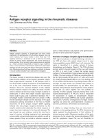
In addition to the association of rs3853839 reported
separately [7], the association of two SNPs in intron 2,
rs179019 and rs179010, was newly detected (Figure 1
and Table 1). Significant association of rs179019 and
rs179010 was observed under the recessive model for
the A and T alleles, respectively (rs179019: P = 0.016,
odds ratio (OR) 2 .02, 95% confidence interval (95% CI)
1.15 to 3.54; rs179010: P = 0.018, OR 1.75, 95% CI 1.10
to 2.80). LD was present between rs179019 and
rs179010 (r
2
= 0.53), while LD between rs3853839 and
each of the intronic SNPs was modest (r
2
=0.02and
0.04) (Figure 1).
To examine the contribution of each SNP to suscept-
ibility to SLE, conditional logistic regression analysis was
conducted. As shown in Table 2, the association of
rs3853839 remained significant after adjustment for the
intronic SNP g enotyp es. Adjusted P values (P
adjusted
)for
rs3853839 under the codominant model were 0.040 and
0.047 after adjustment for rs179019 and rs179010,
respectively. The association of rs179019 and rs179010
also remained significant after adjustment for rs3853839
(rs179019: P
adjusted
= 0.026; rs179010: P
adjusted
= 0.042).
These results suggest that rs3853839 and the i ntronic
SNPs are independently associated with SLE. In con-
trast, the association o f rs179019 and rs179010 was
eliminated wh en they were adjusted for each other as
expected on the basis of LD between the two (Table 2
and Figure 1).
In agreement with these findings, when only the
patients and controls carrying the risk genotypes of the
3’ UTR SNP were analyzed, possession of both of the
intronic SNP risk genotypes was significantly associated
with SLE (P = 0.0043, OR 2.45, 95% CI 1.31 to 4.60)
(Table 3).
SLE-associated SNPs rs179019, rs179010 and
rs38538 39 were estimated to form five ma jor haplotypes
(Table 4). When haplotype frequencies were compared
between female SLE patients and healthy controls, ten-
dencies for an increase of haplotype 3 containing all of
the SLE risk alleles and a decrease of haplotype 2 con-
taining none of them were observed, although the differ-
ences did not reach statis tical signi ficance (permutation
P, haplotype 3 = 0.081; permutation P , haploty pe 2 =
0.068). We next examined the haplotype association
under the recessive model. Individuals homozygous for
all three SNPs were considered to be homozygous for
the haplotype. A significant a ssociation of haplotype 3
was detected under the recessive model (haplotype 3/3
versus others: P = 0.016, OR 2 .37, 95% CI 1 .17 to 4.80),
but haplotype 1 (P = 0.21, OR 1.32, 95% CI 0.86 to
2.05) and haplotype 4 ( P = 1.0, OR 0.80, 95% CI 0.11 to
5.68), which also contained the 3’UTR risk allele but not
both of the intronic SNPs, were not associated. These
results suggest that the combination of the intronic and
3’UTR risk alleles may be associated with higher SLE
risk.
Association of TLR7 SNPs with clinical subsets of SLE
We examined whether TLR7 SNPs were associated with
clinical phenotypes such as the presence of anti-Sm
antibodies, anti-double-stranded DNA antibodies and
renal disorder. Association was tested between SLE
patients with each phenotype and healthy controls. The
OR of rs179019 was slightly higher in the subset with
renal disorder (P = 0.011, OR 2.25, 95% CI 1.21 to 4.18)
than in all SLE patients (P = 0.016, OR 2.02, 95% CI
1.15 to 3.54) (Table 5), although no statistically signifi-
cant associati on was observed in case-only analysis (SLE
patients with renal disorder versus those without). The
Kawasaki et al. Arthritis Research & Therapy 2011, 13:R41
/>Page 3 of 8
association of rs179019 with renal disorder remained
significant after adjustment for rs3853839 on the basis
of logistic regression analysis (P
adjusted
= 0.019, OR 2.10,
95% CI 1.13 to 3.93 under the recessive model).
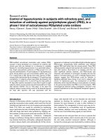
Analysis of association between TLR7 SNPs andTLR7
mRNA levels
To investigate the functional significance of the TLR7
SNPs, we analyzed the association betweenTLR7 SNPs
and TLR7 mRNA levels (Figure 2). The TLR7 mRNA
levels in PBMNCs from Japanese female SLE patients
were measured using RT-PCR assay and were compared
among individuals carrying each genotype. Although not
statistically significant because of the limited sample
size, a tendency toward an association of rs3853839G
with elevated TLR7 mRNA levels was observed (P =
0.20 by Kruskal-Wallis test). This tendency was consis-
tent with the observations in the Chinese population [7],
which demonstrated increased TLR7 transcripts in indi-
viduals carrying rs3853839G. On the other hand, evi-
dence for an association of the intronic SNPs with
mRNA levels was not observed.
Discussion
In the recently reported multicenter study, an association
of rs3853839 was originally found by screening the
TLR7-TLR8 region in Chinese and Korean populations
and was subsequently replicated in Chinese and Japanese
populations [7]. In the process of the study, some popula-
tion difference was noted for rs3853839 and other SNPs,
Figure 1 Association of tag single-nucleotide polymorphisms in the Toll-like receptor 7gene with systemic lupus erythematosus. Top :
P values under the recessive model for minor alleles are indicated. Association was tested by c
2
analysis using 2 × 2 contingency tables.
Bottom: r
2
values based on data from 274 healthy Japanese women are shown.
Kawasaki et al. Arthritis Research & Therapy 2011, 13:R41
/>Page 4 of 8
even among these East Asian populations. Because asso-
ciation between TLR7 and SLE had not been examined in
a systematic manner in a Japanese populatio n, we
thought that TLR7 SN Ps other than rs3853839 might
also contribute to SLE.
To explore such a possibilit y, we analyzed the asso-
ciation of eight tag SNPs in TLR7 and the newly
detected association of two SNPs in i ntron 2, rs179019
and rs179010. Conditional logistic regression analysis
indicated that the association of the intronic SNPs can-
not be explained by LD with rs3853839. In agreement
with these results, the association of the intronic SNPs
remained significant after excluding the effect of the
3’UTR SNP by testing the association only among indi-
viduals carrying the 3’ UTR risk allele. Furthermore,
haplotype analysis showed significant association of the
haplotype containing all of the three SLE risk alleles,
but not of the other haplotypes. All of these results
support the possibility that the possession of both the
3’ UTR and intronic risk alleles may confer further risk
for SLE.
Although rs179019 and rs179010 were also investi-
gated in the Discovery Panel in the previous study, the
majority of whom were Chinese and Korean partici-
pants, no significant association was detected [7]. The
Japanese patients and controls analyzed in this study
were not included in the Discovery Panel. Population
difference was also observed for rs3853839 between the
Chinese and Korean populations, as this SNP was
strongly associated with SLE in Chinese, but not in
Koreans [7], suggesting that the genetic background
with respect to TLR7 association with SLE might be
somewhat different, even among the closely relat ed East
Asian populations. Minor allele frequencies of rs179019
and rs179010 in the HapMap CHB (Han Chinese in
Beijing) samples (rs1 79019: 30.9%, rs179010: 37.3%)
available in the International HapMap database [9] are
similar to those in the Japanese observ ed in thi s study
(rs179019: 28.5%, rs179010: 35.2%). Thus, the difference
in the association cannot be explained by differences in
the minor allele frequencies. We cannot rule out the
possibility that another SNP tagged by rs179019 and
rs179010 in Japanese, but not in Chinese or Koreans
because of difference in the LD status, might play a cau-
sative role. Such a possibility would be addressed by
resequencing the entire TLR7 region.
There i s growing evidence to support involvement of
type I IFN in the development of SLE. TLR7 is crucial
for the production of type I IFN. Thus, the most plausi-
ble role of TLR7 SNPs in SLE pathogenesis is likely to
Table 1 Association of TLR7 SNPs with SLE in a Japanese population
a
Allelic association Dominant model Recessive model
Study population Genotype, n (%) Risk allele, n (%) P OR (95% CI) P OR (95% CI) P OR (95% CI)
rs3853839 G/G G/C C/C G
SLE 197 (57.3) 125 (36.3) 22 (6.4) 519 (75.4) 0.017 1.36
(1.06 to 1.75)
0.030 1.87
(1.05 to 3.31)
0.072 1.34
(0.97 to 1.84)
Controls 137 (50.0) 106 (38.7) 31 (11.3) 380 (69.3)
rs179019 A/A C/A C/C A
SLE 45 (13.1) 131 (38.1) 168 (48.8) 221 (32.1) 0.17 1.19
(0.93 to 1.52)
0.77 1.05
(0.76 to 1.44)
0.016
b
2.02
(1.15 to 3.54)
Controls 19 (6.9) 118 (43.1) 137 (50.0) 156 (28.5)
rs179010 T/T C/T C/C T
SLE 61 (17.7) 156 (45.3) 127 (36.9) 278 (40.4) 0.062 1.25
(0.99 to 1.57)
0.36 1.16
(0.84 to 1.61)
0.018 1.75
(1.10 to 2.80)
Controls 30 (10.9) 133 (48.5) 111 (40.5) 193 (35.2)
a
TLR7, Toll-like receptor 7 gene; SNP, si ngle-nucleotide polymorphism; 95% CI, confidence interval; OR, odds ratio; SLE, systemic lupus erythematosus. Genotype
and allele frequencies are shown in parentheses (%). Association was tested by c
2
analysis or Fisher’s exact test using 2 × 2 contingency tables under the
indicated models for rs3853839G, rs179019A and rs179010 T alleles.
b
Fisher’s exact test was used.
Table 2 Conditional logistic regression analysis of TLR7 SNPs
a
P
adjusted
b
SNP Risk allele Model P
c
rs3853839 rs179019 rs179010
rs3853839 G Codominant 0.021 NA 0.040 0.047
rs179019 A Recessive 0.014 0.026 NA 0.24
rs179010 T Recessive 0.019 0.042 0.42 NA
a
TLR7, Toll-like receptor 7 gene; SNP, si ngle-nucleotide polymorphism; NA, not applicable;
b
P value adjusted for each SNP by conditional logistic regression
analysis using the indicated model;
c
P value for each SNP calculated by logistic regression analysis. The indicated model showed the lowest P value for each SNP.
Kawasaki et al. Arthritis Research & Therapy 2011, 13:R41
/>Page 5 of 8
Table 3 Independent effect of intron 2 SNPs in the carriers of the 3’ UTR risk genotypes
a
Risk genotype Study group, n (%)
rs3853839 rs179019 rs179010 SLE (N = 322) Controls (N = 243) P OR 95% CI
G/G or G/C A/A T/T 42 (13.0) 14 (5.8)
+ + + 280 (87.0) 229 (94.2) 0.0043 2.45 1.31 to 4.60
+ Others Reference
a
SNP, single-nucleotide polymorphism; 3’ UTR, 3’ untranslated region; OR, odds ratio; 95% CI, 95% confidence interval. Genotype frequencies are shown in
parentheses (%). P value was calculated using Fisher’s exact test.
Table 4 Estimated haplotype frequencies in SLE and controls
a
Haplotype rs179019 rs179010 rs3853839 SLE Controls Permutation P value
1 C C G 40.6% 38.0% 0.94
2 C C C 18.2% 24.1% 0.068
3
b
A T G 26.1% 20.3% 0.081
4 C T G 8.5% 8.8% 1.0
5 A T C 5.2% 5.6% 1.0
a
SLE, systemic lupus erythematosus; P values were calculated by permutation test (100,000 perm utations) using HaploView version 4.0 software;
b
each haplotype
was also tested for association under the recessive model. Individ uals homozygous at all three SNPs were considered homozygous for the haplotype. Only
haplotype 3 was significantly associated with SLE under the recessive model (SLE, 31 (9.0%) of 344; control, 11 (4.0%) of 274; P = 0.016 by Fisher’s exact test;
odds ratio 2.37, 95% confidence interval 1.17 to 4.80).
Table 5 Association study of TLR7 SNPs with clinical characteristics of SLE
a
SLE total Anti-Sm antibodies Anti-dsDNA antibodies Renal disorder
SNP Model P OR (95% CI) P OR (95% CI) P OR (95% CI) P OR (95% CI)
rs3853839 Allele 0.017 1.36 (1.06 to 1.75) 0.032 1.65 (1.04 to 2.62) 0.014 1.40 (1.07 to 1.84) 0.025 1.40 (1.04 to 1.89)
rs179019 Recessive 0.016
b
2.02 (1.15 to 3.54) 1.0
b
0.89 (0.29 to 2.73) 0.029
b
1.93 (1.07 to 3.48) 0.011
b
2.25 (1.21 to 4.18)
rs179010 Recessive 0.018 1.75 (1.10 to 2.80) 0.67
b
1.16 (0.51 to 2.67) 0.030 1.72 (1.05 to 2.83) 0.042 1.73 (1.02 to 2.95)
a
TLR7, Toll-like receptor 7 gene; SNP, si ngle-nucleotide polymorphism; SLE, systemic lupus erythematosus; anti-Sm, anti-Smith; dsDNA, double-stranded DNA; OR,
odds ratio, 95% CI, confidence interval;
b
Fisher’s exact test was used. Association was tested by c
2
analysis or Fisher’s exact test using 2 × 2 contingency tables
under the indicated model for rs3853839G, rs179019A and rs179010 T allele. All SLE as well as each SLE subset were compared with healthy controls.
Figure 2 Association analysis of To ll-like receptor 7 genotypes with mRNA expression in peripheral blood mononuclear cells.
Association between Toll-like receptor 7 (TLR7) single-nucleotide polymorphisms (SNPs) and TLR7 mRNA levels was examined by using the
Kruskal-Wallis test. Relative quantitative levels of TLR7 mRNA were normalized to b-actin (ACTB) mRNA levels. Bars indicate median values in each
group. The experiments were performed in triplicate.
Kawasaki et al. Arthritis Research & Therapy 2011, 13:R41
/>Page 6 of 8
be explained by elevated type I IFN production. The
sera of SLE patients displayed elevated levels of type I
IFN, and expression of IFN-inducible genes in PBMNCs
was also upregu lated in SLE [10]. Occasional occurrence
of SLE symptoms following treatment with IFNa in
patients with cancer or hepatitis underscored the rele-
vanceoftypeIIFN[10].TypeIIFNisthoughttobea
potential therapeutic target for SLE, and clinical trials of
anti-IFNa antibodies in SLE are currently underway
[11].
Recent genetic studies have identified an association of
type I IFN pathway-related genes, IFN regulatory factor
5(IRF5)andSTAT4, with SLE in various populations
[10,12-16]. An IRF5 SLE risk haplotype has been shown
to be associated with high serum IFNa activity i n SLE
patients [17], whereas the STAT4 SLE risk variant was
associated with increased sensitivity to IFNa in vivo
[18]. These observations, as well as the previous study
on TLR7 showing upregulation of TLR7 in the risk gen-
otyp e [7], suggest that SLE-associated alleles in the t ype
I IFN pathway are gain-of-function alleles in nature.
Another potential role of TLR7 polymorphism s may
be related to the induction of proinflammatory cyto-
kines. IRF5 is activated by T LR7 signaling and regulates
the expression of many genes, including type I IFN and
proinflammatory cytokines [19]. STAT4 is activated by
type I IFN as well as interleukin 12 and plays a role in
Th1 differentiation [20]. In view of these observations,
the associat ion between TLR7 SNPs and SLE might also
be explained by overproduction of proinflammatory
cytokines in addition to type I IFN.
There are conflicting reports about copy number var-
iation (CNV) of TLR7.Initially,theexistenceofCNV
was reported by Kelley et al. [21]. They showed that,
although common CNV was observed in Caucasians
and African-Americans, no association with SLE was
detected [21]. Recently, García-Ortiz et al. [22] reported
an association o f CNV with childhood-onset SLE in a
Mexican population. In contrast to these observations,
Shen et al. [7] did not find common TLR7 CNV in mul-
tiple populations, including Asians. The latter observa-
tion is consistent with the fact that no CNV was
registered in the Database of Genomic Variants [23],
which includes results derived from the HapMap JPT
(Japanese in Tokyo) samples.
Although our observation in the expression analysis
supported the previous report that indicated the associa-
tion between the risk allele of the 3’ UTR SNP and ele-
vated expression of TLR7 [7], evidence for the
association of the intronic SNPs with levels of TLR7
mRNA was not observed, and therefore the molecular
mechanism of the intronic SNPs requires further study.
TLR7 is mainly expressed in pDCs and B cells. pDCs
represent the major source of type I I FN, but constitute
less than 1% of PBMNCs. If the intronic SNPs have a
regulatory role in a cell type-specific fashion and influ-
ence the expression level of TLR7 in pDCs but not in
other white blood cells, such an effect may not have
been detected in the analysis of total PBMNCs. In addi-
tion, the sample size of this study may not have been
large enough for us to conclude that the intronic SNPs
have no effect on the expression of TLR7.
Because we focused only on the Japanese population,
thesamplesizeofthisstudywaslimitedandthe
observed statistical association wa s modest. Therefore,
the association of the intronic SNPs should be con-
firmed in future independent studies.
Conclusions
TLR7 intronic SNPs rs179019 and rs179010 are asso-
ciated with SLE independently of 3’ UTR SNP rs3853839
in Japanese women. Our findings su pport the genetic
role of TLR7 SNPs in Asian populations with SLE.
Abbreviations
95% CI: 95% confidence interval; CNV: copy number variation; CpG: cytidine-
phosphate-guanosine; IFN: interferon; LD: linkage disequilibrium; OR: odds
ratio; PBMNCs: peripheral blood mononuclear cells; pDCs: plasmacytoid
dendritic cells; RT-PCR: reverse transcription polymerase chain reaction; SLE:
systemic lupus erythematosus; SNP: single-nucleotide polymorphism; ssRNA:
single-stranded RNA; TLR: Toll-like receptor; UTR: untranslated region; Yaa: Y
chromosome-linked autoimmune accelerator.
Acknowledgements
This work was supported by Grant-in-Aid for Scientific Research (B)
(22390199) and Grant-in-Aid for Young Scientists (B) (21790935) from the
Japan Society for the Promotion of Science (JSPS), Health and Labour
Science Research Grants for the Research on intractable diseases from the
Ministry of Health, Labour and Welfare of Japan, Japan Rheumatism
Foundation, and Takeda Science Foundation.
Author details
1
Molecular and Genetic Epidemiology Laboratory, Doctoral Program in
Biomedical Sciences, Graduate School of Comprehensive Human Sciences,
University of Tsukuba, 1-1-1 Tennodai, Tsukuba 305-8575, Japan.
2
Department of Rheumatology, Clinical Research Center for Allergy and
Rheumatology, Sagamihara National Hospital, National Hospital Organization,
18-1 Sakuradai, Minami-ku, Sagamihara 252-0392, Japan.
3
Division of Clinical
Immunology, Doctoral Program in Clinical Sciences, Graduate School of
Comprehensive Human Sciences, University of Tsukuba, 1-1-1 Tennodai,
Tsukuba 305-8575, Japan.
4
Department of Rheumatology, Niigata Rheumatic
Center, 1-2-8 Hon-cho, Shibata 957-0054, Japan.
5
Division of Rheumatology,
Department of Internal Medicine, Juntendo University, 2-1-1 Hongo, Bunkyo-
ku, Tokyo 113-8421, Japan.
6
Juntendo University School of Medicine, 2-1-1
Hongo, Bunkyo-ku, Tokyo 113-8421, Japan.
Authors’ contributions
AK participated in the study design; carried out all genotyping, expression
analysis and statistical analyses; and wrote the manuscript. HF, YK, SI, TH, MK,
IM, ST, YT, HH and TS recruited the patients and controls and collected
clinical information. NT designed and coordinated the study and helped in
the manuscript preparation. All authors read and approved the final
manuscript.
Competing interests
The authors declare that they have no competing interests.
Kawasaki et al. Arthritis Research & Therapy 2011, 13:R41
/>Page 7 of 8
Received: 20 November 2010 Revised: 7 February 2011
Accepted: 11 March 2011 Published: 11 March 2011
References
1. Kawai T, Akira S: The role of pattern-recognition receptors in innate
immunity: update on Toll-like receptors. Nat Immunol 2010, 11:373-384.
2. Marshak-Rothstein A: Toll-like receptors in systemic autoimmune disease.
Nat Rev Immunol 2006, 6:823-835.
3. Pisitkun P, Deane JA, Difilippantonio MJ, Tarasenko T, Satterthwaite AB,
Bolland S: Autoreactive B cell responses to RNA-related antigens due to
TLR7 gene duplication. Science 2006, 312:1669-1672.
4. Subramanian S, Tus K, Li QZ, Wang A, Tian XH, Zhou J, Liang C, Bartov G,
McDaniel LD, Zhou XJ, Schultz RA, Wakeland EK: A Tlr7 translocation
accelerates systemic autoimmunity in murine lupus. Proc Natl Acad Sci
USA 2006, 103:9970-9975.
5. Christensen SR, Shupe J, Nickerson K, Kashgarian M, Flavell RA,
Shlomchik MJ: Toll-like receptor 7 and TLR9 dictate autoantibody
specificity and have opposing inflammatory and regulatory roles in a
murine model of lupus. Immunity 2006, 25:417-428.
6. Komatsuda A, Wakui H, Iwamoto K, Ozawa M, Togashi M, Masai R, Maki N,
Hatakeyama T, Sawada K: Up-regulated expression of Toll-like receptors
mRNAs in peripheral blood mononuclear cells from patients with
systemic lupus erythematosus. Clin Exp Immunol 2008, 152:482-487.
7. Shen N, Fu Q, Deng Y, Qian X, Zhao J, Kaufman KM, Wu YL, Yu CY, Tang Y,
Chen JY, Yang W, Wong M, Kawasaki A, Tsuchiya N, Sumida T, Kawaguchi Y,
Howe HS, Mok MY, Bang SY, Liu FL, Chang DM, Takasaki Y, Hashimoto H,
Harley JB, Guthridge JM, Grossman JM, Cantor RM, Song YW, Bae SC,
Chen S, et al: Sex-specific association of X-linked Toll-like receptor 7
(TLR7) with male systemic lupus erythematosus. Proc Natl Acad Sci USA
2010, 107:15838-15843.
8. Hochberg MC: Updating the American College of Rheumatology revised
criteria for the classification of systemic lupus erythematosus. Arthritis
Rheum 1997, 40:1725.
9. International HapMap Project. [ />en].
10. Kyogoku C, Tsuchiya N: A compass that points to lupus: genetic studies
on type I interferon pathway. Genes Immun 2007, 8:445-455.
11. Rönnblom L, Elkon KB: Cytokines as therapeutic targets in SLE. Nat Rev
Rheumatol 2010, 6:339-347.
12. Graham RR, Kyogoku C, Sigurdsson S, Vlasova IA, Davies LR, Baechler EC,
Plenge RM, Koeuth T, Ortmann WA, Hom G, Bauer JW, Gillett C, Burtt N,
Cunninghame Graham DS, Onofrio R, Petri M, Gunnarsson I, Svenungsson E,
Rönnblom L, Nordmark G, Gregersen PK, Moser K, Gaffney PM, Criswell LA,
Vyse TJ, Syvänen AC, Bohjanen PR, Daly MJ, Behrens TW, et al: Three
functional variants of IFN regulatory factor 5 (IRF5) define risk and
protective haplotypes for human lupus. Proc Natl Acad Sci USA 2007,
104
:6758-6763.
13. International Consortium for Systemic Lupus Erythematosus Genetics
(SLEGEN), Harley JB, Alarcón-Riquelme ME, Criswell LA, Jacob CO,
Kimberly RP, Moser KL, Tsao BP, Vyse TJ, Langefeld CD, Nath SK,
Guthridge JM, Cobb BL, Mirel DB, Marion MC, Williams AH, Divers J,
Wang W, Frank SG, Namjou B, Gabriel SB, Lee AT, Gregersen PK,
Behrens TW, Taylor KE, Fernando M, Zidovetzki R, Gaffney PM, Edberg JC,
Rioux JD, et al: Genome-wide association scan in women with systemic
lupus erythematosus identifies susceptibility variants in ITGAM, PXK,
KIAA1542 and other loci. Nat Genet 2008, 40:204-210.
14. Han JW, Zheng HF, Cui Y, Sun LD, Ye DQ, Hu Z, Xu JH, Cai ZM, Huang W,
Zhao GP, Xie HF, Fang H, Lu QJ, Xu JH, Li XP, Pan YF, Deng DQ, Zeng FQ,
Ye ZZ, Zhang XY, Wang QW, Hao F, Ma L, Zuo XB, Zhou FS, Du WH,
Cheng YL, Yang JQ, Shen SK, Li J, et al: Genome-wide association study in
a Chinese Han population identifies nine new susceptibility loci for
systemic lupus erythematosus. Nat Genet 2009, 41:1234-1237.
15. Kawasaki A, Kyogoku C, Ohashi J, Miyashita R, Hikami K, Kusaoi M,
Tokunaga K, Takasaki Y, Hashimoto H, Behrens TW, Tsuchiya N: Association
of IRF5 polymorphisms with systemic lupus erythematosus in a
Japanese population: support for a crucial role of intron 1
polymorphisms. Arthritis Rheum 2008, 58:826-834.
16. Kawasaki A, Ito I, Hikami K, Ohashi J, Hayashi T, Goto D, Matsumoto I, Ito S,
Tsutsumi A, Koga M, Arinami T, Graham RR, Hom G, Takasaki Y,
Hashimoto H, Behrens TW, Sumida T, Tsuchiya N: Role of STAT4
polymorphisms in systemic lupus erythematosus in a Japanese
population: a case-control association study of the STAT1-STAT4 region.
Arthritis Res Ther 2008, 10:R113.
17. Niewold TB, Kelly JA, Flesch MH, Espinoza LR, Harley JB, Crow MK:
Association of the IRF5 risk haplotype with high serum interferon-α
activity in systemic lupus erythematosus patients. Arthritis Rheum 2008,
58:2481-2487.
18. Kariuki SN, Kirou KA, MacDermott EJ, Barillas-Arias L, Crow MK, Niewold TB:
Cutting edge: autoimmune disease risk variant of STAT4 confers
increased sensitivity to IFN-α in lupus patients in vivo. J Immunol 2009,
182:34-38.
19. Savitsky D, Tamura T, Yanai H, Taniguchi T: Regulation of immunity and
oncogenesis by the IRF transcription factor family. Cancer Immunol
Immunother 2010, 59:489-510.
20. Watford WT, Hissong BD, Bream JH, Kanno Y, Muul L, O’Shea JJ: Signaling
by IL-12 and IL-23 and the immunoregulatory roles of STAT4. Immunol
Rev 2004, 202:139-156.
21. Kelley J, Johnson MR, Alarcón GS, Kimberly RP, Edberg JC: Variation in the
relative copy number of the TLR7 gene in patients with systemic lupus
erythematosus and healthy control subjects. Arthritis Rheum 2007,
56
:3375-3378.
22. García-Ortiz H, Velázquez-Cruz R, Espinosa-Rosales F, Jiménez-Morales S,
Baca V, Orozco L: Association of TLR7 copy number variation with
susceptibility to childhood-onset systemic lupus erythematosus in
Mexican population. Ann Rheum Dis 2010, 69:1861-1865.
23. The Database of Genomic Variants. [ />doi:10.1186/ar3277
Cite this article as: Kawasaki et al.: TLR7 single-nucleotide
polymorphisms in the 3’ untranslated region and intron 2 independently
contribute to systemic lupus erythematosus in Japanese women: a case-
control association study. Arthritis Research & Therapy 2011 13:R41.
Submit your next manuscript to BioMed Central
and take full advantage of:
• Convenient online submission
• Thorough peer review
• No space constraints or color figure charges
• Immediate publication on acceptance
• Inclusion in PubMed, CAS, Scopus and Google Scholar
• Research which is freely available for redistribution
Submit your manuscript at
www.biomedcentral.com/submit
Kawasaki et al. Arthritis Research & Therapy 2011, 13:R41
/>Page 8 of 8