Báo cáo y học: "Nociceptive tolerance is improved by bradykinin receptor B1 antagonism and joint morphology is protected by both endothelin type A and bradykinin receptor B1 antagonism in a surgical model of osteoarthritis" pdf
Bạn đang xem bản rút gọn của tài liệu. Xem và tải ngay bản đầy đủ của tài liệu tại đây (3.79 MB, 11 trang )
RESEARCH ARTIC LE Open Access
Nociceptive tolerance is improved by bradykinin
receptor B1 antagonism and joint morphology
is protected by both endothelin type A and
bradykinin receptor B1 antagonism in a surgical
model of osteoarthritis
Gabriel N Kaufman
1*
, Charlotte Zaouter
1
, Barthélémy Valteau
2
, Pierre Sirois
3
and Florina Moldovan
1,4*
Abstract
Introduction: Endothelin-1, a vasoconstrictor peptide, influences cartilage metabolism mainly via endothelin
receptor type A (ETA). Along with the in flammatory nonapeptide vasodilator bradykinin (BK), which acts via
bradykinin receptor B1 (BKB1) in chronic inflammatory conditions, these vasoactive factors potentiate joint pain and
inflammation. We describe a preclinical study of the efficacy of treatment of surgically induced osteoarthritis with
ETA and/or BKB1 specific peptide antagonists. We hypothesize that antagonism of both receptors will diminish
osteoarthritis progress and articular nociception in a synergistic manner.
Methods: Osteoarthritis was surgically induced in male rats by transection of the right anterior cruciate ligament.
Animals were subsequently treated with weekly intra-articular injections of speci fic peptide antagonists of ETA
and/or BKB1. Hind limb nociception was measured by static weight bearing biweekly for two months post-
operatively. Post-mortem, right knee joints were analyzed radiologically by X-ray and magnetic resonance, and
histologically by the OARSI histopathology assessment system.
Results: Single local BKB1 antagonist treatment diminished overall hind limb nociception, and accelerated
post-operative recovery after disease induction. Both ETA and/or BKB1 antagonist treatments protected joint
radiomorphology and histomorphology. Dual ETA/BKB1 antagonism was slightly more protective, as measured by
radiology and histology.
Conclusions: BKB1 antagonism impr oves nociceptive tolerance, and both ETA and/or BKB1 antagonism prevents
joint car tilage degradation in a surgical model of osteoarthritis. Therefore, they represent a novel therapeutic
strategy: specific receptor antagonism may prove beneficial in disease management.
Introduction
Osteoarthritis (OA) is characterized by a progressive
destruction of articular cartilage accompanied by sub-
chondral bone remodeling, osteophyte formation, and
synovial membrane inflammation [1]. Clinically, this dis-
ease progresses slowly and principally affects the hands
and large weight-bearing joints. Pain is the primary
complaint of patients with OA. Its etiology is multifac-
torial: subchondral bone can have micro-fractures,
osteophytes can cause stretching of peri-osteal nerve
endings, ligaments may be stretched, the joint capsule
can be inflamed or distended, the synovium may be
inflamed, and muscles may spasm [2]. Furthe rmore,
neo-innervation of joint tissue concurrent with angio-
genesis [3,4] may contribute to deep joint pain. Further
understanding of the molecular mechanisms behind
these effects should provide avenues towards targeted
disease-modifying or -slowing treatments [5,6].
* Correspondence: ; florina.
1
Orthopaedic Molecular Biology Laboratory, Sainte-Justine Hospital Research
Centre, 3175 Côte Sainte-Catherine, Montreal, QC, H3T 1C5, Canada
Full list of author information is available at the end of the article
Kaufman et al. Arthritis Research & Therapy 2011, 13:R76
/>© 2011 Kaufman et al.; licensee BioMed Central Ltd. Thi s is an open access article distributed under the terms of the Creative
Commons Attribution License (http://creativecommons. org/licenses/by/2.0), which permits unrestricted u se, distribution, and
reproduction in any medium, provided the original work is properly cited.
We have previously shown that endothelin-1 (ET-1), a
21-amino-acid potent vasoconstricto r peptide, plays a
major role in OA pa thogenesis. It reduces cartilage ana-
bolism by inhibiting collagen and proteoglycan synthesis
[7]. It causes matrix metalloproteinases one and thirteen
to be synthesized and activated in OA cartilage [8]. ET-
1 also causes exc essive production of ni tric oxide, which
is generated as the result of an increase in inducible
nitric oxide synthase levels [9]. These effects occur
mainly via endothelin receptor type A (ETA) [10]: it is
expressed in articular tissue by chondrocytes, synovio-
cytes, and endothelial cells, where it plays a significant
role in cartilage and bone metabolism [11,12]; ETA also
potentiates inflammatory joint pain induced by ET-1
[13,14].
ET-1 affects vascul ar homeostasis via the renin-angio-
ten sin-aldostero ne system [15]. Through cross-talk with
the kallikrein-kinin system [16], it can also mediate
kinin-induced pain and inflammation. Bradykinin (BK),
the inflammatory nonapeptide vasodilator, ha s also been
implicated in OA pain and inflammation. It is generated
in OA synovium, as in a ll inflamed tissue; it a lso is
released due to the increased vascular pressure in sub-
chondral bone [17]. BK binds two receptors, bradykinin
rece ptor B1 (BKB1) and bradykinin receptor B2 (BKB2).
The effects of BK in OA occur largely via BKB1, a
receptor implicated in articular nociception [18,19] and
pro-inflammatory reactions [20]. BKB1 also potentiates
the effects of othe r pro-inflammatory mediators such as
cytokines and prostaglandins. BKB2, though it has been
implicated in nociceptor sensitization in OA [17,19],
may be less relevant as a therapeutic target in the con-
text of a chronic inflammatory response. It is constitu-
tively expressed to a large extent, and is primarily
involved in the acute phase of inflammation [21,22]. In
contrast, BKB1 is up-regulated in chronic inflammatory
conditions, its expression often induced secondary to
inflammatory mediator release [22-24].
Antagonism of ETA an d/or BKB1 may represent a
novel therapeutic option to alleviate, and perhaps prevent
or reverse, the pain, inflammation, and tissue damage
that occur as OA progresses from an acute to a chronic
state. We hypothesi ze that E TA and BKB1 antagonism
will diminish OA progress in a synergistic manner. In the
present work, we describe a preclinical study of the effi-
cacy of treatment of surgically induced OA with ETA
and/or BKB1 peptide antagonists, using an established
rat model of the disease. We found that BKB1 antagonist
treatment diminished hind limb nociception, and both
ETA and/or BKB1 antagon ism protected joint radiomor-
phology and histomorphology. This demonstrates that
ETA and BKB1 receptor expression is involved in OA
pathog enesis, and that specific receptor anta gonism may
prove beneficial in OA disease management.
Materials and methods
Rat model of osteoarthritis
Animals
Eight-week-old male L ewis rats were purchased from
Charles River Canada (Sain t-Constant, Quebec) and
housed under standard conditions. All procedures were
approved by the Sainte-Justine Hospital Research Centre
animal ethics committee and conformed to Canadian
Council on Animal Care guidelines [25].
Study design
The study was conducted as a fractional factorial experi-
ment. Animals were randomly assigned to one of three
surgery conditions: anterior cruciate li gament transec-
tion (ACLT), sham surgery, or no surgery (negative con-
trol). Subsequently, animals were assigned to one of
four treatment groups, as detailed below (Table 1). Sam-
ple size was n = 6 per group.
Surgical technique
OA was induced by surgical transection of the right ante-
rior cruciate ligame nt. The procedure was modified from
previously published reports [26-29], and is described in
detail in Additional file 1. Briefly, animals were an aesthe-
tized and subjected to either anteri or cruciate ligament
transection or sham surgery. One group of animals, ke pt
as negative controls, were not operated upon.
Drug treatment
Over the course of two months post-operatively, animals
were treated by weekly intra-articular injections of ETA
and/or BKB1 specific peptide antagonists: BQ-123 (ETA
antagonist; Sigma-Aldrich, O akville, Ontario) [30,31], R-954
(BKB1 antagonist; a k ind gift from P ierre Sirois, I PS Théra -
peutique, Sherbrooke, Quebec) [32,33], both, or saline vehi-
cle,wasinjectedintotherightkneeatadoseof30nmolin
a volume of 50 μL. Injections were performed under iso-
flurane anaesthesi a, using a 28G needle; the procedure is
described in detail in Additional file 1. Chemical structures
of the antagonists are depicted in Additional file 2. Doses
were based upon previously published reports [ 14,19].
Static weight bearing
Over the course of the study, animal nociception
was evaluated biweekly by the static weight bearing test.
Table 1 Experimental groups
Group number Surgery Treatment
1 None Saline
2 Sham Saline
3 ACLT Saline
4 ACLT BQ-123
5 ACLT R-954
6 ACLT BQ-123+R-954
Six experimental groups were designated in the fractional factorial study, with
six subjects per group.
Kaufman et al. Arthritis Research & Therapy 2011, 13:R76
/>Page 2 of 11
A static weight bearing apparatus was reverse-engi-
neered from previously published reports [34-36],
designed, and machined by Usinage FB (Le Gardeur,
Quebec). Design diagrams and photos are appended in
Additional files 3 and 4.
After conditioning, animals were introduced to the
apparatus and restra ined in a plexiglass chamber with
an angled base, such that each hind paw rested on a
separate force plate connected to a load cell. The weight
in grams distributed on each hind limb was recorded by
a computer software interface (Futek USB software
interface v ersion 2.10). The static weight b earing distri-
bution of each animal was recorded for 30 seconds;
each data point was then taken as the mean of three 30-
second readings. Data were transferred off-line to a per-
sonal computer, and the weight bearin g on the right
hind limb as a percentage of total weight bearing on
both hind limbs was calculated by the following equa-
tion [37]:
% Weight on right leg =
Weight on right leg
Wei
g
ht on ri
g
ht le
g
+Wei
g
ht on left le
g
× 10
0
All values are given as mean ± standard deviation (SD)
per experimental group.
Statistics
Static weight bearing data were analyzed by repeated
measures analysis of varia nce (ANOVA), which com-
pares the global differences between groups of response
profiles measured on the same subjects repeatedly over
the course of the study [38,39]. Test values w ere taken
as the dependent variable and treatment group as the
independent variable, with the animal as the grouping
factor. Sphericity was confirmed with Mauchly’s W test.
Tukey multiple comparisons testing was used to estab-
lish significance in between groups, with directio nality
taken from the sign of the mean difference. P -values less
than 0.0 5 were considered statistically signific ant. Ana-
lyses were conducted using R (version 2.12.1) [40].
Euthanasia and sample preparation
At four or eight weeks post-surgery, animals were sacri-
ficed by cardiac puncture under deep isoflurane anaes-
thesia. The right knee was dissected, and 40-mm-long
sampleswerecutandstoredinphosphate-bufferedsal-
ine until imaged by digital micro-X-ray (DX) and/or
micro-magnetic resonance (MR). Samples were dissected
the same day as the radiological scans.
Digital micro-X-ray
All knee samples were X-rayed using a Faxitron MX-20
specimen X-ray system (Faxitron X-Ray Corporation,
Lincolnshire, IL). Anteroposterior and lateral views were
acquired at 5 × magnification (10 × 10 μmpixelsize)
using a dose of 26 kV for 6 seconds. Images were
analyzed using OsiriX software (version 3.7.1) [41]. Radi-
ological evidence of joint degradation was scored by two
blinded examiners using an OA radiological score modi-
fied from Clark et al. [42] and Esser et al. [43]. Bone
demineralization, subchondral bone erosion, and hetero-
topic ossification were all scored on a scale from zero
(normal) to three (marked degenerati ve changes) . Total
scores were calcu lated by summing the individual scores
for each index, with a maximum possible score of nine.
Statistics
OA radiological scor es were statisticall y analyzed by
one-way ANOVA, with total scores taken as the depen-
dent variable and treatment group as the independent
variable. Pai rwise post-hoc testing with Holm correction
was used to establish significance in between treatment
groups. P-values less than 0.05 were considered statisti-
cally significant. Analyses were conducted using R (ver-
sion 2.12.1) [40].
Micro-magnetic resonance imaging
Image acquisition
A subset of animals were sacrificed four weeks post-
operatively and their right knees were imaged by micro-
MR. Imaging was performed using a Bruker PharmaS-
can (Ettlingen, Germany) 7 Tesla MR scanner at the
McGill University Small Animal Imaging Lab (Montreal,
Quebec). Knee samples were placed in a custom-made
support inside a 15-mL centrifuge tube, which was then
filled with the MR-inert buffer FC- 770 (3M Fluorinert
Electronic L iquid). Samples were introduced into a
1
H
mouse brain radio frequency (RF) coil (inner diameter
22 mm), and centered in the magnet. The RF coil was
tuned and matched to the sample, and the magnet wa s
then shimmed. The system was controlled via Bruker
ParaVision software (version 5.0).
Positioning was confirmed with a tri-pilot rapid scan,
which was then used to place 14 coronal slices for two-
dimensional anatomical scanning of the joint using a
rapid acquisition with relaxation enhancement (RARE)
multiecho spi n echo pul se sequence (TurboRARE). Scan
param eters were as follows: repetition time (TR) = 3500
milliseconds (ms), echo time (TE) = 36 ms, echo train
length (ETL) = 8, slice thickness = 500 μm, acquisition
matr ix = 384 × 384, and number of averages = 4. Voxel
size was
140.1
¯
6 × 140.1
¯
6 × 500
µm
. These scans were
then repeated in the sagittal projection.
Once these scans were acquired, one 1-mm-thick axi al
slice was placed in the center of the knee joint in order to
scan the articular cartilage with a series of multislice mul-
tiecho (MSME) T
2
-wei ghted pulse sequences . Scan para-
meters were TR = 3500 ms, ETL = 1, acquisition matrix =
192 × 256, with voxel size of 156.25 × 156.25 × 1000 μm.
16 different TE were used: 10, 20, 30, 40, 50, 60, 70, 80,
90, 100, 110, 120, 130, 140, 150, and 160 ms.
Kaufman et al. Arthritis Research & Therapy 2011, 13:R76
/>Page 3 of 11
Total scan time was roughly 1 hour per sample. Scan
sequences were based on previously published reports
[44].
Image processing and analysis
After acquisition, images were analyzed using OsiriX
software (version 3.7.1) [41]. Anatomical TurboRARE
images were ex amined for corre ct depict ion of anatomi-
cal features of the knee joint, and to confirm ACLT
where applicable. As well, images were analyz ed for
signs of cartilage decay, indicated by lower signal i nten-
sity of the articular surfaces. The MSME-T
2
images
were aligned into an image stack, and regions of inter-
est, corresponding to the articular cartilage, were manu-
ally drawn and propagated throughout the stack. A
mean T
2
fit map was then automatically generated by
fitting the signal intensity to the spin-spin relaxation sig-
nal decay equation:
S(TE)=M
0
exp −
TE
T
2
where signal intensity S is defined as a function of
echo time TE, and is relat ed to the spin density M
0
and
the transverse relaxation time T
2
. The equation was
solved for the mean T
2
value over the 16-image stack by
using least-squares single-expo nential curve-fitting, with
initial guesses of M
0
= signal intensity at 10 ms and T
2
= 30 ms, in order to guarantee rapid convergence [44].
OsiriX then generated a T
2
fit map graph with r egres-
sion line and values for T
2
and M
0
.
Histology
After radiological examination, knee samples were fixed in
10% neutral buffered formalin for two weeks, decalcified
with RDO Rapid Decalcifier (Apex Engineering Products,
Aurora, Illinois) for three days, circulated, and embedded
in paraff in. Fiv e-micron sagittal sections were acquired
from the middle of the knee joint. Histomorphological
staining was performed as previously described [45]: slides
were deparaffinized, rehydrated, stained with Safranin O
(which colors proteoglycans red), counterstained with Fast
Green FCF (which colors proteins green) and with Wei-
gert’s hematoxylin (which colors nuclei black), dehydrated,
cleared, and mounted in Permount. Representative digital
photomicrog raphs were acquired with a Leica DM R
microscope (Wetzlar, Germany) fitted with a QImaging
Retiga 1300 B camera (Surrey , British Columbia), con-
trolled by QCapture software (version 2.95.0). Images
were captured at 50 × (low-power) or 200 × (high-power)
magnification, and subsequently color-matched and
balanced using Adobe Photoshop CS3.
Histopathological scoring
Four slides from each condition were scored by two
blinded examiners using the Osteoarthritis Research
Society International (OARSI) histopathology assessment
system [46], which assigns numeric values to grade, or
depth progression into cartilage (0-6), and stage, or
extent of joint involvement (0-4); multipl ying grade and
stage yields a total OA score with a maximum value of
24. Scores were averaged in between the two examiners;
inter-examiner variation was within ± 5%.
Statistics
OARSI scores were statistically analyzed by one-way
ANOVA, with total scores taken as the dependent vari-
able and treatment group as the independent variable.
Pairwise post-hoc testing with Holm correction was
used to establish significance in between treatment
groups. P-values less than 0.05 were considered statisti-
cally significant. Analyses were conducted using R (ver-
sion 2.12.1) [40].
Immunohistochemistry
Additional 5-micron sections were processed for immu-
nohistochemical detection of type II collagen. Slides
were deparaffinized, rehydrated, and washed in phos-
phate-buffered saline (PBS). Section s were incubated in
2 mg/mL hyaluronidase for 30 minutes at 37°C, followed
by permeabilization with 0.3% Triton X-100 for 30 min-
utes at room temperature. Endogenous peroxidase activ-
ity was then quenched with 2% hydrogen peroxide in
PBS for 15 minute s. Sections were blocked with normal
mouse serum (Vector Laboratories, Burlingame, Califor-
nia) for 1 hour, after which they were blotted and then
incubated with monoclonal mouse anti-rat type II col-
lagen (clone SPM239; Spring Bioscience, Pleasanton,
California) for 18 hours at 4°C. Sections were then
washed in PBS, incubated with biotinylated anti-mouse
IgG (Vector) for 1 hour at room temperature, and
stained using the avidin-biotin complex method (Vectas-
tain ABC kit; Vector). Color was developed using 3,3’ -
diaminobenzidine (Dako Diagnostics, Mississauga,
Ontario) containing hydrogen peroxide. Slides were
counterstained with Harris modified hematoxyli n, dehy-
drated, cleared, mounted, and examined by light micro-
scopy as described above.
Results
ETA and BKB1 antagonism ameliorates OA nociceptive
tolerance
To determine the effects of ETA and/or BKB1 local
antagonist treatment on nociception in a surgical OA
model, the static weight bearing asymmetry of the ani-
mals was measured repeatedly over the course of the
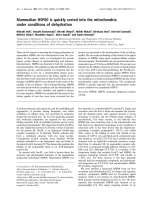
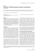
study (Figure 1). Pre-operative baseline values for all
groups indicated hind limb weight bearing symmetry
(49.89 ± 0.42%). Unoperated vehicle-treated animals
showed no important changes in hind limb weight bear-
ing from baseline pre-operative values over the course
Kaufman et al. Arthritis Research & Therapy 2011, 13:R76
/>Page 4 of 11
of the study, staying roughly within ±4% of even weight
distribu tion. Sham-operated vehicle-treated animals dis-
played an initial weight bearing imbalance 14 days post-
operatively (36.47 ± 1.12%), but recovered weight bear-
ing symmetry quickly thereafter (44.84 ± 0.3 3% by day
26 post-operatively). ACLT saline-treated animals
showed significant weight bearing imbalance two weeks
post-operatively, down to 33.66 ± 2.05% weight on the
right leg, suggesting sever e nocice ption. All animals had
similar n ociceptive tolerance at the last measured t ime-
point (day 50 post-operatively), indicating nociceptive
adaptation, but drug-treated animals were able to
recover faster than saline-treat ed animals (up to 40.54 ±
3.36% weig ht on right leg by day 40 post-operatively, for
BQ-123 and R-954 dual treatment).
Repeated measures analysis of variance of the static
weight bearing data, followed by Tukey post-hoc
hypothesis tests (Table 2), demonstrated that treatment
with R-954, or both BQ-123 and R-954, significantly
ameliorate d nociceptive tolerance in ACLT animals over
the study period, as compared to saline-treated positive
controls (0.0001 ≤ P ≤ 0.0002). When administered
alone, BQ-123 did not result in statistically significant
increased nociceptive tolerance (P = 0.1847). Sham sur-
gery was found to be slightly less nociceptive than ACL
transection (P = 0.019), confirming that ACLT is neces-
sary for a maximal nociceptive response. Furthermore,
nociception i n the sham-operated animals was compar-
able to unoperated animals, with no statistically signifi-
cant difference calculated (P = 0.8746).
Antagonist treatment improved radiological indices of OA
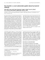
Rightkneejointsweredissectedattheendofthestudy
period and imaged by DX (Figure 2) and MR (Figure 3) to
examine the radiological effects of antagonist treat ments.
ACLT rapidly i nduced radiological evidence of OA: knee
joints showed signs of degradation such as subchondral
bone r emodeling, osteophyt e fo rmation (Figure 2c an d
Table 3), cartilage layer thinning (Figure 3c), and length-
ened cartilage T
2
(Table 4). Neither sham surgery nor
intra-articular injection affected joint radiomorphology
(Figures 2a,b and 3a,b). DX analysis of antagonist-treated
25
30
35
40
45
50
55
0 14264050
% weight on right leg
Days post-surgery
None/Saline Sham/Saline ACLT/Saline ACLT/BQ-123 ACLT/R-954 ACLT/BQ-123+R-954
Figure 1 ETA and/or BKB1 antagonist treatment improves static weight bearing tolerance. Static weight bearing tolerance was measured
repeatedly at defined time points over the course of the study. Data are presented as mean ± SD per experimental group (n = 6), of weight on
the right leg as a percentage of total weight on both hind limbs. Day 0, baseline pre-operative values. Repeated measures analysis of variance
with Tukey post-hoc (Table 2) indicated that BKB1 antagonist treatment significantly ameliorated nociceptive tolerance in ACLT animals over the
study period, as compared to saline-treated positive controls.
Kaufman et al. Arthritis Research & Therapy 2011, 13:R76
/>Page 5 of 11
knee joints showed less subchondral bone remodeling and
heterotopic ossification than saline-treated animals (Figure
2d,e,f and Table 3). Dual ETA/BKB1 antagonism appeared
to be slightly more protective than single antagonism: less
subchondral bone remodeling and greater trabecular
integrity was observed in the dual-an tagonist-treated ani-
mals than in the single-antagonist-treated animals. Radi-
ological scoring of the DX views for a p anel of OA joint
degenerative changes (Table 3 and Additional file 5)
demonstrated that treatment with BQ-123, R-954, or both,
significantly ameliorated radiological indices of disease
progression in ACLT animals, as compared to saline-trea-
ted positive controls (0.0020 ≤ P ≤ 0.0214, one-way
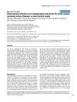
ANOVA with Holm post-hoc). MR analysis of knee joints
revealed that antagonist-treated animals had greater
cartilage thickness and fewer cartilage lesions (Figure 3d,e,
f), as well as shorter cartilage T
2
(Table 4, statistical signifi-
cance not achieved) than saline-treated ACLT animals.
These data suggest that antagonist treatmen t protected
joint radiomorphology after ACLT.
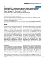
Antagonism protects joint histomorphology
To investigate the effects of ETA and/or BKB1 antago-
nist treatment on histological indic es of disease, rat
knee joints were processed to as sess cartilage proteogly-
can content and joint histomorphology (Figure 4 left
and middle columns). ACLT saline-treated animals lost
most proteogl ycan staining when examin ed at eight
weeks post-operatively, with severe articular surface dis-
ruptions and osteophyte formation (Figure 4g,h). I n
contrast, cartilage proteoglycans were detected in the
knees of ETA and/or BKB1 antagonist-treated animals
(Figures 4j,k,m,n and 4p,q), indicating that treatment
protects cartilage structural components. As well,
articular surface inte grity was prese rved to a greater
extent, with dual antagonism appearing to be most pro-
tective (Figure 4p,q). Neither sham surgery nor intra-
articular injection of saline vehicle negatively affected
joint histomorphology (F igures 4a,b and 4d,e). Mean
OARSI scores (Table 5 and Additional file 6) indicate
that ETA and/or BKB1 antagonist treatment signifi-
cantly reduced the amount of affected joint tissue and
the degree of histopathology, as compared to saline-
treated positive controls (P < 0.0001 for all compari-
sons, one-way ANOVA with Holm post-hoc).
Table 2 Static weight bearing post-hoc tests
Contrast Estimate Standard
error
z-score P(>|z|)
None/Saline vs Sham/Saline - 5.1697 1.9903 - 2.597 0.8746
Sham/Saline vs ACLT/Saline 6.5667 2.0155 3.258 0.019
ACLT/BQ-123 vs
ACLT/Saline
2.5845 1.8841 1.372 0.1847
ACLT/R-954 vs ACLT/Saline 0.6951 1.9669 0.353 0.0002
ACLT/BQ-123+R-954 vs
ACLT/Saline
0.2784 1.8503 0.15 0.0001
Tukey post-hoc tests were conducted following repeated measures ANOVA of
the static weight bearing data. From left to right, the table columns present
the contrast of interest, the parameter estimate from the linear matrix model,
the standard error of that estimate, the standard z-score, and the associated
P-value.
Figure 2 Antagonist treatment improves radiological indices of OA: X-ray results. (a) No surgery and saline treatment; (b) sham surgery
and saline treatment; (c) ACLT and saline treatment; (d) ACLT and BQ-123 treatment; (e) ACLT and R-954 treatment; (f) ACLT and BQ-123+R-954
dual treatment. Blue arrows indicate tibial plateau, purple arrows indicate subchondral bone, and green arrows indicate osteophytes. Sagittal
views. Scale bar, 1 cm.
Kaufman et al. Arthritis Research & Therapy 2011, 13:R76
/>Page 6 of 11
Type II collagen, the major structural collagen of car-
tilage, was detected by immunohistochemistry (Figure 4
right column). ACLT saline-treated animals displayed
significant losses of articular surface type II collagen
(Figure 4i) with some localization in the deep zones of
car tilage, reflecting cartilage remodeling processes. Ani-
mals treated with E TA and/or BKB1 antagon ists (Figure
4l,o,r) displayed varying degrees of protection, retaining
some type II collagen staining. Neither sham surgery
nor i ntra-articular injection of saline vehicle negatively
affected joint type II collagen expression (Figur es 4c and
4f): protein was localized in the superficial zone of
articular cartilage, indicating functional joint tissue.
Discussion
In the present study, we investigated whether antagon-
ism of ETA and/or BKB1 could slow and/or prevent
osteoarthritic cartilage degradation and joint nociception
in a rat surgical model of OA. We provide several lines
of evidence that suggest protective effects of ETA and/
or BKB1 antagonism in vivo: BKB1 antagonist treatment
improved hind limb nociceptive tolerance, and both
ETA and/or BKB1 antagonist treatment ameliorated
radiological indices of disease, and protected articular
cartilage and bone histomorphometry.
The most interesting finding of our study is that noci-
ceptive tolerance was augmented in our model after
BKB1 antagonist treatment, with faster post-operative
recovery than vehicle-treated controls. These results are
consistent with other reports [19], where local treatment
with BKB1 receptor antagonists reduced overt acute
joint nociception. We extend this finding to the dual
antagonist treatment approach to male animals in a
model of chronic pain, as well as relating it to me asures
of joint integrity by radiology and histology. Low-grade
joint pain is the most common reason for patient pre-
sentation, and is often the major debilitating factor in
OA cases [47,48]. Thus, the anti-nociceptive effects of
Figure 3 Antagonist treatment improves radiological indices of OA: MR results. (a) No surgery and saline treatment; (b) sham surgery and
saline treatment; (c) ACLT and saline treatment; (d) ACLT and BQ-123 treatment; (e) ACLT and R-954 treatment; (f) ACLT and BQ-123+R-954 dual
treatment. Red arrows indicate articular cartilage. Sagittal views. Scale bar, 1 cm.
Table 3 OA radiological scores
Group
number
Surgery Treatment Mean total radiological
score
SD
1 None Saline 0.25 0.50
2 Sham Saline 1.16 0.75
3 ACLT Saline 4.86 1.68
4 ACLT BQ-123 2.83 1.47
a
5 ACLT R-954 2.50 1.22
b
6 ACLT BQ-123+R-
954
2.67 1.03
c
Radiological scoring of the DX views of the knee joints [42,43] indicated that
antagonist treatment protected joint radiomorphology after ACLT. One-way
ANOVA with Holm post-hoc:
a
P = 0.0214, ACLT/BQ-123 treatment versus
ACLT/saline treatment;
b
P = 0.0020, ACLT/R-954 treatment versus ACLT/saline
treatment;
c
P = 0.0125, ACLT/BQ-123+R-954 dual treatment versus ACLT/saline
treatment.
Table 4 Cartilage mean T
2
values
Group number Surgery Treatment Mean T
2
value (ms)
1 None Saline 51.60
2 Sham Saline 52.12
3 ACLT Saline 64.38
4 ACLT BQ-123 63.23
5 ACLT R-954 61.13
6 ACLT BQ-123+R-954 56.57
Cartilage mean T
2
values in milliseconds were calculated for all conditions
using OsiriX software (version 3.7.1) [41]. Statistical significance in between
groups was not achieved.
Kaufman et al. Arthritis Research & Therapy 2011, 13:R76
/>Page 7 of 11
Figure 4 Antagonist treatment protects joint histomorphometry. Sagittal sections. Left and middle columns, Safranin O/Fast Green FCF
staining. Right column, type II collagen immunohistochemistry. Left column, low-power magnification: scale bar, 200 μm; original magnification
50 ×. Middle and right columns, high-power magnification: scale bar, 50 μ m; original magnification 200 ×. Conditions by rows: (a), (b), (c) no
surgery and saline treatment; (d) , (e), (f) sham surgery and saline treatment; (g), (h), (i) ACLT and saline treatment; (j), (k), (l) ACLT and BQ-123
treatment; (m), (n), (o) ACLT and R-954 treatment; (p), (q), (r) ACLT and BQ-123+R-954 dual treatment. Yellow arrows indicate loss of Safranin O
staining, purple arrows indicate cartilage notching, and red arrow indicates an osteophyte. Black arrows indicate type II collagen immunostaining.
Kaufman et al. Arthritis Research & Therapy 2011, 13:R76
/>Page 8 of 11
BKB1 antagonism make this treatment strategy attrac-
tive. Surprisingly, single ETA antagonism was relatively
ineffective a t diminishing joint nociception i n our
model. This finding, which contradicts our initial
hypothesis, suggests that ETA potentiation of ET-1-
induced joint pain [14] may not be direct, especially in a
chronic inflammatory state.
WefoundthatsingleanddualETA/BKB1antagonist
treatments decreased radiological disease indices, in
terms of osteophyte formation, cartilage thinning, and
subchondral bone remodeling, with dual antagonism
being most protecti ve. As well , cartilage T
2
, increased i n
ACLT animals, was decreased by antagonist treatment,
which indicates a cartilage-preserving effect. Longer car-
tilage transverse relaxation times are an indicator of car-
tilage degradation; this MR parameter is indicative of
cartilage composition and integrity [49-51]. Radiographic
evidence is the main criterion for OA diagnosis and pro-
gression [52,53]. The most common clinical diagnostic
test is via X-ray of the affected joint: joint space narrow-
ing as measured on X-ray is ofte n used as a longitudinal
marker of disease evolution. It i s difficult to directly
compare radiological parameters b etween human and
rat knees due to the quadrupedal nature of the animal
and the markedly different radiological anatomy that
this entails [54]. However, we were able to detect radi-
ological evidence of OA progression in ACLT animals,
as has been described in similar studies [44,55].
OA induction in rat knees lead s to a rapid decrease in
cartilag e proteoglycan staining, along with articular sur-
face disruption and osteophyte formation [26,27]. ETA/
BKB1 antagonist treatment protected the proteoglycan
content of the j oint and preserved articular surface
integrity. Furthermore, there was some protection of
type II collagen protein expression. This allowed the
joint cartilage to retain its normal biophysical properties,
as cartilage proteoglycans are responsible, along with
collagen, for retaining water in the tissue, which pro-
vides spring and resilience [56,57]. These findings likely
suggest that the protection of cartilage proteoglycans,
collagens, and articular surface histomorpho logy may be
one explanation for the increased pain tolerance
obs erved in antagonist-treated animals; our results con-
cur with those of other reports, which correlated the
preservation of articular cartilage proteoglycan staining
with pain tolerance behavior [26].
The ET-1 and BK systems are involved in joint tissue
inflammation and nociception, conco mitant with pro-
inflammatory mediators. However, exploration of potential
therapeutic targets in these systems has been modest: the
main classes of disease-modifying osteoarthritis drugs cur-
rently in development include cytokine and matrix metal-
loproteinase inhibitors, anti-resorptives, and growth factors
[58]. To o ur knowledge, the only clinical trial o f a drug tar-
geting a vasoactive factor in OA is the bradykinin receptor
B2 antagonist Icatibant, by Sanofi-Aventis [59]. This drug
is no longer in clinical development [60], due to mixed
results: while it provided loc al analgesia in knee OA, no
anti-inflammatory effect could be detected [61 ]. Our results
suggest that ETA and BKB1 represent novel therapeutic
targets in O A. Specific receptor a ntagonists c ould be t e sted
in clinical tria ls for OA pain and ti ssue damage.
Conclusions
Using a rat surgically induced model of OA, we d emon-
strated that local treatment with specific peptide antago-
nists of ETA and/or BKB1 may slow or stabilize the
development of radiomorphological and histomorphologi-
cal changes occurring in OA p athogenesis. Furthermore,
we showed that BKB1 a ntagonist treatment acc elerated
recovery of, and improved longitudinally, nociceptive tol-
erance in ACLT animals. Taken together, our results indi-
cate that blocking ETA and BKB1 improves OA
prognostic indices, which implies th at defective signali ng
might play a role in chronic OA pain. Our results also
raise the possibility of targeted receptor antagonism as a
relevant therapeutic option. Further studies are required
to understand the mechanisms underlying th e exact nat-
ure of receptor cross-regulation and synergism.
Additional material
Additional file 1: Rat anterior cruciate ligament transection and
intra-articular injection. Detailed descriptions and macro photographs
of rat anterior cruciate ligament transection and intra-articular injection.
PDF file named Rat ACLT and IA injection.pdf (3 pages).
Additional file 2: Chemical structures of BQ-123 and R-954.2D
chemical structures of selective ETA peptide antagonist BQ-123 (left) and
selective BKB1 peptide antagonist R-954 (right). PDF file named
antagonist structures.pdf (1 page).
Additional file 3: Design diagrams for static weight bearing
apparatus. Original design diagrams for static weight bearing apparatus.
Labels in French. Auto-drafted using CATIA V5 R19. PDF file named
static weight bearing apparatus design diagrams.pdf
(4 pages).
Table 5 OARSI histopathology scores
Group number Surgery Treatment Mean OARSI score SD
1 None Saline 0.43 0.53
2 Sham Saline 0.50 1.00
3 ACLT Saline 17.00 5.77
4 ACLT BQ-123 4.75 0.96
a
5 ACLT R-954 4.25 2.02
b
6 ACLT BQ-123+R-954 3.50 2.89
c
Four slides per condition were scored by two blinded examiners using the
OARSI histopathology assessment system [46]. Results were averaged and are
presented as mean scores per condition. Inter-examiner variation was within
± 5%. One-way ANOVA with Holm post-hoc:
a
P = 0.000017, ACLT/BQ-123
treatment versus ACLT/saline treatment;
b
P = 0.00001, ACLT/R-954 treatment
versus ACLT/saline treatment;
c
P = 0.0000048, ACLT/BQ-123+R-954 dual
treatment versus ACLT/saline treatment.
Kaufman et al. Arthritis Research & Therapy 2011, 13:R76
/>Page 9 of 11
Additional file 4: Static weight bearing apparatus in use. Static
weight bearing apparatus with rat positioned for measurements. A, side
view; B, angle view; C, front view. PDF file named static weight
bearing apparatus photos.pdf (1 page).
Additional file 5: OA radiological scores. Unblinded raw data for the
OA radiological scores, presented as averaged scores for each parameter.
CSV file named radiological scores.csv.
Additional file 6: OARSI histopathology scores. Unblinded raw data
for the OARSI histopathology scores, presented as averaged scores for
each parameter. CSV file named OARSI scores.csv.
Abbreviations
ACLT: anterior cruciate ligament transection; ANOVA: analysis of variance; BK:
bradykinin; BKB1: bradykinin receptor B1; BKB2: bradykinin receptor B2; DX:
digital micro-X-ray; ET-1: endothelin-1; ETA: endothelin receptor type A; ETL:
echo train length; MR: magnetic resonance; MSME: multislice multiecho; OA:
osteoarthritis; OARSI: Osteoarthritis Research Society International; PBS:
phosphate-buffered saline; RARE: rapid acquisition with relaxation
enhancement; RF: radio frequency; SD, standard deviation; TE: echo time; TR:
repetition time.
Acknowledgements
We thank Archana Sangole for valuable help with the static weight bearing
apparatus design. We thank Saadallah Bouhanik of the Viscogliosi Laboratory
for Molecular Genetics of Musculoskeletal Disorders (Montreal, Quebec) for
providing access to and help with the Faxitron micro-X-ray system, as well
as Jason Cakiroglu (MR engineer) and Barry J. Bedell (director) of the Small
Animal Imaging Lab at McGill University (Montreal, Quebec) for providing
access to their 7 Tesla micro-MR scanner. We thank Stéphane Faubert and
Serge Nadeau for their surgical instruction, as well as Denise Carrier
(director) and the staff of the Sainte-Justine Hospital Research Centre animal
facility for their technical assistance. Finally, we thank Kessen Patten for his
writing and editing suggestions.
This work was supported by operating grants from The Arthritis Society
(William T. Holland Arthritis Research Grant, RG05/084) and the Canadian
Institutes of Health Research (IMH-94011). Publication costs were payed by
the Yvon Roberge publication support fund of the Faculty of Dentistry,
Université de Montréal. GNK held a Sainte-Justine Hospital Foundation/
Foundation of the Stars bursary.
Author details
1
Orthopaedic Molecular Biology Laboratory, Sainte-Justine Hospital Research
Centre, 3175 Côte Sainte-Catherine, Montreal, QC, H3T 1C5, Canada.
2
Paediatric Mechanobiology Laboratory, Sainte-Justine Hospital Research
Centre, 3175 Côte Sainte-Catherine, Montreal, QC, H3T 1C5, Canada.
3
IPS
Thérapeutique, 3201 Jean-Mignault, Sherbrooke, QC, J1E 4K8, Canada.
4
Faculty of Dentistry, Université de Montréal, PO Box 6128 Stn CV, Montreal,
QC, H3C 3J7, Canada.
Authors’ contributions
GNK designed the in vivo study, performed the surgeries, injections, static
weight bearing measurements, dissections, and radiological analyses,
analyzed the data, and wrote the paper. CZ assisted with surgeries and
dissections, performed histological studies, and revised the paper. BV
reverse-engineered the static weight bearing apparatus and assisted with
data analysis. PS contributed the BKB1 antagonist R-954. FM conceived the
study and supervised the research group. GNK gabriel.kaufman@umontreal.
ca takes responsibility for the integrity of the work as a whole. All authors
read and approved the final manuscript.
Competing interests
Intellectual property rights (GNK, PS, FM) of the dual-antagonist treatment
strategy are protected through Univalor, the technology transfer corporation
of Université de Montréal. PS holds patents relating to the preparation and
use of R-954. CZ and BV declare that they have no competing interests.
Received: 28 July 2010 Accepted: 16 May 2011 Published: 16 May 2011
References
1. Goldring MB, Goldring SR: Osteoarthritis. J Cell Physiol 2007, 213:626-34.
2. Martel-Pelletier J, Lajeunesse D, Pelletier JP: Etiopathogenesis of
osteoarthritis. In Arthritis and Allied Conditions. A Textbook of Rheumatology.
Volume Chap 109 15 edition. Edited by: Koopman WJ, Moreland LW.
Williams 2005:2199-2226.
3. Ashraf S, Walsh DA: Angiogenesis in osteoarthritis. Current opinion in
rheumatology 2008, 20:573-80.
4. Bonnet CS, Walsh DA: Osteoarthritis, angiogenesis and inflammation.
Rheumatology (Oxford) 2005, 44:7-16.
5. Alcaraz MJ, Megías J, García-Arnandis I, Clérigues V, Guillén MI: New
molecular targets for the treatment of osteoarthritis. Biochemical
Pharmacology 2010, 80:13-21.
6. Mandelbaum B, Waddell D: Etiology and pathophysiology of
osteoarthritis. Orthopedics 2005, 28:S207-214.
7. Khatib AM, Siegfried G, Messai H, Moldovan F, Mitrovic DR: Mechanism of
inhibition of endothelin-1-stimulated proteoglycan and collagen
synthesis in rat articular chondrocytes. Cytokine 2002, 17:254-61.
8. Roy-Beaudry M, Martel-Pelletier J, Pelletier JP, M’Barek KN, Christgau S,
Shipkolye F, Moldovan F: Endothelin 1 promotes osteoarthritic cartilage
degradation via matrix metalloprotease 1 and matrix metalloprotease 13
induction. Arthritis Rheum 2003, 48:2855-64.
9. Manacu CA, Martel-Pelletier J, Roy-Beaudry M, Pelletier JP, Fernandes JC,
Shipkolye FS, Mitrovic DR, Moldovan F: Endothelin-1 in osteoarthritic
chondrocytes triggers nitric oxide production and upregulates
collagenase production. Arthritis Res Ther 2005, 7:R324-32.
10. Khatib AM, Lomri A, Mitrovic RD, Moldovan F: Articular chondrocyte aging
and endothelin-1. Cytokine 2007, 37:6-13.
11. Lodhi KM, Sakaguchi H, Hirose S, Shibabe S, Hagiwara H: Perichondrial
localization of ETA receptor in rat tracheal and xiphoid cartilage and in
fetal rat epiphysis. Am J Physiol 1995, 268:C496-502.
12. Messai H, Panasyuk A, Khatib A, Barbara A, Mitrovic DR: Endothelin-1
receptors on cultured rat articular chondrocytes: regulation by age,
growth factors, and cytokines, and effect on cAMP production. Mech
Ageing Dev 2001, 122:519-31.
13. Khodorova A, Montmayeur JP, Strichartz G: Endothelin receptors and pain.
J Pain 2009, 10:4-28.
14. De-Melo JD, Tonussi CR, D’Orléans-Juste P, Rae GA:
Articular nociception
induced
by endothelin-1, carrageenan and LPS in naive and previously
inflamed knee-joints in the rat: inhibition by endothelin receptor
antagonists. Pain 1998, 77:261-9.
15. Rossi GP, Sacchetto A, Cesari M, Pessina AC: Interactions between
endothelin-1 and the renin-angiotensin-aldosterone system. Cardiovasc
Res 1999, 43:300-7.
16. Shen B, El-Dahr SS: Cross-talk of the renin-angiotensin and kallikrein-kinin
systems. Biol Chem 2006, 387:145-50.
17. Meini S, Maggi CA: Knee osteoarthritis: a role for bradykinin? Inflamm Res
2008, 57:351-61.
18. Sainz IM, Uknis AB, Isordia-Salas I, Cadena RAD, Pixley RA, Colman RW:
Interactions between bradykinin (BK) and cell adhesion molecule (CAM)
expression in peptidoglycan-polysaccharide (PG-PS)-induced arthritis.
FASEB J 2004, 18:887-9.
19. Tonussi CR, Ferreira SH: Bradykinin-induced knee joint incapacitation
involves bradykinin B2 receptor mediated hyperalgesia and bradykinin
B1 receptor-mediated nociception. Eur J Pharmacol 1997, 326:61-5.
20. Benton HP, Jackson TR, Hanley MR: Identification of a novel inflammatory
stimulant of chondrocytes. Early events in cell activation by bradykinin
receptors on pig articular chondrocytes. Biochem J 1989, 258:861-7.
21. Moreau ME, Garbacki N, Molinaro G, Brown NJ, Marceau F, Adam A: The
kallikrein-kinin system: current and future pharmacological targets. J
Pharmacol Sci 2005, 99:6-38.
22. Hall JM: Bradykinin receptors. Gen Pharmacol 1997, 28:1-6.
23. Calixto JB, Cabrini DA, Ferreira J, Campos MM: Inflammatory pain: kinins
and antagonists. Curr Opin Anaesthesiol 2001, 14:519-26.
24. Calixto JB, Medeiros R, Fernandes ES, Ferreira J, Cabrini DA, Campos MM:
Kinin B1 receptors: key G-protein-coupled receptors and their role in
inflammatory and painful processes. Br J Pharmacol 2004, 143:803-18.
25. Olfert ED, Cross BM, McWilliam AA, (Eds): Guide to the Care and Use of
Experimental Animals Ottawa, Ontario: Canadian Council on Animal Care; 1993.
26. Appleton CTG, McErlain DD, Pitelka V, Schwartz N, Bernier SM, Henry JL,
Holdsworth DW, Beier F: Forced mobilization accelerates pathogenesis:
Kaufman et al. Arthritis Research & Therapy 2011, 13:R76
/>Page 10 of 11
characterization of a preclinical surgical model of osteoarthritis. Arthritis
Res Ther 2007, 9:R13.
27. Hayami T, Pickarski M, Zhuo Y, Wesolowski GA, Rodan GA, Duong LT:
Characterization of articular cartilage and subchondral bone changes in
the rat anterior cruciate ligament transection and meniscectomized
models of osteoarthritis. Bone 2006, 38:234-43.
28. Stoop R, Buma P, van der Kraan PM, Hollander AP, Billinghurst RC,
Meijers TH, Poole AR, van den Berg WB: Type II collagen degradation in
articular cartilage fibrillation after anterior cruciate ligament transection
in rats. Osteoarthr Cartil 2001, 9:308-15.
29. Williams JM, Felten DL, Peterson RG, O’Connor BL: Effects of surgically
induced instability on rat knee articular cartilage. J Anat 1982, 134:103-9.
30. Ihara M, Noguchi K, Saeki T, Fukuroda T, Tsuchida S, Kimura S, Fukami T,
Ishikawa K, Nishikibe M, Yano M: Biological profiles of highly potent novel
endothelin antagonists selective for the ETA receptor. Life Sci 1992,
50:247-55.
31. Ihara M, Ishikawa K, Fukuroda T, Saeki T, Funabashi K, Fukami T, Suda H,
Yano M: In vitro biological profile of a highly potent novel endothelin
(ET) antagonist BQ-123 selective for the ETA receptor. J Cardiovasc
Pharmacol 1992, 20(Suppl 12):S11-4.
32. Gabra BH, Benrezzak O, Pheng LH, Duta D, Daull P, Sirois P, Nantel F,
Battistini B: Inhibition of type 1 diabetic hyperalgesia in streptozotocin-
induced Wistar versus spontaneous gene-prone BB/Worchester rats:
efficacy of a selective bradykinin B1 receptor antagonist. J Neuropathol
Exp Neurol 2005, 64:782-9.
33. Neugebauer W, Blais PA, Hallé S, Filteau C, Regoli D, Gobeil F: Kinin B1
receptor antagonists with multi-enzymatic resistance properties. Can J
Physiol Pharmacol 2002, 80:287-92.
34. Bove SE, Calcaterra SL, Brooker RM, Huber CM, Guzman RE, Juneau PL,
Schrier DJ, Kilgore KS: Weight bearing as a measure of disease
progression and efficacy of anti-inflammatory compounds in a model of
monosodium iodoacetate-induced osteoarthritis. Osteoarthr Cartil 2003,
11:821-30.
35. Bove SE, Laemont KD, Brooker RM, Osborn MN, Sanchez BM, Guzman RE,
Hook KE, Juneau PL, Connor JR, Kilgore KS: Surgically induced
osteoarthritis in the rat results in the development of both
osteoarthritis-like joint pain and secondary hyperalgesia. Osteoarthr Cartil
2006, 14:1041-8.
36. Vermeirsch H, Biermans R, Salmon PL, Meert TF: Evaluation of pain
behavior and bone destruction in two arthritic models in guinea pig
and rat. Pharmacol Biochem Behav 2007, 87:349-59.
37. Pomonis JD, Boulet JM, Gottshall SL, Phillips S, Sellers R, Bunton T, Walker K:
Development and pharmacological characterization of a rat model of
osteoarthritis pain. Pain 2005, 114:339-46.
38. Ware JH: Linear Models for the Analysis of Longitudinal Studies. Am Stat
1985, 39:95-101.
39. Louis TA: General methods for analysing repeated measures. Stat Med
1988, 7:29-45.
40. R Development Core Team: R: A Language and Environment for Statistical
Computing Vienna, Austria: R Foundation for Statistical Computing; 2010.
41. Rosset A, Spadola L, Ratib O: OsiriX: an open-source software for
navigating in multidimensional DICOM images. J Digit Imaging 2004,
17:205-16.
42. Clark RL, Cuttino JT Jr, Anderle SK, Cromartie WJ, Schwab JH: Radiologic
analysis of arthritis in rats after systemic injection of streptococcal cell
walls. Arthritis Rheum 1979, 22:25-35.
43. Esser RE, Hildebrand AR, Angelo RA, Watts LM, Murphey MD, Baugh LE:
Measurement of radiographic changes in adjuvant-induced arthritis in
rats by quantitative image analysis. Arthritis Rheum 1995, 38:129-38.
44. Chou MC, Tsai PH, Huang GS, Lee HS, Lee CH, Lin MH, Lin CY, Chung HW:
Correlation between the MR T2 value at 4.7 T and relative water
content in articular cartilage in experimental osteoarthritis induced by
ACL transection. Osteoarthr Cartil 2009, 17:441-7.
45. Rosenberg L: Chemical basis for the histological use of safranin O in the
study of articular cartilage. J Bone Jt Surg Amer Vol 1971, 53:69-82.
46. Pritzker KPH, Gay S, Jimenez SA, Ostergaard K, Pelletier JP, Revell PA,
Salter D, van den Berg WB: Osteoarthritis cartilage histopathology:
grading and staging. Osteoarthr Cartil 2006, 14:13-29.
47. Dray A, Read SJ: Arthritis and pain. Future targets to control
osteoarthritis pain. Arthritis Res Ther 2007, 9:212.
48. Felson DT: The sources of pain in knee osteoarthritis. Curr Opin
Rheumatology 2005, 17:624-8.
49. Blumenkrantz G, Majumdar S: Quantitative magnetic resonance imaging
of articular cartilage in osteoarthritis. Eur Cells Mater 2007, 13:76-86.
50. Bolbos RI, Zuo J, Banerjee S, Link TM, Ma CB, Li X, Majumdar S: Relationship
between trabecular bone structure and articular cartilage morphology
and relaxation times in early OA of the knee joint using parallel MRI at
3T.Osteoarthr Cartil 2008, 16:1150-9.
51. Stahl R, Luke A, Li X, Carballido-Gamio J, Ma CB, Majumdar S, Link TM:
T1rho, T2 and focal knee cartilage abnormalities in physically active and
sedentary healthy subjects versus early OA patients-a 3.0-Tesla MRI
study. Eur Radiol 2009, 19:132-43.
52. Abadie E, Ethgen D, Avouac B, Bouvenot G, Branco J, Bruyere O, Calvo G,
Devogelaer JP, Dreiser RL, Herrero-Beaumont G, Kahan A, Kreutz G,
Laslop A, Lemmel EM, Nuki G, Van De Putte L, Vanhaelst L, Reginster JY:
Recommendations for the use of new methods to assess the efficacy of
disease-modifying drugs in the treatment of osteoarthritis. Osteoarthr
Cartil 2004, 12:263-268.
53. Ornetti P, Brandt K, Hellio-Le Graverand MP, Hochberg M, Hunter DJ,
Kloppenburg M, Lane N, Maillefert JF, Mazzuca SA, Spector T, Utard-
Wlerick G, Vignon E, Dougados M: OARSI-OMERACT definition of relevant
radiological progression in hip/knee osteoarthritis. Osteoarthr Cartil 2009,
17
:856-863.
54. Wang YXJ, Bradley DP, Kuribayashi H, Westwood FR: Some aspects of rat
femorotibial joint microanatomy as demonstrated by high-resolution
magnetic resonance imaging. Lab Anim 2006, 40:288-95.
55. Wang YX: In vivo magnetic resonance imaging of animal models of knee
osteoarthritis. Lab Anim 2008, 42:246-64.
56. Muir H: Proteoglycans of cartilage. J Clin Pathol Suppl (R Coll Pathol) 1978,
12:67-81.
57. Roughley PJ: The structure and function of cartilage proteoglycans. Eur
Cells Mater 2006, 12:92-101.
58. Qvist P, Bay-Jensen AC, Christiansen C, Dam EB, Pastoureau P, Karsdal MA:
The disease modifying osteoarthritis drug (DMOAD): Is it in the horizon?
Pharmacol Res 2008, 58:1-7.
59. Sanofi-Aventis. Efficacy and Safety Study of Intra-Articular Multiple
Doses of Icatibant in Patients With Painful Knee Osteoarthritis. 2006-
2007. Clinicaltrials.gov Identifier: NCT00303056. [ />show/NCT00303056].
60. Read SJ, Dray A: Osteoarthritic pain: a review of current, theoretical and
emerging therapeutics. Expert Opin Investig Drugs 2008, 17:619-40.
61. Song IH, Althoff CE, Hermann KG, Scheel AK, Knetsch T, Burmester GR,
Backhaus M: Contrast-enhanced ultrasound in monitoring the efficacy of
a bradykinin receptor 2 antagonist in painful knee osteoarthritis
compared with MRI. Annals of the Rheumatic Diseases 2009, 68:75-83.
doi:10.1186/ar3338
Cite this article as: Kaufman et al.: Nociceptive tolerance is improved by
bradykinin receptor B1 antagonism and joint morphology is protected
by both endothelin type A and bradykinin receptor B1 antagonism in a
surgical model of osteoarthritis. Arthritis Research & Therapy 2011 13:R76.
Submit your next manuscript to BioMed Central
and take full advantage of:
• Convenient online submission
• Thorough peer review
• No space constraints or color figure charges
• Immediate publication on acceptance
• Inclusion in PubMed, CAS, Scopus and Google Scholar
• Research which is freely available for redistribution
Submit your manuscript at
www.biomedcentral.com/submit
Kaufman et al. Arthritis Research & Therapy 2011, 13:R76
/>Page 11 of 11