Báo cáo y học: "Phenotypical and functional characterization of alveolar macrophage subpopulations in the lungs of NO2-exposed rats" pot
Bạn đang xem bản rút gọn của tài liệu. Xem và tải ngay bản đầy đủ của tài liệu tại đây (566.58 KB, 11 trang )
BioMed Central
Page 1 of 11
(page number not for citation purposes)
Respiratory Research
Open Access
Research
Phenotypical and functional characterization of alveolar
macrophage subpopulations in the lungs of NO
2
-exposed rats
Holger Garn*
1
, Anette Siese
2
, Sabine Stumpf
2
, Anka Wensing
1
, Harald Renz
1
and Diethard Gemsa
2
Address:
1
Department of Clinical Chemistry and Molecular Diagnostics, Philipps University of Marburg, Biomedical Research Center, Hans-
Meerwein-Str., 35043 Marburg, Germany and
2
Institute of Immunology, Philipps University of Marburg, Robert-Koch-Str. 17, 35037 Marburg,
Germany
Email: Holger Garn* - ; Anette Siese - ; Sabine Stumpf - ;
Anka Wensing - ; Harald Renz - ; Diethard Gemsa -
marburg.de
* Corresponding author
Abstract
Background: Alveolar macrophages (AM) are known to play an important role in the regulation of
inflammatory reactions in the lung, e.g. during the development of chronic lung diseases. Exposure of rats
to NO
2
has recently been shown to induce a shift in the activation type of AM that is characterized by
reduced TNF-α and increased IL-10 production. So far it is unclear, whether a functional shift in the
already present AM population or the occurrence of a new, phenotypically different AM population is
responsible for these observations.
Methods: AM from rat and mice were analyzed by flow cytometry for surface marker expression and in
vivo staining with PKH26 was applied to characterize newly recruited macrophages. Following magnetic
bead separation, AM subpopulations were further analyzed for cytokine, inducible NO synthase (iNOS)
and matrix metalloproteinase (MMP) mRNA expression using quantitative RT-PCR. Following in vitro
stimulation, cytokines were quantitated in the culture supernatants by ELISA.
Results: In untreated rats the majority of AM showed a low expression of the surface antigen ED7
(CD11b) and a high ED9 (CD172) expression (ED7
-
/ED9
high
). In contrast, NO
2
exposure induced the
occurrence of a subpopulation characterized by the marker combination ED7
+
/ED9
low
. Comparable
changes were observed in mice and by in vivo labeling of resident AM using the dye PKH26 we could
demonstrate that CD11b positive cells mainly comprise newly recruited AM. Subsequent functional
analyses of separated AM subpopulations of the rat revealed that ED7
+
cells showed an increased
expression and production of the antiinflammatory cytokine IL-10 whereas TNF-α production was lower
compared to ED7
-
AM. However, iNOS and IL-12 expression were also increased in the ED7
+
subpopulation. In addition, these cells showed a significantly higher mRNA expression for the matrix
metalloproteinases MMP-7, -8, -9, and -12.
Conclusion: NO
2
exposure induces the infiltration of an AM subpopulation that, on the one hand may
exert antiinflammatory functions by the production of high amounts of IL-10 but on the other hand may
contribute to the pathology of NO
2
-induced lung damage by selective expression of certain matrix
metalloproteinases.
Published: 06 January 2006
Respiratory Research 2006, 7:4 doi:10.1186/1465-9921-7-4
Received: 15 August 2005
Accepted: 06 January 2006
This article is available from: />© 2006 Garn et al; licensee BioMed Central Ltd.
This is an Open Access article distributed under the terms of the Creative Commons Attribution License ( />),
which permits unrestricted use, distribution, and reproduction in any medium, provided the original work is properly cited.
Respiratory Research 2006, 7:4 />Page 2 of 11
(page number not for citation purposes)
Background
The special situation in the lung, that exposes an epithelial
surface of about 200 m
2
to the environment, requires
effective defense mechanisms to safe the organism from
the entry of foreign substances including pathogenic
microorganisms. Indeed, the mammalian lung is
equipped with a variety of defense systems that include
mechanical and chemical barriers (e.g. cough reflex,
mucociliary escalator, mucus, surfactant, lysozyme,
defensins) as well as mechanisms of the innate and adap-
tive immunity (e.g. macrophages, dendritic cells, secretory
IgA, bronchus-associated lymphatic tissue) [1,2]. Invad-
ing foreign materials may pass into different parts of the
airways or even the lung parenchyma due to different
physical and chemical properties. Therfore, certain com-
ponents of the pulmonary defense system are localized at
different quantities in the several parts of the lung and
within the distal airways and the lung parenchyma macro-
phages comprise the most important cellular structures of
this system [3].
Even though macrophages may occur in different localiza-
tions in the lung, alveolar macrophages (AM) are the best
characterized pulmonary macrophage population [4,5].
Their special localization outside the epithelial barrier
requires a specific adaptation to this environment and,
indeed, AM differ in certain phenotypical and functional
parameters not only from macrophages from other organs
[6,7] but also from interstitial pulmonary macrophages
[4,8]. On the one hand they are characterized by a higher
capacity to phagocytose foreign material, increased pro-
duction of reactive oxygen and nitrogen species and of the
pleiotropic cytokine TNF-α. In contrast, they release
reduced amounts of the proinflammatory cytokines IL-1β
and IL-6 and show only a weak surface expression of
MHC-class-II molecules and costimulatory molecules
such as CD80 and CD86 [9]. These properties imply, that
AM are very effective in the defense of microbial invaders,
however, do not necessarily induce an inflammatory reac-
tion or initiate an adaptive immune response [10]. With
this respect, AM fulfill rather "classical" macrophage func-
tions, i.e. direct defense of microorganisms and show only
poor immunostimulating properties. In fact, they may
even induce reversible anergy in T lymphocytes [11].
The situation may change significantly when an inflam-
matory reaction is induced. For example, AM with a rather
monocytic phenotype appear following intratracheal
administration of LPS or the CXC chemokine MCP-1 [12].
Using a rat NO
2
exposure model, we recently demon-
strated a reduced capacity of AM from exposed animals to
produce superoxide radicals following in vitro stimula-
tion with zymosan as phagocytic stimulus [13]. Moreover,
AM from these animals showed a shift to an alternatively
activated phenotype, mainly characterized by a reduced
expression of the proinflammatory cytokines TNF-α and
IL-1β and a significantly increased expression and produc-
tion of the antiinflammatory cytokine IL-10 [14]. So far, it
is not clear whether these changes are due to the appear-
ance of a phenotypical different AM subpopulation or due
to a functional shift in the already present AM population.
Therefore, the aim of the present study was to investigate
whether phenotypically different AM subpopulations are
present in the lung following NO
2
exposure and whether
these AM subpopulations show distinct functional prop-
erties. In fact we are able to show, that a phenotypically
different AM subpopulation occurs in the lungs of NO
2
-
exposed animals due to new infiltration. These cells show
functional differences to already present AM with respect
to mediator mRNA expression and production as well as
mRNA expression for several matrix metalloproteinases.
Materials and methods
Animal exposure
Fischer344 rats were obtained from Charles River Wiga
(Sulzfeld, Germany) at a body weight of about 120 g and
C57BL/6 mice were purchased through Harlan Winkel-
mann (Borchen, Germany) at an age of 6 – 8 weeks. The
animals were housed in wire cages at room temperatures
in a 12-12 hours light-dark cycle and given food and water
ad libitum.
Groups of rats were continuously exposed to 10 ppm NO
2
for 24 h, 3 and 20 days, control animals breathed normal
air. Exposure regimes were designed that animals of all
exposure groups could be analyzed simultaneously. Mice
were exposed for 7 days. Exposure was carried out in air-
tight chambers having a total volume of 60 l and
equipped with in- and outlet for the gas mixture and a
ventilator to ensure equal distribution of the gas atmos-
phere throughout the whole chamber. NO
2
(Messer-
Griesheim, Duisburg, Germany) was adjusted to a final
concentration of 10 ppm by mixing with compressed air
and directed through the chambers at a constant gas flow
of 15 l/min. NO
2
concentration was controlled at least
twice a day using a NO
2
-sensitive electrochemical element
(ECS 102-1, MPSensor Systems, Munich, Germany).
Exposures were performed at temperatures of 22 ± 2°C
and a relative humidity of 50 ± 5 %. Animal housing con-
ditions and NO
2
exposure met German and International
Guidelines.
Bronchoalveolar lavage
Following anesthetization by intraperitoneal application
of sodium pentobarbital (100 mg/kg body weight; Nar-
coren
®
, Merial GmbH, Hallbergmoos, Germany) mixed
with 100 IU heparin (Liquemin
®
N, Roche, Mannheim,
Germany) the tracheas were cannulated and the animals
were thoracotomized. The lungs were perfused via the
Respiratory Research 2006, 7:4 />Page 3 of 11
(page number not for citation purposes)
pulmonary artery with prewarmed (37°C) perfusion
buffer (PBS + Ca
2+
, Mg
2+
supplemented with 10 mM
HEPES, 50 µg/ml gentamicin and 10 U/ml penicillin, pH
7.4) until they became white and hearts and lungs were
removed en bloc. Finally, lungs were lavaged extracorpor-
ally 6 times with 8 ml lavage buffer (Ca
2+
/Mg
2+
-free PBS
with 10 mM Hepes, 0.2 mM EGTA, 50 µg/ml gentamicin
and 10 U/ml penicillin, pH 7.4) which was allowed to
passively run out after each instillation while gentle mas-
saging the lung. Bronchoalveolar lavage fluid was centri-
fuged at 300 × g for 10 min at 4°C to obtain alveolar cells.
Contaminating red blood cells were eliminated by hypot-
onic lysis for 30 seconds with double-distilled water.
Remaining cells were washed twice in PBS.
FACS analysis
Surface marker expression of AM was investigated by labe-
ling of the cells with several primary antibodies directed to
rat myeloid cell epitopes (kindly provided by Dr. Steini-
ger, Institute of Anatomy, Philipps University of Marburg;
see Table 2) combined with a signal amplification system
to overcome draw-backs evoked by the high AM autofluo-
rescence and subsequent flow cytometric analysis. Briefly,
cells were suspended in FACS buffer (PBS supplemented
with 1% fetal calf serum and 0.1% sodium azide) at a con-
centration of 2 × 10
6
cells/ml and 250 µl of the cell sus-
pensions were labeled with 50 µl of the appropriately
diluted, unlabeled primary antibody. Bound antibodies
were than detected by addition of a biotinylated goat anti-
mouse antibody (Becton Dickinson – Pharmingen, Hei-
delberg, Germany) followed by phycoerythrin (PE)-con-
jugated streptavidin (Becton Dickinson – Pharmingen).
This complex was then incubated with a biotinylated anti-
streptavidin antibody (Vector, Burlingame, CA) and,
finally, all free biotin binding sites were labeled by
repeated addition of PE-labeled streptavidin.
Mouse AM were labeled with anti-mouse CD11b-biotin
and fluorescein isothiocyanate (FITC)-labeled strepatvi-
din as secondary reagent (both purchased from Becton
Dickinson – Pharmingen) and the macrophage-specific
antibody F4/80 conjugated to Alexa647 (Caltag, Ham-
burg, Germany).
All incubations were performed at 4°C for 30 min and
after each incubation, unbound reagents were washed out
by three washing steps with FACS buffer. Stained cells
were finally suspended in 250 µl FACS fixation buffer
(FACS buffer plus 1% formaldehyde) and 250 µl of azide
free Diluid
®
(J.T. Baker B.V., Deventer, The Netherlands)
were added prior to FACS analysis. Appropriate controls
were performed to ensure the specificity of the labeling
reactions including use of irrelevant isotype control
immunoglobulins and omission of key reagents.
Flow cytometric analysis of stained cells was carried out
using a FACScan (Becton Dickinson). A forward scatter
life gate was set and 5,000 events were measured for each
sample using FACScan Plus software. Data analysis was
performed with the PC-compatible FlowMate software
(Dako A/S, Glostrup, Denmark).
Preparation of purified AM subpopulations by magnetic
bead separation
AM subpopulations were separated by a two-step purifica-
tion protocol using the MACS magnetic cell sorting sys-
tem (Miltenyi Biotec, Bergisch Gladbach, Germany). In
the first step, neutrophils and T cells were removed to
obtain purified total AM that were further separated in a
second step in ED7
-
and ED7
+
AM. Therefore, BAL cells
were resuspended in 5 ml MACS buffer (PBS without
Ca
2+
/Mg
2+
+ 2 mM EDTA + 0.5% bovine serum albumin)
and subsequently filtered through 75 µm and 30 µm fil-
ters to remove cell clumps. After washing and resuspen-
sion in 5 ml MACS buffer, 10 µl of HIS-48-biotin (labels
rat neutrophil granulocytes; Becton Dickinson – Pharmin-
gen) antibody solution were added. Cell suspensions were
incubated at 4°C on a roller shaker for 20 min and
washed twice in MACS buffer. Subsequently, cells were
suspended in 80 µl MACS buffer plus 10 µl streptavidin-
beads and 10 µl rat pan T cell beads. After another 20 min
of incubation, cells were washed, suspended in 0.5 ml
MACS buffer and applied to MACS-MS columns that were
placed in an OctoMACS separation unit (all materials
from Miltenyi). Subsequently, the columns were washed
three times with 0.5 ml MACS buffer. Cells in the pooled
flow throughs represented purified total AM with a purity
of >99 %. Similar to the first step protocol, these cells were
than labeled with the ED-7 antibody (Serotec, Duessel-
dorf, Germany) followed by anti-mouse-IgG beads
(Miltenyi) and separated on MACS-MS columns. Cells in
the flow throughs were collected as ED7
-
AM, and ED7
+
AM were obtained by washing the columns after removal
from the magnet. Finally, cells were washed and resus-
pended in the respective buffer or medium for subsequent
applications.
In vivo labeling of resident AM with PKH26
Three days prior to the initiation of NO
2
- or sham-expo-
sure, 100 µl of a 300 µM solution of PKH26 dissolved in
Diluent C (PKH26 Red Fluorescent Phagocytic Cell Linker
Kit, Sigma, Deisenhofen, Germany) were intravenously
injected into mice, resulting in an estimated serum con-
centration of 15 µM according to Maus et al. [12].
Quantitative reverse transcriptase polymerase chain
reaction
Total RNA from purified AMs was prepared using the
RNeasy Total RNA Mini Kit (Qiagen, Hilden, Germany)
according to manufacturer's protocol. For first-strand
Respiratory Research 2006, 7:4 />Page 4 of 11
(page number not for citation purposes)
cDNA synthesis, RNA was treated with DNase I (Gibco –
Invitrogen, Groningen, The Netherlands) and subse-
quently reverse-transcribed using an oligo(dT)
20
primer
(MWG Biotech, Ebersberg, Germany) and Omniscript
Reverse Transcriptase (Qiagen). All procedures were car-
ried out according to supplier's recommendations.
Primer sequences were generated from the respective
mRNA sequences obtained from the European Molecular
Biology Laboratory (EMBL) gene bank and primers were
synthesized by MWG Biotech. Primer sequences are sum-
marized in Table 1. Quantitative LightCycler PCR was per-
formed by use of the QuantiTect
®
SYBR
®
Green PCR Kit
(Qiagen). Therefore, 12.5 µl QuantiTect
®
SYBR
®
Green
Master Mix, 0.5 µl of each primer at a concentration of 50
pmol/µl and 10.5 µl water were added to 1 µl of cDNA,
standard or water (negative control). 20 µl of each mix
were transferred into LightCycler capillaries (Roche, Man-
nheim, Germany) that were subjected to the following
temperature profile within the LightCycler equipment
(Roche): initial 15 min at 95°C to activate the enzyme,
and 55 cycles of 95°C (15 sec) – 60°C (30 sec) – 72°C
(15 sec). Finally, product identity was verified by melting
curve analysis. Calculation of crossing points was per-
formed using the second derivative maximum method
(included in LightCycler software) for the unknown sam-
ples and for DNA standards of known concentrations gen-
erated from purified PCR-products of the respective gene.
Unknown sample concentrations were than calculated
from the standard curve. Sample equality was confirmed
by comparable expression of the housekeeping gene L32.
In vitro stimulation of BAL cells
Separated ED7
-
and ED7
+
AM were washed twice in Ca
2+
/
Mg
2+
-free PBS and were suspended in RPMI 1640 (Linaris,
Bettingen, Germany) supplemented with 2 mM L-
glutamine, 10 mM HEPES, 1 mM sodium pyruvate, 1 ×
non-essential amino acids, 100 U/ml penicillin and 100
µg/ml streptomycin (all purchased from Life Technolo-
gies, Gaithersburg, MD) and 1 % fetal calf serum (FCS,
Biochrom, Berlin, Germany). The number of living cells
was determined using the CASY
®
1 Cell Counting System
(Schärfe Systems, Reutlingen, Germany) and AMs were
incubated at a final concentration of 1 × 10
6
cells/ml in
48-well cell culture plates (Costar, Corning, NY) at a total
volume of 250 µl. Cell cultures were performed in the
absence or presence of LPS from E. coli O127:B8 (Difco
Laboratories, Chicago, MI) at 37°C in a humid atmos-
phere containing 5% CO
2
. Cells were allowed to adhere to
the culture plate surface for about 1 hour before LPS (100
ng/ml) was added. Cell culture supernatants were col-
lected after 24 hours of culture and stored until use for
mediator quantitation at -20°C.
Cytokine quantitation in cell culture supernatants
Cell culture supernatant TNF-α and IL-10 were measured
with rat specific enzyme-linked immunosorbent assays
(ELISAs) using matched antibody pairs with monoclonal
capture and biotinylated detection antibodies and recom-
binant cytokines (all purchased from Becton Dickinson –
Pharmingen) as standards. ELISAs were performed
according to a recently described protocol [15] using per-
oxidase-labeled streptavidin (Roche, Heidelberg, Ger-
many) and o-phenylendiamine (Sigma, Deisenhofen,
Germany) as substrate.
IL-12 p70 was quantitated using a commercially available
ELISA to rat IL-12 p70 obtained from Biosource (Nivelles,
Belgium) that was carried out according to the instruc-
tions of the manufacturer.
Results
Phenotypical characterization of AM of NO
2
-exposed rats
First we analyzed by flow cytometry the expression of sev-
eral surface molecules on AM obtained from rats exposed
to NO
2
for different times. Since AM are known to exert a
high degree of autofluorescence that often interferes with
the detection of surface molecules by FACS analysis we
developed an amplifying system to improve the signal to
background (autofluorescence) ratio. For this method,
cells were initially labeled with the respective unconju-
gated primary antibody (all generated in the mouse) that
was then detected by a biotinylated secondary antibody
(goat anti-mouse IgG) followed by streptavidin-PE. This
complex was now incubated with an anti-streptavidin
antibody also labeled with biotin and, finally, streptavi-
din-PE was added again to cover all free biotin binding
Table 1: Primer sequences.
Gene Primer Sequence
TNF-α sense 5'- TCC CAA ATG GGC TCC CTC TC -3'
antisense 5'- AAA TGG CAA ACC GGC TGA CG -3'
IL-10 sense 5'- CCA TGG CCC AGA AAT CAA GG -3'
antisense 5'- TCT TCA CCT GCT CCA CTG CC -3'
iNOS sense 5'- TTG CCA CGG AAG AGA CGC AC -3'
antisense 5'- CAG GCA CAC GCA ATG ATG GG -3'
IL-12 p40 sense 5'- GTT CTT CGT CCG CAT CCA GC -3'
antisense 5'- GCA TTG GAC TTC GGC AGA GG -3'
MMP-2 sense 5'- AGT TCC CGT TCC GCT TCC AG -3'
antisense 5'- CCA CAC CTT GCC ATC GCT TC -3'
MMP-7 sense 5'- TGC CGG AGA CTG GAA AGC TG -3'
antisense 5'- GGT GCA AAG GCA TGG CCT AG -3'
MMP-8 sense 5'- TGC CCG ACT CTG GTG ATT TC -3'
antisense 5'- GGG TTG ATG GCA CAC TCC AG -3'
MMP-9 sense 5'- ACT TGC CGC GAG ACG TGA TC -3'
antisense 5'- TTG CCG TCG AAG GGA TAC CC -3'
MMP-12 sense 5'- TCG ATG TGG AGT GCC TGA TG -3'
antisense 5'- ATC CGC ACG CTT CAT GTC TG -3'
L32 sense 5'- AAG CGA AAC TGG CGG AAA CC -3'
antisense 5'- CTG GCG TTG GGA TTG GTG AC -3'
Respiratory Research 2006, 7:4 />Page 5 of 11
(page number not for citation purposes)
sites. The application of this method enabled us to dem-
onstrate the expression of surface molecules on alveolar
macrophages that were not to be detected with conven-
tional staining methods.
Having this method available we characterized normal
AM of the rat using a number of antibodies that have been
described or assumed to react with cells of the myeloid
hematopoetic lineage and could demonstrate the surface
expression of different molecules on AM as summarized
in Table 2. In addition, for certain markers we were able
to detect differences in the expression level in AM
obtained from NO
2
-exposed rats in comparison to those
obtained from untreated control animals (see Table 2 and
Figure 1). With exception of ED9, AM from NO
2
-exposed
animals showed always a higher expression of the respec-
tive surface marker when compared to cells from controls.
The most remarkable differences were demonstrated
using the antibodies ED7, ED9, RM-4 and OX6. Staining
with ED7 clearly revealed the increasing occurrence of a
second AM subpopulation that was characterized by a
higher ED7 antigen expression, perhaps themselves repre-
senting two populations with medium and high ED7
expression. In contrast, ED9 showed a strong staining of
all AM from treated and untreated animals, however, a
subpopulation showing a slightly lower ED9 expression
was found the longer the animals had been exposed to
NO
2
. An increased surface expression was also found for
the marker RM-4 and for MHC-class-II molecules, as
detected by the antibody OX-6, in AM from exposed rats
(Figure 1).
The major disadvantage of the applied signal amplifica-
tion method is that double staining of cells is not possi-
ble. To further characterize the observed AM
subpopulations we, therefore, separated AM obtained
from 3 days exposed animals that show a low expression
of ED7 (further referred as ED7
-
) from those showing a
Table 2: Overview of cell surface expression of several cell surface molecules on rat alveolar macrophages and detection of differential
expression in AM from NO
2
-exposed rats in comparison to AM from untreated controls. Expression analysis was performed by flow
cytometry following staining of cells with the respective primary antibody and a signal amplification system.
Antibody Antigen/Cell population Expression Differences
1A29 ICAM-1 (CD54) medium medium
1C7 mononuclear phagocytes (CD68 ?) medium medium
3.2.3. NKR-P1 (CD161) weak no
3A12 PECAM-1 (CD31) weak no
5F10 VCAM-1 no
ART18 IL-2 receptor no
ART65 IL-2 receptor no
ED2 macrophage subset (no monocytes) no
ED3 macrophage subset (no monocytes) no
ED4 macrophages medium no
ED7 macrophage subset (CD11/CD18; CR3) medium strong
ED8 macrophage subset (CD11/CD18; CR3) medium small
ED9 macrophage subset (SIRP
α
, CD172a) strong medium
KIM2R mature tissue macrophages no
MAR3 macrophage subset no
Ox2 CD200 no
Ox26 transferrin receptor (CD71) no
Ox3 MHC-II (I-A like) weak small
Ox4 MHC-II (I-A like) weak small
Ox41 macrophages, DCs, PMNs (SIRP) no
Ox50 hyaluronic acid receptor (CD44) medium small
Ox52 activated monocytes
Ox6 MHC-II (RT1.B; I-A) weak medium
Ox62 DC subpopulation no
Ox8 CD8
α
no
Ox85 L-selectin (CD62L) no
RM-1 monocytes/macrophages/DCs/PMNs strong small
RM-4 all macrophages (no monocytes) medium strong
RMA macrophage subset (120 kDa antigen) medium medium
RP-1 neutrophiles (intracellular) no
RP-3 neutrophiles (intracellular) no
W3/13 leukosialin (CD43) no
WT/1 LFA-1 (CD11a) weak no
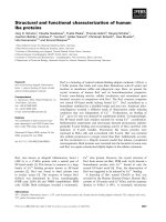
Respiratory Research 2006, 7:4 />Page 6 of 11
(page number not for citation purposes)
high ED7 expression (ED7
+
) by use of magnetic bead sep-
aration after removing contaminating neutrophils and
lymphocytes. As shown in the left panel of Figure 2 we
obtained very pure AM subpopulations. These cells were
now stained with the ED9 antibody combined with the
described amplification system. Interestingly, we found
that those AM showing a high level of ED7 expression are
characterized by a reduced ED9 expression whereas the
ED7
-
AM show the higher ED9 surface expression (Figure
2, right panel). Thus, two AM subpoplations were demon-
strated in the lungs of NO
2
-exposed rats that are character-
ized by the marker combinations ED7
+
/ED9
low
and ED7
-
/
ED
high
, in the following still referred as ED7
+
and ED7
-
AM, respectively.
Origin of AM subpopulations in NO
2
-exposed animals
The occurrence of phenotypically different AM subpopu-
lations may either be explained by a functional shift of
already present AM or by the infiltration of macrophages
that already represent the different phenotype. To address
this question we applied the recently described method of
in vivo labeling of resident AM by use of the fluorescent
cell tracer PKH26 [12]. When intravenously applied in
combination with a specific diluent, this dye is able to
label phagocytic cells within the organs, e.g. AM of the
lungs, without a significant staining of blood monocytes.
However, since this model is only applicable for the
mouse, we switched to the mouse model for these investi-
gations. Since we have recently shown that mice show a
slower development of the inflammatory reaction
towards NO
2
[16], mice were exposed for 7 days for these
analyses. Following this treatment, also in mice an AM
subpopulation was observed that revealed an increased
expression level of CD11b, the mouse homologue to ED7
(Fig. 3B). To analyze the origin of these cells, mice were
treated with PKH26 three days prior to the onset of the
NO
2
- or sham-exposure. At this time point, almost 100 %
of the AM were positively stained whereas blood mono-
cytes appeared PKH26-negative (data not shown).
Whereas this situation did not change in the sham-
exposed control group, a significant portion of PKH26-
negative, newly recruited AM were observed in the lungs
of NO
2
-exposed mice (Fig. 3C). A separate analysis of
PKH26-positive and PKH26-negative cells revealed that
the latter population was indeed characterized by a higher
expression of the surface marker CD11b indicating that
Flow cytometric analysis of AM from NO
2
-exposed and con-trol ratsFigure 1
Flow cytometric analysis of AM from NO
2
-exposed and con-
trol rats. Rats were exposed to NO
2
for the indicated times
and BAL cells were stained with antibodies to ED7, ED9,
RM-4, and OX-6. To overcome autofluorescence signals, pri-
mary antibodies were detected using a biotin-PE/streptavidin-
anti-streptavidin enhancing system and labeling of AM was
analyzed by flow cytometry following gating by help of for-
ward and sideward scatter properties. Shown are represent-
ative results of at least six animals per group.
Flow cytometric analysis of ED7 and ED9 expression of AM following magnetic bead separationFigure 2
Flow cytometric analysis of ED7 and ED9 expression of AM
following magnetic bead separation. AM of 3 days NO
2
-
exposed rats were separated due to their expression of the
cell surface molecule ED7 using magnetic bead separation.
Susbsequently, ED7 (left) and ED9 (right) expression was
analyzed in unseparated AM (A), ED7-positive AM (B), and
ED7-negative AM (C). Numbers right of each histogram rep-
resent the mean fluorescence of the respective cell popula-
tion. The figure clearly demonstrates that ED7-positive AM
show a lower ED9 expression compared to ED7-negative
AM. Shown is a representative data set of more than twenty
animals.
Respiratory Research 2006, 7:4 />Page 7 of 11
(page number not for citation purposes)
the CD11b-positive AM subpopulation mainly originated
from newly recruited macrophages (Figure 3D).
Cytokine and iNOS mRNA expression in separated AM
subpopulations
For functional analysis of the two phenotypically different
AM subpopulations we first compared the mRNA expres-
sion for several macrophage-derived mediators that are
involved in the regulation of inflammatory responses.
Therefore, ED7
+
and ED7
-
AM of the rat were separated
from the lungs of 3 days exposed animals. Total RNA was
immediately isolated and following cDNA synthesis
mediator mRNA expression was analyzed by quantitative
PCR. As shown in Figure 4, no differences were observed
between the two AM subpopulations in the expression of
the proinflammatory cytokine TNF-α. However, signifi-
cantly increased mRNA levels were found in the ED7
+
population for IL-12 p40 and iNOS. Interestingly, the
expression of the antiinflammatory cytokine IL-10 was
also higher in the ED7
+
AM subpopulation (Fig. 4).
Cytokine release by AM subpopulations following in vitro
stimulation
To confirm the importance of the gene expression data we
stimulated separated AM in vitro with 100 ng/ml LPS and
analyzed the release of cytokines in the 24 h culture super-
natants. When investigating proinflammatory cytokines
we found that TNF-α was released at significantly higher
amounts by AM of the ED7
-
subpopulation whereas IL-12
p70 was released at higher levels by the ED7
+
subpopula-
tion. However, the most important difference was
observed for IL-10 that was detected in more than 100-
FACS analysis of CD11b and PKH26 labeling of AM from NO
2
-expsoed C57BL/6 miceFigure 3
FACS analysis of CD11b and PKH26 labeling of AM from
NO
2
-expsoed C57BL/6 mice. Mice were intravenously given
PKH26 in combination with diluent C three days prior to
onset of a seven days NO
2
-exposure. Afterwards, AM were
stained with F4/80-Alexa647 and CD11b-FITC. Isotype con-
trol (A), CD11b (B) and PKH26 (C) staining was subse-
quently analyzed by flow cytometry within the F4/80-positive
cell population. (C) The proportion of PKH26-negative cells
is shown in blue. Part (D) shows a separate analysis of
CD11b-expression in PKH26-negative (blue histogram) and
PKH26-positive AM (pink histogram) thereby clearly demon-
strating that the CD11b-positive cell population mainly con-
sists of PKH26-negative, newly recruited AM. Shown are
representative results of eight animals per group.
Cytokine and iNOS mRNA expression in AM subpopulations of NO
2
-exposed ratsFigure 4
Cytokine and iNOS mRNA expression in AM subpopulations
of NO
2
-exposed rats. ED7-positive and ED7-negative AM
were separated from 3 days NO
2
-exposed rats and total
RNA was prepared immediately after cell separation.
Cytokine (TNF-α, IL-10, IL-12 p40) and iNOS mRNA
expression was analyzed by quantitative RT-PCR with L32 as
house-keeping gene control in ED7-negative (blank bars) and
ED7-positive AM (hatched bars). Data are presented as rela-
tive expression with mean expression in ED7-negative AM
was 100 %. Shown are mean ± SD of six animals per group.
Significance of differences was tested using the U-test
according to Mann and Whitney and is indicated by * for p <
0.05 or ** for p < 0.01.
Respiratory Research 2006, 7:4 />Page 8 of 11
(page number not for citation purposes)
fold amounts in the supernatants of ED7
+
AM when com-
pared to the ED7
-
subpopulation (Fig. 5).
MMP mRNA expression in separated AM subpopulations
In the context of an oxidant-induced inflammatory reac-
tion in the lung AM are not only involved in the regula-
tion of the inflammatory reaction by release of respective
mediators but may also produce factors such as MMPs
that may contribute to tissue remodelling and also lung
damage under these conditions. We therefore investigated
whether a specific subpopulation of AM is responsible for
the expression of several metalloproteinases. The results
of these analyses are summarized in Figure 6. With excep-
tion of MMP-2, that showed a comparable expression in
both AM subpopulations, mRNA for all other tested
MMPs (MMP-7, MMP-8, MMP-9, and MMP-12) were
almost not detectable in the ED7
-
subpopulation but were
found at significantly elevated levels in the ED7
+
AM sub-
population.
Discussion
Exposure of rodents to NO
2
have been shown to induce
inflammatory reactions in the lung that have several fea-
tures in common with the situation observed in patients
that suffer from inflammatory lung diseases such as
chronic obstructive lung disease (COPD). Due to it's poor
water solubility NO
2
may reach distal parts of the lung
including small airways and lung parenchyma where it
causes histopathological and functional changes. These
alterations comprise histomorphological changes in lung
parenchyma and vasculature [17,18] with increased vas-
cular permeability [14], loss of cilia in the airway epithe-
lium [19], hypertrophy of bronchial epithelial cells [20],
and mucus hypersecretion due to a hyperplasia of goblet
cells. In addition, several changes in surfactant metabo-
lism were described [21,22] and a replacement of type-I-
pneumocytes by type-II-cells was observed [20]. Moreo-
ver, prolonged exposure to NO
2
may also cause changes in
lung function such as limitation of airflow and increased
expiration time that are indicative for the occurrence of
airway obstruction [23] and may finally even lead to the
development of emphysema [24,25]. Especially the last
features are major characteristics of human COPD. As also
observed in these patients, macrophages and neutrophil
granulocytes are the most important inflammatory cell
populations [25,26]. Using the identical NO
2
exposure
model as applied for the investigations described here we
could recently demonstrate that neutrophils show an
immediate infiltration and their number peaks in the BAL
already at three days after onset of the exposure in rats
[14]. Even though mice show a slower development of
inflammatory changes [16], macrophages play the domi-
nant role over the whole observation period in both spe-
cies. With exception of day one in rats, significantly
increased alveolar macrophage numbers have been
observed over the whole observation period in rat and
mice, thereby representing the major cell population at all
time points [14]. However, only little is known about the
role that AM play in the pathogenesis of chronic inflam-
matory lung diseases especially at early stages of their
development.
mRNA expression for several MMPs in AM subpopulations of NO
2
-exposed ratsFigure 6
mRNA expression for several MMPs in AM subpopulations of
NO
2
-exposed rats. ED7-positive and ED7-negative AM were
separated from 3 days NO
2
-exposed rats and total RNA was
prepared immediately after cell separation. MMP-2, -7, -8, -9,
and -12 mRNA expression was analyzed by quantitative RT-
PCR with L32 as house-keeping gene control in ED7-negative
(blankbars) and ED7-positive AM (hatched bars). Data are
presented as relative expression with mean expression in
ED7-negative AM was 100 %. Shown are mean ± SD of six
animals per group. Significance of differences was tested
using the U-test according to Mann and Whitney and is indi-
cated by * for p < 0.05 or ** for p < 0.01.
Differential cytokine production by LPS-stimulated AM sub-populations of NO
2
-exposed ratsFigure 5
Differential cytokine production by LPS-stimulated AM sub-
populations of NO
2
-exposed rats. ED7-positive and ED7-
negative AM were separated from 3 days NO
2
-exposed rats
and cultured in vitro for 24 hours in the presence of 100 µg
LPS. Subsequently, TNF-α, IL-10, and IL-12 p70 were quanti-
tated in the culture supernatants of ED7-negative (blank
bars) and ED7-positive AM (hatched bars) by ELISA. Data are
presented as mean ± SD of at least six animals per group. Sig-
nificance of differences was tested using Students t-test and is
indicated by ** for p < 0.01 or *** for p < 0.001.
Respiratory Research 2006, 7:4 />Page 9 of 11
(page number not for citation purposes)
In the present study we could clearly demonstrate that a
new phenotypically different AM subpopulation occurs in
the lungs of rats and mice under the influence of oxida-
tive/nitrosative stress exerted by exposure of the animals
to NO
2
. Using PKH labeling of resident AM in mice we
were able to show, that these macrophages represent
newly recruited macrophages, a mechanism that is
assumed to be similar in rats. These macrophages differ
from already present AM by a higher expression of the sur-
face marker ED7 (in rat) or its murine homologue CD11b.
Interestingly, an increased expression of CD11b was also
observed in AM from smokers [27]. In addition, other sur-
face markers are also differentially expressed in AM from
control and NO
2
-exposed animals, e.g. ED9, RM-4 and
MHC-class-II molecules, at least in the rat. AM are known
to normally show a low expression of CD11b even though
this molecule is a typical surface marker of cells of the
monocyte/macrophage lineage in the blood and other tis-
sues [28]. Thus, the limited CD11b expression seems to be
a sign of tissue specific activation of AM that also show an
elevated expression of the transcription factor PU.1 [29],
a differential expression of protein kinase C isoforms [30]
and a decreased DNA binding capacity of the transcrip-
tion factor AP-1 [31] when compared to macrophages
from other tissues. In addition, the proteome of AM dif-
fers significantly from that of blood monocytes [32]. Per-
haps, AM-specific differentiation signals are
underrepresented during an inflammatory process in the
lung or these signals may not properly influence newly
infiltrating macrophages under these circumstances. As a
consequence, these alterations may lead to a different
phenotype of AM that enter the lung during an inflamma-
tory process in comparison to macrophages that infiltrate
under non-pathological conditions. However, very recent
data also suggest the existence of two phenotypically dif-
ferent monocyte populations that selectively enter healthy
or inflamed tissue areas [33,34]. This would imply that
the described AM subpopulations originated from already
different monocyte subpopulations.
In the model presented here, newly recruited AM seem to
have a dual role with respect to regulatory and effector
functions. A major feature of these cells is their high
expression and production of IL-10 which is in contrast to
resident AM that do only poorly produce this cytokine
even following LPS stimulation [35]. IL-10 is known to
exert antiinflammatory properties [36] and, therefore,
ED7
+
AM seem to play a role in the control of the inflam-
matory reaction. On the other hand these ED7
+
AM also
produce higher amounts of IL-12, a cytokine that is
involved in the activation of T helper 1 (Th1) lymphocytes
[37] that in turn may amplify a macrophage-dominated
inflammatory reaction. The latter mechanism is sup-
ported by observations in CCR2 knock-out mice that lack
the receptor for the CC-chemokine CCL2 (MCP-1; mono-
cyte chemotactic protein-1). These animals show dimin-
ished inflammatory reactions due to an impaired
migration of monocytes into inflammatory sites associ-
ated with decreased Th1 activities [38]. In line with these
findings it has also been demonstrated that these mice
exert enhanced Th2 responses [39,40]. In conclusion, our
findings clearly suggest that the newly recruited ED7
+
AM
are involved in the regulation of the ongoing inflamma-
tory process. Whether the antiinflammatory effects of IL-
10 or the proinflammatory role of IL-12 (or even addi-
tional regulatory molecules) will dominate the regulatory
function of ED7
+
AM in our model has to be investigated
in future studies.
In addition, ED7
+
AM are not only involved in regulatory
processes but may also directly act as effector cells. With
this respect the selective expression of several MMPs by
these macrophages was a quite interesting finding. It is
known that activated granulocytes and macrophages are
major producers of these proteases [41], however, to our
best knowledge this is the first description that a specific
inflammatory macrophage subpopulation is almost selec-
tively responsible for the production of certain MMPs,
among them MMP-9 and MMP-12. Interestingly, lung
macrophages from human smokers and COPD patients
have also been reported to show an increased expression
of MMP-9 [42] but macrophage subpopulations were not
investigated. MMP-12 seems to play an important role in
the development of emphysema at least in the mouse
model since absence of this specific MMP inhibits the gen-
eration of cigarette-smoke induced emphysema in MMP-
12 knock out mice [43]. More recent investigations pro-
vide evidence that both, elastase activities, such as MMP-
12, and collagenolytic activities, as exerted by MMP-2 and
MMP-9, in combination lead to an effective destruction of
lung parenchymal tissue that finally results in the genera-
tion of emphysema [44,45]. In addition, certain MMPs
may also be involved in the regulation of inflammatory
processes, e.g. by activation or inactivation of inflamma-
tory mediators [46-48]. Thus, by expression of important
MMPs ED7
+
AM may contribute to the pathology of NO
2
-
induced lung damage and are further involved in the reg-
ulation of the inflammatory process.
Conclusion
Exposure of rodents to the oxidative/nitrosative agent
NO
2
leads to the infiltration of a new AM subpopulation
that phenotypically and functionally differs from resident
AM. There is no doubt that these AM by release of regula-
tory mediators and expression of MMPs strongly influence
the mechanisms that regulate the inflammatory response
to the inducing agent and are directly involved in the
pathologic processes induced by NO
2
. Since NO
2
and
related molecules are major components of tobacco
smoke it is likely that similar processes may occur in
Respiratory Research 2006, 7:4 />Page 10 of 11
(page number not for citation purposes)
smokers and even patients suffering from COPD. Indeed,
phenotypically and functionally different macrophages
have been observed in sputum of those patients [49].
These macrophages represent a different compartment of
the lung, however, their occurrence implicates that similar
processes as described in our animal model may also
occur in humans following oxidative/nitrosative stress. If
so, these newly recruited macrophages may represent an
interesting target for therapeutic approaches for the treat-
ment of chronic inflammatory diseases of the lung.
Competing interests
The author(s) declare that they have no competing inter-
ests.
Authors' contributions
HG conceived of and designed the study, was involved in
animal exposure and cell preparation, performed FACS
analysis and drafted the manuscript.
AS was involved in animal exposure and cell preparation,
carried out MACS separation of AM subpopulations and
performed in vitro cell stimulation and mediator analysis.
SS was responsible for animal preparation, performed
mRNA expression analyses, and helped in FACS and
MACS procedures.
AW performed the PKH26 experiments and was involved
in subsequent FACS analyses. In addition she helped car-
rying out mRNA-expression analyses.
HR helped in study design and coordination as well as in
preparation of the manuscript.
DG participated in the design of the experiments, its coor-
dination and manuscript preparation.
Acknowledgements
The study was funded by the German Ministry of Education and Research
Grant No. 01GC0103.
References
1. Crapo JD, Harmsen AG, Sherman MP, Musson RA: Pulmonary
immunobiology and inflammation in pulmonary diseases.
Am J Respir Crit Care Med 2000, 162:1983-1986.
2. Nicod LP: Pulmonary defense mechanisms. Respiration 1999,
66:2-11.
3. Shapiro SD: The macrophage in chronic obstructive pulmo-
nary disease. Am J Resp Crit Care Med 1999, 160:S29-S32.
4. Lohmann-Matthes ML, Steinmüller C, Franke-Ullmann G: Pulmo-
nary macrophages. Eur Respir J 1994, 7:1678-1689.
5. Lavnikova N, Prokhorova S, Helyar L, Laskin DL: Isolation and par-
tial characterization of subpopulations of alveolar macro-
phages, granulocytes, and highly enriched interstitial
macrophages from rat lung. Am J Respir Cell Mol Biol 1993,
8:384-392.
6. Laskin DL, Weinberger B, Laskin JD: Functional heterogeneity in
liver and lung macrophages. J Leukoc Biol 2001, 70:163-170.
7. Dorger M, Munzing S, Allmeling AM, Messmer K, Krombach F: Phe-
notypic and functional differences between rat alveolar,
pleural, and peritoneal macrophages. Exp Lung Res 2001,
27:65-76.
8. Ferrari-Lacraz S, Nicod LP, Chicheportiche R, Welgus HG, Dayer JM:
Human lung tissue macrophages, but not alveolar macro-
phages, express matrix metalloproteinases after direct con-
tact with activated T lymphocytes. Am J Respir Cell Mol Biol 2001,
24:442-451.
9. Chelen CJ, Fang Y, Freeman GJ, Secrist H, Marshall JD, Hwang PT,
Frankel LR, DeKruyff RH, Umetsu DT: Human alveolar macro-
phages present antigen ineffectively due to defective expres-
sion of B7 costimulatory cell surface molecules. J Clin Invest
1995, 95:1415-1421.
10. Knapp S, Leemans JC, Florquin S, Branger J, Maris NA, Pater J, van
Rooijen N, van der PT: Alveolar macrophages have a protective
antiinflammatory role during murine pneumococcal pneu-
monia. Am J Respir Crit Care Med 2003, 167:171-179.
11. Strickland D, Kees UR, Holt PG: Regulation of T-cell activation
in the lung: aveolar macrophages induce reversible T-cell
anergy in vitro associated with inhibition of interleukin-2
receptor signal transduction. Immunology 1996, 87:250-258.
12. Maus U, Herold S, Muth H, Maus R, Ermert L, Ermert M, Weissmann
N, Rosseau S, Seeger W, Grimminger F, et al.: Monocytes recruited
into the alveolar air space of mice show a monocytic pheno-
type but upregulate CD14. Am J Physiol Lung Cell Mol Physiol 2001,
280:L58-L68.
13. Olker C, Siese A, Stumpf S, Muller B, Gemsa D, Garn H: Impaired
superoxide radical production by bronchoalveolar lavage
cells from NO(2)-exposed rats. Free Radic Biol Med 2004,
37:977-987.
14. Garn H, Siese A, Stumpf S, Barth PJ, Müller B, Gemsa D: Shift
towards an alternatively activated macrophage response in
lungs of NO
2
-exposed rats. Am J Respir Cell Mol Biol 2003,
28:386-396.
15. Garn H, Friedetzky A, Kirchner A, Jäger R, Gemsa D: Experimental
silicosis: A shift to a preferential IFN-γ-based Th1 response in
thoracic lymph nodes. Am J Physiol Lung Cell Mol Physiol 2000,
278:L1221-L1230.
16. Wegmann M, Fehrenbach A, Heimann S, Fehrenbach H, Renz H, Garn
H, Herz U: NO2-induced airway inflammation is associated
with progressive airflow limitation and development of
emphysema-like lesions in C57BL/6 mice. Exp Toxicol Pathol
2005, 56:341-350.
17. Barth PJ, Uhlarik S, Bittinger A, Wagner U, Ruschoff J: Diffuse alve-
olar damage in the rat lung after short and long term expo-
sure to nitrogen dioxide. Pathol Res Pract 1994, 190:33-41.
18. Barth PJ, Müller B, Wagner U, Bittinger A: Quantitative analysis of
parenchymal and vascular alterations in NO
2
-induced lung
injury in rats. Eur Respir J 1995, 8:1115-1121.
19. Chitano P, Rado V, Di Stefano A, Papi A, Boniotti A, Zancuoghi G,
Boschetto P, Romano M, Salmona M, Ciaccia A, et al.: Effect of
subchronic in vivo exposure to nitrogen dioxide on lung tis-
sue inflammation, airway microvascular leakage, and in vitro
bronchial muscle responsiveness in rats. Occup Environ Med
1996, 53:379-386.
20. Rombout PJ, Dormans JA, Marra M, van Esch GJ: Influence of expo-
sure regimen on nitrogen dioxide-induced morphological
changes in the rat lung. Environ Res 1986, 41:466-480.
21. Müller B, Barth PJ, Wichert Pv: Structural and functional impair-
ment of surfactant protein A after exposure to nitrogen
dioxide in rats. Am J Physiol 1992, 263:L177-L184.
22. Müller B, Schäfer H, Barth PJ, Wichert Pv: Lung surfactant compo-
nents in bronchoalveolar lavage after inhalation of NO
2
as
markers of altered surfactant metabolism. Lung 1994,
172:61-72.
23. Wegmann M, Renz H, Herz U: Long-term NO2 exposure
induces pulmonary inflammation and progressive develop-
ment of airflow obstruction in C57BL/6 mice: a mouse
model for chronic obstructive pulmonary disease? Pathobiol-
ogy 2002, 70:284-286.
24. Blank J, Glasgow JE, Pietra GG, Burdette L, Weinbaum G: Nitrogen-
dioxide-induced emphysema in rats. Lack of worsening by
beta- aminopropionitrile treatment. Am Rev Respir Dis 1988,
137:376-379.
Publish with BioMed Central and every
scientist can read your work free of charge
"BioMed Central will be the most significant development for
disseminating the results of biomedical research in our lifetime."
Sir Paul Nurse, Cancer Research UK
Your research papers will be:
available free of charge to the entire biomedical community
peer reviewed and published immediately upon acceptance
cited in PubMed and archived on PubMed Central
yours — you keep the copyright
Submit your manuscript here:
/>BioMedcentral
Respiratory Research 2006, 7:4 />Page 11 of 11
(page number not for citation purposes)
25. Glasgow JE, Pietra GG, Abrams WR, Blank J, Oppenheim DM, Wein-
baum G: Neutrophil recruitment and degranulation during
induction of emphysema in the rat by nitrogen dioxide. Am
Rev Respir Dis 1987, 135:1129-1136.
26. Pagani P, Romano M, Erroi A, Ferro M, Salmona M: Biochemical
effects of acute and subacute nitrogen dioxide exposure in
rat lung and bronchoalveolar fluid. Arch Environ Contam Toxicol
1994, 27:426-430.
27. Schaberg T, Lauer C, Lode H, Fischer J, Haller H: Increased
number of alveolar macrophages expressing adhesion mole-
cules of the leukocyte adhesion molecule family in smoking
subjects. Association with cell-binding ability and superoxide
anion production. Am Rev Respir Dis 1992, 146:1287-1293.
28. Gordon S: Pattern recognition receptors: doubling up for the
innate immune response. Cell 2002, 111:927-930.
29. Shibata Y, Berclaz PY, Chroneos ZC, Yoshida M, Whitsett JA, Trap-
nell BC: GM-CSF regulates alveolar macrophage differentia-
tion and innate immunity in the lung through PU.1. Immunity
2001, 15:557-567.
30. Monick MM, Carter AB, Gudmundsson G, Geist LJ, Hunninghake
GW: Changes in PKC isoforms in human alveolar macro-
phages compared with blood monocytes. Am J Physiol 1998,
275:L389-L397.
31. Monick MM, Carter AB, Hunninghake GW: Human alveolar mac-
rophages are markedly deficient in REF-1 and AP-1 DNA
binding activity. J Biol Chem 1999, 274:18075-18080.
32. Jin M, Opalek JM, Marsh CB, Wu HM: Proteome comparison of
alveolar macrophages with monocytes reveals distinct pro-
tein characteristics. Am J Respir Cell Mol Biol 2004, 31:322-329.
33. Geissmann F, Jung S, Littman DR: Blood monocytes consist of
two principal subsets with distinct migratory properties.
Immunity 2003, 19:71-82.
34. Sunderkotter C, Nikolic T, Dillon MJ, Van Rooijen N, Stehling M,
Drevets DA, Leenen PJ: Subpopulations of mouse blood mono-
cytes differ in maturation stage and inflammatory response.
J Immunol 2004, 172:4410-4417.
35. Salez L, Singer M, Balloy V, Creminon C, Chignard M: Lack of IL-10
synthesis by murine alveolar macrophages upon lipopolysac-
charide exposure. Comparison with peritoneal macro-
phages. J Leukoc Biol 2000, 67:545-552.
36. Mosmann TR: Properties and function of IL-10. Adv Immunol
1994, 56:1-26.
37. Brombacher F, Kastelein RA, Alber G: Novel IL-12 family mem-
bers shed light on the orchestration of Th1 responses. Trends
Immunol 2003, 24:207-212.
38. Boring L, Gosling J, Chensue SW, Kunkel SL, Farese RV Jr, Broxmeyer
HE, Charo IF: Impaired monocyte migration and reduced type
1 (Th1) cytokine responses in C-C chemokine receptor 2
knockout mice. J Clin Invest 1997, 100:2552-2561.
39. Blease K, Mehrad B, Standiford TJ, Lukacs NW, Gosling J, Boring L,
Charo IF, Kunkel SL, Hogaboam CM: Enhanced pulmonary aller-
gic responses to Aspergillus in CCR2-/- mice. J Immunol 2000,
165:2603-2611.
40. Kim Y, Sung S, Kuziel WA, Feldman S, Fu SM, Rose CE Jr: Enhanced
airway Th2 response after allergen challenge in mice defi-
cient in CC chemokine receptor-2 (CCR2). J Immunol 2001,
166:5183-5192.
41. Parks WC, Shapiro SD: Matrix metalloproteinases in lung biol-
ogy. Respir Res 2001, 2:10-19.
42. Finlay GA, O'Driscoll LR, Russell KJ, D'Arcy EM, Masterson JB, Fit-
zGerald MX, O'Connor CM: Matrix metalloproteinase expres-
sion and production by alveolar macrophages in
emphysema. Am J Respir Crit Care Med 1997, 156:240-247.
43. Hautamaki RD, Kobayashi DK, Senior RM, Shapiro SD: Require-
ment for macrophage elastase for cigarette smoke-induced
emphysema in mice. Science 1997, 277:2002-2004.
44. Karkmann U, Radbruch A, Holzel V, Scheffold A: Enzymatic signal
amplification for sensitive detection of intracellular antigens
by flow cytometry. J Immunol Methods 1999, 230:113-120.
45. Lanone S, Zheng T, Zhu Z, Liu W, Lee CG, Ma B, Chen Q, Homer RJ,
Wang J, Rabach LA, et al.: Overlapping and enzyme-specific con-
tributions of matrix metalloproteinases-9 and -12 in IL-13-
induced inflammation and remodeling. J Clin Invest 2002,
110:463-474.
46. Haro H, Crawford HC, Fingleton B, Shinomiya K, Spengler DM, Mat-
risian LM: Matrix metalloproteinase-7-dependent release of
tumor necrosis factor- alpha in a model of herniated disc
resorption. J Clin Invest 2000, 105:143-150.
47. Yu Q, Stamenkovic I: Cell surface-localized matrix metallopro-
teinase-9 proteolytically activates TGF-beta and promotes
tumor invasion and angiogenesis. Genes Dev 2000, 14:163-176.
48. Schonbeck U, Mach F, Libby P: Generation of biologically active
IL-1 beta by matrix metalloproteinases: a novel caspase-1-
independent pathway of IL-1 beta processing. J Immunol 1998,
161:3340-3346.
49. Frankenberger M, Menzel M, Betz R, Kassner G, Weber N, Kohlhaufl
M, Haussinger K, Ziegler-Heitbrock L: Characterization of a pop-
ulation of small macrophages in induced sputum of patients
with chronic obstructive pulmonary disease and healthy vol-
unteers. Clin Exp Immunol 2004, 138:507-516.