Báo cáo y học: "Can HRCT be used as a marker of airway remodelling in children with difficult asthma?" docx
Bạn đang xem bản rút gọn của tài liệu. Xem và tải ngay bản đầy đủ của tài liệu tại đây (496.04 KB, 9 trang )
BioMed Central
Page 1 of 9
(page number not for citation purposes)
Respiratory Research
Open Access
Research
Can HRCT be used as a marker of airway remodelling in children
with difficult asthma?
S Saglani
1,4
, G Papaioannou
2
, L Khoo
3
, M Ujita
3
, PK Jeffery
4
, C Owens
2
,
DM Hansell
3
, DN Payne
1
and A Bush*
1
Address:
1
Respiratory Paediatrics, Royal Brompton Hospital, London, UK,
2
Department of Radiology, Great Ormond Street Hospital, London, UK,
3
Department of Radiology, Royal Brompton Hospital, London, UK and
4
Lung Pathology, Imperial College London at the Royal Brompton
Hospital, London, UK
Email: S Saglani - ; G Papaioannou - ; L Khoo - ;
M Ujita - ; PK Jeffery - ; C Owens - ; DM Hansell - ;
DN Payne - ; A Bush* -
* Corresponding author
Abstract
Background: Whole airway wall thickening on high resolution computed tomography (HRCT) is
reported to parallel thickening of the bronchial epithelial reticular basement membrane (RBM) in
adult asthmatics. A similar relationship in children with difficult asthma (DA), in whom RBM
thickening is a known feature, may allow the use of HRCT as a non-invasive marker of airway
remodelling. We evaluated this relationship in children with DA.
Methods: 27 children (median age 10.5 [range 4.1–16.7] years) with DA, underwent
endobronchial biopsy from the right lower lobe and HRCT less than 4 months apart. HRCTs were
assessed for bronchial wall thickening (BWT) of the right lower lobe using semi-quantitative and
quantitative scoring techniques. The semi-quantitative score (grade 0–4) was an overall assessment
of BWT of all clearly identifiable airways in HRCT scans. The quantitative score (BWT %; defined
as [airway outer diameter – airway lumen diameter]/airway outer diameter ×100) was the average
score of all airways visible and calculated using electronic endpoint callipers. RBM thickness in
endobronchial biopsies was measured using image analysis. 23/27 subjects performed spirometry
and the relationships between RBM thickness and BWT with airflow obstruction evaluated.
Results: Median RBM thickness in endobronchial biopsies was 6.7(range 4.6 – 10.0) µm. Median
qualitative score for BWT of the right lower lobe was 1(range 0 – 1.5) and quantitative score was
54.3 (range 48.2 – 65.6)%. There was no relationship between RBM thickness and BWT in the right
lower lobe using either scoring technique. No relationship was found between FEV
1
and BWT or
RBM thickness.
Conclusion: Although a relationship between RBM thickness and BWT on HRCT has been found
in adults with asthma, this relationship does not appear to hold true in children with DA.
Published: 27 March 2006
Respiratory Research2006, 7:46 doi:10.1186/1465-9921-7-46
Received: 12 September 2005
Accepted: 27 March 2006
This article is available from: />© 2006Saglani et al; licensee BioMed Central Ltd.
This is an Open Access article distributed under the terms of the Creative Commons Attribution License ( />),
which permits unrestricted use, distribution, and reproduction in any medium, provided the original work is properly cited.
Respiratory Research 2006, 7:46 />Page 2 of 9
(page number not for citation purposes)
Background
Thickening of the epithelial reticular basement membrane
(RBM) is one characteristic feature of airway remodelling
in asthma. It has been reported in both adults and school-
aged children [1-3]. However, the clinical significance of
RBM thickening, and the mechanisms involved in its
pathogenesis remain unclear. In particular, it is not
known at what age RBM thickening begins.
RBM thickness can be measured in endobronchial biopsy
(EB), but this requires an invasive procedure, and the
opportunities for obtaining EB in children are therefore
limited. The potential to investigate the timing and natu-
ral history of RBM thickening, and other features of airway
remodelling in children, would be increased by the devel-
opment of non-invasive techniques, thus providing the
opportunity to monitor changes over time and in
response to treatment. A number of non-invasive tech-
niques have been developed for the study of airway
inflammation in asthma [4]. In comparison, there has
been little interest in the development of similar tech-
niques to study airway structural changes. One exception
is the use of high-resolution computed tomography
(HRCT) to study airway wall changes in asthma [5]. Bron-
chial wall thickening (BWT) on HRCT has been shown to
be a consistent finding in children with difficult asthma
[6] and a relationship between BWT and RBM thickness
has been demonstrated in adults with asthma, following
treatment with oral corticosteroids and short-acting β
2
-
agonists [7]. The demonstration of a similar relationship
in children with difficult asthma would therefore allow
HRCT to be used as a surrogate marker of RBM thickening.
Airway remodelling is often considered to contribute to
the element of irreversible airflow obstruction, which is a
feature of some patients with asthma. Kasahara and col-
leagues reported a significant negative correlation
between post-bronchodilator forced expiratory volume in
one second (FEV
1
) and both BWT on HRCT and RBM
thickness in EB [7]. However, other cross-sectional studies
have failed to demonstrate an association between FEV
1
and either BWT [6,8] or RBM thickness [3,9].
The aims of the present study were therefore to investigate
i) whether BWT, as shown on HRCT, can be used as a non-
invasive indicator of RBM thickness in EB, in a group of
children with difficult asthma, and ii) the association
between the degree of airflow limitation, assessed by FEV
1
and BWT or RBM thickness.
Methods
Subjects
Twenty-seven children (median age 10.5 [range 4.1–16.7]
years) with difficult asthma, who underwent bronchos-
copy, EB and HRCT between January 2000 and November
2002 were identified and studied retrospectively. Subjects
underwent bronchoscopy, bronchoalveolar lavage and EB
as part of their clinical assessment in order to help con-
firm the diagnosis of asthma and to exclude any other
associated abnormalities such as structural airway abnor-
malities or significant infection. They underwent HRCT to
exclude bronchiectasis or any other airways disease such
as obliterative bronchiolitis that may have been an alter-
native explanation for their disease severity. Difficult
asthma was defined as persistent symptoms requiring res-
cue bronchodilator therapy > 3 days per week, despite ≥
800 micrograms per day of inhaled budesonide (or equiv-
alent), and long acting β
2
agonists, and/or regular oral
steroids. All subjects that had a bronchoscopy and EB in
the defined time period were identified. Not all had a
biopsy of sufficient quality (defined as a biopsy contain-
ing recognisable epithelium, RBM and subepithelium,
with at least 1 mm of RBM) to quantify the RBM [3,10].
Therefore only those with a good quality biopsy were
included in this study. There was no difference in age, sex
or disease severity between subjects with and without
good quality biopsies. The clinical details of subjects
included are summarised in table 1.
Twenty-three of 27 subjects also performed spirometry in
accordance with American Thoracic Society guidelines
Table 1: Clinical characteristics of children with difficult asthma
Number 27
Age* 10.5 (4.1 – 16.7)
Male/Female 17/10
FEV
1
(% predicted)*, pre-bronchodilator
a
82.6 (32.1 – 118)
Atopic 21 (78%)
Treatment:
Daily dose budesonide/equivalent* 2000 (800 – 4000) µm
Number on LABA 20 (74%)
Number on regular orals steroids 50(18.5%)
* median (range)
a – only 23/27 able to perform satisfactory spirometry
Respiratory Research 2006, 7:46 />Page 3 of 9
(page number not for citation purposes)
[11]. Four subjects, aged between 4.1 and 5.2 years were
unable to perform spirometry with a satisfactory tech-
nique. Sixteen of the 23 subjects that performed spirome-
try had received a 2-week course of oral corticosteroids
before spirometry and 9 of those 16 performed spirome-
try before and after inhaled bronchodilator (short acting
β
2
agonist).
Informed parental consent was obtained prior to perform-
ance of EB and HRCT in all cases. Ethical approval was
obtained to study all biopsies and HRCTs.
Endobronchial biopsies
Flexible bronchoscopy was performed under general
anaesthetic, as previously described [12]. Up to six EB
were taken from the sub-carinae of the right lower lobe.
Biopsies were fixed and processed into paraffin blocks.
Step sections (5 µm thick) were cut 50 µm apart and
stained with haematoxylin and eosin. At least one meas-
urable section, which was well orientated and had identi-
fiable epithelium, RBM and subepithelium, was chosen
from each patient. If more than one biopsy, satisfying the
above criteria, was obtained from the same patient, the
between biopsy variability was assessed. RBM thickness
was measured using computer-aided image analysis (NIH
image 1.55; National Institute of Health, Bethesda, MD)
as previously described [13]. Briefly, at a magnification of
×400, at least 40 measurements of RBM thickness were
made 20 µm apart. A minimum length of 1 mm of RBM
was assessed. The geometric mean of all measurements
was calculated to represent thickness for that section.
Measurements of RBM thickness were made without
knowledge of the HRCT assessments.
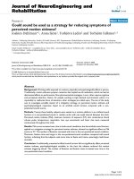
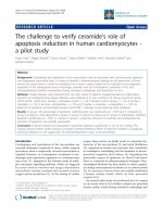
Outer and inner bronchial diametersFigure 1
Outer and inner bronchial diameters. Magnified area of an axial HRCT of a child with difficult asthma showing a circular
bronchus that was quantified. 1a) outer (Do = 0.5 cm) and 1b) inner (Di = 0.3 cm) bronchial diameters were measured as out-
lined.
1a
1b
Respiratory Research 2006, 7:46 />Page 4 of 9
(page number not for citation purposes)
HRCT
All HRCTs were obtained at near total lung capacity with
breath holding rehearsed before commencement of the
HRCT. 1.5 mm thick sections were acquired at 10 mm
intervals in the supine position using an electron beam
ultrafast scanner (Imatron Inc., San Francisco, California)
and images were reconstructed using a high spatial resolu-
tion reconstruction algorithm. Images were photographed
using window settings optimised for paediatric lungs
(centre: -500 H.U., width: 1500 H.U.) [14].
HRCT scoring
A quantitative score that has previously been used to
assess BWT in HRCT in adult asthmatics [7] was used.
However, more recently a paediatric study comparing
BWT in HRCT with endobronchial biopsies has used a
semi-quantitative score [15]. Also, for clinical purposes,
application of a quantitative score is time consuming.
Therefore, in order to compare findings to previously pub-
lished data and to assess whether there is any advantage in
using a quantitative scoring system, both semi-quantita-
tive and quantitative scores of BWT on HRCT were
applied. HRCT images were assessed by two radiologists
(LK, UM) for the semi-quantitative scoring and then by a
third radiologist (GP) for the quantitative scoring; all radi-
ologists were unaware of the clinical status of the subjects.
Semi-quantitative score
HRCT images were assessed by two radiologists (LK, UM)
independently. A semi-quantitative score for BWT in all
sections of the HRCT was recorded. The evaluation of
BWT was confined to clearly identifiable segmental and
sub-segmental airways. A separate score was given to each
lobe. Scores ranged from 0 to 4. 0 was normal wall thick-
ness, 1 was minimal wall thickening, 2 was bronchial wall
thickness half of the diameter of the adjacent blood vessel,
3 was bronchial wall thickness half to the same diameter
of the adjacent vessel, and 4 was bronchial wall thickness
greater than the diameter of the adjacent vessel. This score,
which in the context of this study was only used to assess
bronchial wall thickness has been used previously to
assess the relationship between CT features of bron-
chiectasis and lung function [16]. Bronchial dilatation
was not assessed. The mean of the two scores ascribed was
used to assess the relationship between BWT and RBM
thickness and FEV
1
.
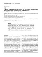
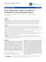
Diagram of outer and inner diametersFigure 2
Diagram of outer and inner diameters. Diagramatic draft of the measurement techniques applied to magnified cross-sec-
tional images. The obvious round-shaped artery (A) and bronchus (B) pairs were identified and the outer (Do) and inner (Di)
diameter of the bronchus were measured (solid and dotted lines respectively). WT% was calculated as [(Do-Di)/Do] × 100.
Respiratory Research 2006, 7:46 />Page 5 of 9
(page number not for citation purposes)
Quantitative score
The quantitative scoring system was based on that previ-
ously used in adult studies that have assessed BWT in
HRCTs from asthmatics [7]. HRCT scans were loaded to a
PACS workstation (mv1000, Siemens) and all images
were analysed electronically. A magnification factor of 7
was applied in all images that were displayed in HRCT
window settings. All clearly visible segmental and sub-seg-
mental airway/vessel pairs that had a rounded cross-sec-
tional circumference were measured manually by using
electronic caliper endpoints (figures 1a and 1b). By esti-
mating the Hounsfield units using the ROI (region of
interest) tool, the endpoints were placed at the cut-off
edge between the wall and the air. For each airway with an
obvious circular appearance, the outer diameter was
measured in the x and y axis. The shorter of these two, was
termed the airway outer diameter (Do) (figure 1a). In the
same axis, the airway inner diameter, or lumen diameter,
(Di) was measured (figure 1b). The percentage of airway
wall thickness (BWT) was calculated (BWT % = [Do-Di]/
Do × 100) (figure 2).
Statistical analysis
A weighted kappa coefficient was calculated to determine
the level of agreement for the semi-quantitative score
between the two HRCT observers [17]. The Kruskal-Wallis
test was used to look for a relationship between numerical
and categorical variables. The relationship between RBM
thickness and % BWT, and RBM thickness and predicted
FEV
1
were assessed using Spearman's correlation (r
s
). The
variability of RBM thickness within and between biopsies
was calculated as the % coefficient of variation, by divid-
ing the standard deviation of the measurements by the
mean. All analyses were performed using the Statistical
Package for the Social Sciences (SPSS) version 11.5.
Results
Twenty-seven children had EB and HRCT performed no
more than 4 months apart. Eighteen of 27 had both inves-
tigations on the same day (table 2). Median RBM thick-
ness was 6.7 (range 4.6–10.0) µm. The coefficient of
variation for within-biopsy measurements ranged from
1.6 – 7.4%, and that for variability between biopsies
ranged from 6 – 21.6%.
Semi-quantitative BWT score and RBM thickness
There was a moderate level of agreement between observ-
ers for HRCT scores for BWT of the right lower lobe
(weighted κ = 0.54). The average of the two scores
ascribed for the right lower lobe bronchus, near the site of
EB, was used for subsequent analyses. Median score for
BWT in the right lower lobe was 1 (range 0–1.5). None of
the HRCTs had evidence of bronchiectasis. Subjects
grouped according to BWT score showed there was no
relationship between RBM thickness and median BWT
score on HRCT scan (figure 3a). The result was the same
when only those patients (18/27) who had EB and HRCT
on the same day were included in the analysis (figure 3b).
For the patients who also performed spirometry, median
pre-bronchodilator FEV
1
% predicted was 79.7% (range
32.1–118.0%). There was no difference in pre-bronchodi-
lator FEV
1
between the groups, based on BWT score (figure
5a).
Quantitative BWT score and RBM thickness
A score was obtained for the average BWT for all lobes and
also just for the right lower lobe (as this is where biopsies
were taken). Median BWT for the whole scan was 55.5
(range 48.7 – 58.5)% and that for just the right lower lobe
was 54.3 (range 48.2 – 65.6)%. There was no correlation
between BWT for the whole scan and RBM thickness on
EB (r
s
= 0.066, p = 0.75) (figure 4a) and there was also no
relationship between BWT for the right lower lobe and
RBM thickness (r
s
= 0.03, p = 0.89), (figure 4b). There was
a good correlation between BWT score for the whole
HRCT scan and that just for the right lower lobe (r
s
= 0.64,
p < 0.001).
RBM thickness, BWT and lung function
There was no relationship between pre-bronchodilator
FEV
1
and RBM thickness (r
s
= -0.155, p = 0.48). There was
also no relationship between FEV
1
and BWT on HRCT,
measured using both techniques (figure 5a and 5b). 10/
27 patients had HRCT, EB and spirometry on the same
day (table 2). When they were analysed separately, there
was no relationship between BWT and RBM thickness or
pre-bronchodilator FEV
1
. Similarly, no relationship was
seen between BWT and RBM or post-bronchodilator FEV
1
Table 2: Patients who had investigations performed on same and different days
HRCT & EB same day HRCT & EB different days Total
FEV
1
available* 17 6 23
No FEV
1
134
Total 18 9 27
* 10/17 had all 3 tests on the same day, 7/17 had spirometry on a different day
HRCT: high-resolution computed tomography
EB: endobronchial biopsy
FEV
1
: forced expiratory volume in 1 second
Respiratory Research 2006, 7:46 />Page 6 of 9
(page number not for citation purposes)
when the 9 patients who had performed spirometry pre
and post bronchodilator were analysed.
Discussion
There was no relationship between RBM thickness in EB
and BWT on HRCT in children with difficult asthma. Also,
no relationship was found between FEV
1
(% predicted)
and BWT on HRCT or RBM thickness in EB.
In keeping with our previous findings from a group of
children with difficult asthma, we found no relationship
between RBM thickness and % predicted FEV
1
[3]. Also, in
agreement with Marchac and colleagues we found no rela-
tionship between BWT on HRCT and % predicted FEV
1
,
nor did we find any evidence of bronchiectasis [6]. Our
findings are in contrast to those of Kasahara and col-
leagues in adults [7] and de Blic and colleagues [15] in
children.
A limitation of this study compared to that by Kasahara
and colleagues in adults [7], was that there were no HRCT
measurements from control subjects. However, given the
ethical implications of unnecessary radiation exposure, it
was not possible to justify performing HRCT in healthy
children. It is especially important to consider measure-
ments of both RBM thickness and BWT on HRCT in
healthy children because of the influence of normal air-
way development in this age group [18]. However, a pae-
diatric study that has reported a relationship between
RBM thickness on EB and BWT on HRCT in difficult asth-
matics also did not include healthy controls [15]. A fur-
ther limitation of the current study was that patients were
identified retrospectively, and lung function data was not
available in all cases. This resulted in only a small number
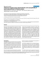
RBM thickness and HRCT bronchial wall thickening using quantitative scoreFigure 4
RBM thickness and HRCT bronchial wall thickening
using quantitative score. Relationship between RBM
thickness in endobronchial biopsy and 4a) bronchial wall
thickening on whole HRCT and 4b) right lower lobe bron-
chial wall thickening, measured using a quantitative score.
47.5 50.0 52.5 55.0 57.5 60.0
2.5
5.0
7.5
10.0
BWT (%) - all lobes
RBM (
µ
µ
µ
µ
m)
45 50 55 60 65
2.5
5.0
7.5
10.0
BWT (%) right lower lobe
RBM(
µ
µ
µ
µ
m)
4a
4b
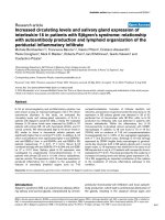
RBM thickness and HRCT bronchial wall thickening using semi-quantitative scoreFigure 3
RBM thickness and HRCT bronchial wall thickening
using semi-quantitative score. Relationship between
RBM thickness in endobronchial biopsy and bronchial wall
thickening on HRCT measured using a semi-quantitative
score. 3a) all HRCTs and bronchial biopsies, 3b) only HRCTs
and bronchial biopsies performed on the same day.
0 0.5 1.0 1.5
0.0
2.5
5.0
7.5
10.0
BWT score for right lower lobe
RBM (
µ
µ
µ
µ
m)
0 0.5 1.0
0.0
2.5
5.0
7.5
10.0
BWT score right lower lobe
RBM (
µ
µ
µ
µ
m)
3a
3b
Respiratory Research 2006, 7:46 />Page 7 of 9
(page number not for citation purposes)
of patients with all data present. In some patients, tests
(EB, HRCT and lung function) were not performed on the
same day because the investigations were all clinically
indicated and therefore were performed only when neces-
sary.
As only 9/23 patients performed spirometry pre and post-
bronchodilator, the relationship between BWT and lung
function was assessed for all 23 patients using pre-bron-
chodilator FEV
1
. This is in contrast to the study by Kasa-
hara and colleagues who compared post-bronchodilator
FEV
1
with BWT and RBM thickness [7], and may account
for the discrepancy between the results of their study and
the present one. When these 9 patients were analysed sep-
arately, no relationship was seen. However, the small
number limits the ability to draw firm conclusions. In the
present study, only 10/27 patients had HRCT, EB and
spirometry performed on the same day (table 2). No rela-
tionship was found between any of the parameters when
these patients were analysed separately, providing support
for the results found for the group as a whole.
Two-thirds (18/27) of the subjects had HRCT and EB on
the same day (table 2), while the remainder had a period
of up-to 4 months between the tests. As EB was performed
after a two-week course of oral steroids, it may be that this
affected the results for those who had the tests separately.
However, previous studies that have assessed the effect of
steroid therapy on RBM thickness have shown a reduction
in thickness after prolonged therapy for several months,
not weeks [19], so a short course, even when given system-
ically is unlikely to have affected RBM thickness. Impor-
tantly, all subjects studied by Kasahara and colleagues did
have pre-treatment with 2 weeks of prednisolone prior to
HRCT in order to minimise the effects of any airway
oedema. However, in the current study, when the patients
who had both tests on the same day, immediately after
completion of the steroid course, were analysed sepa-
rately, there was still no relationship between BWT and
RBM thickness.
In order to ensure that the failure to show a relationship
between BWT and the other parameters was not due to the
scoring technique used, and to ensure the scoring tech-
niques used in previously published studies were used,
[7,15] HRCTs were scored using both semi-quantitative
[16] and quantitative techniques [7]. However, despite
using 2 separate techniques and using independent
observers to score the scans and ensuring an adequate
level of agreement between observers for the semi-quanti-
tative technique (weighted kappa > 0.5), there was still no
relationship found between BWT and RBM thickness or
FEV
1
. Furthermore, as the biopsies were taken from the
right lower lobe, the CT score for that lobe alone was used
in the analysis. It may be proposed that quantitative meth-
ods are more accurate than semi-quantitative scoring.
However, we have demonstrated that with a moderate
level of agreement between observers, the use of the semi-
quantitative technique gives similar results to the quanti-
tative technique. Although the quantitative technique
might appear to be the more objective of the two, it also
involves some degree of subjective bias, since the identifi-
cation of the boundaries of the inner lumen and outer
wall requires a judgement by the investigator. Impor-
tantly, there was a very good relationship between the
quantitative BWT score for all lobes and that for just the
right lower lobe, suggesting it may not be necessary to
score all lobes for future studies.
Of note, in the present study, the median HRCT scores for
BWT overall was only 1 (minimal wall thickening), sug-
gesting that the extent of wall thickening was relatively
mild. James and colleagues showed a relationship
between RBM thickening and airway wall thickness in
HRCT bronchial wall thickening and FEV
1
Figure 5
HRCT bronchial wall thickening and FEV
1
. 5a) Rela-
tionship between bronchial wall thickening on HRCT and
FEV
1
using a semi-quantitative score and 5b) using a quantita-
tive scoring technique.
0 0.5 1.0
0
25
50
75
100
125
BWT score for right lower lobe
FEV
1
(% predicted)
50 60 70
25
50
75
100
125
% BWT right lower lobe
FEV
1
(% predicted)
5a
5b
Respiratory Research 2006, 7:46 />Page 8 of 9
(page number not for citation purposes)
lung tissue obtained post-mortem from adults [20].
Therefore, it may be that unlike RBM thickening, whole
airway wall thickening, as a reflection of remodelling,
increases with age and is thus a later phenomenon. Data
from Bai and colleagues, who found an increase in airway
wall thickness in older, but not younger, subjects with
fatal asthma would support this suggestion [21]. Further-
more, RBM thickening is only one structural airway
change seen as part of the process of remodelling in
asthma. Other changes such as adventitial thickening [21]
or increase in smooth muscle [22], which have not yet
been quantified in children with asthma [23], may con-
tribute more to the thickness of the whole airway wall,
and may occur later.
It might be proposed that a relationship was not found
between RBM thickness and BWT because all patients
included were relatively similar clinically, in terms of dis-
ease severity. They were all on high dose inhaled steroids
and long acting beta agonists and despite this were still
symptomatic on at least 3 days per week. Data concerning
the relationship between RBM thickness and disease
severity are controversial, whereby some have reported
equal thickening in both mild and severe disease [3,24]
whereas others have suggested a relationship between
RBM thickness and disease severity [25]. This suggests that
disease severity alone is unlikely to be the explanation for
the lack of relationship between HRCT BWT and RBM
thickness in the present study. However, a positive rela-
tionship between RBM thickness in EB and BWT on HRCT
has been reported by de Blic and colleagues in a group of
children with difficult asthma, all with similar disease
severity [15]. Importantly, this was a weak relationship
that could only be applied to the group as a whole. If indi-
viduals were considered, then even from their data BWT
on HRCT cannot be used as a surrogate for RBM thickness
on EB.
Conclusion
In summary, these data demonstrate that measurements
of BWT on HRCT cannot be used as a surrogate marker for
RBM thickness in EB in children with difficult asthma. In
addition, BWT measurements are not associated with the
degree of airflow limitation in this group of patients.
Competing interests
The author(s) declare that they have no competing inter-
ests.
Authors' contributions
SS identified the subjects, analysed the biopsies, per-
formed the data analysis, and prepared the manuscript.
GP performed the quantitative HRCT measurements. LK
and MU performed the semi-quantitative HRCT measure-
ments. PKJ was involved in biopsy preparation and
guided biopsy measurements. CO guided the quantitative
HRCT measurements. DMH guided the semi-quantitative
measurements. DNP and AB provided biopsies, and
guided data analysis and manuscript preparation.
References
1. Barbato A, Turato G, Baraldo S, Bazzan E, Calabrese F, Tura M, Zuin
R, Beghe B, Maestrelli P, Fabbri LM, Saetta M: Airway inflammation
in childhood asthma. Am J Respir Crit Care Med 2003, 168:798-803.
2. Jeffery P: Inflammation and remodeling in the adult and child
with asthma. Pediatr Pulmonol Suppl 2001, 21:3-16.
3. Payne DN, Rogers AV, Adelroth E, Bandi V, Guntupalli KK, Bush A,
Jeffery PK: Early thickening of the reticular basement mem-
brane in children with difficult asthma. Am J Respir Crit Care Med
2003, 167:78-82.
4. Stick SM: Non-invasive monitoring of airway inflammation.
Med J Aust 2002, 177 Suppl:S59-S60.
5. Nakano Y, Muller NL, King GG, Niimi A, Kalloger SE, Mishima M, Pare
PD: Quantitative assessment of airway remodeling using
high-resolution CT. Chest 2002, 122:271S-275S.
6. Marchac V, Emond S, Mamou-Mani T, Bihan-Benjamin C, Le Bour-
geois M, de Blic J, Scheinmann P, Brunelle F: Thoracic CT in pedi-
atric patients with difficult-to-treat asthma. AJR Am J
Roentgenol 2002, 179:1245-1252.
7. Kasahara K, Shiba K, Ozawa T, Okuda K, Adachi M: Correlation
between the bronchial subepithelial layer and whole airway
wall thickness in patients with asthma. Thorax 2002,
57:242-246.
8. Little SA, Sproule MW, Cowan MD, Macleod KJ, Robertson M, Love
JG, Chalmers GW, McSharry CP, Thomson NC: High resolution
computed tomographic assessment of airway wall thickness
in chronic asthma: reproducibility and relationship with lung
function and severity. Thorax 2002, 57:247-253.
9. Chu HW, Halliday JL, Martin RJ, Leung DY, Szefler SJ, Wenzel SE:
Collagen deposition in large airways may not differentiate
severe asthma from milder forms of the disease. Am J Respir
Crit Care Med 1998, 158:1936-1944.
10. Saglani S, Malmstrom K, Pelkonen AS, Malmberg LP, Lindahl H,
Kajosaari M, Turpeinen M, Rogers AV, Payne DN, Bush A, Haahtela
T, Makela MJ, Jeffery PK: Airway remodeling and inflammation
in symptomatic infants with reversible airflow obstruction.
Am J Respir Crit Care Med 2005, 171:722-727.
11. Standardization of Spirometry, 1994 Update. American
Thoracic Society. Am J Respir Crit Care Med 1995, 152:1107-1136.
12. Payne D, McKenzie SA, Stacey S, Misra D, Haxby E, Bush A: Safety
and ethics of bronchoscopy and endobronchial biopsy in dif-
ficult asthma. Arch Dis Child 2001, 84:423-426.
13. Sullivan P, Stephens D, Ansari T, Costello J, Jeffery P: Variation in
the measurements of basement membrane thickness and
inflammatory cell number in bronchial biopsies. Eur Respir J
1998, 12:811-815.
14. Owens C: Radiology of diffuse interstitial pulmonary disease
in children. Eur Radiol 2004, 14 Suppl 4:L2-12.
15. de Blic J, Tillie-Leblond I, Emond S, Mahut B, Dang Duy TL, Schein-
mann P: High-resolution computed tomography scan and air-
way remodeling in children with severe asthma. J Allergy Clin
Immunol 2005, 116:750-754.
16. Roberts HR, Wells AU, Milne DG, Rubens MB, Kolbe J, Cole PJ,
Hansell DM: Airflow obstruction in bronchiectasis: correlation
between computed tomography features and pulmonary
function tests. Thorax 2000, 55:198-204.
17. Altman DG: Some common problems in medical research. In
Practitcal statistics for medical research Edited by: Altman DG. London,
Chapman & Hall/CRC; 1991:396-435.
18. de Jong PA, Long FR, Wong JC, Merkus PJ, Tiddens HA, Hogg JC,
Coxson HO: Computed tomographic estimation of lung
dimensions throughout the growth period. Eur Respir J 2006,
27:261-267.
19. Ward C, Pais M, Bish R, Reid D, Feltis B, Johns D, Walters EH: Air-
way inflammation, basement membrane thickening and
bronchial hyperresponsiveness in asthma. Thorax 2002,
57:309-316.
20. James AL, Maxwell PS, Pearce-Pinto G, Elliot JG, Carroll NG: The
relationship of reticular basement membrane thickness to
Publish with BioMed Central and every
scientist can read your work free of charge
"BioMed Central will be the most significant development for
disseminating the results of biomedical research in our lifetime."
Sir Paul Nurse, Cancer Research UK
Your research papers will be:
available free of charge to the entire biomedical community
peer reviewed and published immediately upon acceptance
cited in PubMed and archived on PubMed Central
yours — you keep the copyright
Submit your manuscript here:
/>BioMedcentral
Respiratory Research 2006, 7:46 />Page 9 of 9
(page number not for citation purposes)
airway wall remodeling in asthma. Am J Respir Crit Care Med
2002, 166:1590-1595.
21. Bai TR, Cooper J, Koelmeyer T, Pare PD, Weir TD: The effect of
age and duration of disease on airway structure in fatal
asthma. Am J Respir Crit Care Med 2000, 162:663-669.
22. Woodruff PG, Dolganov GM, Ferrando RE, Donnelly S, Hays SR, Sol-
berg OD, Carter R, Wong HH, Cadbury PS, Fahy JV: Hyperplasia of
smooth muscle in mild to moderate asthma without changes
in cell size or gene expression. Am J Respir Crit Care Med 2004,
169:1001-1006.
23. McKay KO, Hogg JC: The contribution of airway structure to
early childhood asthma. Med J Aust 2002, 177 Suppl:S45-S47.
24. Jeffery PK, Godfrey RW, Adelroth E, Nelson F, Rogers A, Johansson
SA: Effects of treatment on airway inflammation and thicken-
ing of basement membrane reticular collagen in asthma. A
quantitative light and electron microscopic study. Am Rev
Respir Dis 1992, 145:890-899.
25. Chetta A, Foresi A, Del Donno M, Bertorelli G, Pesci A, Olivieri D:
Airways remodeling is a distinctive feature of asthma and is
related to severity of disease. Chest 1997, 111:852-857.