Báo cáo y học: " Role of acetylcholine and polyspecific cation transporters in serotonin-induced bronchoconstriction in the mouse" potx
Bạn đang xem bản rút gọn của tài liệu. Xem và tải ngay bản đầy đủ của tài liệu tại đây (2.79 MB, 12 trang )
Respiratory Research
BioMed Central
Open Access
Research
Role of acetylcholine and polyspecific cation transporters in
serotonin-induced bronchoconstriction in the mouse
Wolfgang Kummer*1, Silke Wiegand1, Sibel Akinci1, Ignatz Wessler2,
Alfred H Schinkel3, Jürgen Wess4, Hermann Koepsell5,
Rainer V Haberberger1,6 and Katrin S Lips1
Address: 1Institute for Anatomy and Cell Biology, Justus-Liebig-University, 35385 Giessen, Germany, 2Department of Pathology, University of
Mainz, Germany, 3Division of Experimental Therapy, The Netherlands Cancer Institute, 1066 CX Amsterdam, The Netherlands, 4Laboratory of
Bioorganic Chemistry, National Institute of Diabetes and Digestive and Kidney Diseases, Bethesda, Maryland 20892, USA, 5Institute for Anatomy
and Cell Biology, Julius-Maximilians-University, 97070 Würzburg, Germany and 6Department of Anatomy and Histology, Flinders University,
50001 Adelaide, Australia
Email: Wolfgang Kummer* - ; Silke Wiegand - ;
Sibel Akinci - ; Ignatz Wessler - ; Alfred H Schinkel - ;
Jürgen Wess - ; Hermann Koepsell - ; Rainer V Haberberger - ;
Katrin S Lips -
* Corresponding author
Published: 12 April 2006
Respiratory Research2006, 7:65
doi:10.1186/1465-9921-7-65
Received: 29 November 2005
Accepted: 12 April 2006
This article is available from: />© 2006Kummer et al; licensee BioMed Central Ltd.
This is an Open Access article distributed under the terms of the Creative Commons Attribution License ( />which permits unrestricted use, distribution, and reproduction in any medium, provided the original work is properly cited.
Abstract
Background: It has been proposed that serotonin (5-HT)-mediated constriction of the murine
trachea is largely dependent on acetylcholine (ACh) released from the epithelium. We recently
demonstrated that ACh can be released from non-neuronal cells by corticosteroid-sensitive
polyspecific organic cation transporters (OCTs), which are also expressed by airway epithelial cells.
Hence, the hypothesis emerged that 5-HT evokes bronchoconstriction by inducing release of ACh
from epithelial cells via OCTs.
Methods: We tested this hypothesis by analysing bronchoconstriction in precision-cut murine
lung slices using OCT and muscarinic ACh receptor knockout mouse strains. Epithelial ACh
content was measured by HPLC, and the tissue distribution of OCT isoforms was determined by
immunohistochemistry.
Results: Epithelial ACh content was significantly higher in OCT1/2 double-knockout mice (42 ±
10 % of the content of the epithelium-denuded trachea, n = 9) than in wild-type mice (16.8 ± 3.6
%, n = 11). In wild-type mice, 5-HT (1 µM) caused a bronchoconstriction that slightly exceeded that
evoked by muscarine (1 µM) in intact bronchi but amounted to only 66% of the response to
muscarine after epithelium removal. 5-HT-induced bronchoconstriction was undiminished in M2/
M3 muscarinic ACh receptor double-knockout mice which were entirely unresponsive to
muscarine. Corticosterone (1 µM) significantly reduced 5-HT-induced bronchoconstriction in wildtype and OCT1/2 double-knockout mice, but not in OCT3 knockout mice. This effect persisted
after removal of the bronchial epithelium. Immunohistochemistry localized OCT3 to the bronchial
smooth muscle.
Conclusion: The doubling of airway epithelial ACh content in OCT1/2-/- mice is consistent with
the concept that OCT1 and/or 2 mediate ACh release from the respiratory epithelium. This effect,
Page 1 of 12
(page number not for citation purposes)
Respiratory Research 2006, 7:65
/>
however, does not contribute to 5-HT-induced constriction of murine intrapulmonary bronchi.
Instead, this activity involves 1) a non-cholinergic epithelium-dependent component, and 2) direct
stimulation of bronchial smooth muscle cells, a response which is partly sensitive to acutely
administered corticosterone acting on OCT3. These data provide new insights into the
mechanisms involved in 5-HT-induced bronchoconstriction, including novel information about
non-genomic, acute effects of corticosteroids on bronchoconstriction.
Background
Serotonin (5-hydroxytryptamine, 5-HT) causes constriction of murine airways that is sensitive to atropine both in
vivo and in vitro [1,2]. This response is markedly reduced
after removal of the epithelium in the isolated mouse trachea [3]. Hence, it has been suggested that stimulation of
epithelial 5-HT2A receptors on mouse tracheal epithelial
cells triggers the release of acetylcholine (ACh) from these
cells, which then causes airway constriction [3]. In line
with this notion, the presence of ACh, its synthesizing
enzyme choline acetyltransferase, and of the high-affinity
choline transporter, CHT1, that mediates the rate-limiting
step of ACh synthesis, has been demonstrated in the airway epithelium of several mammalian species [4-7,3]. It
remains unclear, however, by which molecular mechanism ACh is released from airway epithelial cells. In
cholinergic neurons, ACh is synthesized in the cytosol by
choline acetyltransferase (ChAT), translocated into synaptic vesicles by the vesicular ACh transporter (VAChT) and
then released by exocytosis. VAChT expression has been
detected in some airway epithelial cells [7,8]. However,
since 5-HT-induced constriction of the mouse trachea is
insensitive to botulinum toxin A [3], it is unlikely that
exocytotic ACh release is involved in this activity.
Recently, polyspecific organic cation transporters (OCTs)
have emerged as alternative mediators for the release of
ACh. All known OCT isoforms (OCT1-3) are expressed by
rat and human airway epithelia [8]. OCT inhibitors and
pre-treatment with OCT-anti-sense-oligonucleotides
diminish ACh release from human placental villi [9].
Recently, we demonstrated that rat and human OCT1 and
OCT2 expressed by Xenopus oocytes mediate ACh transport, and that this effect could be blocked by corticosteroids [8].
Hence, we speculated that corticosteroid-sensitive OCTs
may mediate 5-HT-induced ACh release from airway epithelial cells, thus leading to airway constriction in the
mouse. In order to test this hypothesis, 5-HT-induced
bronchoconstriction of small intrapulmonary airways
and the sensitivity of this response to corticosterone were
studied videomorphometrically in precision-cut lung
slices (PCLS) [10-12] taken from OCT1-3-deficient mice
[13,14]. PCLS offer the advantage to study smallest bronchi whose bronchoconstrictor response can, otherwise,
not directly been visualised. The presence of ACh in
murine respiratory epithelium was validated by biochemical techniques and ChAT-immunohistochemistry, and
we obtained evidence for a significant role of OCT1 and 2
in the release of ACh from airway surface epithelium. The
potential involvement of ACh in 5-HT-induced bronchoconstriction was tested by using mice deficient in both M2
and M3 muscarinic ACh receptors (M2/3R-/- mice). We
demonstrated previously that muscarinic agonists are
unable to constrict bronchi taken from M2/3R-/- mice
[11]. Surprisingly, the data obtained with these mutant
strains revealed that ACh is not involved in 5-HT-induced
bronchoconstriction. On the other hand, we uncovered a
direct involvement of smooth muscular OCT3 in 5-HTinduced bronchoconstriction which proved to be corticosterone-sensitive.
Methods
Animals
Lungs were taken from 8–12 wk old male M2/3R-/- mutant
mice and M2/3R+/+ wild-type mice of the same genetic
background [129/J1 (25 %) × 129SvEv (50 %) × CF1 (25
%)], OCT1/2-/- mice, OCT3-/- mice, and their corresponding wild-type strain (FVB) (all age- and gender-matched).
The generation of the mutant mouse strains used in this
study has been described previously [11]. M2/3R-/- mice
and the corresponding wild-type strain were kept under
specified pathogen-free conditions, whereas the remaining animals were kept in a standard animal facility.
ACh assay
FVB and OCT1/2-/- mice were killed by isoflurane inhalation. Tracheas were carefully cleaned from adhering tissue
and fixed in a Petri dish with the luminal surface facing
upwards. A cotton-tipped applicator (Q-tip) was gently
rubbed along the luminal surface as described earlier [5]
and thereafter placed in 1 ml 15% formic acid in acetone
(v/v). Epithelium-intact or denuded tracheas were also
placed in 1 ml 15% formic acid in acetone (v/v) and
minced with scissors. After a 30 min incubation on ice, Qtips were removed and the extraction medium was centrifuged (2 min; 10 000 rpm), and the supernatant was evaporated to dryness by nitrogen. The dried sample was
resuspended in 800 µl of the mobile phase of the HPLC
system, and 20 µl were injected.
Page 2 of 12
(page number not for citation purposes)
Respiratory Research 2006, 7:65
ACh was measured by cationic exchange HPLC combined
with bioreactors and electrochemical detection as
described elsewhere [15,4]. The BAS 481 microbore system was used (Bioanalytical Systems Inc., West Lafayette,
USA). ACh and choline were separated on an analytical
SepStik column (1 × 530 mm; BAS, Axel Semrau, Sprockhövel, Germany) using a mobile phase of 45 mM phosphate buffer and 0.3 mM EDTA (adjusted to pH 8.5). The
analytical column was followed by an immobilized
enzyme reactor containing acetylcholinesterase to hydrolyze ACh and choline oxidase to produce H2O2 from the
breakdown product choline. H2O2 flowing across a platinum electrode is oxidized producing a current which is
proportional to the amount of ACh in the sample. Twenty
µl samples were injected by an automatic injector. The
amount of ACh was calculated by comparison with external standard containing 1 pmol/20 µl of both ACh and
choline.
Videomorphometry
PCLS were prepared using a slightly modified version of
the protocol described by Martin et al. [10], as reported in
full detail earlier [11,12]. Very briefly, mice were killed by
cervical dislocation, the pulmonary vasculature was
flushed blood-free via the right ventricle, and the airways
were filled via the cannulated trachea with low melting
point agarose (Sigma, Taufkirchen, Germany). Lungs and
heart were dissected in toto, cooled, and PCLS were cut
(vibratome VT1000S, Leica, Bensheim, Germany) at a
thickness of 200 µm from the left lobe of the lung and
incubated in minimal essential medium (MEM; GIBCO,
Karlsruhe, Germany) at 37°C for 4–7 h to remove the agarose. Experiments were performed in HEPES-Ringer buffer
in a lung slice superfusion chamber (Hugo Sachs Elektronik, March, Germany) mounted on an inverted
microscope. Images of bronchi of about 200 µm in diameter were recorded with a CCD camera and analyzed with
Optimas 6.5 software (Stemmer Imaging, Puchheim, Germany). Only those bronchi were included in the final data
analysis which responded to a test stimulus of 10-6 M muscarine (or, in case of M2/3R-/- mice, 10-5 M U44619, a
thromboxane analogue) with a reduction of luminal area
of at least 25 %.
Epithelia were removed after preparation of PCLS and
wash-out of agarose. PCLS were placed in HEPES-Ringer
buffer in a Petri dish on a binocular stage and immobilized with a mesh of nylon strings connected to a platinum ring. Under microscopic control, the lumen of
selected bronchi was manually rubbed with a fine steelneedle (0.15 mm diameter; Faber, Berlin, Germany)
mounted onto a wooded rod, until the epithelium could
be seen floating off. The position of treated bronchi
within PCLS was recorded to assure subsequent re-identification. PCLS were returned for 2–8 h into the equilib-
/>
rium medium in the incubator before the start of the
experiments. After completion of the videomorphometric
recordings, PCLS were placed on microscopic slides and
cover-slipped. The efficiency of epithelium removal was
then assessed microscopically. Only those bronchi were
included in the analysis in which at least 75 % of the luminal circumference was found to be devoid of epithelial
cells. Epithelium denudation of the entire circumference
could not be achieved.
Muscarine, atropine, 5-HT, U44619, and corticosterone
were purchased from Sigma, Taufkirchen, Germany. Corticosterone was dissolved in ethanol at 10-2 M, and diluted
in water to the desired experimental concentration immediately before use.
Immunofluorescence
OCTs. Thoraxes of wild-type FVB mice (n = 5) and OCT1/
2-/- mice (n = 3) were dissected, the lungs were filled with
Tissue-Tek (Sakura Finetek, Zoeterwoude, Netherlands),
and the tissues were shock-frozen in melting isopentane.
Cryosections (10 µm) were fixed in acetone for 10 min at
-20°C, preincubated for 1 h in phosphate-buffered saline
(PBS) containing 50 % horse serum, and then covered for
12–16 h with primary antibodies diluted in PBS. The
affinity-purified antibody against OCT1 (dilution 1:20;
Alpha Diagnostic, San Antonio, TX, USA) was raised
against a 21 amino acid sequence near the C-terminus of
rat OCT1, which shares 95 % amino acid identity with
mouse OCT1. Two affinity-purified antibodies against
OCT2 were used. One was raised against amino acids
533–547 (near the C-terminus) of human OCT2 (dilution
1:100; [8]) that share 82 % amino acid identity with
mouse OCT2, and the other one was raised against a 21
amino acid sequence near the C-terminus of rat OCT2
(1:400; Alpha Diagnostic) sharing 76 % amino acid identity with mouse OCT2. The affinity-purified antibody
against OCT3 was raised against amino acids 297–313 of
human OCT3 (dilution 1: 500; [8]) that share 82 % identity with mouse-OCT3. Since the OCT3 antibody apparently labelled smooth muscle cells, it was also applied in
combination with a mouse monoclonal marker antibody
for this cell type, i.e. anti-α-smooth muscle actin antibody
directly conjugated to fluorescein-isothiocyanate (clone
1A4; Sigma, Taufkirchen, Germany; dilution 1:500) to
ascertain muscular localization. After washing in PBS, the
sections were incubated for 1 h at room temperature with
Cy3-coupled donkey anti-rabbit IgG (1:2000 in PBS
diluted; Chemicon, Hofheim, Germany) and coverslipped with carbonate-buffered glycerol (pH 8.6). The
sections were evaluated by epifluorescence microscopy
(BX60, Olympus, Hamburg, Germany) or with a confocal
laser scanning microscope (TCS SP2; Leica, Mannheim,
Germany).
Page 3 of 12
(page number not for citation purposes)
Respiratory Research 2006, 7:65
/>
We have recently demonstrated the specificity of the primary antibodies in OCT1-3 overexpressing cell lines [8].
On the present material, it was further validated by (a)
omission of the primary antibody, (b) preabsorption with
the corresponding antigen (40 µg/ml) for 1 h at room
temperature prior to use in immunofluorescence, and (c)
evaluation of immunofluorescence in OCT-deficient
mice.
ChAT. Lungs from 4 FVB mice were prepared as described
above. Cryosections (10 µm) were dipped in phosphatebuffered 15 % picric acid/2 % paraformaldehyde, preincubated for 1 h in PBS containing 0.5 % Tween 20 (Sigma)
and 0.1 % bovine serum albumin (Sigma), and covered
overnight with a rabbit antiserum (dilution 1:8000)
raised against a synthetic peptide corresponding to amino
acids 282–295 of the predicted rat ChAT protein [16].
This antiserum specifically recognizes the "common type"
of ChAT [16]. After PBS washes, the sections were incubated for 1 h at room temperature with Cy3-coupled donkey anti-rabbit IgG (1:1000; Chemicon), postfixed for 10
min in 4 % buffered paraformaldehyde, washed, and
cover-slipped with carbonate-buffered glycerol (pH 8.6).
Micropgraphs were taken by confocal laser scanning
microscopy.
Control sections were incubated with antiserum that had
been preincubated with its corresponding peptide (20 µg/
ml) for 1 h at room temperature prior to use in immunofluorescence.
Statistical analysis
Data are presented as mean ± standard error of the mean.
Non-matched groups were compared by Mann-Whitney
U-test. In case of more than two groups, analysis was done
first by global Kruskal-Wallis rank sum test, and if significant (p < 0.05) differences were observed, comparison
between two groups was made by Mann-Whitney U-test.
Throughout, differences were considered as statistically
significant when p < 0.05.
Results
ACh in murine trachea and respiratory epithelium
We used an HPLC procedure to determine ACh levels separately in epithelium and underlying tissues in wild-type
(FVB strain) and OCT1/2-/- mice. Using wet weight of the
sample as reference, ACh content of the epitheliumdenuded trachea was not significantly different in these
strains (FVB: 17.34 ± 4.07 pmol/mg; n = 11; OCT1/2-/-:
15.90. ± 4.0 pmol/mg, n = 9). The relative proportion of
epithelial ACh, however, was significantly (p < 0.01)
higher in OCT1/2-/- mice (42 ± 10 % of that in the
denuded specimens) than in corresponding wild-type
(FVB) mice (16.8 ± 3.6 %). In a few additional samples,
tracheal specimens with intact epithelium were analysed,
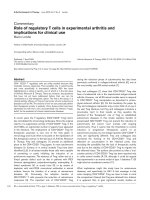
Figure 1
eral bronchi
Immunohistochemical localization of ChAT in murine periphImmunohistochemical localization of ChAT in murine peripheral bronchi. Respiratory epithelial cells are strongly ChATimmunoreactive in wild-type FVB mice (A). The specificity of
this labelling is indicated by its absence after preabsorption of
the antiserum with its corresponding antigenic peptide (B).
Bar represents 50 µm.
yielding 36.5 ± 4.4 pmol/mg in FVB mice (n = 4) and 28.5
± 3.50 pmol/mg in OCT1/2-/- mice (n = 3).
Bronchi of about 200 µm in diameter were too small to
dissect the respiratory epithelium for biochemical ACh
analysis. The ACh synthesizing enzyme, ChAT, was demonstrated in epithelial cells of these bronchi by immunohistochemistry (Fig. 1).
Role of the epithelium and of ACh in 5-HT-induced
bronchoconstriction
Small intrapulmonary bronchi from M2/3R+/+ wild-type
mice strongly constricted in response to both muscarine
(10-6 M) and to 5-HT (10-6 M; Fig. 2). The magnitude of
the 5-HT-induced bronchoconstriction even surpassed
that evoked by muscarine (Fig. 2). Mechanical (partial)
removal of the epithelium diminished the constriction to
muscarine (Fig. 2), consistent with the results of a previous study involving the chemical (Triton X-100) ablation
of the murine tracheal epithelium [3]. Removal of the airway epithelium also led to a significant reduction in the 5HT-induced bronchoconstriction response (Fig. 2). However, removal of the epithelium had a more pronounced
effect on 5-HT- than on muscarine-induced bronchoconstriction. Thus, in contrast to intact bronchi, the magnitude of the 5-HT response was smaller than that evoked by
muscarine after epithelium removal.
Bronchi from M2/3R-/- mice were entirely unresponsive to
muscarine (10-6 M; Fig. 3), as reported earlier [11]. In
striking contrast, 5-HT (10-6 M) induced indistinguishable
bronchoconstrictor responses in M2/3R-/- mutant and
M2/3R+/+ wild-type mice, both in absolute values and
expressed as percent response evoked by the thromboxane
analogue, U46610 (10-5 M) (Fig. 3).
Page 4 of 12
(page number not for citation purposes)
/>
Mu
s
140
5-H
T
10 -6
10 -6
M
M
Respiratory Research 2006, 7:65
wash
wash
120
Area [%]
100
80
60
40
20
0
0
5
10
Bronchoconstriction [%]
A
B
15
20
Control (n=15/5)
25
30
35
Time [min]
40
45
50
55
Denuded (n=7/2)
Control
Denuded
200
150
*** ** *
100
50 µm
50
Denuded
Control
1'
5'
max.
1'
5'
max.
50 µm
C
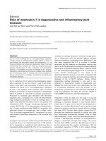
Effect of2
Figure epithelium removal on constriction of peripheral bronchi in PCLS of M2/3R+/+ mice
Effect of epithelium removal on constriction of peripheral bronchi in PCLS of M2/3R+/+ mice. (A) Reduction of luminal area of
intact (control, blue) and epithelium-denuded (denuded, red) peripheral bronchi in response to muscarine (Mus, 10-6 M) and 5HT (10-6 M). The numbers in parentheses refer to the numbers of bronchi/number of lungs from which they were taken. Panel
(B) illustrates the magnitude of the response to 5-HT (10-6 M) compared to that to muscarine (10-6 M) which was set as 100 %.
Control bronchi react slightly stronger to 5-HT than to muscarine, whereas the 5-HT response is significantly smaller that the
muscarine response after epithelium removal, particularly at 1 min (1') after agonist application. The box plots shows percentiles 0, 25, 50 (median), 75, and 100; individual data points beyond 3× S.D. are indicated by * or °. ***p < 0.001, **p < 0.01, *p
< 0.05 (comparison of corresponding time points by Mann-Whitney U-test). (C) Microscopic appearance of control and epithelium-denuded bronchi. In the left panel, arrowheads indicate thickness of the epithelial layer in a control bronchus. In the
right panel, the arrowhead points to a small remnant of epithelium after mechanical denudation of the epithelium.
Page 5 of 12
(page number not for citation purposes)
/>
M
10 -6
M
120
5-H
T
10 -6
Mu
s
140
U4
66
19
10 -5
M
Respiratory Research 2006, 7:65
wash
wash
Area [%]
100
80
60
40
20
0
A
0
5
10 15 20 25 30 35 40 45 50 55 85 90 95 100 105
Time [min]
+/+ (n=16/5)
M2/3
M2/3-/- (n=9/5)
Bronchoconstriction [%]
M2/3+/+
/
150
140
130
Mus 10-6 M
120
110
5-HT 10-6 M
100
90
80
B
M2/3-/-
U46619 10-5 M
M2/3+/+
M2/3-/-
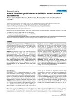
C
Figure 3in luminal area of peripheral bronchi in response to muscarine (Mus, 10-6 M), 5-HT (10-6 M), and U44619 (10-5 M) in
Changes (M2/3R+/+) and M2/3R-/- mice
wild-type
Changes in luminal area of peripheral bronchi in response to muscarine (Mus, 10-6 M), 5-HT (10-6 M), and U44619 (10-5 M) in
wild-type (M2/3R+/+) and M2/3R-/- mice. (A) 5-HT induces similar responses in both strains. The numbers in parentheses refer
to the numbers of bronchi/number of lungs from which they were taken. Panel (B) expresses the 5-HT-induced constriction in
percent of that evoked by U44619 in the first min after agonist application. The box plots shows percentiles 0, 25, 50 (median),
75, and 100; * indicates an individual data point beyond 3× S.D. (C) Original images of a peripheral bronchus of a wild-type and
an M2/3R-/- double-knockout animal before and after agonist application. As depicted in (A), there is no constriction in
response to muscarine in M2/3R-/- mice. On the other hand, both strains show identical responses to 5-HT and U44619.
Page 6 of 12
(page number not for citation purposes)
10 -6
M
wash
At
ro
120
Area [%]
pin
e
0 -6
T1
5-H
140
At
r
5-H opin
T1 e1
0 -6 0 -6
M M+
/>
M
Respiratory Research 2006, 7:65
wash
100
80
60
40
20
5
pin
e
10 -6
wash
wash
At
ro
140
5-H
T
Mu
sc
ari
n
e
M
10 -6
M
A
10 15 20 25 30 35 40 45 50 55 60 65 70 75
Time [min]
M2/3+/+ (n=5/3)
+
0
10 -4
M
At
rop
5-H in
T1 e1
0 -6 0 -4
M M
0
wash
Area [%]
120
100
80
60
40
20
0
0
Bronchoconstriction [%]
B
60
40
20
0
-20
N=
C
5 10 15 20 25 30 35 40 45 50 55 60 65 70 75 80 85 90
Time [min]
M2/3-/- (n=5/4)
5
6
OCT1-3+/+ OCT1/2-/-
4
OCT3-/-
4
M2/3+/+
5
M2/3-/-
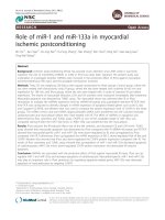
Figure atropine on 5-HT-induced bronchoconstriction (reduction of bronchial luminal area) in PCLS
Effect of4
Effect of atropine on 5-HT-induced bronchoconstriction (reduction of bronchial luminal area) in PCLS. Atropine blocks 5-HTinduced constriction partially at 10-6 M (A), and nearly completely at 10-4 M, even in absence of both M2 and M3 muscarinic
receptors (B). The numbers in parentheses refer to the numbers of bronchi/number of lungs from which they were taken. (C)
Persisting bronchoconstriction in response to 5-HT (10-6 M) in the presence of 10-4 M atropine in different wild-type and
knockout strains. The initial 5-HT-induced bronchoconstriction was set as 100 %.
Page 7 of 12
(page number not for citation purposes)
Respiratory Research 2006, 7:65
/>
Figure 5
Immunohistochemical localization of OCTs in murine bronchi
Immunohistochemical localization of OCTs in murine bronchi. OCT1-immunolabelling is localized to the apical membrane of
ciliated epithelial cells in wild-type FVB mice (arrows in A). The specificity of this labelling is indicated by its absence after preabsorption of the antiserum with its corresponding antigenic peptide (B) and the lack of labelling in OCT1/2-/- mice (C). Neither
of the two OCT2-antibodies used in this study showed specific labelling of mouse bronchi (D, E). The spotty labelling of epithelial cells observed with the OCT2-antibody raised against the human sequence (E) was also observed in OCT1/2-/- mice (F),
indicating that this signal is non-specific. Specific OCT3-immunolabelling, documented by its absence in the preabsorption control (inset in G), is observed primarily on the bronchial smooth muscle (sm) and, less intensely, on epithelial cells (G). OCT3localization in smooth muscle cells is confirmed by double-labelling immunofluorescence with OCT3-antibody and a monoclonal antibody against α-smooth muscle actin (SMA) (G') yielding the yellow signal in the merged image (G'). Bar represents 10
µm in A-F and 20 µm in G-G".
Page 8 of 12
(page number not for citation purposes)
Respiratory Research 2006, 7:65
In preparations from M2/3R+/+ wild-type mice, atropine
(10-6 M) partially inhibited 5-HT-induced constriction
(Fig. 4A). The same concentration of atropine fully
blocked muscarine-induced bronchoconstriction (data
not shown, see our previous report [11]). At a higher concentration (10-4 M), however, atropine reduced 5-HTinduced bronchoconstriction by approximately 80 % in
all strains tested, including M2/3R-/-, OCT1/2-/-, OCT3-/-,
and corresponding wild-type mice (Fig. 4B, C).
Distribution of OCTs in murine bronchi
Immunohistochemistry revealed OCT1-immunoreactivity in the apical membrane of ciliated cells (Fig. 5A). This
labelling was OCT1-specific since it was absent when the
antiserum was preabsorbed with the corresponding antigenic peptide and when tissue from OCT1/2-/- mice was
used for immunohistochemistry (Fig. 5B, C). No specific
OCT2-immunolabelling was observed in the bronchial
wall (Fig. 5D–F). Specific OCT3-immunoreactivity was
most intense in the bronchial smooth muscle and weaker
on epithelial cells (Fig. 5G–G").
Role of OCTs in 5-HT-induced bronchoconstriction
Small intrapulmonary bronchi from OCT1/2-/-, OCT3-/-,
and OCT1-3+/+ wild-type mice reacted with a strong constriction to muscarine (10-6 M) and to 5-HT (10-6 M) (Fig.
6A, B). The absence of OCT1/2 or OCT3 had no significant effect on the 5-HT bronchoconstrictor response. Corticosterone (10-6 M) significantly reduced the 5-HTinduced bronchoconstriction both in wild-type and in
OCT1/2-/- mice but was ineffective in OCT3-/- mice (Fig.
6C, D). The effect of epithelium removal on the inhibitory
action of corticosterone on 5-HT-induced bronchoconstriction was investigated in M2/3R+/+ wild-type mice. In
intact bronchi from this strain, 86 ± 5 % (mean ± S.E.M.;
7 PCLS from 7 lungs) of the 5-HT-induced contraction
remained in the presence of corticosterone, so that the
corticosterone effect was not as marked as in OCT1-3+/+
wild-type (FVB) mice. This small, but significant reduction of 5-HT-induced contraction by corticosterone in
M2/3R+/+ wild-type mice was still present after epithelium
removal (remaining contraction: 72 ± 5 %; mean ± S.E.M.;
7 PCLS from 7 lungs).
Discussion
The present data clearly demonstrate an epitheliumdependent component of 5-HT-induced bronchoconstriction in the mouse, consistent with the results of a previous
study on the mouse trachea [3]. It has been suggested that
this activity is dependent on the release of ACh from airway epithelial cells [3]. In the Xenopus oocyte expression
system, both OCT1 and 2, but not OCT3, proved to be
able to translocate ACh across the plasma membrane [8].
In the present study, we found that the airway epithelial
ACh content was twice as high in OCT1/2-/- than in wild-
/>
type mice. This observation supports the concept that
OCT1/2 may also play a role in the release of ACh from
airway epithelia. However, to our surprise, the magnitude
of 5-HT-induced bronchoconstrictor responses was
unchanged in PCLS preparations from OCT1/2-/- mice,
indicating that 5-HT-induced bronchoconstriction does
not require the presence of OCT1 and 2. Moreover, videomorphometric studies showed that PCLS from M2/3R-/mice remained fully responsive to 5-HT. In contrast, PCLS
from M2/3R-/- mice do no longer show a bronchoconstrictor response following cholinergic stimulation, as shown
in this and in an earlier study [11]. These data clearly indicate that the release of epithelial ACh is not involved in
the 5-HT-induced bronchoconstrictor response, but that
another epithelium-derived constrictory factor contributes to this activity.
In previous studies, ACh emerged as a candidate for mediating 5-HT-induced airway constriction in the mouse
because this effect could be inhibited by atropine [1-3]. In
the present study, we found a large reduction of 5-HTinduced bronchoconstriction only after application of an
unusually high concentration of atropine (10-4 M). On the
other hand, a much smaller concentration of atropine
(10-6 M) was sufficient to fully block muscarine-induced
bronchoconstriction. Interestingly, Eum et al. [2] also did
not observe a significant inhibition of 5-HT-induced contraction of the isolated mouse trachea at 10-6 M atropine.
The inhibition of 5-HT-induced bronchoconstriction by
10-4 M atropine persisted in M2/3R-/- mice, clearly indicating that this high concentration of atropine inhibits airway smooth muscle contractility via non-specific effects
that are not due to muscarinic receptor blockade. Indeed,
atropine has been described as a competitive antagonist at
the 5-HT3-receptor [17]. Taken together, the present data
demonstrate that 5-HT releases an epithelium-derived
bronchoconstrictory factor that is OCT-independent and
different from ACh.
We made the striking observation that corticosterone
exerted an acute inhibitory effect on 5-HT-induced bronchoconstriction. This acute effect of corticosterone was
mediated by OCT3, as demonstrated by its absence in
OCT3-/- mice. This finding is of potential clinical relevance
since rapid therapeutical effects of a bolus of inhaled glucocorticoids have been reported in asthmatic patients
where they reverse airway subsensitivity to β2-agonists
[18,19]. In our model, the inhibitory action of corticosterone on 5-HT-induced bronchoconstriction is epitheliumindependent since it persisted after epithelium removal.
In line with this observation, immunohistochemistry
demonstrated that OCT3 is located directly on bronchial
smooth muscle cells. In principle, all OCT isoforms tested
so far are sensitive to corticosteroids that are not substrates for transport by themselves but inhibit transport of
Page 9 of 12
(page number not for citation purposes)
Respiratory Research 2006, 7:65
/>
5-H
T1
0 -6
M
M
Mu
s1
0 -6
140
wash
120
/
wash
5-HT
OCT1-3+/+
Area [%]
100
80
OCT1/2-/-
60
40
20
OCT3-/-
0
140
120
wash
40
45
wash
50
55
OCT3-/- (n=17/13)
B
125
wash
Area [%]
100
80
60
40
Bronchoconstriction [%]
Mu
s1
0 -6
M
A
25
30
35
Time [min]
OCT1/2-/- (n=20/13)
Cs
5-H 10 -6
T1 M+
0 -6
M
OCT1-3+/+ (n=19/14)
20
M
15
Cs
10 -6
10
10 -6
M
5
5-H
T
0
100
**
75
50
25
20
0
0
0
C
5
10 15 20
OCT1-3+/+ (n=7/6)
25 30 35 40 45 50 55
Time [min]
OCT1/2-/- (n=7/5)
60 65 70 75
OCT1-3+/+ OCT1/2-/- OCT3-/-
80 85 90
OCT3-/- (n=7/4)
D
Figure 6
to corticosterone
5-HT-induced reduction of bronchial luminal area (bronchoconstriction) in OCT-deficient mice and sensitivity of this response
5-HT-induced reduction of bronchial luminal area (bronchoconstriction) in OCT-deficient mice and sensitivity of this response
to corticosterone. (A, B) Wild-type FVB mice (OCT1-3+/+), OCT1/2-/- mice and OCT3-/- mice exhibit no differences in their
response to 5-HT (10-6 M). The numbers in parentheses refer to the numbers of bronchi/number of lungs from which they
were taken. (C, D) In wild-type and OCT1/2-/- mice, but not in OCT3-/- mice, the bronchoconstriction in response to 5-HT is
significantly reduced by corticosterone (Cs, 10-6 M). Panel (D) depicts the bronchoconstrictor response to 5-HT (10-6 M, 1 min
after administration) in the presence of corticosterone (10-6 M), as compared to the response to 5-HT alone (set as 100%). **p
< 0.01, Mann-Whitney U-test.
other substances [20]. OCT3, which we identified as being
responsible for the acute inhibitory effect of corticosterone on 5-HT-induced bronchoconstriction, has the highest affinity for corticosteroids [20]. It also clears
monoamines, including catecholamines and 5-HT, from
the extracellular space [21], and hence its blockade is
expected to increase the extracellular concentrations of
these agents. Indeed, acute human bronchial vasocon-
striction elicited by corticosteroids has been explained by
inhibition of OCT3 with subsequent rise of extracellular
noradrenaline and prolonged activation of α1-adrenoreceptors [22]. However, a separate, specific serotonin transporter (SERT) is highly expressed in the lung [23,24]. As a
result, deficiency or blockade of OCT3 may have little
impact on 5-HT turnover. In agreement with this notion,
the magnitude of the bronchoconstrictor response to 5-
Page 10 of 12
(page number not for citation purposes)
Respiratory Research 2006, 7:65
HT remained unchanged in bronchi from OCT3-/- mice
and the 5-HT response was reduced rather than augmented by corticosterone. It is therefore unlikely that the
observed OCT3-mediated inhibition of 5-HT-induced
bronchoconstriction by acutely administered corticosterone involves direct interference with 5-HT transport. In
view of the electrogenic properties of OCTs [20], the acute
inhibitory effect of corticosterone on 5-HT-induced bronchoconstriction might be caused by modulation of membrane potential, but the underlying signal transduction
cascade still awaits to be clarified.
Conclusion
5-HT-induced constriction of murine intrapulmonary
bronchi involves two independent pathways. One pathway is dependent on the release of an epithelium-derived
constrictory factor that is different from ACh. The second
pathway involves the direct stimulation of bronchial
smooth muscle cells. This latter pathway is partly sensitive
to acutely administered corticosterone acting on OCT3.
These data provide new insights into the mechanisms
involved in 5-HT-induced bronchoconstriction, including
novel information about non-genomic, acute pulmonary
effects of corticosteroids.
Competing interests
The author(s) declare that they have no competing interests.
/>
References
1.
2.
3.
4.
5.
6.
7.
8.
9.
10.
11.
Authors' contributions
WK carried out the epithelium removal, evaluated immunohistochemistry, participated in the design of the study
and drafted the manuscript. SW carried out epithelium
removal, videomorphometric and statistical analyses. SA
carried out videomorphometric and statistical analyses.
IW performed the ACh assay and revised the manuscript
critically for important intellectual content. AHS provided
OCT-deficient mice and revised the manuscript critically
for important intellectual content. JW provided M2R/
M3R-deficient mice and revised the manuscript critically
for important intellectual content. HK provided antibodies, added to the design of the study and revised the manuscript critically for important intellectual content. RVH
coordinated the videomorphometric setup and breeding
of genetically deficient mice strains, and revised the manuscript critically for important intellectual content. KSL
performed and evaluated immunohistochemistry, and
participated in the design of the study and drafting of the
manuscript. The data presented in this manuscript are part
of the doctoral thesis of SA.
12.
13.
14.
15.
16.
17.
18.
19.
Acknowledgements
We thank Mr M. Bodenbenner, Ms U. Butz-Schiller and Ms K. Michael for
skilful technical assistance.
20.
Levitt RC, Mitzner W: Autosomal recessive inheritance of airway hyperreactivity to 5-hydroxytryptamine. J Appl Physiol
1989, 67:1125-1132.
Eum SY, Norel X, Lefort J, Labat C, Vargaftig BB, Brink C: Anaphylactic bronchoconstriction in BP2 mice: interactions
between serotonin and acetylcholine. Br J Pharmacol 1999,
126:312-316.
Moffatt JD, Cocks TM, Page CP: Role of the epithelium and acetylcholine in mediating the contraction to 5-hydroxytryptamine in the mouse isolated trachea. Br J Pharmacol 2004,
141:1159-1166.
Reinheimer T, Bernedo P, Klapproth H, Oelert H, Zeiske B, Racke K,
Wessler I: Acetylcholine in isolated airways of rat, guinea pig,
and human: species differences in role of airway mucosa. Am
J Physiol 1996, 270:L722-L728.
Reinheimer T, Munch M, Bittinger F, Racke K, Kirkpatrick CJ, Wessler
I: Glucocorticoids mediate reduction of epithelial acetylcholine content in the airways of rats and humans. Eur J Pharmacol
1998, 349:277-284.
Pfeil U, Lips KS, Eberling L, Grau V, Haberberger RV, Kummer W:
Expression of the high-affinity choline transporter, CHT1, in
the rat trachea. Am J Respir Cell Mol Biol 2003, 28:473-477.
Proskocil BJ, Sekhon HS, Jia Y, Savchenko V, Blakely RD, Lindstrom J,
Spindel ER: Acetylcholine is an autocrine or paracrine hormone synthesized and secreted by airway bronchial epithelial cells. Endocrinology 2004, 145:2498-2506.
Lips KS, Volk C, Schmitt BM, Pfeil U, Arndt P, Miska D, Ermert L,
Kummer W, Koepsell H: Polyspecific cation transporters mediate luminal release of acetylcholine from bronchial epithelium. Am J Respir Cell Mol Biol 2005, 33:79-88.
Wessler I, Roth E, Deutsch C, Brockerhoff P, Bittinger F, Kirkpatrick
CJ, Kilbinger H: Release of non-neuronal acetylcholine from
the isolated human placenta is mediated by organic cation
transporters. Br J Pharmacol 2001, 134:951-956.
Martin C, Uhlig S, Ullrich V: Videomicroscopy of methacholineinduced contraction of individual airways in precision-cut
lung slices. Eur Respir J 1996, 9:2479-2487.
Struckmann N, Schwering S, Wiegand S, Gschnell A, Yamada M, Kummer W, Wess J, Haberberger RV: Role of muscarinic receptor
subtypes in the constriction of peripheral airways: studies on
receptor-deficient mice. Mol Pharmacol 2003, 64:1444-1451.
Pfaff M, Powaga N, Akinci S, Schutz W, Banno Y, Wiegand S, Kummer
W, Wess J, Haberberger RV: Activation of the SPHK/S1P signalling pathway is coupled to muscarinic receptor-dependent
regulation of peripheral airways. Respir Res 2005, 6:48-61.
Zwart R, Verhaagh S, Buitelaar M, Popp-Snijders C, Barlow DP:
Impaired activity of the extraneuronal monoamine transporter system known as uptake-2 in Orct3/Slc22a3-deficient
mice. Mol Cell Biol 2001, 21:4188-4196.
Jonker JW, Wagenaar E, van Eijl S, Schinkel AH: Deficiency in the
organic cation transporters 1 and 2 (Oct1/Oct2 [Slc22a1/
Slc22a2]) in mice abolishes renal secretion of organic cations. Mol Cell Biol 2003, 23:7902-7908.
Wessler I, Bender H, Harle P, Hohle KD, Kirdorf G, Klapproth H,
Reinheimer T, Ricny J, Schniepp-Mendelssohn KE, Racke K: Release
of [3H]acetylcholine in human isolated bronchi. Effect of
indomethacin on muscarinic autoinhibition. Am J Respir Crit
Care Med 1995, 151:1040-1046.
Pfeil U, Vollerthun R, Kummer W, Lips KS: Expression of the
cholinergic gene locus in the rat placenta. Histochem Cell Biol
2004, 122:121-130.
Fan P, Weight FF: The effect of atropine on the activation of 5hydroxytryptamine3 channels in rat nodose ganglion neurons. Neuroscience 1994, 62:1287-1292.
Aziz I, Lipworth BJ: A bolus of inhaled budesonide rapidly
reverses airway subsensitivity and β2-adrenoceptor downregulation after regular inhaled formoterol. Chest 1999,
115:623-628.
Lipworth BJ, Aziz I: Bronchodilator response to albuterol after
regular formoterol and effects of acute corticosteroid
administration. Chest 2000, 117:156-162.
Koepsell H, Schmitt BM, Gorboulev V: Organic cation transporters. Rev Physiol Biochem Pharmacol 2003, 150:36-90.
Page 11 of 12
(page number not for citation purposes)
Respiratory Research 2006, 7:65
21.
22.
23.
24.
/>
Gründemann D, Schechinger B, Rappold GA, Schömig E: Molecular
identification of the corticosterone-sensitive extraneuronal
catecholamine transporter. Nat Neurosci 1998, 1:349-351.
Horvath G, Sutto Z, Torbati A, Conner GE, Salathe M, Wanner A:
Norepinephrine transport by the extraneuronal monoamine
transporter in human bronchial arterial smooth muscle
cells. Am J Physiol Lung Cell Mol Physiol 2003, 285:L829-L837.
Chang AS, Chang SM, Starnes DM, Schroeter S, Bauman AL, Blakely
RD: Cloning and expression of the mouse serotonin transporter. Brain Res Mol Brain Res 1996, 43:185-192.
James KM, Bryan-Lluka LJ: Efflux studies allow further characterisation of the noradrenaline and 5-hydroxytryptamine transporters in rat lungs. Naunyn Schmiedebergs Arch Pharmacol 1997,
356:126-133.
Publish with Bio Med Central and every
scientist can read your work free of charge
"BioMed Central will be the most significant development for
disseminating the results of biomedical researc h in our lifetime."
Sir Paul Nurse, Cancer Research UK
Your research papers will be:
available free of charge to the entire biomedical community
peer reviewed and published immediately upon acceptance
cited in PubMed and archived on PubMed Central
yours — you keep the copyright
BioMedcentral
Submit your manuscript here:
/>
Page 12 of 12
(page number not for citation purposes)